Article

The Isolation of Vibrio crassostreae and V. cyclitrophicus in Lesser-Spotted Dogfish (Scyliorhinus canicula) Juveniles Reared in a Public Aquarium

1
Istituto Zooprofilattico Sperimentale del Piemonte, Liguria e Valle d’Aosta, Via Bologna 148, 10154 Torino, Italy
2
Acquario di Cala Gonone, Via La Favorita, 08022 Dorgali, Italy
*
Authors to whom correspondence should be addressed.
These authors contributed equally to this work.
J. Mar. Sci. Eng. 2022, 10(1), 114; https://doi.org/10.3390/jmse10010114
Submission received: 10 November 2021 / Revised: 12 January 2022 / Accepted: 13 January 2022 / Published: 15 January 2022
(This article belongs to the Special Issue Microbial Diseases of Marine Organisms)

Abstract

:
The genus Vibrio currently contains 147 recognized species widely distributed, including pathogens for aquatic organisms. Vibrio infections in elasmobranchs are poorly reported, often with identifications as Vibrio sp. and without detailed diagnostic insights. The purpose of this paper is the description of the isolation and identification process of Vibrio spp. following a mortality event of Scyliorhinus canicula juvenile reared in an Italian public aquarium. Following investigations aimed at excluding the presence of different pathogens of marine fish species (parasites, bacteria, Betanodavirus), several colonies were isolated and subjected to species identification using the available diagnostic techniques (a biochemical test, MALDI-TOF MS, and biomolecular analysis). Discrepancies were observed among the methods; the limits of biochemistry as a unique tool for Vibrio species determination were detected through statistical analysis. The use of the rpoB gene, as a diagnostic tool, allowed the identification of the isolates as V. crassostreae and V. cyclotrophicus. Although the pathogenic role of these microorganisms in lesser-spotted dogfish juveniles has not been demonstrated, and the presence of further pathogens cannot be excluded, this study allowed the isolation of two Vibrio species in less-studied aquatic organisms, highlighting the weaknesses and strengths of the different diagnostic methods applied.

1. Introduction

The bacteria of the Vibrionaceae family are Gram-negative curved rods that globally occur in marine, estuarine and freshwater ecosystems. They occupy habitats ranging from the deep-sea to shallow aquatic environments [1]. Among them, some species are important for natural systems, including carbon cycle and osmoregulation, and as free-living inhabitants in the water column or associated with particulate matter [2]. The genus Vibrio currently contains 147 recognized species widely distributed in aquatic environments [3]. Some vibrios cause water- and seafood-related outbreaks of gastrointestinal infections in humans [4], and many can be pathogenic for marine vertebrates [5,6] and invertebrates [7,8]. Paillard et al. recognized that the emergence of vibrios as etiological agents of diseases is likely to increase over the coming years due to ocean warming [9]. Classical vibriosis is generally characterized by lethargic movement of affected fish [10,11,12], the presence of various skin ulcerations [12,13,14,15], rotting fins [14,16,17], and pigmentation of the body [10,13,14,18]. Internal organs appear enlarged, hemorrhagic and congested, particularly the liver, kidney, and spleen [11,12,18]. Clinical signs demonstrated during vibriosis outbreaks are different from case to case and are influenced by multiple factors such as host species, age, exposure time, and strain virulence factors. Epidemiologically, vibriosis is often acute in young fish larvae and fry. The disease spread rapidly following infection and most infected young fish die without showing clinical signs. Thus, sudden death is often noticed in young fish that died with less severe or no external symptoms. In contrast, infected adult fish usually develop a chronic infection with evident skin ulcerations and pigmentation [19]. Stress is a major factor in increasing fish susceptibility to pathogens [15]. Regarding marine fish aquaculture, the most hazardous species of the Vibrionaceae family are V. anguillarum, V. ordalii, V. salmonicida, V. vulnificus, V. alginolyticus, V. harveyi, V. parahaemolyticus, V. ponticus for the genera Vibrio, and Photobacterium damselae subsp. damselae (formerly known as Vibrio damselae) [20,21,22,23,24]. Lesser-reported species of the genus Vibrio in other marine organisms are poorly described; this is presumably due to the high diversity of Vibrionaceae in environments and from the natural environment itself, which does not present the typical stressors of intensive farming. Grimes et al. [25] reported a mortality case of a single brown shark (Carcharhinus plumbeus) reared in captivity due to an infection by a Vibrio species, identified as V. carchariae. However, this isolate was later recognized as a V. harveyi strain based on whole-genome sequencing data [26].
Elasmobranchs are considered a fundamental element in the trophic chain of marine environments [27,28]. Currently, the population of elasmobranchs in the Mediterranean Sea is in decline due to habitat degradation and consequent to the direct impacts of fishing [29,30,31]. At the time of writing, 41% of the species are considered at risk (classified as either critically endangered, endangered, or vulnerable) by the regional assessment of the International Union for the Conservation of Nature (IUCN), and 33% are considered as lacking in data [32]. Focusing on the lesser-spotted dogfish Scyliorhinus canicula (Linnaeus, 1758), a small demersal shark (Carcharhiniformes: Scyliorhinidae) classified as LC by IUCN, it is a broad generalist, and in terms of diet and habitat requirements, it is an opportunistic feeder, and cannibalism is frequently recorded [33,34]. It is also possible to find this species in public aquaria because of its easy reproduction in captivity [35]. However, artificial housing conditions are often linked to the onset of diseases, and limited literature is available for sharks. To date, there are few reports on infectious diseases affecting sharks reported in the literature [36,37]. For this reason, this study aimed to clarify the cause of tank mortality that occurred in S. canicula juveniles reared in an Italian public aquarium.

2. Materials and Methods

2.1. Outbreak Description and Conditions of Sampling Procedure

Once laid, lesser-spotted dogfish eggs were moved from the exhibition tank to interconnecting tanks in a recirculated aquaculture system (RAS) (1 m3 each), in accordance with the current legislation on animal welfare. The water introduced into a closed circuit, technically known as RAS, was subjected to different types of treatment—namely, mechanical, biological, thermal, gaseous, chemical rebalancing, and knocking down the bacterial charge. Specifically, disinfection of water occurred by means of special fluorescent lamps capable of producing ultraviolet (UV) light with wavelengths included in the UV–C band (λ 100–280 nm) [38]. Since organisms are exposed to UV light, UV rays affect nucleic acids (DNA and RNA), damaging the aromatic structures with the formation of double bonds in stable pyrimidine molecules and preventing the cellular development of microbes [39]. Only microorganisms such as algae, viruses, bacteria, and other pathogens floating in the water are impacted. Nevertheless, nitrifying bacteria (with key roles in the nitrogen cycle) are not carried to the UV sterilizer and therefore do not come in range and will not be lost. The following water physicochemical parameters were measured by a multiparametric field probe (HACH, HQd Field Case): temperature (17 ± 1 °C); salinity (36 ± 1 PSU); dissolved oxygen (8.5 ± 0.4 mg/L); pH (8.0 ± 0.3). The specimens (Figure 1) showed the first symptoms a few months after hatching, i.e., anomalies in swimming, fast breathing, and lack of appetite. For the analysis, symptomatic and moribund fish were sent inside a double plastic bag (containing ⅓ water and ⅔ air/oxygen) and sent refrigerated to the fish diseases laboratory of the Istituto Zooprofilattico Sperimentale of Piemonte, Liguria and Valle d’Aosta, Turin, Italy.

2.2. Necropsy

In the laboratory, the fish were euthanatized using an overdose of ethyl 3-aminobenzoate methanesulfonate (MS-222, Sigma Aldrich, St. Louis, USA) following the current legislation. Before necropsy, biometrical parameters of each animal were recorded (total length (TL) and total weight (TW)). The fish were then macroscopically examined to highlight external alterations. After dissection with sterile tools, the coelomic cavity and the internal organs were subjected to visual inspection for the evaluation of anatomopathological changes.

2.3. Parasitological Examination

A general approach was used for parasitological examination in order to detect eso- and ectoparasites. Gills, skin, visceral cavity, and digestive tract were analyzed, both macroscopically and microscopically, with the use of a microscope. The microscope used for the analysis is an OLYMPUS BX40, with magnification ranging from 4× to 40×.

2.4. Virological Examination

For virological analysis, a sample of a portion of the brain was taken for the search for viral encephalopathy and retinopathy virus (Betanodavirus). The total RNA was extracted using the AllPrep DNA/RNA Micro Kit (QIAGEN, Hilden, Germany) following the manufacturer's protocol. Subsequently, cDNA was synthesized from extracted RNA using the QuantiTect Reverse Transcription Kit (QIAGEN, Hilden, Germany). A real-time RT–PCR targeting the viral encephalopathy and retinopathy virus RNA2 portion was used for viral diagnosis, following the protocol described by Panzarin et al. [40]. The reactions were carried out using the BioRad CFX96 Touch Real-Time PCR Detection System (Bio-Rad, Hercules, USA), and the data were analyzed by Bio-Rad CFX Maestro Software version 1.1.

2.5. Bacteriological and Biochemical Analyses

Bacteriological sampling was carried out from the head kidney, brain, and blood. The organs samples were taken using a 1 μL sterile calibrated loop, while blood sampling was performed immediately after the euthanasia procedure, taking it directly from the sharks’ hearts with an insulin syringe, inoculated on plates, and spread using a 1 μL sterile loop. The first isolation was performed on Columbia blood agar (BA), tryptic soy agar (TSA) supplemented with 2% NaCl, and Monsur medium (thiosulfate citrate bile sucrose, TCBS); the latter medium was used for its selectivity toward Vibrionaceae. Plates were incubated at 22 ± 2 °C for a total of 72 h, and growth was checked daily. After growth, colonies were cloned on BA for identification analyses. After Gram staining, pure isolates were identified by API® 20E tests, matrix-assisted laser desorption ionization–time of flight mass spectrometry (MALDI–TOF MS) using VITEK-MS (BioMerieux, Marcy-l'Étoile, France), and molecular analyses. API® tests were also used for the biochemical characterization of strains. API® galleries were incubated for 48 h at 22 ± 2 °C; tests were read every 24 h, and reagents were added according to the manufacturer’s instructions. The results were issued using the specific API® test reading software (APIWEBTM) available on the BioMerieux website (https://apiweb.biomerieux.com, accessed on 30 September 2021).

2.6. Biomolecular Analysis for Bacterial Identification

DNA extraction was performed using the boiling and freeze–thawing protocol as described by Pastorino et al. [41]. RNA polymerase beta subunit (rpoB) gene was amplified to discriminate among different Vibrio species. Specific primers for the rpoB gene, developed by Ki et al. [42], and the protocol suggested by the same authors were used. Amplicons were run on 2% GelGreen (Biotium, Landing Pkwy, USA) stained agarose gel and then visualized under UV exposure. A 50–2000 kb ladder (Amplisize Molecular Ruler, Bio-Rad, Hercules, USA) was used as a molecular marker. The amplicons were purified with an ExtractMe DNA Clean-Up and Gel-Out Kit (Blirt, Gdańsk, Poland), according to the manufacturer’s instructions. The purified PCR products were bidirectionally sequenced using Big Dye 3.1 (Applied Biosystems, Waltham, USA) chemistry and the same primers used for PCR amplification. Cycle-sequencing products were purified using Dye Ex 2.0 Spin Kit (QIAGEN, Hilden, Germany) and sequenced in an ABI3130xl Genetic analyzer (Applied Biosystems, Waltham, USA). Contig assembly of the DNA sequences was performed using the Lasergene Software package (DNASTAR, Madison, USA). The rpoB sequences were compared with nucleotide sequences in the GenBank database using the Basic Local Alignment Search Tool (BLAST) search algorithm. Subsequently, rpoB sequences were deposited on the GenBank database.

2.7. Phylogenetic Analysis

A neighbor-joining analysis was performed [43] using Molecular Evolutionary Genetics Analysis software (MEGAX) [44]. The rpoB gene sequences of 19 isolated strains (sequences with 100% identity with the reference sequence on BLAST were not used in the analysis), 10 rpoB sequences of different Vibrio species, and 4 genomic sequences for Vibrio species for which rpoB gene sequences were not available alone (V. crassostreae, V. cyclitrophicus, V. celticus, and V. gigantis) were considered for the phylogenetic analysis. Evolutionary distances were ascertained via the maximum composite likelihood method [45]. A bootstrap test of 1000 replicates was performed; a cut-off of 50% was set for the computation of the bootstrap condensed tree.

2.8. Statistical Analysis

Principal component analysis (PCA) was performed to reduce the dimensionality of the biochemical dataset, increasing interpretability but, at the same time, minimizing information loss. PCA was performed to illustrate the clustering of bacteria species based on biochemical features. This was accomplished considering both phenotypic (API® 20E) and molecular (PCR) species identification. Statistical analysis was performed using R software (version 1.1.463, RStudio, Inc., Boston, MA, USA).

3. Results

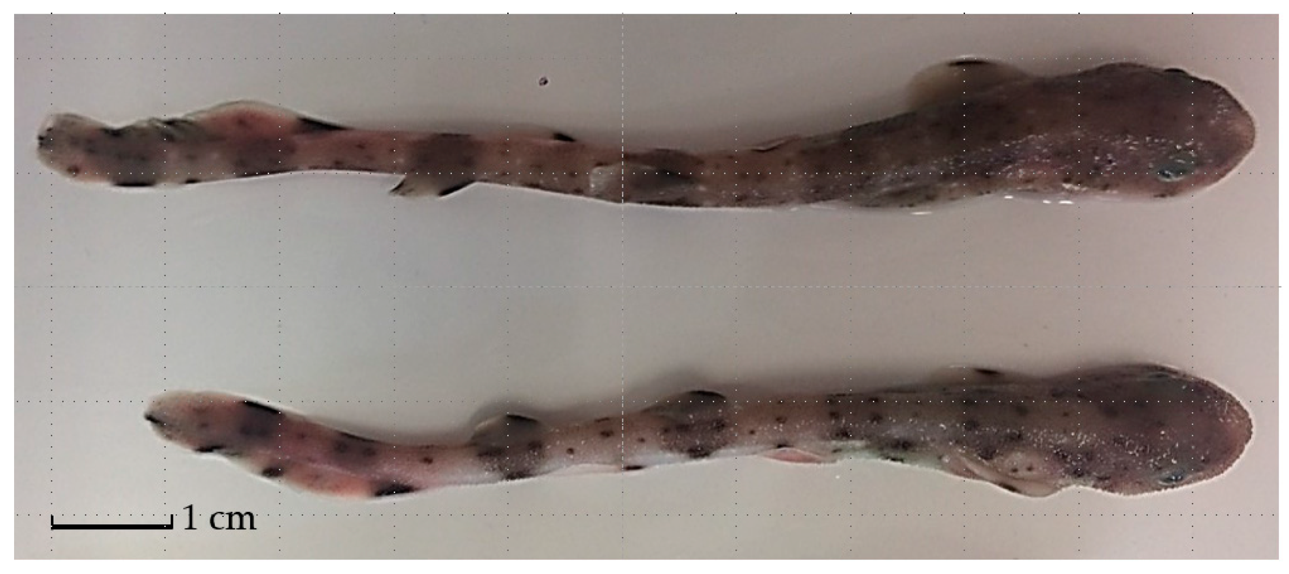
3.1. Necropsy, Parasitological, and Virological Examinations

A total of 20 lesser-spotted dogfish with an average weight of 2.16 ± 0.29 g (minimum weight: 1.70 g; maximum weight: 2.78 g) and an average length of 94.60 ± 4.03 mm (minimum length: 87 mm; maximum length: 103 mm) were analyzed. No external lesions were found, and the necropsy of the sharks did not show macroscopic lesions of the viscera. Parasitological (internal and external examination) and virological (Betanodavirus RT–PCR) tests yielded negative results.

3.2. Bacteriological Analysis

Out of 20 fish tested, 12 (12/20; 60%) tested positive from at least one matrix (head kidney, brain, or blood). A first visual examination of the plates was conducted, leading to the identification of 24 morphologically different isolates (Table 1). Each selected colony was subcloned to BA for subsequent tests. Following Gram staining, all isolates were identified as Gram-negative curved rods. Based on morphological and coltural characteristics, the identification by API® test envisaged the use of the API® 20E galleries. The results of the biochemical analyses carried out in a micro-method are summarized in Figure 2. On the other hand, identification by VITEK-MS did not produce any valid results according to the instrument cut-offs, so it was not possible to identify the isolates with this method.

3.3. Biomolecular Analysis for Bacterial Identification

All the 24 strains isolated showed a fragment of 730 bp from the rpoB gene amplification. Of those, 11 isolates (11/24; 45.8%) were identified at species level as Vibrio cyclitrophicus and 13 (13/24; 54.2%) as V. crassostreae (BLASTn nucleotide sequence identity value ranging from 98 to 100%). Results are reported in Table 2.
All obtained sequences, except for sequences with 100% identity value with the reference sequences in BLAST, were deposited on GenBank, a freely available online database (Accession Numbers: OM158211-OM158229).

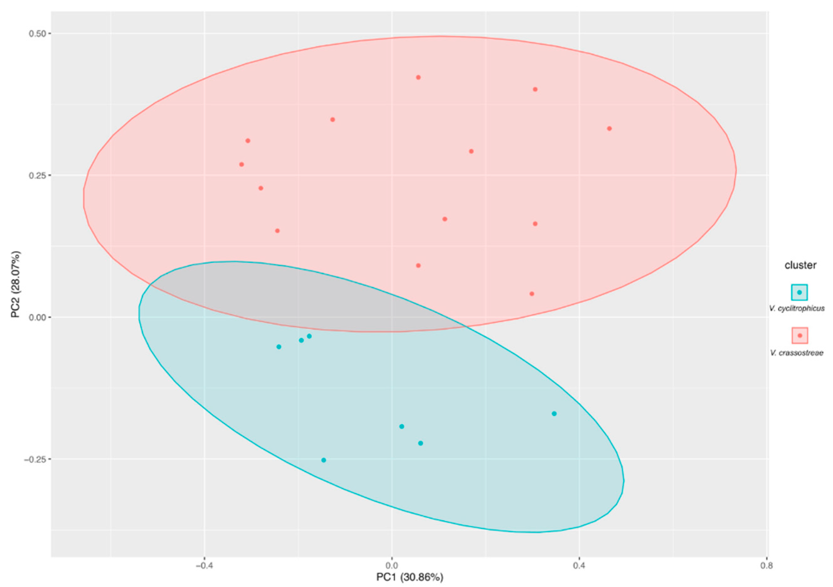
3.4. Phylogenetic Analysis

The bootstrap condensed tree is shown in Figure 3. Two main clusters are present: one containing the 10 rpoB sequences of different Vibrio species subdivided into three different subclusters, the second containing V. cyclitrophicus sequences with a high bootstrap value (97%). V. crassostreae isolates are not shown to be clustering with other Vibrio species, considering a bootstrap value cut-off of 50%.

3.5. Biochemical Features of Isolates

Morphological and biochemical analysis showed that all isolates were Gram-negative bacteria, positive for cytochrome oxidase, produced indole from tryptophan, and liquefied gelatin from enzyme gelatinase. None of the isolated strains produced lysine decarboxylase, ornithine decarboxylase, hydrogen sulfide, urease, nor were they able to utilize citrate. The enzyme tryptophan deaminase was negative in almost all strains. Generally, the isolates produced acid from glucose, arabinose, amygdalin, and mannitol. No acid was produced from amygdalin and arabinose. It is important to highlight a certain variability in biochemical features for the following test: decarboxylation of the amino acid arginine by arginine dihydrolase, detection of acetoin (acetyl methylcarbinol) produced by fermentation of glucose by bacteria utilizing the butylene glycol pathway (Voges–Proskauer test), and fermentation of sucrose and melibiose (Figure 2). The first PCA (Figure 4) based on phenotypic identification (API® 20E) showed that the first (PC1) and second (PC2) components accounted for meaningful amounts of the total variance (87.96%). PC1 explained 78.1% of the total variance, and PC2, 9.86%. PCA of bacteria strains yielded three main clusters. The blue cluster contains most of the bacteria species (Vibrio celticus, V. gigantis, and V. gallium) that were similar in biochemical features. The red cluster contains only V. fluvialis. Finally, the green cluster contains only Aeromonas hydrophila and V. splendidus. The second PCA cluster analysis (Figure 5) based on molecular species identification showed that the first (PC1) and second (PC2) components accounted for meaningful amounts of the total variance (58.93%). PC1 explained 30.86% of the total variance, and PC2, 28.07%. PCA of bacteria strains yielded two main clusters. The red cluster contains most of the strains that belonged to V. crassostreae, whereas the blue cluster contains V. cyclitrophicus. It is possible to observe a partial overlap of the two bacteria clusters, confirmed by the overlap of the confidence ellipses (95%) of each one, indicating similar biochemical features for certain tests.

4. Discussion

This study reports the finding of Vibrio crassostreae and V. cyclotrophicus in Scyliorhinus canicula isolated from different matrices (brain, blood, and kidney) and provides a description of the isolated strains. Notably, there are only a few reports in the literature on the isolation of pathogens from sharks. Therefore, our findings could clarify the pathogenic role of these bacteria despite the absence of gross pathology lesions.
The isolation process of Vibrio spp. from sharks started following the observation of external clinical signs common to many infectious diseases. For this reason, the presence of parasites, bacteria, and viruses was investigated. Parasitological and virological analyses tested negative, although most cases of diseases in sharks reported in the literature were of parasitic etiology [37]. In relation to the symptoms highlighted in the tank, a possible viral cause different from Betanodavirus cannot be excluded. However, it was not possible to carry out in-depth analyses due to the low number of samples, the limited amount of biological material available (in relation to shark size), and the scarce presence of information regarding elasmobranch viruses.
On the contrary, colonial growth was observed on agar media. Once pure cultures of the morphologically different colonies were obtained, the diagnostic techniques available in the laboratory clearly identified the isolates. Therefore, the isolates were tested by a biochemical test (API® 20E), MALDI-TOF MS, and molecular biology techniques. Upon Gram staining, the isolates were all found to be Gram-negative rods. Since the colonies grew on TCBS and had the typical morphology of Vibrio spp., it was decided to use the API® 20E galleries for species identification and characterization based on a biochemical test.
Biochemical tests led to the identification of bacteria of the genus Vibrio, as presumed, but also of Aeromonas hydrophila isolates. Therefore, the identification process continued by VITEK-MS, which did not lead to the identification of any of the isolates. The possible explanation of this was probably due to the absence in the database used by the instrument of reference strains comparable to those of the study. The implementation of these databases with Vibrio spp. strains not belonging from food or clinical isolates could make MALDI-TOF MS a rapid and efficient method for diagnosing these bacteria. Moreover, the identification of strains identified as A. hydrophila by biochemical methods was not confirmed, although it is reported that VITEK-MS is able to distinguish Aeromonas spp. from some species of Vibrio (V. cholerae) [46]. For this reason, it was decided to use molecular biology techniques to obtain a more accurate identification.
For genetic identification, the 16S subunit of ribosomal RNA (16S rRNA) gene is commonly used for the study of phylogenetic relationship among bacterial taxa and identification at the species level; however, the 16S rRNA genes of vibrios are not sufficiently polymorphic to ensure a reliable identification [47,48,49]. For this reason, species-specific PCRs have been developed, and different genetic markers have been considered. Some examples are the gene encoding the α-subunit of bacterial ATP synthase (atpA) [50] and rpoB [42]; considering that the rpoB gene led to more suitable results than the 16S rRNA gene for Vibrio species identification; thus, this tool was chosen in this study for the molecular identification. The mentioned analysis allowed the identification of the isolates, respectively, as V. crassostreae (13/24; 54.2%) and V. cyclitrophicus (11/24; 45.8%). The first species was reported as a pathogen of mollusks and fish [51], while V. cyclitrophicus was reported in mollusks and in the microbiome of marine copepods [52,53]. There are no reports of these species in sharks, although bacteria of the genus Vibrio have already been reported in elasmobranchs but not specifically for S. canicula.
As further support of these results, a statistical analysis was carried out on the results of biochemical and molecular tests. The biochemical test (API® 20E) allowed the identification of three main clusters of Vibrio species, based on biochemical features. In particular, the first PCA revealed that strains belonging to different Vibrio species (V. celticus, V. gigantis, and V. gallicus) had the same biochemical features. Considering this, it is clear how such identification is not a good diagnostic tool in fish pathology. On the contrary, the second PCA performed on biochemical features of Vibrio species identified throughout molecular tools allowed the discrimination of the biochemical features of V. crassostreae and V. cyclotrophicus, even though a partial overlap of the two main clusters was observed, indicating similar features for a certain aspect of the biochemical test (e.g., negative for decarboxylations of the amino acid ornithine, decarboxylation of the amino acid lysine urease, H2S production). These results support what has already been indicated in the description of the two Vibrio species isolated in this survey [54,55].
The phylogenetic analysis showed a clear separation between V. cyclitrophicus, characterized by a cluster with a high bootstrap value (97%), and V. crassostreae, even if a partial overlap was detected in the PCA analysis.
Similarly, Aeromonas hydrophila, V. splendidus, and V. fluvialis in the phylogenetic tree were clearly separated from V. crassostreae and V. cyclitrophicus even if sometimes identified as them by the API system.
Vibrio species identified in the study are generally linked to mortality in bivalve mollusks or found at the environmental level. A study by Petton et al. [56] shows how Vibrio infection is related to water temperature: In this article, it is shown that temperatures higher or lower than 16 °C affect oyster mortality due to the growth of Vibrio spp. As indicated by Sims [57], S. canicula fits perfectly in a thermal range between 14.9 °C and 17.7 °C. Thus, the tank’s water temperature is ideal both for the growth of isolated Vibrio species and for S. canicula. Nevertheless, it has been found that in natural environments, the lesser-spotted dogfish prefers temperatures lower than 17 °C, for optimization of its metabolism [57].
Currently, few studies are available in the literature on elasmobranchs infectious diseases. Probably, these shortcomings are due to the lack of general elasmobranch studies, as supported by Abdul Malak et al. [32]. Among bacterial diseases, Vibrio spp. infections are reported, although in a few reports and with several shortcomings in the identification process. The present study, therefore, has the function of a report of the presence of vibrios, similar to previous research; in-depth studies will be required for details on the pathogenic mechanism of these bacteria in sharks. Nevertheless, our study made it possible to identify specifically V. crassostreae and V. cyclitrophicus, underlining the criticalities in the identification process in relation to the techniques adopted (API® 20E, MALDI-TOF MS, molecular biology) and the need for developing specific guidelines for the identification of nonmajor pathogenic Vibrio species.

Author Contributions

Conceptualization: M.T., G.E., D.M., P.P., M.P. and S.C.; methodology: M.T., G.E., D.M., P.P., M.P. and S.C.; investigation: M.T., G.E., N.S., V.M., F.G., I.C., A.P., M.P. and S.C.; resources: P.L.A., A.D. and M.P.; data curation: M.T., G.E., P.P., M.P. and S.C.; writing—original draft preparation: M.T. and G.E.; writing—review and editing: D.M., P.P., M.P. and S.C.; visualization: M.T., G.E., D.M., P.P., M.P. and S.C.; supervision: P.L.A., A.D. and M.P. All authors have read and agreed to the published version of the manuscript.

Funding

This research received no external funding.

Institutional Review Board Statement

The Ethics Committee was not included, as all samples were derived from a diagnostic service that the laboratory in which the authors work offers to users (http://www.izsplv.it/) Therefore, the study was derived from a routine activity for which it was not necessary to establish an Ethics Committee.

Informed Consent Statement

Not applicable.

Data Availability Statement

Not applicable.

Conflicts of Interest

The authors declare no conflict of interest.

References

  1. Reen, F.J.; Almagro-Moreno, S.; Ussery, D.; Boyd, E.F. The genomic code inferring Vibrionaceae niche specialization. Nat. Rev. Microbiol. 2006, 9, 697–704. [Google Scholar] [CrossRef] [PubMed]
  2. Johnson, C.N. Fitness factors in vibrios: A mini-review. Microb. Ecol. 2013, 65, 826–851. [Google Scholar] [CrossRef] [PubMed]
  3. Parte, A.C.; Sardà Carbasse, J.; Meier-Kolthoff, J.P.; Reimer, L.C.; Göker, M. List of Prokaryotic names with Standing in Nomenclature (LPSN) moves to the DSMZ. Int. J. Syst. Evol. 2020, 70, 5607–5612. [Google Scholar] [CrossRef] [PubMed]
  4. Colwell, R.R.; Soira, W.M. The ecology of Vibrio Cholera. In Cholera; Barua, D., Greenough, W.B., Eds.; Springer: Boston, MA, USA, 1992; pp. 1–36. ISBN 978-1-4757-9690-2. [Google Scholar]
  5. Sorum, H.; Myhr, E.; Zwicker, B.M.; Lillehaug, A. Comparison by plasmid profiling of Vibrio salmonicida strains isolated from diseased fish from different north European and Canadian coastal areas of the Atlantic Ocean. Can. J. Fish Aquat. Sci. 1993, 50, 247–250. [Google Scholar] [CrossRef]
  6. Diggles, B.K.; Carson, J.; Hine, P.M.; Hickman, R.W.; Tait, M.J. Vibrio species associated with mortalities in hatchery-reared turbot (Colistium nudipinnis) and brill (C. guntheri) in New Zealand. Aquaculture 2000, 183, 1–12. [Google Scholar] [CrossRef]
  7. Goarant, C.; Herlin, J.; Brizard, R.; Marteau, A.L.; Martin, C.; Martin, B. Toxic factors of Vibrio strains pathogenic to shrimp. Dis. Aquat. Org. 2000, 40, 101–107. [Google Scholar] [CrossRef] [PubMed] [Green Version]
  8. Takahashi, K.G.; Nakamura, A.; Mori, K. Inhibitory effects of ovoglobulins on bacillary necrosis in larvae of the Pacific oyster, Crassostrea gigas. J. Invertebr. Pathol. 2000, 75, 212–217. [Google Scholar] [CrossRef] [PubMed]
  9. Paillard, C.; Le Roux, F.; Borrego, J.J. Bacterial disease in marine bivalves, a review of recent studies: Trends and evolution. Aquat. Living Resour. 2004, 17, 477–498. [Google Scholar] [CrossRef] [Green Version]
  10. Liu, P.C.; Lin, J.Y.; Hsiao, P.T.; Lee, K.K. Isolation and characterization of pathogenic Vibrio alginolyticus from diseased cobia Rachycentron canadum. J. Basic Microbiol. 2004, 44, 23–28. [Google Scholar] [CrossRef] [PubMed]
  11. Zhao, D.H.; Sun, J.J.; Liu, L.; Zhao, H.H.; Wang, H.F.; Liang, L.Q.; Liu, L.B.; Li, G.F. Characterization of two phenotypes of Photobacterium damselae subsp. damselae isolated from diseased juvenile Trachinotus ovatus reared in cage mariculture. J. World Aquacult. Soc. 2009, 40, 281–289. [Google Scholar] [CrossRef]
  12. Zhang, X.; Li, Y.W.; Mo, Z.Q.; Luo, X.C.; Sun, H.Y.; Liu, P.; Li, A.X.; Zhou, S.M.; Dan, X.M. Outbreak of a novel disease associated with Vibrio mimicus infection in fresh water cultured yellow catfish, Pelteobagrus fulvidraco. Aquaculture 2014, 432, 119–124. [Google Scholar] [CrossRef]
  13. Rajan, P.R.; Lopez, C.; Lin, J.H.; Yang, H. Vibrio alginolyticus infection in cobia (Rachycentron canadum) cultured in Taiwan. Bull. Eur. Ass. Fish Pathol. 2001, 21, 228–234. [Google Scholar]
  14. Ransangan, J.; Mustafa, S. Identification of Vibrio harveyi isolated from diseased Asian seabass (Lates calcarifer) by use of 16S ribosomal DNA sequencing. J. Aquat. Anim. Health 2009, 21, 150–155. [Google Scholar] [CrossRef] [PubMed]
  15. Austin, B.; Austin, D. Bacterial Fish Pathogens: Diseases of Farmed and Wild Fish, 6th ed.; Springer International Publishing: Dordrecht, The Netherlands, 2016. [Google Scholar]
  16. Akayli, T.; Timur, G.; Albayrak, G.; Aydemir, B. Identification and genotyping of Vibrio ordalii: A comparison of different methods. Isr. J. Aquacult-Bamid 2010, 62, 9–18. [Google Scholar] [CrossRef]
  17. Dong, H.T.; Taengphu, S.; Sangsuriya, P.; Charoensapsri, W.; Phiwsaiya, K.; Sornwatana, T.; Khunrae, P.; Rattanarojpong, T.; Senapin, S. Recovery of Vibrio harveyi from scale drop and muscle necrosis disease in farmed barramundi, Lates calcarifer in Vietnam. Aquaculture 2017, 473, 89–96. [Google Scholar] [CrossRef]
  18. Labella, A.; Vida, M.; Alonso, M.C.; Infante, C.; Cardenas, S.; Lopez–Romalde, S.; Manchado, M.; Borrego, J.J. First isolation of Photobacterium damselae ssp. damselae from cultured redbanded seabream, Pagrus auriga Valenciennes, in Spain. J. Fish Dis. 2006, 2, 175–179. [Google Scholar] [CrossRef]
  19. Nurliyana, M.; Amal, M.N.A.; Zamri-Saad, M.; Ina-Salwany, M.Y. Possible transmission routes of Vibrio spp. in tropical cage-cultured marine fishes. Lett. Appl. Microbiol. 2019, 68, 485–496. [Google Scholar] [CrossRef]
  20. Kim, M.N.; Bang, H.J. Detection of marine pathogenic bacterial Vibrio species by multiplex polymerase chain reaction (PCR). J. Environ. Biol. 2008, 29, 543–546. [Google Scholar]
  21. Sandlund, N.; Rødseth, O.M.; Knappskog, D.H.; Fiksdal, I.U.; Bergh, Ø. Comparative susceptibility of turbot, halibut, and cod yolk-sac larvae to challenge with Vibrio spp. Dis. Aquat. Org. 2010, 89, 29–37. [Google Scholar] [CrossRef]
  22. Haenen, O.L.M.; Van Zanten, E.; Jansen, R.; Roozenburg, I.; Engelsma, M.Y.; Dijkstra, A.; Möller, A.V.M. Vibrio vulnificus outbreaks in Dutch eel farms since 1996: Strain diversity and impact. Dis. Aquat. Org. 2014, 108, 201–209. [Google Scholar] [CrossRef] [PubMed]
  23. Bellos, G.; Angelidis, P.; Miliou, H. Effect of temperature and seasonality principal epizootiological risk factor on vibriosis and photobacteriosis outbreaks for european sea bass in greece (1998–2013). J. Aquac. Res. Dev. 2015, 6, 10–4172. [Google Scholar] [CrossRef] [Green Version]
  24. Liu, C.H.; Wu, K.; Chu, T.W.; Wu, T.M. Dietary supplementation of probiotic, Bacillus subtilis E20, enhances the growth performance and disease resistance against Vibrio alginolyticus in parrot fish (Oplegnathus fasciatus). Aquac. Int. 2018, 26, 63–74. [Google Scholar] [CrossRef]
  25. Grimes, D.J.; Colwell, R.R.; Stemmler, J.; Hada, H.; Maneval, D.; Hetrick, F.M.; Stoskopf, M. Vibrio species as agents of elasmobranch disease. Helgol. Meeresunters. 1984, 37, 309–315. [Google Scholar] [CrossRef] [Green Version]
  26. Heng, S.P.; Letchumanan, V.; Deng, C.Y.; Ab Mutalib, N.S.; Khan, T.M.; Chuah, L.H.; Chan, K.G.; Goh, B.H.; Pusparajah, P.; Lee, L.H. Vibrio vulnificus: An environmental and clinical burden. Front. Microbiol. 2017, 8, 997. [Google Scholar] [CrossRef] [Green Version]
  27. Libralato, S.; Christensen, V.; Pauly, D. A method for identifying keystone species in food web models. Ecol. Modell. 2006, 195, 153–171. [Google Scholar] [CrossRef]
  28. Baum, J.K.; Worm, B. Cascading top-down effects of changing oceanic predator abundances. J. Anim. Ecol. 2009, 78, 699–714. [Google Scholar] [CrossRef]
  29. Ferretti, F.; Myers, R.A.; Serena, F.; Lotze, H.K. Loss of large predatory sharks from the Mediterranean Sea. Biol. Conserv. 2008, 22, 952–964. [Google Scholar] [CrossRef] [PubMed]
  30. Coll, M.; Piroddi, C.; Steenbeek, J.; Kaschner, K.; Ben Rais Lasram, F.; Aguzzi, J.; Voultsiadou, E. The biodiversity of the Mediterranean Sea: Estimates, patterns, and threats. PLoS ONE 2010, 5, e11842. [Google Scholar] [CrossRef] [PubMed] [Green Version]
  31. Coll, M.; Navarro, J.; Palomera, I. Ecological role, fishing impact, and management options for the recovery of a Mediterranean endemic skate by means of food web models. Biol. Conserv. 2013, 157, 108–120. [Google Scholar] [CrossRef]
  32. Malak, D.A. Overview of the Conservation Status of the Marine Fishes of the Mediterranean Sea; IUCN: Gland, Switzerland, 2011. [Google Scholar]
  33. Lyle, J.M. Food and feeding habits of the lesser spotted dogfish, Scyliorhinus canicula (L.) in Isle of Man waters. J. Fish. Biol. 1983, 23, 139–148. [Google Scholar] [CrossRef]
  34. Olaso, I.; Velasco, F.; Pérez, N. Importance of discarded blue whiting (Micromessistius poutassou) in the diet of lesser spotted dogfish (Scyliorhinus canicula) in the Cantabrian Sea. ICES J. Mar. Sci. 1998, 55, 331–341. [Google Scholar] [CrossRef] [Green Version]
  35. Lee, K.A.; Huveneers, C.; Peddemors, V.; Boomer, A.; Harcourt, R.G. Born to be free? Assessing the viability of releasing captive-bred wobbegongs to restock depleted populations. Front. Mar. Sci. 2015, 2, 18. [Google Scholar] [CrossRef] [Green Version]
  36. Terrell, S.P. An Introduction to Viral, Bacterial and Fungal Diseases of Elasmobranchs. In Elasmobranch Husbandry Manual: Captive Care of Sharks, Rays and Their Relatives; Smith, M., Warmolts, D., Thoney, D., Hueter, R., Eds.; Ohio Biological Survey Inc.: Columbus, OH, USA, 2004; p. 589. [Google Scholar]
  37. Bakopoulos, V.; Tsepa, E.; Diakou, A.; Kokkoris, G.; Kolygas, M.; Athanassopoulou, F. Parasites of Scyliorhinus canicula (Linnaeus, 1758) in the north-eastern Aegean Sea. J. Mar. Biolog. Assoc. 2018, 98, 2133–2143. [Google Scholar] [CrossRef]
  38. Gratzek, J.B.; Gilbert, J.P.; Lohr, A.L.; Shotts, E.B., Jr.; Brown, J. Ultraviolet light control of Ichthyophthirius multifiliis Fouquet in a closed fish culture recirculation system. J. Fish Dis. 1983, 6, 145–153. [Google Scholar] [CrossRef]
  39. Pratesi, R. L’interazione radiazione ottica-biomateria: Meccanismi d’azione, danni biologici e cautele d’uso. In Quaderni di Tecniche di Protezione Ambientale a Cura di Adriano Zavatti; Pitagora Editrice: Bologna, Italy, 1993. [Google Scholar]
  40. Panzarin, V.; Patarnello, P.; Mori, A.; Rampazzo, E.; Cappellozza, E.; Bovo, G.; Cattoli, G. Development and validation of a real-time TaqMan PCR assay for the detection of betanodavirus in clinical specimens. Arch. Virol. 2010, 155, 1193–1203. [Google Scholar] [CrossRef]
  41. Pastorino, P.; Colussi, S.; Pizzul, E.; Varello, K.; Menconi, V.; Mugetti, D.; Tomasoni, M.; Esposito, G.; Bertoli, M.; Bozzetta, E.; et al. The unusual isolation of carnobacteria in eyes of healthy salmonids in high-mountain lakes. Sci. Rep. 2021, 11, 2314. [Google Scholar] [CrossRef] [PubMed]
  42. Ki, J.S.; Zhang, R.; Zhang, W.; Huang, Y.L.; Qian, P.Y. Analysis of RNA polymerase beta subunit (rpoB) gene sequences for the discriminative power of marine Vibrio species. Microb. Ecol. 2009, 58, 679–691. [Google Scholar] [CrossRef]
  43. Saitou, N.; Nei, M. The neighbor-joining method: A new method for reconstructing phylogenetic trees. Mol. Biol. Evol. 1987, 4, 406–425. [Google Scholar] [CrossRef] [PubMed]
  44. Kumar, S.; Stecher, G.; Li, M.; Knyaz, C.; Tamura, K. MEGA X: Molecular Evolutionary Genetics Analysis across computing platforms. Mol. Biol. Evol. 2018, 35, 1547–1549. [Google Scholar] [CrossRef] [PubMed]
  45. Tamura, K.; Nei, M.; Kumar, S. Prospects for Inferring Very Large Phylogenies by Using the Neighbor-Joining Method. Proc. Natl. Acad. Sci. USA 2004, 101, 11030–11035. [Google Scholar] [CrossRef] [Green Version]
  46. Rychert, J.; Creely, D.; Mayo-Smith, L.M.; Calderwood, S.B.; Ivers, L.C.; Ryan, E.T.; Boncy, J.; Qadri, F.; Ahmed, D.; Ferraro, M.J.; et al. Evaluation of matrix-assisted laser desorption ionization-time of flight mass spectrometry for identification of Vibrio cholerae. J. Clin. Microbiol. 2015, 53, 329–331. [Google Scholar] [CrossRef] [Green Version]
  47. Tarr, C.L.; Patel, J.S.; Puhr, N.D.; Sowers, E.G.; Bopp, C.A.; Strockbine, N.A. Identification of Vibrio isolates by a multiplex PCR assay and rpoB sequence determination. J. Clin. Microbiol. 2007, 45, 134–140. [Google Scholar] [CrossRef] [PubMed] [Green Version]
  48. Chun, J.; Huq, A.; Colwell, R.R. Analysis of 16S–23S rRNA intergenic spacer regions of Vibrio cholerae and Vibrio mimicus. Appl. Environ. Microbiol. 1999, 65, 2202–2208. [Google Scholar] [CrossRef] [PubMed] [Green Version]
  49. Thompson, F.L.; Gevers, D.; Thompson, C.C.; Dawyndt, P.; Naser, S.; Hoste, B.; Munn, C.B.; Swings, J. Phylogeny and molecular identification of vibrios on the basis of multilocus sequence analysis. Appl. Environ. Microbiol. 2005, 71, 5107–5115. [Google Scholar] [CrossRef] [Green Version]
  50. Thompson, C.C.; Thompson, F.L.; Vicente, A.C.P.; Swings, J. Phylogenetic analysis of vibrios and related species by means of atpA gene sequences. Int. J. Syst. Evol. Microbiol. 2007, 57, 2480–2484. [Google Scholar] [CrossRef] [PubMed]
  51. Sohn, H.; Kim, J.; Jin, C.; Lee, J. Identification of Vibrio species isolated from cultured olive flounder (Paralichthys olivaceus) in Jeju Island, South Korea. Fish. Aquat. Sci. 2019, 22, 14. [Google Scholar] [CrossRef]
  52. Nuttall, R.; Sharma, G.; Moisander, P.H. Draft Genome Sequence of Vibrio cyclitrophicus NCT10V, Cultivated from the Microbiome of a Marine Copepod. Microbiol. Resour. Announc. 2019, 8, e01208-19. [Google Scholar] [CrossRef] [Green Version]
  53. Li, Y.F.; Chen, Y.W.; Xu, J.K.; Ding, W.Y.; Shao, A.Q.; Zhu, Y.T.; Yang, J.L. Temperature elevation and Vibrio cyclitrophicus infection reduce the diversity of haemolymph microbiome of the mussel Mytilus coruscus. Sci. Rep. 2019, 9, 1–10. [Google Scholar] [CrossRef]
  54. Hedlund, B.P.; Staley, J.T. Vibrio cyclotrophicus sp. nov., a polycyclic aromatic hydrocarbon (PAH)-degrading marine bacterium. Int. J. Syst. Evol. Microbiol. 2001, 51, 61–66. [Google Scholar] [CrossRef]
  55. Faury, N.; Saulnier, D.; Thompson, F.L.; Gay, M.; Swings, J.; Roux, F.L. Vibrio crassostreae sp. nov., isolated from the haemolymph of oysters (Crassostrea gigas). Int. J. Syst. Evol. Microbiol. 2004, 54, 2137–2140. [Google Scholar] [CrossRef] [Green Version]
  56. Petton, B.; Boudry, P.; Alunno-Bruscia, M.; Pernet, F. Factors influencing disease-induced mortality of Pacific oysters Crassostrea gigas. Aquac. Environ. Interact. 2015, 6, 205–222. [Google Scholar] [CrossRef] [Green Version]
  57. Sims, D.W. Tractable models for testing theories about natural strategies: Foraging behaviour and habitat selection of free-ranging sharks. J. Fish Biol. 2003, 63, 53–73. [Google Scholar] [CrossRef]
Figure 1. Specimens of lesser-spotted dogfish (Scyliorhinus canicula) analyzed during this survey.
Jmse 10 00114 g001
Figure 2. Biochemical features of isolates (API® 20E). ONPG: test for β-galactosidase enzyme by hydrolysis of the substrate o-nitrophenyl-b-D-galactopyranoside; ADH, decarboxylation of the amino acid arginine by arginine dihydrolase; LDC, decarboxylation of the amino acid lysine by lysine decarboxylase; ODC, decarboxylation of the amino acid ornithine by ornithine decarboxylase; CIT, utilization of citrate as only carbon source; H2S, production of hydrogen sulfide; URE, test for the enzyme urease; TDA, tryptophan deaminase; detection of the enzyme tryptophan deaminase: reagent, ferric chloride; IND: indole test production of indole from tryptophan by the enzyme tryptophanase. Reagent–indole is detected by addition of Kovac’s reagent; VP, the Voges–Proskauer test for the detection of acetoin (acetyl methylcarbinol) produced by fermentation of glucose by bacteria utilizing the butylene glycol pathway; GEL, test for the production of the enzyme gelatinase, which liquefies gelatin; GLU, fermentation of glucose (hexose sugar); MAN, fermentation of mannose (hexose sugar); INO, fermentation of inositol (cyclic polyalcohol); SOR, fermentation of sorbitol (alcohol sugar); RHA, fermentation of rhamnose (methyl pentose sugar); SAC, fermentation of sucrose (disaccharide); MEL, fermentation of melibiose (disaccharide); AMY, fermentation of amygdalin (glycoside); ARA, fermentation of arabinose (pentose sugar). In the last two columns, there is the API identifier and the real identifier provided by the molecular analysis.
Jmse 10 00114 g002
Figure 3. Phylogenetic relationship among Vibrio species isolated from lesser-spotted dogfish and different Vibrio species. Phylogenetic tree was constructed using MEGAX and neighbor-joining method. A bootstrap test of 1000 replicates was performed; the bootstrap condensed tree, using a cut-off value of 50% is shown. The sequences with “G” displayed after the scientific name derived from a deposited genome.
Jmse 10 00114 g003
Figure 4. Principal component analysis of biochemical features of Vibrio spp. strains based on phenotypic species identification (API® 20E test). The blue cluster contains most of the strains that were similar in biochemical features (Vibrio celticus, V. gigantis, and V. gallicus). The red cluster contains V. fluvialis, whereas the green cluster contains Aeromonas hydrophila and V. splendidus. Confidence ellipses (95%) plot convex hull values of each cluster.
Jmse 10 00114 g004
Figure 5. Principal component analysis of biochemical features of Vibrio spp. strains based on molecular species identification. The red cluster contains most of the strains that belonged to V. crassostreae, whereas the blue cluster contains V. cyclitrophicus. Confidence ellipses (95%) plot convex hull values of each cluster.
Jmse 10 00114 g005
Table 1. Characteristics of the isolates. The table shows the number of sharks, the letter linked to the different isolated colonies (A, B, C; only in specimens showing more than one colony), the matrix from which the sampling was made, and the first isolation medium. Columbia blood agar (BA); tryptic soy agar supplemented with 2% NaCl (TSA2); thiosulfate citrate bile sucrose (TCBS).
SpecimenIsolates No.MatrixMedium
1ABrainTCBS
BBrainTSA2
2 BloodBA
3AKidneyTSA2
BBrainTSA2
4ABloodBA
BBrainTCBS
CBrainTCBS
6 KidneyTSA2
8AKidneyTCBS
BKidneyTCBS
9ABloodTSA2
BBloodTSA2
CBrainTCBS
11 BrainTCBS
13AKidneyTSA2
BBloodTSA2
CBrainTSA2
14ABloodTSA2
BBloodTSA2
19AKidneyTSA2
BBrainBA
20AKidneyTSA2
BBloodTSA2
Table 2. Isolates identifier, identification (ID), percentage (%) of identity compared to reference sequences, and accession numbers of sequences deposited on GenBank. *: the sequences with an identity percentage equal to 100% to reference sequences already deposited on GenBank were not submitted.
Isolates No.Genetic ID ID (%)Reference for IdentityGenBank Accession
1AV. crassostreae98.71CP016228OM158222
1BV. crassostreae98.85CP016228OM158223
2V. crassostreae98.69CP016228OM158216
3AV. cyclitrophicus99.86CP039700OM158211
3BV. cyclitrophicus100CP039700not submitted *
4AV. cyclitrophicus99.85CP039700OM158217
4BV. crassostreae98.66CP016228OM158224
4CV. cyclitrophicus99.86CP039700OM158225
6V. crassostreae98.8CP016228OM158212
8AV. crassostreae98.81CP016228OM158213
8BV. crassostreae98.82CP016228OM158214
9AV. crassostreae98.71CP016228OM158218
9BV. cyclitrophicus100CP039700not submitted *
9CV. crassostreae98.71CP016228OM158226
11V. crassostreae98.71CP016228OM158227
13AV. cyclitrophicus100CP039700not submitted *
13BV. cyclitrophicus100CP039700not submitted *
13CV. cyclitrophicus99.86CP039700OM158228
14AV. crassostreae99.2CP016228OM158219
14BV. crassostreae98.72CP016228OM158220
19AV. cyclitrophicus100CP039700not submitted *
19BV. crassostreae98.72CP016228OM158229
20AV. cyclitrophicus99.86CP039700OM158215
20BV. cyclitrophicus99.86CP039700OM158221
Publisher’s Note: MDPI stays neutral with regard to jurisdictional claims in published maps and institutional affiliations.

