Does the Use of the “Proseek® Multiplex Oncology I Panel” on Peritoneal Fluid Allow a Better Insight in the Pathophysiology of Endometriosis, and in Particular Deep-Infiltrating Endometriosis?

 , , ,
, , ,
Abstract
:1. Introduction
2. Experimental Section
2.1. Patients
2.2. Sample Analysis
3. Results
4. Discussion
4.1. Endometriosis vs. Controls
4.2. DIE Patients vs. Endometriosis Patients without DIE
5. Conclusions
Supplementary Materials
Author Contributions
Conflicts of Interest
References
- Burney, R.O.; Giudice, L.C. Pathogenesis and pathophysiology of endometriosis. Fertil. Steril. 2012, 98, 511–519. [Google Scholar] [CrossRef] [PubMed] [Green Version]
- Giudice, L.C. Clinical practice. Endometriosis. New Engl. J. Med. 2010, 362, 2389–2398. [Google Scholar] [CrossRef]
- Sampson, J.A. Metastatic or Embolic Endometriosis, due to the Menstrual Dissemination of Endometrial Tissue into the Venous Circulation. Am. J. Pathol. 1927, 3, 93. [Google Scholar]
- Halme, J.; Hammond, M.G.; Hulka, J.F.; Raj, S.G.; Talbert, L.M. Retrograde menstruation in healthy women and in patients with endometriosis. Obstet. Gynecol. 1984, 64, 151–154. [Google Scholar]
- Liu, D.T.; Hitchcock, A. Endometriosis: Its association with retrograde menstruation, dysmenorrhoea and tubal pathology. Br. J. Obstet. Gynaecol. 1986, 93, 859–862. [Google Scholar] [CrossRef]
- Dorien, F.O.; Roskams, T.; Van den Eynde, K.; Vanhie, A.; Peterse, D.P.; Meuleman, C.; Tomassetti, C.; Peeraer, K.; D’Hooghe, T.; Fassbender, A. The Presence of Endometrial Cells in Peritoneal Fluid of Women With and Without Endometriosis. Reprod. Sci. 2017, 24, 242–251. [Google Scholar]
- Kyama, C.M.; Overbergh, L.; Debrock, S.; Valckx, D.; Vander Perre, S.; Meuleman, C.; Mihalyi, A.; Mwenda, J.; Mathieu, C.; D’Hooghe, T. Increased peritoneal and endometrial gene expression of biologically relevant cytokines and growth factors during the menstrual phase in women with endometriosis. Fertil. Steril. 2006, 85, 1667–1675. [Google Scholar] [CrossRef] [Green Version]
- Kuessel, L.; Wenzl, R.; Proestling, K.; Balendran, S.; Pateisky, P.; Yotova, I.; Yerlikaya, G.; Streubel, B.; Husslein, H. Soluble VCAM-1/soluble ICAM-1 ratio is a promising biomarker for diagnosing endometriosis. Hum. Reprod. 2017, 32, 1–10. [Google Scholar] [CrossRef] [Green Version]
- Wu, M.H.; Yang, B.C.; Hsu, C.C.; Lee, Y.C.; Huang, K.E. The expression of soluble intercellular adhesion molecule-1 in endometriosis. Fertil. Steril. 1998, 70, 1139–1142. [Google Scholar] [CrossRef]
- Vercellini, P.; Viganò, P.; Somigliana, E.; Fedele, L. Endometriosis: Pathogenesis and treatment. Nat. Rev. Endocrinol. 2014, 10, 261–275. [Google Scholar] [CrossRef] [PubMed]
- Yerlikaya, G.; Balendran, S.; Pröstling, K.; Reischer, T.; Birner, P.; Wenzl, R.; Kuessel, L.; Streubel, B.; Husslein, H. Comprehensive study of angiogenic factors in women with endometriosis compared to women without endometriosis. Eur. J. Obstet. Gynecol. Reprod. Biol. 2016, 204, 88–98. [Google Scholar] [CrossRef] [PubMed]
- Jiang, L.; Yan, Y.; Liu, Z.; Wang, Y. Inflammation and endometriosis. Front. Biosci. 2016, 21, 941–948. [Google Scholar]
- Somigliana, E.; Viganò, P.; Gaffuri, B.; Guarneri, D.; Busacca, M.; Vignali, M. Human endometrial stromal cells as a source of soluble intercellular adhesion molecule (ICAM)-1 molecules. Hum. Reprod. 1996, 11, 1190–1194. [Google Scholar] [CrossRef] [PubMed]
- Reis, F.M.; Petraglia, F.; Taylor, R.N. Endometriosis: Hormone regulation and clinical consequences of chemotaxis and apoptosis. Hum. Reprod. Update 2013, 19, 406–418. [Google Scholar] [CrossRef] [PubMed] [Green Version]
- Nisolle, M.; Donnez, J. Peritoneal endometriosis, ovarian endometriosis, and adenomyotic nodules of the rectovaginal septum are three different entities. Fertil. Steril. 1997, 68, 585–596. [Google Scholar] [CrossRef]
- Buggio, L.; Barbara, G.; Facchin, F.; Frattaruolo, M.P.; Aimi, G.; Berlanda, N. Self-management and psychological-sexological interventions in patients with endometriosis: Strategies, outcomes, and integration into clinical care. Int. J. Womens Health 2017, 9, 281–293. [Google Scholar] [CrossRef] [PubMed] [Green Version]
- Zorbas, K.A.; Economopoulos, K.P.; Vlahos, N.F. Continuous versus cyclic oral contraceptives for the treatment of endometriosis: A systematic review. Arch. Gynecol. Obstet. 2015, 292, 37–43. [Google Scholar] [CrossRef]
- Hadfield, R.; Mardon, H.; Barlow, D.; Kennedy, S. Delay in the diagnosis of endometriosis: A survey of women from the USA and the UK. Hum. Reprod. 1996, 11, 878–880. [Google Scholar] [CrossRef] [Green Version]
- Staal, A.H.J.; Van Der Zanden, M.; Nap, A.W. Diagnostic Delay of Endometriosis in the Netherlands. Gynecol. Obstet. Invest. 2016, 81, 321–324. [Google Scholar] [CrossRef]
- Lind, L.; Siegbahn, A.; Lindahl, B.; Stenemo, M.; Sundström, J.; Ärnlöv, J. Discovery of new risk markers for ischemic stroke using a novel targeted proteomics chip. Stroke 2015, 46, 3340–3347. [Google Scholar] [CrossRef] [Green Version]
- Chen, H.; Zucknick, M.; Werner, S.; Knebel, P.; Brenner, H. Head-to-head comparison and evaluation of 92 plasma protein biomarkers for early detection of colorectal cancer in a true screening setting. Clin. Cancer Res. 2015, 21, 3318–3326. [Google Scholar] [CrossRef] [PubMed] [Green Version]
- Mahboob, S.; Ahn, S.B.; Cheruku, H.R.; Cantor, D.; Rennel, E.; Fredriksson, S.; Edfeldt, G.; Breen, E.; Khan, A.; Mohamedali, A.; et al. A novel multiplexed immunoassay identifies CEA, IL-8 and prolactin as prospective markers for Dukes’ stages A-D colorectal cancers. Clin. Proteom. 2015, 12, 1–12. [Google Scholar] [CrossRef] [PubMed] [Green Version]
- Boylan, K.L.M.; Geschwind, K.; Koopmeiners, J.S.; Geller, M.A.; Starr, T.K.; Skubitz, A.P.N. A multiplex platform for the identification of ovarian cancer biomarkers. Clin. Proteom. 2017, 14, 1–21. [Google Scholar] [CrossRef] [PubMed]
- Kris, M.G.; Johnson, B.E.; Berry, L.D.; Kwiatkowski, D.J.; Iafrate, A.J.; Wistuba, I.I.; Varella-Garcia, M.; Franklin, W.; Aronson, S.; Su, P.; et al. Using multiplexed assays of oncogenic drivers in lung cancers to select targeted drugs. JAMA 2014, 311, 1998–2006. [Google Scholar] [CrossRef] [PubMed]
- Hollman-Hewgley, D.; Lazare, M.; Bordwell, A.; Zebadua, E.; Tripathi, P.; Ross, A.S.; Fisher, D.; Adams, A.; Bouman, D.; O’Malley, D.; et al. A single slide multiplex assay for the evaluation of classical Hodgkin lymphoma. Am. J. Surg. Pathol. 2014, 38, 1193–1202. [Google Scholar] [CrossRef] [PubMed]
- Dorien, F.O.; El Aalamat, Y.; Waelkens, E.; De Moor, B.; D’Hooghe, T.; Fassbender, A. Multiplex immunoassays in endometriosis: An array of possibilities. Front. Biosci. 2017, 22, 479–492. [Google Scholar]
- Jørgensen, H.; Hill, A.S.; Beste, M.T.; Kumar, M.P.; Chiswick, E.; Fedorcsak, P.; Isaacson, K.B.; Lauffenburger, D.A.; Griffith, L.G.; Qvigstad, E. Peritoneal fluid cytokines related to endometriosis in patients evaluated for infertility. Fertil. Steril. 2017, 107, 1191–1199. [Google Scholar] [CrossRef] [Green Version]
- Pateisky, P.; Pils, D.; Szabo, L.; Kuessel, L.; Husslein, H.; Schmitz, A.; Wenzl, R.; Yotova, I. hsa-miRNA-154-5p expression in plasma of endometriosis patients is a potential diagnostic marker for the disease. Reprod. Biomed. Online 2018, 37, 449–466. [Google Scholar] [CrossRef]
- Tuttlies, F.; Keckstein, J.; Ulrich, U.; Possover, M.; Schweppe, K.W.; Wustlich, M.; Buchweitz, O.; Greb, R.; Kandolf, O.; Mangold, R.; et al. ENZIAN-Score, eine klassifikation der tief infiltrierenden endometriose. Zentralbl. Gynakol. 2005, 127, 275–281. [Google Scholar] [CrossRef]
- Fassbender, A.; Rahmioglu, N.; Vitonis, A.F.; Viganò, P.; Giudice, L.C.; D’Hooghe, T.M.; Hummelshoj, L.; Adamson, G.D.; Becker, C.M.; Missmer, S.A.; et al. World Endometriosis Research Foundation Endometriosis Phenome and Biobanking Harmonisation Project: IV. Tissue collection, processing, and storage in endometriosis research. Fertil. Steril. 2014, 102, 1244–1253. [Google Scholar] [CrossRef]
- BRB Array Tools Version 4.4.1. Available online: https://brb.nci.nih.gov/BRB-ArrayTools/ (accessed on 1 December 2015).
- Muzii, L.; Bianchi, A.; Bellati, F.; Cristi, E.; Pernice, M.; Zullo, M.A.; Angioli, R.; Panici, P. Histologic analysis of endometriomas: What the surgeon needs to know. Fertil. Steril. 2007, 87, 362–366. [Google Scholar] [CrossRef] [PubMed]
- Tanaka, T.; Narazaki, M.; Kishimoto, T. IL-6 in Inflammation, Immunity, and Disease. Cold Spring Harb. Perspect. Biol. 2014, 6, 1–16. [Google Scholar] [CrossRef] [PubMed]
- Kumari, N.; Dwarakanath, B.S.; Das, A.; Bhatt, A.N. Role of interleukin-6 in cancer progression and therapeutic resistance. Tumor Biol. 2016, 37, 11553–11572. [Google Scholar] [CrossRef] [PubMed]
- Sikora, J.; Smycz-Kubańska, M.; Mielczarek-Palacz, A.; Kondera-Anasz, Z. Abnormal peritoneal regulation of chemokine activation—The role of IL-8 in pathogenesis of endometriosis. Am. J. Reprod. Immunol. 2017, 77, 1–8. [Google Scholar] [CrossRef] [PubMed]
- Lee, E.B. A review of sarilumab for the treatment of rheumatoid arthritis. Immunotherapy 2018, 10, 57–65. [Google Scholar] [CrossRef]
- Khayyamian, S.; Hutloff, A.; Büchner, K.; Gräfe, M.; Henn, V.; Kroczek, R.A.; Mages, H. ICOS-ligand, expressed on human endothelial cells, costimulates Th1 and Th2 cytokine secretion by memory CD4+ T cells. Proc. Natl. Acad. Sci. USA 2002, 99, 6198–6203. [Google Scholar] [CrossRef] [Green Version]
- Durai, V.; Bagadia, P.; Briseño, C.G.; Theisen, D.J.; Iwata, A.; Davidson, J.T.; Gargaro, M.; Fremont, D.; Murphy, T.; Murphy, K. Altered compensatory cytokine signaling underlies the discrepancy between Flt3-/- and Flt3l-/- mice. J. Exp. Med. 2018, 215, 1417–1435. [Google Scholar] [CrossRef] [Green Version]
- Karin, N.; Razon, H. Chemokines beyond chemo-attraction: CXCL10 and its significant role in cancer and autoimmunity. Cytokine 2018, 109, 24–28. [Google Scholar] [CrossRef]
- Ciardiello, F.; Tortora, G. EGFR Antagonists in Cancer Treatment. New Engl. J. Med. 2008, 358, 1160–1174. [Google Scholar] [CrossRef] [Green Version]
- Gutierrez, C.; Schiff, R. HER2: Biology, detection, and clinical implications. Arch. Pathol. Lab Med. 2011, 135, 55–62. [Google Scholar]
- Cheung, A.; Bax, H.J.; Josephs, D.H.; Ilieva, K.M.; Pellizzari, G.; Opzoomer, J.; Bloomfield, J.; Fittall, M.; Grigoriadis, A.; Figini, M.; et al. Targeting folate receptor alpha for cancer treatment. Oncotarget 2016, 7, 52553–52574. [Google Scholar] [CrossRef] [PubMed] [Green Version]
- Simmons, A.R.; Baggerly, K.; Bast, R.C. The emerging role of HE4 in the evaluation of epithelial Ovarian and endometrial carcinomas. Oncology 2013, 27, 548–556. [Google Scholar] [PubMed]
- McLaren, J.; Prentice, A.; Charnock-Jones, D.S.; Millican, S.A.; Müller, K.H.; Sharkey, A.M.; Smith, S. Vascular endothelial growth factor is produced by peritoneal fluid macrophages in endometriosis and is regulated by ovarian steroids. J. Clin. Invest. 1996, 98, 482–489. [Google Scholar] [CrossRef] [PubMed]
- Kang, S.A.; Blache, C.A.; Bajana, S.; Hasan, N.; Kamal, M.; Morita, Y.; Gupta, V.; Tsolmon, B.; Suh, S.; Gorenstein, D.; et al. The effect of soluble E-selectin on tumor progression and metastasis. BMC Cancer 2016, 16, 1–13. [Google Scholar]
- Rakhila, H.; Al-Akoum, M.; Bergeron, M.E.; Leboeuf, M.; Lemyre, M.; Akoum, A.; Pouliot, M. Promotion of angiogenesis and proliferation cytokines patterns in peritoneal fluid from women with endometriosis. J. Reprod. Immunol. 2016, 116, 1–6. [Google Scholar] [CrossRef]
- Wang, X.M.; Ma, Z.Y.; Song, N. Inflammatory cytokines IL-6, IL-10, IL-13, TNF-α and peritoneal fluid flora were associated with infertility in patients with endometriosis. Eur. Rev. Med. Pharmacol. Sci. 2018, 22, 2513–2518. [Google Scholar]
- Luk, J.; Seval, Y.; Kayisli, U.A.; Ulukus, M.; Ulukus, C.E.; Arici, A. Regulation of interleukin-8 expression in human endometrial endothelial cells: A potential mechanism for the pathogenesis of endometriosis. J. Clin. Endocrinol. Metab. 2005, 90, 1805–1811. [Google Scholar] [CrossRef] [Green Version]
- Wolf, J.; Rose-John, S.; Garbers, C. Interleukin-6 and its receptors: A highly regulated and dynamic system. Cytokine 2014, 70, 11–20. [Google Scholar] [CrossRef]
- Galleri, L.; Luisi, S.; Rotondi, M.; Romagnani, P.; Cobellis, L.; Serio, M.; Petraglia, F. Low serum and peritoneal fluid concentration of interferon-γ-induced protein-10 (CXCL10) in women with endometriosis. Fertil. Steril. 2009, 91, 331–334. [Google Scholar] [CrossRef]
- Mimori, K.; Yamashita, K.; Ohta, M.; Yoshinaga, K.; Ishikawa, K.; Ishii, H.; Utsunomiya, T.; Barnard, G.; Inoue, H.; Mori, M. Coexpression of matrix metalloproteinase-7 (MMP-7) and epidermal growth factor (EGF) receptor in colorectal cancer: An EGF receptor tyrosine kinase inhibitor is effective against MMP-7—Expressing cancer cells. Clin. Cancer Res. 2004, 10, 8243–8249. [Google Scholar] [CrossRef] [Green Version]
- Chatterjee, K.; Jana, S.; DasMahapatra, P.; Swarnakar, S. EGFR-mediated matrix metalloproteinase-7 up-regulation promotes epithelial-mesenchymal transition via ERK1-AP1 axis during ovarian endometriosis progression. FASEB J. 2018, 32, 4560–4572. [Google Scholar] [CrossRef] [PubMed] [Green Version]
- Nasu, K.; Hayata, T.; Takai, N.; Kawano, Y.; Sugano, T.; Matsui, N.; Miyakawa, I. Endometriosis: Immunohistochemical study of c-erb b-2 protein expression in endometriosis. Hum. Reprod. 1995, 10, 935–937. [Google Scholar] [CrossRef]
- Senol, S.; Ceyran, A.B.; Aydin, A.; Zemheri, E.; Ozkanli, S.; Kösemetin, D.; Sehitoglu, I.; Akalin, I. Folate receptor α expression and significance in endometrioid endometrium carcinoma and endometrial hyperplasia. Int. J. Clin. Exp. Pathol. 2015, 8, 5633–5641. [Google Scholar] [PubMed]
- Zapardiel, I.; Gorostidi, M.; Ravaggi, A.; Allende, M.T.; Silveira, M.; Abehsera, D.; MacUks, R. Utility serum marker HE4 for the differential diagnosis between endometriosis and adnexal malignancy. Int. J. Gynecol. Cancer 2016, 26, 52–55. [Google Scholar] [CrossRef] [PubMed]
- Daniel, Y.; Baram, A.; Fait, G.; Lessing, J.B.; Geva, E.; Amit, A.; Eshed-Englender, T. Do soluble cell adhesion molecules play a role in endometriosis? Am. J. Reprod. Immunol. 2000, 43, 160–166. [Google Scholar] [CrossRef]
- Schmidt, M.; Regidor, P.A.; Engel, K.; Regidor, M.; Winterhager, E.; Scotti, S.; Schindler, A.E. E- and P-selectin expression in endometriotic tissues and the corresponding endometria. Gynecol. Endocrinol. 2000, 14, 111–117. [Google Scholar] [CrossRef]
- Uzan, C.; Cortez, A.; Dufournet, C.; Fauvet, R.; Siffroi, J.P.; Daraï, E. Endometrium from women with and without endometriosis, and peritoneal, ovarian and bowel endometriosis, show different c-kit protein expression. J. Reprod. Immunol. 2005, 65, 55–63. [Google Scholar] [CrossRef]
- Yan, Y.; Chen, R.; Wang, X.; Hu, K.; Huang, L.; Lu, M.; Hu, Q. CCL19 and CCR7 Expression, Signaling Pathways, and Adjuvant Functions in Viral Infection and Prevention. Front. Cell Dev. Biol. 2019, 7, 1–13. [Google Scholar] [CrossRef]
- Riechers, A.; Bosserhoff, A.K. Melanoma inhibitory activity in melanoma diagnostics and therapy—A small protein is looming large. Exp. Dermatol. 2014, 23, 12–14. [Google Scholar] [CrossRef]
- Keichel, S.; Barcena De Arellano, M.L.; Reichelt, U.; Riedlinger, W.F.J.; Schneider, A.; Khler, C.; Mechsner, S. Lymphangiogenesis in deep infiltrating endometriosis. Hum. Reprod. 2011, 26, 2713–2720. [Google Scholar] [CrossRef] [Green Version]
- Zhu, T.H.; Ding, S.J.; Li, T.T.; Zhu, L.B.; Huang, X.F.; Zhang, X.M. Estrogen is an important mediator of mast cell activation in ovarian endometriomas. Reproduction 2018, 155, 73–83. [Google Scholar] [CrossRef] [PubMed]
- Osuga, Y.; Koga, K.; Tsutsumi, O.; Igarashi, T.; Okagaki, R.; Takai, Y.; Matsumi, H.; Hiroi, H.; Fujiwara, T.; Momoeda, M.; et al. Stem cell factor (SCF) concentrations in peritoneal fluid of women with or without endometriosis. Am. J. Reprod. Immunol. 2000, 44, 231–235. [Google Scholar] [CrossRef] [PubMed]
- Laudański, P.; Szamatowicz, J.; Oniszczuk, M. Profiling of peritoneal fluid of women with endometriosis by chemokine protein array. Adv. Med. Sci. 2006, 51, 148–152. [Google Scholar] [PubMed]
- Diao, R.; Wei, W.; Zhao, J.; Tian, F.; Cai, X.; Duan, Y.G. CCL19/CCR7 contributes to the pathogenesis of endometriosis via PI3K/Akt pathway by regulating the proliferation and invasion of ESCs. Am. J. Reprod. Immunol. 2017, 78, 1–7. [Google Scholar]


| Patient Characteristics | Control Group (n = 31) | Endometriosis (n = 53) | p-Value | Endometriosis without DIE (n = 34) | Endometriosis with DIE (n = 19) | p-Value |
|---|---|---|---|---|---|---|
| General Information | ||||||
| Age (years) | 34.3 ± 6.0 | 33.1 ± 6.2 | 0.277 | 33.5 ± 6.0 | 32.4 ± 6.6 | 0.486 |
| BMI (kg/m2) | 26.2 ± 6.5 | 22.5 ± 4.0 | 0.006 | 23.2 ± 4.5 | 21.4 ± 2.7 | 0.540 |
| Gravidity | 1.5 ± 1.7 | 0.5 ± 1.1 | <0.001 | 0.7 ± 1.3 | 0.2 ± 0.5 | 0.161 |
| Parity | 0.5 ± 0.7 | 0.3 ± 0.8 | 0.039 | 0.4 ± 0.9 | 0.2 ± 0.5 | 0.173 |
| Preoperative pain symptoms | ||||||
| Dysmenorrhea (n, %) | 30 (96.8%) | 50 (94.3%) | 0.613 | 33 (97.1%) | 17 (89.5%) | 0.290 |
| Dysmenorrhea Intensity (VAS range 0–10) | 6 (4–8) | 8 (6–10) | 0.003 | 8 (6–10) | 8 (5.5–10) | 0.917 |
| Dyspareunia (n, %) | 16 (51.6%) | 27 (50.9%) | 0.935 | 16 (47.1%) | 10 (52.6%) | 0.697 |
| Dyspareunia Intensity (VAS range 0–10) | 6 (4.25–8) | 6 (4–8) | 0.414 | 6 (4.25–8) | 5 (4–7.25) | 0.917 |
| Influence of pain on Sex Life (n, %) | 12 (38.7%) | 20 (37.7%) | 0.929 | 11 (32.4%) | 8 (42.1%) | 0.478 |
| Influence of pain on Sex Life Intensity (VAS range 0–10) | 4.5 (3.23–8.75) | 6 (4–10) | 0.915 | 7 (5–10) | 5.5 (2.5–8) | 0.917 |
| Cycle Phase | 0.528 | 0.983 | ||||
| Proliferative (n, %) | 13 (41.9%) | 26 (49.1%) | 16 (47.1%) | 9 (47.4%) | ||
| Secretory (n, %) | 18 (58.1%) | 27 (50.9%) | 18 (52.9%) | 10 (52.6%) | ||
| rAFS score (n, %) | ||||||
| I | NA | 10 (18.9%) | 9 (26.5%) | 1 (5.3%) | ||
| II | NA | 9 (17%) | 8 (23.5%) | 1 (5.3%) | ||
| III | NA | 17 (32.1%) | 10 (29.4%) | 7 (36.8%) | ||
| IV | NA | 17 (32.1%) | 7 (20.6%) | 10 (52.6%) | ||
| Endometrioma (n, %) | NA | 32 (60.4%) | 23 (67.6%) | 9 (47.4%) | 0.148 | |
| ENZIAN score | ||||||
| A 1–3 | NA | 12 (22.6%) | NA | NA | 12 (63.2%) | NA |
| B 1–3 | NA | 18 (34.6%) | NA | NA | 18 (94.7%) | NA |
| C 1–3 | NA | 7 (13.2%) | NA | NA | 7 (36.8%) | NA |
| FA | NA | 0 | NA | NA | 0 | NA |
| FI | NA | 1 (1.9%) | NA | NA | 1 (5.3%) | NA |
| FO | NA | 0 | NA | NA | 0 | NA |
| Target | Geometric Mean of Intensities in | Fold Changein Endometriosis | p-Value | ||
|---|---|---|---|---|---|
| Controls (n = 31) | Endometriosis (n = 53) | ||||
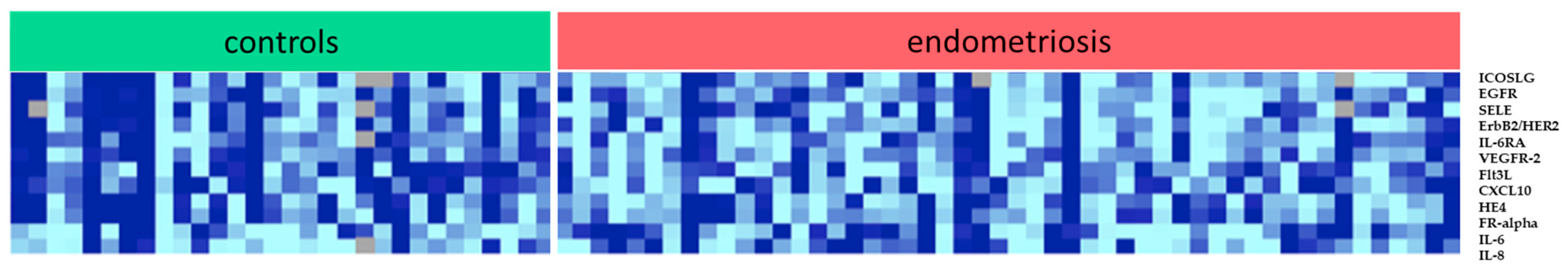
| Total | ICOSLG | 0.72 | 0.56 | 0.78 | 0.027 |
| EGFR | 1.20 | 0.99 | 0.83 | 0.023 | |
| SELE | 1.63 | 1.29 | 0.79 | 0.037 | |
| ErbB2/HER2 | 7.27 | 5.81 | 0.80 | 0.032 | |
| IL-6RA | 2.35 | 1.87 | 0.80 | 0.022 | |
| VEGFR-2 | 3.88 | 3.14 | 0.81 | 0.026 | |
| Flt3L | 37.77 | 28.17 | 0.75 | 0.008 | |
| CXCL10 | 41.80 | 24.43 | 0.58 | 0.032 | |
| HE4 | 68.25 | 42.36 | 0.62 | 0.029 | |
| FR-alpha | 30.51 | 18.25 | 0.60 | 0.049 | |
| IL-6 | 16.55 | 35.53 | 2.13 | 0.045 | |
| IL-8 | 5.69 | 14.52 | 2.56 | 0.045 | |
| Proliferative Cycle Phase (n, %) | 13 (41.9%) | 26 (49.1%) | |||
| Secretory Cycle Phase (n, %) | 18 (58.1%) | 27 (50.9%) | |||
| Target | Geometric Mean of Intensities in | Fold Change in DIE | p-Value | ||
|---|---|---|---|---|---|
| Non-DIE (n = 34) | DIE (n = 19) | ||||
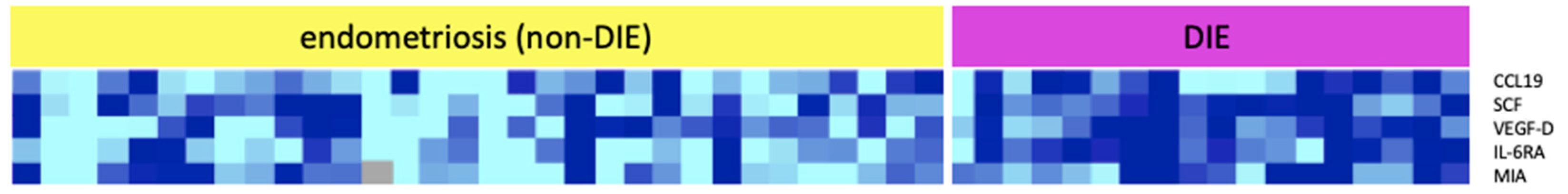
| Total | CCL 19 | 13.97 | 26.53 | 1.90 | 0.038 |
| SCF | 5.57 | 7.42 | 1.33 | 0.033 | |
| VEGF-D | 4.23 | 7.42 | 1.75 | 0.034 | |
| IL-6RA | 1.66 | 2.33 | 1.40 | 0.004 | |
| MIA | 0.94 | 1.24 | 1.32 | 0.040 | |
| Proliferative Cycle Phase (n, %) | 16 (47.1%) | 9 (47.4%) | |||
| Secretory Cycle Phase (n, %) | 18 (52.9%) | 10 (52.6%) | |||
| Target | Involved in: | Relation of Factor in Endometriosis Compared to Controls | Characteristics |
|---|---|---|---|
| Interleukin-6 (IL-6) | IR | ↑ |
|
| Interleukin-8 (IL-8) | IR, AG | ↑ |
|
| Interleukin-6 receptor alpha (IL-6Ralpha) | IR | ↓ |
|
| Inducible Co-Stimulator Ligand (ICOSL) | IR, AA, CP | ↓ |
|
| FMS-like tyrosine kinase 3 ligand (FLT3L) | IR | ↓ |
|
| C-X-C motif chemokine C (CXCL10) | IR | ↓ |
|
| Epidermal growth factor receptor (EGFR) | AA, CP | ↓ |
|
| ErbB2/HER2 | AA, CP | ↓ |
|
| Folate receptor alpha (FR-alpha) | CP | ↓ |
|
| human epididymis protein 4 (HE4) | CP | ↓ |
|
| Vascular endothelial growth factor receptor-2 (VEGFR-2) | AG | ↓ |
|
| Endothelial Selectin (SELE) | CA | ↓ |
|
| Target | Involved in: | Relation of Factor in DIE Compared to Non-DIE | Characteristics |
|---|---|---|---|
| Stem cell factor (SCF) (c-kit ligand) | IR | ↑ |
|
| Interleukin-6 receptor alpha (IL-6R-alpha) | IR | ↑ |
|
| Chemokine ligand 19 (CCL-19) | IR | ↑ |
|
| Melanoma inhibitory activity (MIA) | CP | ↑ |
|
| Vascular endothelial growth factor D (VEGF-D) | AG | ↑ |
|
© 2020 by the authors. Licensee MDPI, Basel, Switzerland. This article is an open access article distributed under the terms and conditions of the Creative Commons Attribution (CC BY) license (http://creativecommons.org/licenses/by/4.0/).
Share and Cite
Perricos, A.; Wenzl, R.; Husslein, H.; Eiwegger, T.; Gstoettner, M.; Weinhaeusel, A.; Beikircher, G.; Kuessel, L. Does the Use of the “Proseek® Multiplex Oncology I Panel” on Peritoneal Fluid Allow a Better Insight in the Pathophysiology of Endometriosis, and in Particular Deep-Infiltrating Endometriosis? J. Clin. Med. 2020, 9, 2009. https://doi.org/10.3390/jcm9062009
Perricos A, Wenzl R, Husslein H, Eiwegger T, Gstoettner M, Weinhaeusel A, Beikircher G, Kuessel L. Does the Use of the “Proseek® Multiplex Oncology I Panel” on Peritoneal Fluid Allow a Better Insight in the Pathophysiology of Endometriosis, and in Particular Deep-Infiltrating Endometriosis? Journal of Clinical Medicine. 2020; 9(6):2009. https://doi.org/10.3390/jcm9062009
Chicago/Turabian StylePerricos, Alexandra, René Wenzl, Heinrich Husslein, Thomas Eiwegger, Manuela Gstoettner, Andreas Weinhaeusel, Gabriel Beikircher, and Lorenz Kuessel. 2020. "Does the Use of the “Proseek® Multiplex Oncology I Panel” on Peritoneal Fluid Allow a Better Insight in the Pathophysiology of Endometriosis, and in Particular Deep-Infiltrating Endometriosis?" Journal of Clinical Medicine 9, no. 6: 2009. https://doi.org/10.3390/jcm9062009