Potential Utility of Neurosonology in Paroxysmal Atrial Fibrillation Detection in Patients with Cryptogenic Stroke

 , , ,
, , ,
Abstract
:1. Introduction
2. Methods
Statistical Analyses
3. Results
4. Discussion
5. Conclusions
Author Contributions
Funding
Conflicts of Interest
References
- Schulz, U.G.; Rothwell, P.M. Differences in vascular risk factors between etiological subtypes of ischemic stroke: Importance of population based studies. Stroke 2003, 34, 2050–2059. [Google Scholar] [CrossRef] [PubMed]
- Tsivgoulis, G.; Patousi, A.; Pikilidou, M.; Birbilis, T.; Katsanos, A.H.; Mantatzis, M.; Asimis, A.; Papanas, N.; Skendros, P.; Terzoudi, A.; et al. Stroke incidence and outcomes in Northeastern Greece: The Evros stroke registry. Stroke 2018, 49, 288–295. [Google Scholar] [CrossRef] [PubMed]
- Adams, H.P., Jr.; Bendixen, B.H.; Kappelle, L.J.; Biller, J.; Love, B.B.; Gordon, D.L.; Marsh, E.E., 3rd. Classification of subtype of acute ischemic stroke. Definitions for use in a multicenter clinical trial. TOAST. Trial of Org 10172 in Acute Stroke Treatment. Stroke 1993, 24, 35–41. [Google Scholar] [CrossRef] [PubMed]
- Murtagh, B.; Smalling, R.W. Cardioembolic stroke. Curr. Atheroscler. 2006, 8, 310–316. [Google Scholar] [CrossRef]
- Khoo, C.W.; Lip, G.Y. Clinical outcomes of acute stroke patients with atrial fibrillation. Expert. Rev. Cardiovasc. Ther. 2009, 7, 371–374. [Google Scholar] [CrossRef]
- Arboix, A.; Vericat, M.C.; Pujades, R.; Massons, J.; García-Eroles, L.; Oliveres, M. Cardioembolic infarction in the Sagrat Cor-Alianza Hospital of Barcelona Stroke Registry. Acta Neurol. Scand. 1997, 96, 407–412. [Google Scholar] [CrossRef]
- Lip, G.Y.; Hee, F.L. Paroxysmal atrial fibrillation. QJM 2001, 94, 665–678. [Google Scholar] [CrossRef]
- Kishore, A.; Vail, A.; Majid, A.; Dawson, J.; Lees, K.R.; Tyrrell, P.J.; Smith, C.J. Detection of atrial fibrillation after ischemic stroke or transient ischemic attack: A systematic review and meta-analysis. Stroke 2014, 45, 520–526. [Google Scholar] [CrossRef]
- Kernan, W.N.; Ovbiagele, B.; Black, H.R.; Bravata, D.M.; Chimowitz, M.I.; Ezekowitz, M.D.; Fang, M.C.; Fisher, M.; Furie, K.L.; Heck, D.V. Guidelines for the prevention of stroke in patients with stroke and transient ischemic attack: A guideline for healthcare professionals from the American Heart Association/American Stroke Association. Stroke 2014, 45, 2160–2236. [Google Scholar] [CrossRef]
- EAFT (European Atrial Fibrillation Trial) Study Group. Secondary prevention in non-rheumatic atrial fibrillation after transient ischaemic attack or minor stroke. Lancet 1993, 342, 1255–1262. [Google Scholar] [CrossRef]
- Hart, R.G.; Benavente, O.; McBride, R.; Pearce, L.A. Antithrombotic therapy to prevent stroke in patients with atrial fibrillation: A meta-analysis. Ann. Intern. Med. 1999, 131, 492–501. [Google Scholar] [CrossRef] [PubMed]
- European Stroke Organisation (ESO); Executive Committee; ESO Writing Committee. Guidelines for management of ischaemic stroke and transient ischaemic attack 2008. Cerebrovasc Dis. 2008, 25, 457–507. [Google Scholar] [CrossRef] [PubMed]
- Masdeu, J.C.; Irimia, P.; Asenbaum, S.; Bogousslavsky, J.; Brainin, M.; Chabriat, H.; Herholz, K.; Markus, H.S.; Martínez-Vila, E.; Niederkorn, K.; et al. EFNS guideline on neuroimaging in acute stroke. Report of an EFNS task force. Eur. J. Neurol. 2006, 13, 1271–1283. [Google Scholar] [CrossRef] [PubMed]
- Qureshi, A.I.; Alexandrov, A.V.; Tegeler, C.H.; Hobson, R.W.; Dennis Baker, J.; Hopkins, L.N.; American Society of Neuroimaging; Society of Vascular and Interventional Neurology. Guidelines for screening of extracranial carotid artery disease: A statement for healthcare professionals from the multidisciplinary practice guidelines committee of the American Society of Neuroimaging; cosponsored by the Society of Vascular and Interventional Neurology. J. Neurol. 2007, 17, 19–47. [Google Scholar]
- Alexandrov, A.V.; Sloan, M.A.; Wong, L.K.; Douville, C.; Razumovsky, A.Y.; Koroshetz, W.J.; Kaps, M.; Tegeler, C.H.; American Society of Neuroimaging Practice Guidelines Committee. Practice standards for transcranial Doppler ultrasound: Part I--test performance. J. Neurol. 2007, 17, 11–18. [Google Scholar]
- Tsivgoulis, G.; Alexandrov, A.V.; Sloan, M.A. Advances in transcranial doppler ultrasonography. Curr. Neurol. Neurosci. Rep. 2009, 9, 46–54. [Google Scholar] [CrossRef]
- Alexandrov, A.V.; Sloan, M.A.; Tegeler, C.H.; Newell, D.N.; Lumsden, A.; Garami, Z.; Levy, C.R.; Wong, L.K.; Douville, C.; Kaps, M.; et al. Practice standards for transcranial Doppler (TCD) ultrasound. Part II. Clinical indications and expected outcomes. J. Neurol. 2012, 22, 215–224. [Google Scholar]
- Liantinioti, C.; Tympas, K.; Katsanos, A.H.; Parissis, J.; Chondrogianni, M.; Zompola, C.; Papadimitropoulos, G.; Ioakeimidis, M.; Triantafyllou, S.; Roussopoulou, A.; et al. Duration of paroxysmal atrial fibrillation in cryptogenic stroke is not associated with stroke severity and early outcomes. J. Neurol. Sci. 2017, 376, 191–195. [Google Scholar] [CrossRef]
- Katsanos, A.H.; Bhole, R.; Frogoudaki, A.; Giannopoulos, S.; Goyal, N.; Vrettou, A.R.; Ikonomidis, I.; Paraskevaidis, I.; Pappas, K.; Parissis, J.; et al. The value of transesophageal echocardiography for embolic strokes of undetermined source. Neurology 2016, 87, 988–995. [Google Scholar] [CrossRef]
- National Institute of Health, National Institute of Neurological Disorders and Stroke. Stroke Scale. Available online: https://www.ninds.nih.gov/sites/default/ files/ NIH_Stroke_ Scale_Booklet.pdf (accessed on 3 September 2019).
- Tsivgoulis, G.; Kargiotis, O.; Katsanos, A.H.; Patousi, A.; Mavridis, D.; Tsokani, S.; Pikilidou, M.; Birbilis, T.; Mantatzis, M.; Zompola, C.; et al. Incidence, characteristics and outcomes in patients with embolic stroke of undetermined source: A population-based study. J. Neurol. Sci. 2019, 401, 5–11. [Google Scholar] [CrossRef]
- Ikonomidis, I.; Frogoudaki, A.; Vrettou, A.R.; Andreou, I.; Palaiodimou, L.; Katogiannis, K.; Liantinioti, C.; Vlastos, D.; Zervas, P.; Varoudi, M.; et al. Impaired Arterial Elastic Properties and Endothelial Glycocalyx in Patients with Embolic Stroke of Undetermined Source. Thromb. Haemost. 2019. [Epub ahead of print]. [Google Scholar] [CrossRef] [PubMed]
- Lang, R.M.; Bierig, M.; Devereux, R.B.; Flachskampf, F.A.; Foster, E.; Pellikka, P.A.; Picard, M.H.; Roman, M.J.; Seward, J.; Shanewise, J.S.; et al. Recommendations for chamber quantification: A report from the American Society of Echocardiography’s Guidelines and Standards Committee and the Chamber Quantification Writing Group, developed in conjunction with the European Association of Echocardiography, a branch of the European Society of Cardiology. J. Am. Soc. Echocardiogr. 2005, 18, 1440–1463. [Google Scholar] [PubMed]
- Heart, R.S.; Zipes, D.P.; Camm, A.J.; Borggrefe, M.; Buxton, A.E.; Chaitman, B.; Fromer, M.; Gregoratos, G.; Klein, G.; Moss, A.J.; et al. ACC/AHA/ESC 2006 Guidelines for the Management of Patients with Atrial Fibrillation: A report of the American College of Cardiology/American Heart Association Task Force on Practice Guidelines and the European Society of Cardiology Committee for Practice Guidelines (Writing Committee to Revise the 2001 Guidelines for the Management of Patients With Atrial Fibrillation): Developed in collaboration with the European Heart Rhythm Association and the Heart Rhythm Society. Circulation 2006, 114, e257–e354. [Google Scholar]
- Liao, J.; Khalid, Z.; Scallan, C.; Morillo, C.; O’Donnell, M. Noninvasive cardiac monitoring for detecting paroxysmal atrial fibrillation or flutter after acute ischemic stroke: A systematic review. Stroke 2007, 38, 2935–2940. [Google Scholar] [CrossRef]
- Sanna, T.; Diener, H.C.; Passman, R.S.; Di Lazzaro, V.; Bernstein, R.A.; Morillo, C.A.; Rymer, M.M.; Thijs, V.; Rogers, T.; Beckers, F.; et al. Cryptogenic Stroke and underlying Atrial Fibrillation. N. Engl. J. Med. 2014, 370, 2478–2486. [Google Scholar] [CrossRef]
- Flint, A.C.; Banki, N.M.; Ren, X.; Rao, V.A.; Go, A.S. Detection of paroxysmal atrial fibrillation by 30-day event monitoring in cryptogenic ischemic stroke: The Stroke and Monitoring for PAF in Real Time (SMART) Registry. Stroke 2012, 43, 2788–2790. [Google Scholar] [CrossRef]
- Seet, R.C.; Friedman, P.A.; Rabinstein, A.A. Prolonged rhythm monitoring for the detection of occult paroxysmal atrial fibrillation in ischemic stroke of unknown cause. Circulation 2011, 26, 477–486. [Google Scholar] [CrossRef]
- Choe, W.C.; Passman, R.S.; Brachmann, J.; Morillo, C.A.; Sanna, T.; Bernstein, R.A.; Di Lazzaro, V.; Diener, H.C.; Rymer, M.M.; Beckers, F.; et al. A Comparison of Atrial Fibrillation Monitoring Strategies After Cryptogenic Stroke (from the Cryptogenic Stroke and Underlying AF Trial). Am. J. Cardiol. 2015, 116, 889–893. [Google Scholar] [CrossRef]
- Hylek, E.M.; Go, A.S.; Chang, Y.; Jensvold, N.G.; Henault, L.E.; Selby, J.V.; Singer, D.E. Effect of intensity of oral anticoagulation on stroke severity and mortality in atrial fibrillation. N. Engl. J. Med. 2003, 349, 1019–1026. [Google Scholar] [CrossRef]
- Evans, A.; Perez, I.; Yu, G.; Kalra, L. Secondary stroke prevention in atrial fibrillation: Lessons from clinical practice. Stroke 2000, 31, 2106–2111. [Google Scholar] [CrossRef]
- Nieuwlaat, R.; Prins, M.H.; Le Heuzey, J.Y.; Vardas, P.E.; Aliot, E.; Santini, M.; Cobbe, S.M.; Widdershoven, J.W.; Baur, L.H.; Lévy, S.; et al. Prognosis, disease progression, and treatment of atrial fibrillation patients during 1 year: Follow-up of the Euro Heart Survey on atrial fibrillation. Eur. Heart J. 2008, 29, 1181–1189. [Google Scholar] [CrossRef] [PubMed]
- Puccio, D.; Novo, G.; Baiamonte, V.; Nuccio, A.; Fazio, G.; Corrado, E.; Coppola, G.; Muratori, I.; Vernuccio, L.; Novo, S. Atrial fibrillation and mild cognitive impairment: What correlation? Minerva Cardioangiol. 2009, 57, 143–150. [Google Scholar] [PubMed]
- Coppola, G.; Manno, G.; Mignano, A.; Luparelli, M.; Zarcone, A.; Novo, G.; Corrado, E. Management of Direct Oral Anticoagulants in Patients with Atrial Fibrillation Undergoing Cardioversion. Medicina 2019, 55, 660. [Google Scholar] [CrossRef] [PubMed]
- Van Walraven, C.; Hart, R.G.; Singer, D.E.; Laupacis, A.; Connolly, S.; Petersen, P.; Koudstaal, P.J.; Chang, Y.; Hellemons, B. Oral anticoagulants vs aspirin in nonvalvular atrial fibrillation: An individual patient meta-analysis. JAM 2002, 288, 2441–2448. [Google Scholar] [CrossRef]
- Elijovich, L.; Josephson, S.A.; Fung, G.L.; Smith, W.S. Intermittent atrial fibrillation may account for a large proportion of otherwise cryptogenic stroke: A study of 30-day cardiac event monitors. J. Stroke Cerebrovasc. Dis. 2009, 18, 185–189. [Google Scholar] [CrossRef]
- Ziegler, P.D.; Glotzer, T.V.; Daoud, E.G.; Wyse, D.G.; Singer, D.E.; Ezekowitz, M.D.; Koehler, J.L.; Hilker, C.E. Incidence of newly detected atrial arrhythmias via implant-able devices in patients with a history of thromboembolic events. Stroke 2010, 41, 256–260. [Google Scholar] [CrossRef] [Green Version]
- Gaillard, N.; Deltour, S.; Vilotijevic, B.; Hornych, A.; Crozier, S.; Leger, A.; Frank, R.; Samson, Y. Detection of paroxysmal atrial fibrillation with transtelephonic EKG in TIA or stroke patients. Neurology 2010, 74, 1666–1670. [Google Scholar] [CrossRef]
- Alhadramy, O.; Jeerakathil, T.J.; Majumdar, S.R.; Najjar, E.; Choy, J.; Saqqur, M. Prevalence and predictors of paroxysmal atrial fibrillation on Holter monitor in patients with stroke or transient ischemic attack. Stroke 2010, 41, 2596–2600. [Google Scholar] [CrossRef] [Green Version]
- Favilla, C.G.; Ingala, E.; Jara, J.; Fessler, E.; Cucchiara, B.; Messé, S.R.; Mullen, M.T.; Prasad, A.; Siegler, J.; Hutchinson, M.D.; et al. Predictors of finding occult atrial fibrillation after cryptogenic stroke. Stroke 2015, 46, 1210–1215. [Google Scholar] [CrossRef] [Green Version]
- Tsang, T.S.; Abhayaratna, W.P.; Barnes, M.E.; Miyasaka, Y.; Gersh, B.J.; Bailey, K.R.; Cha, S.S.; Seward, J.B. Prediction of cardiovascular outcomes with left atrial size: Is volume superior to area or diameter? J. Am. Coll. Cardiol. 2006, 47, 1018–1023. [Google Scholar] [CrossRef] [Green Version]
- Jordan, K.; Yaghi, S.; Poppas, A.; Chang, A.D.; Mac Grory, B.; Cutting, S.; Burton, T.; Jayaraman, M.; Tsivgoulis, G.; Sabeh, M.K.; et al. Left Atrial Volume Index Is Associated With Cardioembolic Stroke and Atrial Fibrillation Detection After Embolic Stroke of Undetermined Source. Stroke 2019, 50, 1997–2001. [Google Scholar] [CrossRef] [PubMed]
- Hart, R.G.; Diener, H.C.; Coutts, S.B.; Easton, J.D.; Granger, C.B.; O’Donnell, M.J.; Sacco, R.L.; Connolly, S.J.; Cryptogenic Stroke/ESUS International Working Group. Embolic strokes of undetermined source: The case for a new clinical construct. Lancet Neurol. 2014, 13, 429–438. [Google Scholar] [CrossRef]
- Tsivgoulis, G.; Katsanos, A.H.; Köhrmann, M.; Caso, V.; Lemmens, R.; Tsioufis, K.; Paraskevas, G.P.; Bornstein, N.M.; Schellinger, P.D.; Alexandrov, A.V.; et al. Embolic strokes of undetermined source: Theoretical construct or useful clinical tool? Ther. Adv. Neurol. Disord. 2019, 12, 1756286419851381. [Google Scholar] [CrossRef] [PubMed] [Green Version]
- Hansson, A.; Madsen-Härdig, B.; Olsson, S.B. Arrhythmia-provoking factors and symptoms at the onset of paroxysmal atrial fibrillation: A study based on interviews with 100 patients seeking hospital assistance. BMC Cardiovasc. Disord. 2004, 4, 13. [Google Scholar] [CrossRef] [PubMed] [Green Version]
- Severino, P.; Mariani, M.V.; Maraone, A.; Piro, A.; Ceccacci, A.; Tarsitani, L.; Maestrini, V.; Mancone, M.; Lavalle, C.; Pasquini, M.; et al. Triggers for Atrial Fibrillation: The Role of Anxiety. Cardiol. Res. Pract. 2019, 2019, 1208505. [Google Scholar] [CrossRef] [PubMed]
- Khurshid, S.; Choi, S.H.; Weng, L.C.; Wang, E.Y.; Trinquart, L.; Benjamin, E.J.; Ellinor, P.T.; Lubitz, S. Frequency of Cardiac Rhythm Abnormalities in a Half Million Adults. Circ. Arrhythm. Electrophysiol. 2018, 11, e006273. [Google Scholar] [CrossRef]
- Hamer, M.E.; Wilkinson, W.E.; Clair, W.K.; Page, R.L.; McCarthy, E.A.; Pritchett, E.L. Incidence of symptomatic atrial fibrillation in patients with paroxysmal supraventricular tachycardia. J. Am. Coll. Cardiol. 1995, 25, 984–988. [Google Scholar] [CrossRef] [Green Version]
- Kwong, C.; Ling, A.Y.; Crawford, M.H.; Zhao, S.X.; Shah, N.H. A Clinical Score for Predicting Atrial Fibrillation in Patients with Cryptogenic Stroke or Transient Ischemic Attack. Cardiology 2017, 138, 133–140. [Google Scholar] [CrossRef]
- Lip, G.Y.; Nieuwlaat, R.; Pisters, R.; Lane, D.A.; Crijns, H.J. Refining clinical risk stratification for predicting stroke and thromboembolism in atrial fibrillation using a novel risk factor-based approach: The euro heart survey on atrial fibrillation. Chest 2010, 137, 263–272. [Google Scholar] [CrossRef]


| Variable | Overall |
|---|---|
| Age, years (mean ± SD) | 60 ± 11 |
| Female sex (%) | 122 (33%) |
| NIHSS-Score, points (median, IQR) | 4 (3–10) |
| Hypertension (%) | 230 (62%) |
| Diabetes (%) | 82 (22%) |
| Hyperlipidemia (%) | 215 (58%) |
| Current Smoking (%) | 158 (22.5%) |
| Coronary Artery Disease (%) | 58 (16%) |
| Excessive Alcohol Intake (%) | 37 (10%) |
| Previous History of TIA or Stroke (%) | 74 (20%) |
| Heart Failure (%) | 17 (5%) |
| Peripheral Arterial Disease (%) | 15 (4%) |
| Vascular Disease (%) | 70 (19%) |
| CHA2DS2-VASc Score, Points (mean ± SD) | 3.8 ± 1.3 |
| Left Atrial Enlargement (%) | 155 (42%) |
| Mild | 133 (36%) |
| Moderate | 17 (5%) |
| Severe | 5 (1%) |
| Cortical Location of Infarction (%) | 76 (20%) |
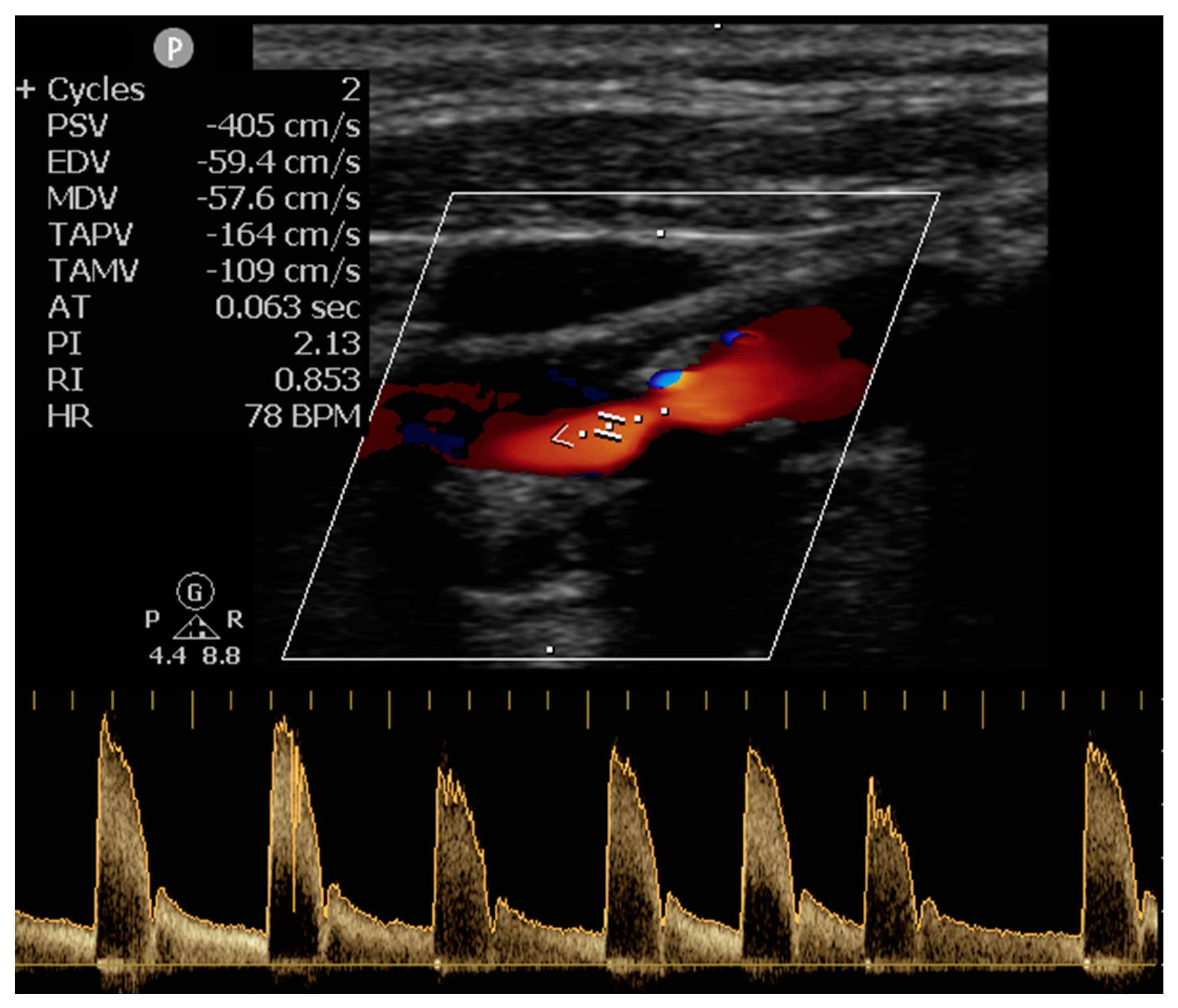
| Cardiac Arrhythmia Detected during Neurosonology Evaluation (%) | 66 (18%) |
| Number of 24-h Holter Recordings (mean ± SD) | 1.5 ± 1.5 |
| 1 | 254 (68%) |
| 2 | 85 (23%) |
| ≥3 | 34 (9%) |
| Left Atrial Enlargement | Atrial Fibrillation (−) | Atrial Fibrillation (+) | p-Value * | p-Value for Linear Trend ** |
|---|---|---|---|---|
| None (%) | 93% | 7% | <0.001 | <0.001 |
| Mild (%) | 88% | 12% | ||
| Moderate or Severe (%) | 64% | 36% |
| Number of 24-h Holter ECG Recordings | Atrial Fibrillation (−) | Atrial Fibrillation (+) | p Value * | p-Value for Linear Trend ** |
|---|---|---|---|---|
| 1 (%) | 92% | 8% | 0.048 | 0.014 |
| 2 (%) | 86% | 14% | ||
| ≥3 (%) | 79% | 21% |
| Univariable Logistic Regression Analysis | Multivariable Logistic Regression Analysis | |||
|---|---|---|---|---|
| Variable | Odds Ratio (95%CI) | p * | Odds Ratio (95%CI) | p |
| Age (per 10-year increase) | 1.81 (1.31–2.50) | <0.001 | 1.68 (1.19–2.37) | 0.003 |
| Female Sex | 1.43 (0.73–2.80) | 0.300 | ||
| NIHSS-Score at Admission (per 1-point increase) | 0.97 (0.91–1.03) | 0.295 | ||
| Hypertension | 1.51 (0.74–3.08) | 0.254 | ||
| Diabetes Mellitus | 1.40 (0.67–2.94) | 0.374 | ||
| Hyperlipidemia | 1.60 (0.80–3.21) | 0.185 | ||
| Previous History of TIA or Stroke | 1.20 (0.54–2.64) | 0.655 | ||
| Coronary Artery Disease | 1.17 (0.49–2.80) | 0.719 | ||
| Congestive Heart Failure | 2.74 (0.85–8.83) | 0.093 | 2.74 (0.85–8.83) | 0.165 |
| Current Smoking | 1.22 (0.63–2.35) | 0.558 | ||
| Excessive Alcohol Intake | 1.34 (0.49–3.67) | 0.565 | ||
| Peripheral Arterial Disease | 1.30 (0.28–5.96) | 0.739 | ||
| Vascular Disease | 1.09 (0.48–2.49) | 0.833 | ||
| CHA2DS2-VASc Score (per 1-point increase) | 1.53 (1.20–1.94) | 0.001 | 1.15 (0.80–1.66) | 0.451 |
| ≥3 (24-h) Holter Evaluations | 2.40 (0.97–5.95) | 0.058 | 1.62 (0.58–4.52) | 0.354 |
| Cortical Location of Infarction | 1.35 (0.63–2.90) | 0.444 | ||
| Cardiac Arrhythmia Detected during Neurosonology Evaluation | 3.77 (1.87–7.60) | <0.001 | 3.09 (1.47–6.48) | 0.003 |
| Moderate or Severe Left Atrial Enlargement | 5.70 (2.22–14.61) | <0.001 | 4.81 (1.77–13.03) | 0.002 |
© 2019 by the authors. Licensee MDPI, Basel, Switzerland. This article is an open access article distributed under the terms and conditions of the Creative Commons Attribution (CC BY) license (http://creativecommons.org/licenses/by/4.0/).
Share and Cite
Liantinioti, C.; Palaiodimou, L.; Tympas, K.; Parissis, J.; Theodorou, A.; Ikonomidis, I.; Chondrogianni, M.; Zompola, C.; Triantafyllou, S.; Roussopoulou, A.; et al. Potential Utility of Neurosonology in Paroxysmal Atrial Fibrillation Detection in Patients with Cryptogenic Stroke. J. Clin. Med. 2019, 8, 2002. https://doi.org/10.3390/jcm8112002
Liantinioti C, Palaiodimou L, Tympas K, Parissis J, Theodorou A, Ikonomidis I, Chondrogianni M, Zompola C, Triantafyllou S, Roussopoulou A, et al. Potential Utility of Neurosonology in Paroxysmal Atrial Fibrillation Detection in Patients with Cryptogenic Stroke. Journal of Clinical Medicine. 2019; 8(11):2002. https://doi.org/10.3390/jcm8112002
Chicago/Turabian StyleLiantinioti, Chrissoula, Lina Palaiodimou, Konstantinos Tympas, John Parissis, Aikaterini Theodorou, Ignatios Ikonomidis, Maria Chondrogianni, Christina Zompola, Sokratis Triantafyllou, Andromachi Roussopoulou, and et al. 2019. "Potential Utility of Neurosonology in Paroxysmal Atrial Fibrillation Detection in Patients with Cryptogenic Stroke" Journal of Clinical Medicine 8, no. 11: 2002. https://doi.org/10.3390/jcm8112002