Article

Etiology of Minor Troponin Elevations in Patients with Atrial Fibrillation at Emergency Department–Tropo-AF Study

1
Heart Center, Turku University Hospital and University of Turku, 20521 Turku, Finland
2
Department of Acute Internal Medicine, Turku University Hospital and University of Turku, 20521 Turku, Finland
3
Cardiovascular Medicine, Brigham and Women’s Hospital, Harvard Medical School, Boston, MA 02115, USA
4
Research Unit of Surgery, Anesthesiology and Critical Care, University of Oulu, 90014 Oulu, Finland
*
Author to whom correspondence should be addressed.
Equal contribution.
J. Clin. Med. 2019, 8(11), 1963; https://doi.org/10.3390/jcm8111963
Submission received: 16 October 2019 / Revised: 31 October 2019 / Accepted: 10 November 2019 / Published: 14 November 2019
(This article belongs to the Section Cardiology)

Abstract

:
Patients with atrial fibrillation (AF) presenting to the emergency department (ED) often have elevated cardiac troponin T (TnT) levels without evidence of type 1 myocardial infarction. We sought to explore the causes and significance of minor TnT elevations in patients with AF at the ED. All patients with AF admitted to the ED of Turku University Hospital between 1 March, 2013 and 11 April, 2016, and at least two TnT measurements, were screened. Overall, 2911 patients with a maximum TnT of 100 ng/L during hospitalization were analyzed. TnT was between 15 and 100 ng/L in 2116 patients. The most common primary discharge diagnoses in this group were AF (18.1%), infection (18.3%), ischemic stroke/transient ischemic attack (10.7%), and heart failure (5.0%). Acute coronary syndrome (ACS) was equally uncommon both in patients with normal TnT and elevated TnT (4.4% vs. 4.5%). Age ≥75 years, low estimated glomerular filtration rate (eGFR), high C-reactive protein (CRP), and hemoglobin <10.0 g/dL, were the most important predictors of elevated TnT. Importantly, TnT elevation was a very frequent (>93%) finding in elderly (≥75 years) AF patients with either low eGFR or high CRP. In conclusion, minor TnT elevations carry limited diagnostic value in elderly AF patients with comorbidities.

1. Introduction

Approximately 10–40% of patients with atrial fibrillation (AF) are hospitalized annually and this proportion is expected to rise in the near future [1]. Cardiac troponin measurements have become a part of routine laboratory work-ups in the emergency department (ED) in various clinical scenarios. A common diagnostic challenge with AF patients is the exclusion of type 1 myocardial infarction (MI), referring to infarction caused by plaque rupture, erosion, and/or dissection in coronary arteries, as opposed to type 2 MI, which refers to a mismatch between oxygen supply and demand [2]. The advent of high-sensitive troponin assays enables early detection of acute MI, however, at a cost of false positive diagnoses. Minor troponin elevations are common in a wide variety of medical conditions, such as systemic infection, pulmonary embolism, renal failure, cerebrovascular accident, heart failure, and even after extreme physical stress [3,4,5,6,7,8,9,10]. This multiplicity of causes is problematic in acute care decision-making when the main goal is to detect or rule out type 1 MI as the cause of the ED visit and troponin elevations.
Troponin elevation is common in AF [11,12,13] and patients with AF are a common patient group at the ED. Since little is known about the causes of mild troponin elevations in this scenario, we sought to assess the frequency of various discharge diagnoses in AF patients with minor (15–100 ng/L) cardiac high-sensitivity troponin T (TnT) elevations. Secondly, we sought factors differentiating AF patients with the clinical diagnosis of acute coronary syndrome (ACS) from patients with other causes for minor TnT elevations.

2. Materials and Methods

This study reports the main results from the Troponins in Atrial Fibrillation study (The Tropo-AF Study, ClinicalTrials.gov Identifier: NCT03683836), which belongs to a series of study protocols assessing clinical challenges in the treatment of AF [5,14,15,16].
The study protocol was approved by the Medical Ethics Committee of the Hospital District of Southwest Finland. Informed consent was not required because of the observational nature of the study. The study complies with the Declaration of Helsinki as revised in 2002.
Initially, database searches were performed to identify patients whose TnT levels were measured between 1 March, 2013 and 11 April, 2016 at the Turku University Hospital laboratory. This initial screening lead to a total of 145,837 individual TnT measurements. Patients with a prior diagnosis of AF (ICD-10 code I48) or AF diagnosed at the index visit, living in the hospital catchment area, with an admission electrocardiography (ECG), and two serial TnT measurements within 8–72 h after admission to the ED, were identified. Thereafter, patients with maximum TnT values < 100 ng/L during the entire hospitalization were selected for the present analysis. Dynamic TnT change was defined as the difference between highest and lowest TnT values recorded during hospitalization, in percentages. Admission ECGs were screened for ST-depression ≥1 mm in two adjacent leads.
The diagnosis of AF (either made before or at the time of the ED visit) was confirmed by 12-lead ECG findings. All individual electronic patient records were reviewed by the investigation group using a standardized protocol to collect specific information on the clinical characteristics of patients, clinical conditions, as well as other clinical and laboratory data of interest from the index hospitalization. Mortality data were retrieved from the national statistical institution, Statistics Finland, and therefore we assumed that no patient was missing at follow-up.
The primary measure of this analysis was an increase of TnT levels between 15 and 100 ng/L during the hospital visit. TnT was analyzed using a commercial high-sensitive assay (Roche Diagnostics GmbH, Mannheim, Germany) at the hospital’s certified laboratory. Determined by the manufacturer, the 99th percentile upper reference limit was 14 ng/L for the assay. Two study groups were formed on the basis of maximum TnT values—patients with normal (<15 ng/L) TnT (n = 795) and those with minor (15–100 ng/L) TnT elevations (n = 2116).
This article was prepared following the STROBE guidelines [17] for the reporting of observational studies.

Statistical Analysis
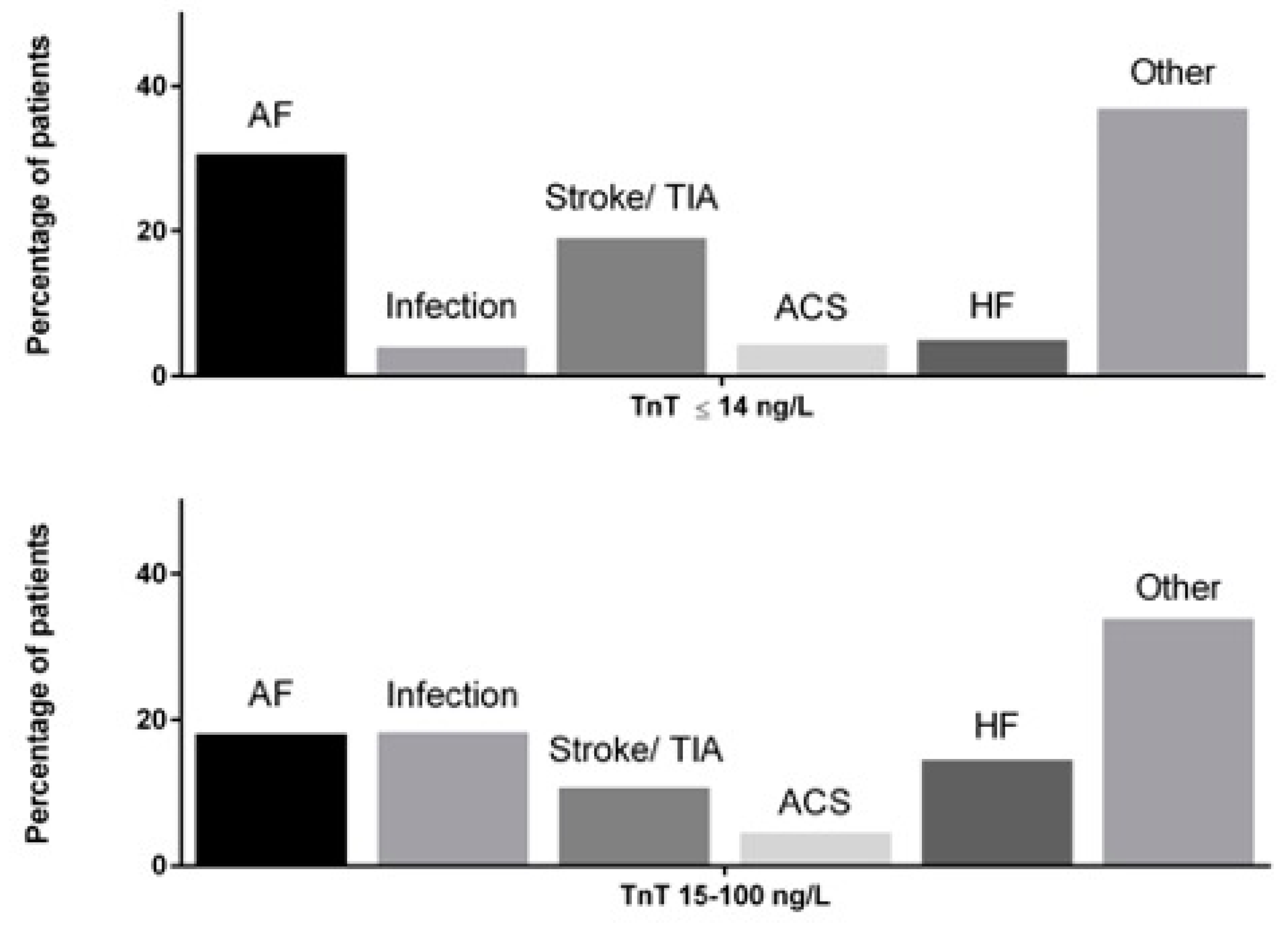
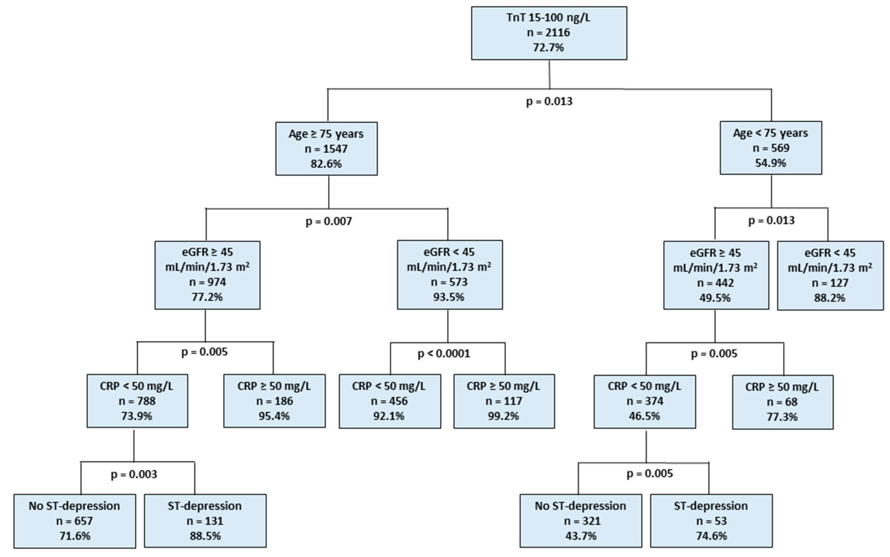
Statistical analyses were performed using SPSS v.25.0 statistical software (IBM Corporation, New York, NY, USA). Continuous variables are reported as mean and standard deviation (SD) if normally distributed, and as median and interquartile range (IQR) if skewed, unless stated otherwise. Missing data were not replaced. Categorical variables are described as counts and percentages. The chi-square test and Fisher’s exact test were used for categorical variables, as appropriate. Logistic regression analysis using the backward Wald method was performed to identify independent predictors of TnT elevations, as well as ACS in patients with elevated TnT. Covariates with p < 0.1 in univariate analyses were entered into the regression model. All tests were two-sided and statistical significance was set at 5%.
Classification and regression tree (CART) analysis was performed to identify independent risk factors for TnT elevations. The classification tree procedure was assessed by cross-validation through 25-fold The minimum number of patients for the parent node was set at 100 and the minimum for the child node was 50. Gini’s method was used to measure impurity, which is the extent to which a node does not represent a homogenous subset of cases. A minimum change in improvement was set at 0.0001. Normalized importance to the model of the included covariates was estimated. Covariates, which were identified as independent risk factors for TnT 15–100 ng/L in logistic regression and with normalized importance >20%, were included in the final CART model. The area under the receiver operating characteristic curve (AUC) was estimated to assess the regression model discrimination.

3. Results

A total of 2911 AF patients were included in the study and 72.7% (n = 2116) of them had minor TnT elevation, ranging from 15 to 100 ng/L. Patient characteristics according to maximum TnT levels are presented in Table 1. The most common symptoms at admission were any kind of chest pain (513 cases, 17.6%), dyspnea (823 cases, 28.3%), and malaise/nausea/dizziness (629 cases, 21.6%). At hospital discharge, the most common primary diagnostic categories were AF (21.6%, n = 628), infections (14.4%, n = 420), ischemic stroke/transient ischemic attack (13.0%, n = 378), decompensated heart failure (11.9%, n = 346), and ACS (4.5%, n = 130), comprising 65.3% of all hospital admissions (Figure 1 and Table S1). Other frequent primary diagnoses were bone fractures, trauma, intoxications, bleeding, renal insufficiency, inflammatory diseases, and various symptomatic diagnoses.
Importantly, ACS was an equally uncommon primary discharge diagnosis, both in patients with normal TnT and minor TnT elevations in the ED (4.4%, n = 35 vs. 4.5%, n = 95, respectively). AF was the most common (30.7%, n = 244) primary discharge diagnosis of the index hospital visit in patients with normal TnT, whereas severe infections (18.3%, n = 388) and AF (18.1%, n = 384) were the most frequent diagnoses at hospital discharge in patients with minor TnT elevations (Figure 1).
Chest pain was reported by 27.0% (n = 215) of patients with normal TnT, while only 14.1% (n = 298) of those with minor TnT elevations reported chest pain. Among patients with ACS, nearly two-thirds (65.4%, n = 85) experienced chest pain, but only 12.8% (n = 38) of patients with chest pain and minor TnT rises were diagnosed with ACS. ST-depressions were observed in 10.7% of patients with ACS and 13.2% of those with other primary diagnoses. A coronary angiogram was performed in 3.0% (n = 87) of all study patients and in 24.6% (n = 32) of patients with the final diagnosis of ACS. Coronary angiography revealed significant coronary artery disease (>50% stenosis) in 31 (96.9%) ACS patients, but resulted in percutaneous coronary intervention in 18 (20.7%) patients, including two patients with normal TnT values. Patients were selected for coronary angiography based on clinical evaluation by the attending physician.
The strongest independent predictors of minor TnT elevation in the whole patient cohort were age ≥ 75 years, estimated glomerular filtration rate (eGFR) < 45 mL/min/1.73 m2, C-reactive protein (CRP) ≥ 50 mg/L, and hemoglobin < 10.0 g/dL (Table 2). CART analysis showed that nearly all (>93%) patients ≥ 75 years of age with either high CRP or low eGFR had elevated TnT concentrations (Figure 2). Age ≥ 75 years (normalized importance, 100%), eGFR < 45 mL/min/1.73 m2 (normalized importance, 57.6%), CRP ≥ 50 mg/L (normalized importance, 30.5%), and ST depression ≥1 mm (normalized importance, 21.0%), were included in the final CART model. The AUC of this CART model was 0.763 (95% CI, 0.745–0.782). A CART model not including patients with a diagnosis of ACS confirmed the findings of the overall cohort (AUC 0.763, 95% CI, 0.744–0.782).
In patients with elevated TnT, chest pain (OR 8.29, 95% CI, 5.20–13.2), previous coronary artery disease (OR 2.33, 95% CI, 1.42–3.81), and low eGFR (OR 1.65, 95% CI, 1.04–2.62) were the independent predictors for the final diagnosis of ACS (Table 3), but only 24.1% of minor TnT elevations in patients with previous coronary artery disease and chest pain were considered to be caused by ACS. A change of at least 100% in absolute TnT value was observed in 12 (12.6%) patients with ACS and in 146 (7.2%) without ACS, p = 0.068.
The 30-day mortality was significantly higher in the TnT elevation group compared to the normal TnT group, both in the whole study cohort and in patients with ACS (8.9% vs. 0.9% and 8.4% vs. 2.9%, p < 0.001 for both). One (0.05%) patient with elevated TnT and no ACS diagnosis during the index hospitalization was treated for ACS during the 30-day follow-up.

4. Discussion

Our study demonstrated that minor TnT elevation is a common finding in AF patients presenting at the ED with a wide variety of acute symptoms. Strikingly, ACS or type 1 MI was considered a rare cause of minor TnT elevations, comprising less than one out of twenty patients. Increasing age and multiple co-morbidities were often associated with minor TnT elevations; severe infections, cerebrovascular events, AFs, and decompensated heart failure were the most common final diagnoses at discharge. Most patients with minor TnT elevations were admitted to hospital and minor TnT elevation was a powerful predictor of short-term mortality regardless of the final diagnosis. Importantly, minor TnT elevation by itself did not predict a new ED admission due to ACS within 30 days.
The use of high-sensitivity troponin assays enables safe and robust early exclusion of acute MI in the ED [18,19]. High-sensitivity assays also allow the use of lower thresholds for the diagnosis of type 1 MI. The use of this improved sensitivity, however, more than doubles the number of patients with type 2 MI or myocardial injury, but does not seem to improve the prognosis of this new patient group with minor troponin elevations [20]. The present results show that the recommended low rule-out threshold for exclusion of type 1 MI is problematic in clinical practice if applied to patients in whom the degree of MI suspicion is low and there are other factors that might contribute to troponin concentration.
At present, cardiac troponin testing is frequently used in the ED in the absence of symptoms suggestive of ACS [21]. In line with earlier reports [22], the vast majority of minor TnT elevations in this scenario reflected myocardial injury or type 2 MI rather than type 1 MI. Earlier reports have shown that if troponin testing is done in all ED patients without selection, elevated cardiac troponin concentrations are frequent, occurring in one in every eight patients [23], and the prevalence of type 1 MI is very low (1.6%) [24]. Furthermore, high-sensitivity troponin is elevated in over 12% of ED patients without any clinical suspicion of ACS [23,24].
Minor TnT elevation was a common finding in AF patients presenting to the ED with a variety of acute symptoms. Older age was a strong predictor of elevated TnT and the majority of AF patients older than 75 years had abnormal TnT. If combined with other strong predictors, i.e., either high CRP or impaired renal function, almost all patients had minor TnT elevations irrespective of their final diagnosis (Figure 2), and minor TnT elevations had no diagnostic value in these patient groups. Taking this into consideration, an age-adjusted TnT threshold might prove useful in reducing the rate of unjustified diagnostic efforts to exclude ACS in patients with non-coronary-type symptoms.
As expected, previous history of coronary artery disease and chest pain in the ED were the strongest predictors of ACS in AF patients with minor TnT elevations. Even in this clinical scenario, TnT elevations were considered to be caused by ACS in only a quarter of cases. Our analysis included only patients with serial TnT testing, and it was supposed that dynamic elevation of TnT would increase the specificity of TnT elevations [25,26,27], although contradictory data has also been published [28]. Disappointingly, dynamic TnT changes (rises or decreases) did not have additional predictive power in the diagnosis of ACS. This observation was, however, not that unexpected since most of the patients with TnT elevations were admitted to hospital because of acute severe illnesses known to cause temporary TnT elevation [29,30,31,32,33,34].
Patients with type 2 MI or myocardial injury have not been shown to benefit from aggressive antithrombotic treatments or invasive investigation and treatment, unlike patients with type 1 MI. Thus, indiscriminate troponin testing in patients without signs or symptoms consistent with ACS is likely to increase diagnostic uncertainty, with no improvements in patient outcomes. Minor troponin elevations may lead to inappropriate diagnostic calibration in the use of coronary angiography, inappropriate resource use, and lengthened hospital admissions. In the present study, only every fifth coronary angiography revealed coronary changes leading to coronary intervention.
A slew of mechanisms of myocardial injury resulting in troponin release, besides myocardial ischemia, have been characterized. Those mechanisms include cell injury, apoptosis, myocardial strain, inflammation, and/or those decreasing troponin clearance, such as acute or chronic renal impairment [35,36]. The causes underlying non-coronary disease-related minor troponin elevation in AF are not fully clear; even minor increases in troponin levels are associated with increased mortality risks in several settings [23,37].
Some limitations of our study should be acknowledged. This is an observational study, including comprehensive data collected from AF patients suffering from a variety of acute symptoms and having at least two TnT measurements during the hospital visit. Patients were treated by a heterogeneous group of emergency medicine physicians with no strict institutional protocol for when to test for TnT, but all measurements require a physician order. The final diagnoses were based on real-life adjudication by the treating physicians in the ED and at wards, with all known uncertainties and potential misjudgments. Our intent was to identify AF patients with ACS among patients with minor TnT elevations in the ED, since the specific diagnosis of type 1 MI is problematic in this study setting. Against our preconceptions, diagnostic coronary angiography was seldom used in the diagnostic work-up, increasing the uncertainty and potential missing of ACS cases. It is, however, noteworthy that ACS was extremely uncommon (0.05%) during the following month in the patients with elevated TnT during the index ED visit.

5. Conclusions

Minor TnT elevations are common in patients with AF presenting at the ED with a variety of symptoms and most of them reflect myocardial injury of type 2 MI. ACS is seldom a cause of minor TnT elevations even in patients with chest pain. Minor TnT elevations do not provide diagnostic value in older patients with multiple co-morbidities. Troponin testing should not be used for unselected ACS screening in ED patients with non-specific symptoms. Clinicians should be aware of the other multiple contributory factors when interpreting elevated cardiac troponin concentrations in their practice.

Supplementary Materials

The following are available online at https://www.mdpi.com/2077-0383/8/11/1963/s1, Table S1: Patient Characteristics According to Primary Diagnosis at Hospital Discharge.

Author Contributions

Conceptualization, K.E.J.A., S.J., I.N., T.O.K., P.P. and T.P.; methodology, I.N, K.E.J.A., S.J. and J.-P.P.; formal analysis, S.J., F.B. and I.N.; investigation, S.J, T.P., F.B., J-P.P., P.P. and K.E.J.A.; resources, K.E.J.A.; data curation, I.N, F.B. and S.J.; writing—original draft preparation, S.J.; writing—review and editing, all authors; visualization, S.J. and F.B.; supervision, K.E.J.A.; project administration, K.E.J.A.; funding acquisition, K.E.J.A.

Funding

This study was funded by The Finnish Foundation for Cardiovascular Research, Helsinki, Finland, and State Clinical Research Fund (EVO) of Turku University Hospital, Turku, Finland.

Acknowledgments

We thank our study coordinator Tuija Vasankari, RN, for her input in study management.

Conflicts of Interest

Samuli Jaakkola: Lecture fees from MSD, Bayer, BMS-Pfizer, AstraZeneca. Tuomas Paana: None. Ilpo Nuotio: None. Tuomas O. Kiviniemi: received research grants from the Finnish Medical Foundation, the Finnish Foundation for Cardiovascular Research, Finnish Cardiac Society, the Emil Aaltonen Foundation, the Maud Kuistila Foundation, and unrestricted grants for investigator-initiated trials from Atricure ltd and Vifor Pharma; received lecture fees from Bayer, Boehringer Ingelheim, MSD, Astra Zeneca, St Jude Medical, and Bristol-Myers-Squibb-Pfizer; a member of the advisory board of Boehringer-Ingelheim, and MSD; Jussi-Pekka Pouru: None. Pekka Porela: Member in the advisory board for Bayer. Fausto Biancari: None. K.E. Juhani Airaksinen: research grants from the Finnish Foundation for Cardiovascular Research, Lecture fees from Bayer, Pfizer and Novartis. Member in the advisory boards for Bayer, Pfizer and Astra Zeneca.

References

  1. Steinberg, B.A.; Kim, S.; Fonarow, G.C.; Thomas, L.; Ansell, J.; Kowey, P.R.; Mahaffey, K.W.; Gersh, B.J.; Hylek, E.; Naccarelli, G.; et al. Drivers of hospitalization for patients with atrial fibrillation: Results from the Outcomes Registry for Better Informed Treatment of Atrial Fibrillation (ORBIT-AF). Am. Heart J. 2014, 167, 735–742. [Google Scholar] [CrossRef] [PubMed]
  2. Thygesen, K.; Alpert, J.S.; Jaffe, A.S.; Chaitman, B.R.; Bax, J.J.; Morrow, D.A.; White, H.D.; Thygesen, K.; Alpert, J.S.; Jaffe, A.S.; et al. Fourth universal definition of myocardial infarction (2018). Eur. Heart J. 2018, 138, 618–651. [Google Scholar]
  3. Carlsson, A.C.; Bandstein, N.; Roos, A.; Hammarsten, O.; Holzmann, M.J. High-sensitivity cardiac troponin T levels in the emergency department in patients with chest pain but no myocardial infarction. Int. J. Cardiol. 2017, 228, 253–259. [Google Scholar] [CrossRef] [PubMed]
  4. Shave, R.; Baggish, A.; George, K.; Wood, M.; Scharhag, J.; Whyte, G.; Gaze, D.; Thompson, P.D. Exercise-Induced Cardiac Troponin Elevation: Evidence, Mechanisms, and Implications. J. Am. Coll. Cardiol. 2010, 56, 169–176. [Google Scholar] [CrossRef]
  5. Paana, T.; Jaakkola, S.; Bamberg, K.; Saraste, A.; Tuunainen, E.; Wittfooth, S.; Kallio, P.; Heinonen, O.J.; Knuuti, J.; Pettersson, K.; et al. Cardiac troponin elevations in marathon runners. Role of coronary atherosclerosis and skeletal muscle injury. The MaraCat Study. Int. J. Cardiol. 2019, 295, 25–28. [Google Scholar] [CrossRef]
  6. Januzzi, J.L.; Filippatos, G.; Nieminen, M.; Gheorghiade, M. Troponin elevation in patients with heart failure: On behalf of the third Universal Definition of Myocardial Infarction Global Task Force: Heart Failure Section. Eur. Heart J. 2012, 33, 2265–2271. [Google Scholar] [CrossRef]
  7. Vaduganathan, M.; Bhatt, D.L. Cardiac Troponins in Chronic Kidney Disease: A Marker of Global Cardiovascular Risk. Am. J. Nephrol. 2017, 45, 301–303. [Google Scholar] [CrossRef]
  8. Douketis, J.D.; Crowther, M.A.; Stanton, E.B.; Ginsberg, J.S. Elevated Cardiac Troponin Levels in Patients With Submassive Pulmonary Embolism. Arch. Intern. Med. 2002, 162, 79–81. [Google Scholar] [CrossRef]
  9. Yaghi, S.; Chang, A.D.; Ricci, B.A.; Jayaraman, M.V.; McTaggart, R.A.; Hemendinger, M.; Narwal, P.; Dakay, K.; Mac Grory, B.; Cutting, S.M.; et al. Early Elevated Troponin Levels After Ischemic Stroke Suggests a Cardioembolic Source. Stroke 2018, 49, 121–126. [Google Scholar] [CrossRef]
  10. Frencken, J.F.; Donker, D.W.; Spitoni, C.; Koster-Brouwer, M.E.; Soliman, I.W.; Ong, D.S.Y.; Horn, J.; van der Poll, T.; van Klei, W.A.; Bonten, M.J.M.; et al. Myocardial Injury in Patients With Sepsis and Its Association With Long-Term Outcome. Circ. Cardiovasc. Qual. Outcomes 2018, 11, 004040. [Google Scholar] [CrossRef]
  11. Hijazi, Z.; Wallentin, L.; Siegbahn, A.; Andersson, U.; Alexander, J.H.; Atar, D.; Gersh, B.J.; Hanna, M.; Harjola, V.P.; Horowitz, J.D.; et al. High-Sensitivity Troponin T and Risk Stratification in Patients With Atrial Fibrillation During Treatment With Apixaban or Warfarin. J. Am. Coll. Cardiol. 2014, 63, 52–61. [Google Scholar] [CrossRef] [PubMed]
  12. Naffaa, M.E.; Nasser, R.; Manassa, E.; Younis, M.; Azzam, Z.S.; Aronson, D. Cardiac troponin-I as a predictor of mortality in patients with first episode acute atrial fibrillation. QJM An. Int. J. Med. 2017, 110, 507–511. [Google Scholar] [CrossRef]
  13. Costabel, J.P.; Burgos, L.M.; Trivi, M. The Significance Of Troponin Elevation In Atrial Fibrillation. J. Atr. Fibrillation 2017, 9, 1530. [Google Scholar] [CrossRef]
  14. Jaakkola, S.; Kiviniemi, T.O.; Nuotio, I.; Hartikainen, J.; Mustonen, P.; Palomäki, A.; Jaakkola, J.; Ylitalo, A.; Hartikainen, P.; Airaksinen, K.E.J. Usefulness of the CHA 2 DS 2 -VASc and HAS-BLED Scores in Predicting the Risk of Stroke Versus Intracranial Bleeding in Patients With Atrial Fibrillation (from the FibStroke Study). Am. J. Cardiol. 2018, 121, 1182–1186. [Google Scholar] [CrossRef]
  15. Nuotio, I.; Hartikainen, J.E.K.; Grönberg, T.; Biancari, F.; Airaksinen, K.E.J. Time to Cardioversion for Acute Atrial Fibrillation and Thromboembolic Complications. JAMA 2014, 312, 647–649. [Google Scholar] [CrossRef] [PubMed]
  16. Airaksinen, K.E.J.; Grönberg, T.; Nuotio, I.; Nikkinen, M.; Ylitalo, A.; Biancari, F.; Hartikainen, J.E.K. Thromboembolic complications after cardioversion of acute atrial fibrillation: The FinCV (Finnish CardioVersion) study. J. Am. Coll. Cardiol. 2013, 62, 1187–1192. [Google Scholar] [CrossRef]
  17. von Elm, E.; Altman, D.G.; Egger, M.; Pocock, S.J.; Gøtzsche, P.C.; Vandenbroucke, J.P. STROBE Initiative The Strengthening the Reporting of Observational Studies in Epidemiology (STROBE) statement: Guidelines for reporting observational studies. Lancet (Lond. Engl.) 2007, 370, 1453–1457. [Google Scholar] [CrossRef]
  18. Roffi, M.; Patrono, C.; Collet, J.-P.; Mueller, C.; Valgimigli, M.; Andreotti, F.; Bax, J.J.; Borger, M.A.; Brotons, C.; Chew, D.P.; et al. 2015 ESC Guidelines for the management of acute coronary syndromes in patients presenting without persistent ST-segment elevation. Eur. Heart J. 2016, 37, 267–315. [Google Scholar] [CrossRef]
  19. Twerenbold, R.; Boeddinghaus, J.; Nestelberger, T.; Wildi, K.; Rubini Gimenez, M.; Badertscher, P.; Mueller, C. Clinical Use of High-Sensitivity Cardiac Troponin in Patients With Suspected Myocardial Infarction. J. Am. Coll. Cardiol. 2017, 70, 996–1012. [Google Scholar] [CrossRef]
  20. Shah, A.S.V.; Anand, A.; Strachan, F.E.; Ferry, A.V.; Lee, K.K.; Chapman, A.R.; Sandeman, D.; Stables, C.L.; Adamson, P.D.; Andrews, J.P.M.; et al. High-sensitivity troponin in the evaluation of patients with suspected acute coronary syndrome: A stepped-wedge, cluster-randomised controlled trial. Lancet 2018, 392, 919–928. [Google Scholar] [CrossRef]
  21. Makam, A.N.; Nguyen, O.K. Use of Cardiac Biomarker Testing in the Emergency Department. JAMA Intern. Med. 2015, 175, 67–75. [Google Scholar] [CrossRef] [PubMed]
  22. Yiadom, M.Y.; Jarolim, P.; Jenkins, C.; Melanson, S.E.F.; Conrad, M.; Kosowsky, J.M. Diagnostic implications of an elevated troponin in the emergency department. Dis. Markers 2015, 2015, 157812. [Google Scholar] [CrossRef] [PubMed]
  23. Lee, K.K.; Noaman, A.; Vaswani, A.; Gibbins, M.; Griffiths, M.; Chapman, A.R.; Strachan, F.; Anand, A.; McAllister, D.A.; Newby, D.E.; et al. Prevalence, Determinants, and Clinical Associations of High-Sensitivity Cardiac Troponin in Patients Attending Emergency Departments. Am. J. Med. 2019, 132, 8–21. [Google Scholar] [CrossRef] [PubMed]
  24. Shah, A.S.V.; Sandoval, Y.; Noaman, A.; Sexter, A.; Vaswani, A.; Smith, S.W.; Gibbins, M.; Griffiths, M.; Chapman, A.R.; Strachan, F.E.; et al. Patient selection for high sensitivity cardiac troponin testing and diagnosis of myocardial infarction: Prospective cohort study. BMJ 2017, 359, j4788. [Google Scholar] [CrossRef] [PubMed]
  25. Morrow, D.A. Clinician’s Guide to Early Rule-Out Strategies With High-Sensitivity Cardiac Troponin. Circulation 2017, 135, 1612–1616. [Google Scholar] [CrossRef] [PubMed]
  26. Morrow, D.A.; Cannon, C.P.; Jesse, R.L.; Newby, L.K.; Ravkilde, J.; Storrow, A.B.; Wu, A.H.B.; Christenson, R.H. National Academy of Clinical Biochemistry National Academy of Clinical Biochemistry Laboratory Medicine Practice Guidelines: Clinical Characteristics and Utilization of Biochemical Markers in Acute Coronary Syndromes. Circulation 2007, 115, 356–375. [Google Scholar]
  27. Mahajan, V.S.; Jarolim, P. How to interpret elevated cardiac troponin levels. Circulation 2011, 124, 2350–2354. [Google Scholar] [CrossRef]
  28. Thelin, J.; Melander, O. Dynamic high-sensitivity troponin elevations in atrial fibrillation patients might not be associated with significant coronary artery disease. BMC Cardiovasc. Disord. 2017, 17, 169. [Google Scholar] [CrossRef]
  29. Wettersten, N.; Maisel, A. Role of Cardiac Troponin Levels in Acute Heart Failure. Card. Fail. Rev. 2015, 1, 102–106. [Google Scholar] [CrossRef]
  30. Zochios, V.; Valchanov, K. Raised cardiac troponin in intensive care patients with sepsis, in the absence of angiographically documented coronary artery disease: A systematic review. J. Intensive Care Soc. 2015, 16, 52–57. [Google Scholar] [CrossRef]
  31. Becattini, C.; Vedovati, M.C.; Agnelli, G. Prognostic Value of Troponins in Acute Pulmonary Embolism. Circulation 2007, 116, 427–433. [Google Scholar] [CrossRef] [PubMed]
  32. Upadhyay, A.; Lopez-Mattei, J.; Kim, P.; Iliescu, C.; Durand, J.-B.; Palaskas, N.; Hassan, S.; Thompson, K.; Mouhayar, E. Diagnostic perfomance of high sensitivity troponin in cancer patients. J. Am. Coll. Cardiol. 2019, 73, 209. [Google Scholar] [CrossRef]
  33. Mochmann, H.-C.; Scheitz, J.F.; Petzold, G.C.; Haeusler, K.G.; Audebert, H.J.; Laufs, U.; Schneider, C.; Landmesser, U.; Werner, N.; Endres, M.; et al. Coronary Angiographic Findings in Acute Ischemic Stroke Patients With Elevated Cardiac Troponin: The Troponin Elevation in Acute Ischemic Stroke (TRELAS) Study. Circulation 2016, 133, 1264–1271. [Google Scholar] [CrossRef] [PubMed]
  34. Agewall, S.; Giannitsis, E.; Jernberg, T.; Katus, H. Troponin elevation in coronary vs. non-coronary disease. Eur. Heart J. 2011, 32, 404–411. [Google Scholar] [CrossRef]
  35. Hammarsten, O.; Mair, J.; Möckel, M.; Lindahl, B.; Jaffe, A.S. Possible mechanisms behind cardiac troponin elevations. Biomarkers 2018, 23, 725–734. [Google Scholar] [CrossRef] [PubMed] [Green Version]
  36. Katus, H.A.; Remppis, A.; Scheffold, T.; Diederich, K.W.; Kuebler, W. Intracellular compartmentation of cardiac troponin T and its release kinetics in patients with reperfused and nonreperfused myocardial infarction. Am. J. Cardiol. 1991, 67, 1360–1367. [Google Scholar] [CrossRef]
  37. Eggers, K.M.; Jernberg, T.; Lindahl, B. Cardiac Troponin Elevation in Patients Without a Specific Diagnosis. J. Am. Coll. Cardiol. 2019, 73, 1–9. [Google Scholar] [CrossRef]
Figure 1. Distribution of different primary discharge diagnoses in patients with normal TnT (top) and minor TnT elevations (bottom). ACS, acute coronary syndrome; AF, atrial fibrillation; HF, heart failure; and TIA, transient ischemic attack. Linear-by-linear association test: statistically significant (p < 0.001) differences in distribution of diagnostic categories between TnT ≤ 14 ng/L and TnT 15–100 ng/L groups.
Jcm 08 01963 g001
Figure 2. Classification and regression tree analysis showing the independent importance of covariates predicting troponin T elevations in patients with atrial fibrillation. %: percentage of patients with TnT 15–100 ng/L among patients with covariates indicated in each box. CRP, C-reactive protein; eGFR, estimated glomerular filtration rate (calculated using the Chronic Kidney Disease Epidemiology Collaboration equation); TnT, high-sensitivity cardiac troponin T. ST-depression: ≥1 mm ST-depression in two adjacent leads.; eGFR, estimated glomerular filtration rate; CRP, C-reactive protein.
Jcm 08 01963 g002
Table 1. Patient characteristics according to troponin T levels at hospital admission.
CharacteristicTnT ≤ 14 ng/L (n = 795)TnT 15–100 ng/L (n = 2116)
No. (%)No. (%)p
Age, median (IQR), y 72.0 (14)81.0 (12)<0.001
Women431 (54.3)1026 (48.5)0.005
CHA2DS2-VASc, mean (SD)3.30 (1.85)4.11 (1.68)<0.001
Heart failure86 (10.8)524 (24.8)<0.001
Hypertension505 (63.5)1485 (70.2)0.001
Diabetes mellitus155 (19.5)590 (27.9)<0.001
Prior stroke119 (15.0)346 (16.4)0.39
Prior myocardial infarction99 (12.5)350 (16.5)0.007
Hypercholesterolemia340 (42.8)811 (38.3)0.03
Coronary artery disease186 (23.4)683 (32.3)<0.001
Heart rate at admission, median (IQR), bpm81 (66–102)87 (72–109)<0.001
Systolic blood pressure, median (IQR), mm Hg * 144 (126–163)140 (122–160)0.002
ST depression in admission ECG *,†42 (5.8)305 (15.9)<0.001
Laboratory tests at admission
TnT, median (IQR), ng/L9 (6–11)30 (21–48)<0.001
Hemoglobin, median (IQR), g/dL13.8 (12.8–14.7)12.8 (11.5–14.2)<0.001
Creatinine, median (IQR), μmol/L 79.6 (70.7–97.2)97.2 (79.6–124)<0.001
eGFR, median (IQR), mL/min/1.73 m2 71.3 (58.8–83.8)55.1 (40.0–72.3)<0.001
ProBNP, median (IQR), ng/L * 998 (454–2335)3370 (1580–6355)<0.001
CRP, median (IQR), mg/L3 (2–7)10 (3–37)<0.001
CHA2DS2-VASc, 1 point each for congestive heart failure, hypertension, diabetes mellitus, vascular disease, age 65–74 years, and female sex, and 2 points for prior stroke, transient ischemic attack, or thromboembolism, and age ≥ 75 years; CRP, C-reactive protein; eGFR, estimated glomerular filtration rate (calculated using the Chronic Kidney Disease Epidemiology Collaboration equation); TnT, high-sensitivity cardiac troponin T, ProBNP, pro-brain natriuretic peptide. *, The number of patients with missing data for CRP was n = 261 (9.0%), for ST-depression n = 265 (9.1%), for systolic blood pressure n = 352 (12.1%), and for proBNP n = 1507 (51.8%). , ≥1 mm ST-depression in two adjacent leads; IQR, interquartile range; SD, standard deviation.
Table 2. Predictors of troponin T elevation in patients with atrial fibrillation.
UnivariableOdds Ratio (95% CI)pMultivariableOdds Ratio (95% CI)p
Age ≥ 75 years3.89 (3.28–4.62)<0.001Age ≥ 75 years4.77 (3.77–6.04)<0.001
Male gender1.26 (1.07–1.49)0.005Male gender2.54 (2.01–3.21)<0.001
Heart failure2.71 (2.13–3.47)<0.001Chest pain0.58 (0.44–0.76)<0.001
Hypertension1.35 (1.14–1.61)0.001ST depression in admission ECG *2.71 (1.85–3.97)<0.001
Diabetes mellitus1.60 (1.31–1.95)<0.001Ventricular rate ≥ 100 bpm at admission1.71 (1.36–2.17)<0.001
Prior stroke or TIA0.98 (0.80–1.20)0.83eGFR < 45 mL/min/1.73 m25.06 (3.58–7.14)<0.001
Hypercholesterolemia0.83 (0.71–0.98)0.03CRP ≥ 50 mg/L3.79 (2.49–5.77)<0.001
Coronary artery disease1.56 (1.29–1.89)<0.001Hemoglobin < 10.0 g/dL4.95 (2.33–10.55)<0.001
Active malignancy1.30 (0.85–1.98)0.23
Chest pain0.44 (0.36–0.54)<0.001
ST depression in admission ECG *,†3.06 (2.19–4.27)<0.001
Ventricular rate ≥ 100 bpm at admission1.37 (1.14–1.64)0.001
AF at admission1.79 (1.51–2.12)<0.001
eGFR < 45 mL/min/1.73 m26.40 (4.82–8.51)<0.001
CRP ≥ 50 mg/L 5.04 (3.46–7.35)<0.001
Hemoglobin < 10.0 g/dL6.19 (3.34–11.4)<0.001
AF, atrial fibrillation; CI, confidence interval; CRP, C-reactive protein; ECG, electrocardiography. eGFR, estimated glomerular filtration rate (calculated using the Chronic Kidney Disease Epidemiology Collaboration equation); TIA, transient ischemic attack. * ≥1 mm ST-depression in two adjacent leads. The number of patients with missing data for CRP was n = 261 (9.0%) and for ST-depression n = 265 (9.1%)
Table 3. Predictors of acute coronary syndrome among patients with atrial fibrillation and elevated cardiac troponin T.
Univariable PredictorsOdds Ratio (95% CI)pMultivariable PredictorsOdds Ratio (95% CI)p
Age ≥ 75 years0.88 (0.56–1.37)0.561Chest pain8.29 (5.20–13.2)<0.001
Male gender1.73 (1.13–2.66)0.012Coronary artery disease2.33 (1.42–3.81)0.001
Hypertension1.43 (0.88–2.33)0.148eGFR < 45 mL/min/1.73 m2 *1.65 (1.04–2.63)0.035
Diabetes mellitus1.69 (1.11–2.59)0.015
Hypercholesterolemia2.52 (1.66–3.84)<0.001
Prior myocardial infarction3.83 (2.50–5.87)<0.001
Prior stroke or TIA0.67 (0.37–1.21)0.186
Coronary artery disease4.68 (3.01–7.26)<0.001
Heart failure0.91 (0.56–1.48)0.711
Chest pain11.1 (7.19–17.1)<0.001
eGFR < 45 mL/min/1.73 m21.57 (1.03–2.37)0.035
CRP ≥ 50 mg/L0.38 (0.18–0.80)0.009
Troponin T, ≥100% change1.86 (0.99–3.48)0.054
Hemoglobin < 100 g/L1.06 (0.51–2.23)0.873
ST depression in ECG *0.73 (0.37–1.43)0.355
CI, confidence interval; CRP, C-reactive protein; ECG, electrocardiogram; eGFR, estimated glomerular filtration rate (calculated using the Chronic Kidney Disease Epidemiology Collaboration equation); TIA, transient ischemic attack. *, ≥1 mm ST-depression in two adjacent leads.

