
Cardiac Sarcoidosis—Diagnostic and Therapeutic Challenges

 , , ,
, , ,
Abstract
:1. Introduction
2. Clinical Manifestations

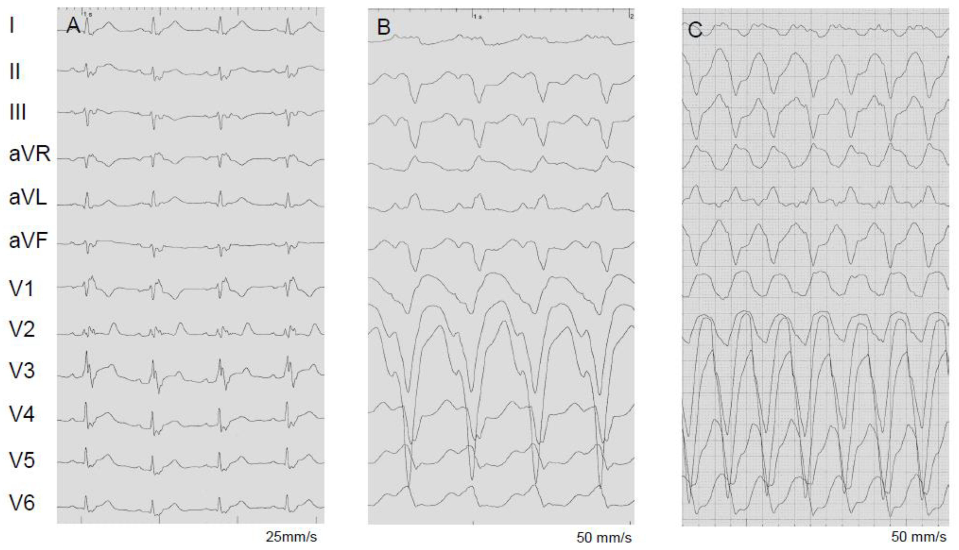
| Clinical Manifestations at Presentation | % |
|---|---|
| Atrioventricular block, 3rd degree or 2nd degree Mobitz II | 46 |
| Sustained ventricular tachycardia | 17 |
| Non-sustained ventricular tachycardia or ectopy | 7 |
| Aborted sudden cardiac death | 4 |
| Heart Failure with reduced LVEF | 18 |
| Atrial tachyarrhythmia | 1 |
| Syndrome mimicking acute coronary syndrome | 4 |
3. Diagnosis
3.1. Advanced Imaging: Cardiovascular Magnetic Resonance (CMR)
3.2. Advanced Imaging: FDG-PET
3.3. Biopsy and Histopathology
3.4. Differential Diagnosis and Screening
4. Management
4.1. Immunosuppression
4.2. Management of Heart Failure
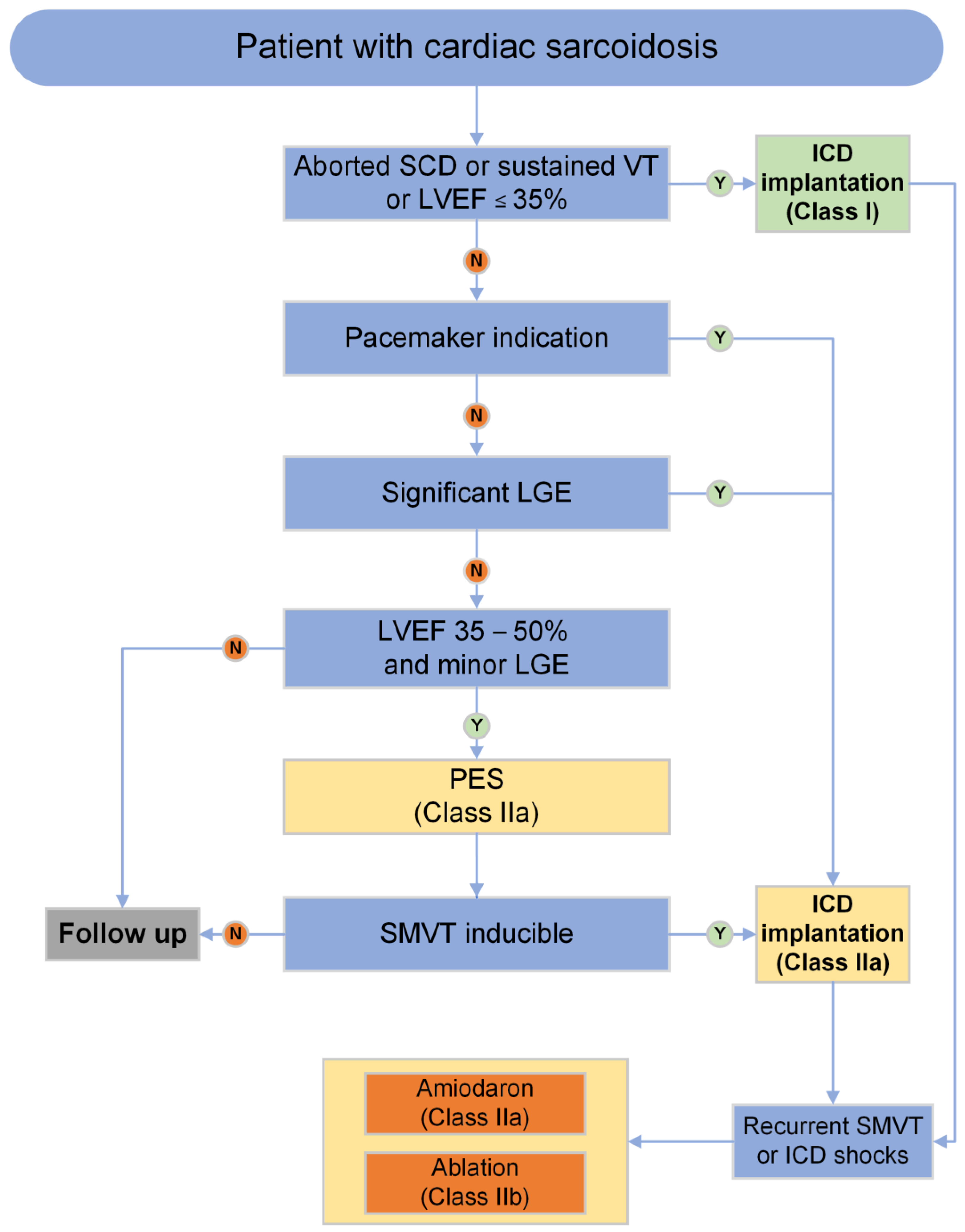
4.3. Management of Arrhythmias and Conduction Disease
4.4. Management of Ventricular Arrhythmias
5. Risk Stratification and Prevention of SCD
6. Prognosis
7. Future Developments
Funding
Conflicts of Interest
References
- Drent, M.; Crouser, E.D.; Grunewald, J. Challenges of Sarcoidosis and Its Management. N. Engl. J. Med. 2021, 385, 1018–1032. [Google Scholar] [CrossRef] [PubMed]
- Kandolin, R.; Lehtonen, J.; Airaksinen, J.; Vihinen, T.; Miettinen, H.; Ylitalo, K.; Kaikkonen, K.; Tuohinen, S.; Haataja, P.; Kerola, T.; et al. Cardiac sarcoidosis: Epidemiology, characteristics, and outcome over 25 years in a nationwide study. Circulation 2015, 131, 624–632. [Google Scholar] [CrossRef] [PubMed]
- Takaya, Y.; Nakamura, K.; Nishii, N.; Ito, H. Clinical outcomes of patients with isolated cardiac sarcoidosis confirmed by clinical diagnostic criteria. Int. J. Cardiol. 2021, 345, 49–53. [Google Scholar] [CrossRef] [PubMed]
- Zeppenfeld, K.; Tfelt-Hansen, J.; de Riva, M.; Winkel, B.G.; Behr, E.R.; Blom, N.A.; Charron, P.; Corrado, D.; Dagres, N.; de Chillou, C.; et al. 2022 ESC Guidelines for the management of patients with ventricular arrhythmias and the prevention of sudden cardiac death. Eur. Heart J. 2022, 43, 3997–4126. [Google Scholar] [CrossRef] [PubMed]
- Könemann, H.; Dagres, N.; Merino, J.L.; Sticherling, C.; Zeppenfeld, K.; Tfelt-Hansen, J.; Eckardt, L. Spotlight on the 2022 ESC guideline management of ventricular arrhythmias and prevention of sudden cardiac death: 10 novel key aspects. Europace 2023, 25, euad091. [Google Scholar] [CrossRef] [PubMed]
- Ekström, K.; Lehtonen, J.; Nordenswan, H.-K.; Mäyränpää, M.I.; Räisänen-Sokolowski, A.; Kandolin, R.; Simonen, P.; Pietilä-Effati, P.; Alatalo, A.; Utriainen, S.; et al. Sudden death in cardiac sarcoidosis: An analysis of nationwide clinical and cause-of-death registries. Eur. Heart J. 2019, 40, 3121–3128. [Google Scholar] [CrossRef]
- Rosenbaum, A.N.; Kolluri, N.; Elwazir, M.Y.; Kapa, S.; Ezzeddine, O.F.A.; Bois, J.P.; Chareonthaitawee, P.; Schmidt, T.J.; Cooper, L.T. Identification of a novel presumed cardiac sarcoidosis category for patients at high risk of disease. Int. J. Cardiol. 2021, 335, 66–72. [Google Scholar] [CrossRef]
- Nordenswan, H.; Lehtonen, J.; Ekström, K.; Räisänen-Sokolowski, A.; Mäyränpää, M.I.; Vihinen, T.; Miettinen, H.; Kaikkonen, K.; Haataja, P.; Kerola, T.; et al. Manifestations and Outcome of Cardiac Sarcoidosis and Idiopathic Giant Cell Myocarditis by 25-Year Nationwide Cohorts. J. Am. Heart Assoc. 2021, 10, e019415. [Google Scholar] [CrossRef]
- Kusano, K.; Ishibashi, K.; Noda, T.; Nakajima, K.; Nakasuka, K.; Terasaki, S.; Hattori, Y.; Nagayama, T.; Mori, K.; Takaya, Y.; et al. Prognosis and Outcomes of Clinically Diagnosed Cardiac Sarcoidosis without Positive Endomyocardial Biopsy Findings. JACC Asia 2021, 1, 385–395. [Google Scholar] [CrossRef]
- Skowasch, D.; Gaertner, F.; Marx, N.; Meder, B.; Müller-Quernheim, J.; Pfeifer, M.; Schrickel, J.W.; Yilmaz, A.; Grohé, C. Diagnostik und Therapie der kardialen Sarkoidose. Kardiologe 2020, 14, 14–25. [Google Scholar] [CrossRef]
- Birnie, D.H.; Sauer, W.H.; Bogun, F.; Cooper, J.M.; Culver, D.A.; Duvernoy, C.S.; Judson, M.A.; Kron, J.; Mehta, D.; Nielsen, J.C.; et al. HRS expert consensus statement on the diagnosis and management of arrhythmias associated with cardiac sarcoidosis. Heart Rhythm 2014, 11, 1305–1323. [Google Scholar] [CrossRef]
- Yoshinaga, K.; Miyagawa, M.; Kiso, K.; Ishida, Y. Japanese Guidelines for Cardiac Sarcoidosis. Ann. Nucl. Cardiol. 2017, 3, 121–124. [Google Scholar] [CrossRef]
- Dechering, D.G.; Kochhäuser, S.; Wasmer, K.; Zellerhoff, S.; Pott, C.; Köbe, J.; Spieker, T.; Piers, S.R.; Bittner, A.; Mönnig, G.; et al. Electrophysiological characteristics of ventricular tachyarrhythmias in cardiac sarcoidosis versus arrhythmogenic right ventricular cardiomyopathy. Heart Rhythm 2013, 10, 158–164. [Google Scholar] [CrossRef]
- Wichter, T.; Paul, T.M.; Eckardt, L.; Gerdes, P.; Kirchhof, P.; Böcker, D.; Breithardt, G. Arrhythmogenic right ventricular cardiomyopathy. Antiarrhythmic drugs, catheter ablation, or ICD? Herz 2005, 30, 91–101. [Google Scholar] [CrossRef]
- Nielsen, J.C.; Lin, Y.-J.; De Oliveira Figueiredo, M.J.; Shamloo, A.S.; Alfie, A.; Boveda, S.; Dagres, N.; Di Toro, D.; Eckhardt, L.L.; Ellenbogen, K.; et al. European Heart Rhythm Association (EHRA)/Heart Rhythm Society (HRS)/Asia Pacific Heart Rhythm Society (APHRS)/Latin American Heart Rhythm Society (LAHRS) expert consensus on risk assessment in cardiac arrhythmias: Use the right tool for the right outcome, in the right population. Europace 2020, 22, 1147–1148. [Google Scholar] [CrossRef] [PubMed]
- Vignaux, O.; Dhote, R.; Duboc, D.; Blanche, P.; Devaux, J.-Y.; Weber, S.; Legmann, P. Detection of myocardial involvement in patients with sarcoidosis applying T2-weighted, contrast-enhanced, and cine magnetic resonance imaging: Initial results of a prospective study. J. Comput. Assist. Tomogr. 2002, 26, 762–767. [Google Scholar] [CrossRef] [PubMed]
- Smedema, J.-P.; Snoep, G.; van Kroonenburgh, M.P.; van Geuns, R.-J.; Dassen, W.R.; Gorgels, A.P.; Crijns, H.J. Evaluation of the accuracy of gadolinium-enhanced cardiovascular magnetic resonance in the diagnosis of cardiac sarcoidosis. J. Am. Coll. Cardiol. 2005, 45, 1683–1690. [Google Scholar] [CrossRef] [PubMed]
- Youssef, G.; Leung, E.; Mylonas, I.; Nery, P.; Williams, K.; Wisenberg, G.; Gulenchyn, K.Y.; Dekemp, R.A.; DaSilva, J.; Birnie, D.; et al. The use of 18F-FDG PET in the diagnosis of cardiac sarcoidosis: A systematic review and metaanalysis including the Ontario experience. J. Nucl. Med. 2012, 53, 241–248. [Google Scholar] [CrossRef] [PubMed]
- Stevenson, A.; Bray, J.J.; Tregidgo, L.; Ahmad, M.; Sharma, A.; Ng, A.; Siddiqui, A.; Khalid, A.A.; Hylton, K.; Ionescu, A.; et al. Prognostic Value of Late Gadolinium Enhancement Detected on Cardiac Magnetic Resonance in Cardiac Sarcoidosis. JACC Cardiovasc. Imaging 2023, 16, 345–357. [Google Scholar] [CrossRef] [PubMed]
- Blankstein, R.; Osborne, M.; Naya, M.; Waller, A.; Kim, C.K.; Murthy, V.L.; Kazemian, P.; Kwong, R.Y.; Tokuda, M.; Skali, H.; et al. Cardiac positron emission tomography enhances prognostic assessments of patients with suspected cardiac sarcoidosis. J. Am. Coll. Cardiol. 2014, 63, 329–336. [Google Scholar] [CrossRef]
- Bietenbeck, M.; Meier, C.; Korthals, D.; Theofanidou, M.; Stalling, P.; Dittmann, S.; Schulze-Bahr, E.; Eckardt, L.; Yilmaz, A. Possible Causes and Clinical Relevance of a “Ring-Like” Late Gadolinium Enhancement Pattern. JACC Cardiovasc. Imaging 2024, 17, 104–106. [Google Scholar] [CrossRef] [PubMed]
- Nordenswan, H.-K.; Pöyhönen, P.; Lehtonen, J.; Ekström, K.; Uusitalo, V.; Niemelä, M.; Vihinen, T.; Kaikkonen, K.; Haataja, P.; Kerola, T.; et al. Incidence of Sudden Cardiac Death and Life-Threatening Arrhythmias in Clinically Manifest Cardiac Sarcoidosis with and without Current Indications for an Implantable Cardioverter Defibrillator. Circulation 2022, 146, 964–975. [Google Scholar] [CrossRef]
- Birnie, D.; Ha, A.C.T.; Gula, L.J.; Chakrabarti, S.; Beanlands, R.S.B.; Nery, P. Cardiac Sarcoidosis. Clin. Chest Med. 2015, 36, 657–668. [Google Scholar] [CrossRef]
- Kupari, M.; Lehtonen, J. POINT: Should Isolated Cardiac Sarcoidosis Be Considered a Significant Manifestation of Sarcoidosis? Yes. Chest 2021, 160, 36–38. [Google Scholar] [CrossRef]
- Okada, D.R.; Bravo, P.E.; Vita, T.; Agarwal, V.; Osborne, M.T.; Taqueti, V.R.; Skali, H.; Chareonthaitawee, P.; Dorbala, S.; Stewart, G.; et al. Isolated cardiac sarcoidosis: A focused review of an under-recognized entity. J. Nucl. Cardiol. 2018, 25, 1136–1146. [Google Scholar] [CrossRef]
- Kawai, H.; Sarai, M.; Kato, Y.; Naruse, H.; Watanabe, A.; Matsuyama, T.; Takahashi, H.; Motoyama, S.; Ishii, J.; Morimoto, S.; et al. Diagnosis of isolated cardiac sarcoidosis based on new guidelines. ESC Heart Fail. 2020, 7, 2662–2671. [Google Scholar] [CrossRef]
- Silverman, K.J.; Hutchins, G.M.; Bulkley, B.H. Cardiac sarcoid: A clinicopathologic study of 84 unselected patients with systemic sarcoidosis. Circulation 1978, 58, 1204–1211. [Google Scholar] [CrossRef] [PubMed]
- Patel, M.R.; Cawley, P.J.; Heitner, J.F.; Klem, I.; Parker, M.A.; Jaroudi, W.A.; Meine, T.J.; White, J.B.; Elliott, M.D.; Kim, H.W.; et al. Detection of myocardial damage in patients with sarcoidosis. Circulation 2009, 120, 1969–1977. [Google Scholar] [CrossRef] [PubMed]
- Kouranos, V.; Tzelepis, G.E.; Rapti, A.; Mavrogeni, S.; Aggeli, K.; Douskou, M.; Prasad, S.; Koulouris, N.; Sfikakis, P.; Wells, A.; et al. Complementary Role of CMR to Conventional Screening in the Diagnosis and Prognosis of Cardiac Sarcoidosis. JACC Cardiovasc. Imaging 2017, 10, 1437–1447. [Google Scholar] [CrossRef]
- Rosen, N.S.; Pavlovic, N.; Duvall, C.; Wand, A.L.; Griffin, J.M.; Okada, D.R.; Chrispin, J.; Tandri, H.; Mathai, S.C.; Stern, B.; et al. Cardiac sarcoidosis outcome differences: A comparison of patients with de novo cardiac versus known extracardiac sarcoidosis at presentation. Respir. Med. 2022, 198, 106864. [Google Scholar] [CrossRef]
- Okada, D.R.; Smith, J.; Derakhshan, A.; Gowani, Z.; Misra, S.; Berger, R.D.; Calkins, H.; Tandri, H.; Chrispin, J. Ventricular Arrhythmias in Cardiac Sarcoidosis. Circulation 2018, 138, 1253–1264. [Google Scholar] [CrossRef] [PubMed]
- Kumar, S.; Barbhaiya, C.; Nagashima, K.; Choi, E.-K.; Epstein, L.M.; John, R.M.; Maytin, M.; Albert, C.M.; Miller, A.L.; Koplan, B.A.; et al. Ventricular tachycardia in cardiac sarcoidosis: Characterization of ventricular substrate and outcomes of catheter ablation. Circ. Arrhythmia Electrophysiol. 2015, 8, 87–93. [Google Scholar] [CrossRef] [PubMed]
- Korthals, D.; Eckardt, L. Die neue ESC-Leitlinie (ESC: European Society of Cardiology) zum Management von Kardiomyopathien: Schlüsselbotschaft für kardiale Elektrophysiologen (The new European Society of Cardiology guideline for the management of cardiomyopathies: Key messages for cardiac electrophysiologists). Herzschrittmacherther. Elektrophysiol. 2023, 34, 311–323. [Google Scholar] [CrossRef] [PubMed]
- Hoogendoorn, J.C.; Venlet, J.; Out, Y.N.; Man, S.; Kumar, S.; Sramko, M.; Dechering, D.G.; Nakajima, I.; Siontis, K.C.; Watanabe, M.; et al. The precordial R’ wave: A novel discriminator between cardiac sarcoidosis and arrhythmogenic right ventricular cardiomyopathy in patients presenting with ventricular tachycardia. Heart Rhythm 2021, 18, 1539–1547. [Google Scholar] [CrossRef] [PubMed]
- Vasaiwala, S.C.; Finn, C.; Delpriore, J.; Leya, F.; Gagermeier, J.; Akar, J.G.; Santucci, P.; Dajani, K.; Bova, D.; Picken, M.M.; et al. Prospective study of cardiac sarcoid mimicking arrhythmogenic right ventricular dysplasia. J. Cardiovasc. Electrophysiol. 2009, 20, 473–476. [Google Scholar] [CrossRef]
- Vogler, J.; Breithardt, G.; Eckardt, L. Bradyarrhythmias and Conduction Blocks. Rev. Española Cardiol. (Engl. Ed.) 2012, 65, 656–667. [Google Scholar] [CrossRef]
- Willy, K.; Dechering, D.G.; Reinke, F.; Bögeholz, N.; Frommeyer, G.; Eckardt, L. The ECG in sarcoidosis—A marker of cardiac involvement? Current evidence and clinical implications. J. Cardiol. 2021, 77, 154–159. [Google Scholar] [CrossRef]
- Niemelä, M.; Uusitalo, V.; Pöyhönen, P.; Schildt, J.; Lehtonen, J.; Kupari, M. Incidence and Predictors of Atrial Fibrillation in Cardiac Sarcoidosis: A Multimodality Imaging Study. JACC Cardiovasc. Imaging 2022, 15, 1622–1631. [Google Scholar] [CrossRef]
- Willy, K.; Dechering, D.G.; Wasmer, K.; Köbe, J.; Bögeholz, N.; Ellermann, C.; Leitz, P.; Reinke, F.; Frommeyer, G.; Eckardt, L. Outcome of catheter ablation of supraventricular tachyarrhythmias in cardiac sarcoidosis. Clin. Cardiol. 2019, 42, 1121–1125. [Google Scholar] [CrossRef]
- Yafasova, A.; Fosbøl, E.L.; Schou, M.; Gustafsson, F.; Rossing, K.; Bundgaard, H.; Lauridsen, M.D.; Kristensen, S.L.; Torp-Pedersen, C.; Gislason, G.H.; et al. Long-Term Adverse Cardiac Outcomes in Patients with Sarcoidosis. J. Am. Coll. Cardiol. 2020, 76, 767–777. [Google Scholar] [CrossRef] [PubMed]
- Rosenthal, D.G.; Fang, C.D.; Groh, C.A.; Nah, G.; Vittinghoff, E.; Dewland, T.A.; Vedantham, V.; Marcus, G.M. Heart Failure, Atrioventricular Block, and Ventricular Tachycardia in Sarcoidosis. J. Am. Heart Assoc. 2021, 10, e017692. [Google Scholar] [CrossRef]
- Lam, C.S.P.; Tolep, K.A.; Metke, M.P.; Glockner, J.; Cooper, L.T. Coronary sarcoidosis presenting as acute coronary syndrome. Clin. Cardiol. 2009, 32, E68–E71. [Google Scholar] [CrossRef]
- Ward, E.V.; Nazari, J.; Edelman, R.R. Coronary artery vasculitis as a presentation of cardiac sarcoidosis. Circulation 2012, 125, e344–e346. [Google Scholar] [CrossRef] [PubMed]
- Kandolin, R.; Ekström, K.; Simard, T.; Hibbert, B.; Nery, P.; Lehtonen, J.; Kupari, M.; Birnie, D. Spontaneous coronary artery dissection in cardiac sarcoidosis. Oxf. Med. Case Rep. 2019, 2019, omz033. [Google Scholar] [CrossRef]
- Mehta, D.; Lubitz, S.A.; Frankel, Z.; Wisnivesky, J.P.; Einstein, A.J.; Goldman, M.; Machac, J.; Teirstein, A. Cardiac involvement in patients with sarcoidosis: Diagnostic and prognostic value of outpatient testing. Chest 2008, 133, 1426–1435. [Google Scholar] [CrossRef] [PubMed]
- Martusewicz-Boros, M.M.; Boros, P.W.; Wiatr, E.; Zych, J.; Piotrowska-Kownacka, D.; Roszkowski-Śliż, K. Prevalence of cardiac sarcoidosis in white population: A case-control study: Proposal for a novel risk index based on commonly available tests. Medicine 2016, 95, e4518. [Google Scholar] [CrossRef]
- Terasaki, F.; Azuma, A.; Anzai, T.; Ishizaka, N.; Ishida, Y.; Isobe, M.; Inomata, T.; Ishibashi-Ueda, H.; Eishi, Y.; Kitakaze, M.; et al. JCS 2016 Guideline on Diagnosis and Treatment of Cardiac Sarcoidosis—Digest Version. Circ. J. 2019, 83, 2329–2388. [Google Scholar] [CrossRef] [PubMed]
- Kandolin, R.; Lehtonen, J.; Kupari, M. Cardiac sarcoidosis and giant cell myocarditis as causes of atrioventricular block in young and middle-aged adults. Circ. Arrhythmia Electrophysiol. 2011, 4, 303–309. [Google Scholar] [CrossRef]
- Nery, P.B.; Beanlands, R.S.; Nair, G.M.; Green, M.; Yang, J.; Mcardle, B.A.; Davis, D.; Ohira, H.; Gollob, M.H.; Leung, E.; et al. Atrioventricular block as the initial manifestation of cardiac sarcoidosis in middle-aged adults. J. Cardiovasc. Electrophysiol. 2014, 25, 875–881. [Google Scholar] [CrossRef]
- Nordenswan, H.-K.; Lehtonen, J.; Ekström, K.; Kandolin, R.; Simonen, P.; Mäyränpää, M.; Vihinen, T.; Miettinen, H.; Kaikkonen, K.; Haataja, P.; et al. Outcome of Cardiac Sarcoidosis Presenting with High-Grade Atrioventricular Block. Circ. Arrhythmia Electrophysiol. 2018, 11, e006145. [Google Scholar] [CrossRef]
- Nery, P.B.; Mc Ardle, B.A.; Redpath, C.J.; Leung, E.; Lemery, R.; Dekemp, R.; Yang, J.; Keren, A.; Beanlands, R.S.; Birnie, D.H. Prevalence of cardiac sarcoidosis in patients presenting with monomorphic ventricular tachycardia. Pacing Clin. Electrophysiol. 2014, 37, 364–374. [Google Scholar] [CrossRef]
- Kiko, T.; Yoshihisa, A.; Kanno, Y.; Yokokawa, T.; Abe, S.; Miyata-Tatsumi, M.; Misaka, T.; Oikawa, M.; Kobayashi, A.; Ishida, T.; et al. A Multiple Biomarker Approach in Patients with Cardiac Sarcoidosis. Int. Heart J. 2018, 59, 996–1001. [Google Scholar] [CrossRef]
- Eurelings, L.E.M.; Miedema, J.R.; Dalm, V.A.S.H.; van Daele, P.L.A.; van Hagen, P.M.; van Laar, J.A.M.; Dik, W.A. Sensitivity and specificity of serum soluble interleukin-2 receptor for diagnosing sarcoidosis in a population of patients suspected of sarcoidosis. PLoS ONE 2019, 14, e0223897. [Google Scholar] [CrossRef]
- Riedy, K.; Fisher, M.R.; Belic, N.; Koenigsberg, D.I. MR imaging of myocardial sarcoidosis. AJR Am. J. Roentgenol. 1988, 151, 915–916. [Google Scholar] [CrossRef]
- Schulz-Menger, J.; Wassmuth, R.; Abdel-Aty, H.; Siegel, I.; Franke, A.; Dietz, R.; Friedrich, M.G. Patterns of myocardial inflammation and scarring in sarcoidosis as assessed by cardiovascular magnetic resonance. Heart 2006, 92, 399–400. [Google Scholar] [CrossRef]
- Abdel-Aty, H.; Boyé, P.; Zagrosek, A.; Wassmuth, R.; Kumar, A.; Messroghli, D.; Bock, P.; Dietz, R.; Friedrich, M.G.; Schulz-Menger, J. Diagnostic performance of cardiovascular magnetic resonance in patients with suspected acute myocarditis: Comparison of different approaches. J. Am. Coll. Cardiol. 2005, 45, 1815–1822. [Google Scholar] [CrossRef]
- Puntmann, V.O.; Isted, A.; Hinojar, R.; Foote, L.; Carr-White, G.; Nagel, E. T1 and T2 Mapping in Recognition of Early Cardiac Involvement in Systemic Sarcoidosis. Radiology 2017, 285, 63–72. [Google Scholar] [CrossRef] [PubMed]
- Mahrholdt, H.; Wagner, A.; Judd, R.M.; Sechtem, U.; Kim, R.J. Delayed enhancement cardiovascular magnetic resonance assessment of non-ischaemic cardiomyopathies. Eur. Heart J. 2005, 26, 1461–1474. [Google Scholar] [CrossRef] [PubMed]
- Greulich, S.; Deluigi, C.C.; Gloekler, S.; Wahl, A.; Zürn, C.; Kramer, U.; Nothnagel, D.; Bültel, H.; Schumm, J.; Grün, S.; et al. CMR imaging predicts death and other adverse events in suspected cardiac sarcoidosis. JACC Cardiovasc. Imaging 2013, 6, 501–511. [Google Scholar] [CrossRef] [PubMed]
- Trivieri, M.G.; Spagnolo, P.; Birnie, D.; Liu, P.; Drake, W.; Kovacic, J.C.; Baughman, R.; Fayad, Z.A.; Judson, M.A. Challenges in Cardiac and Pulmonary Sarcoidosis: JACC State-of-the-Art Review. J. Am. Coll. Cardiol. 2020, 76, 1878–1901. [Google Scholar] [CrossRef] [PubMed]
- Yang, S.; Chen, X.; Li, J.; Sun, Y.; Song, J.; Wang, H.; Zhao, S. Late gadolinium enhancement characteristics in giant cell myocarditis. ESC Heart Fail. 2021, 8, 2320–2327. [Google Scholar] [CrossRef]
- Pöyhönen, P.; Nordenswan, H.-K.; Lehtonen, J.; Syväranta, S.; Shenoy, C.; Kupari, M. Cardiac magnetic resonance in giant cell myocarditis: A matched comparison with cardiac sarcoidosis. Eur. Heart J. Cardiovasc. Imaging 2023, 24, 404–412. [Google Scholar] [CrossRef]
- Hulten, E.; Agarwal, V.; Cahill, M.; Cole, G.; Vita, T.; Parrish, S.; Bittencourt, M.S.; Murthy, V.L.; Kwong, R.; Di Carli, M.F.; et al. Presence of Late Gadolinium Enhancement by Cardiac Magnetic Resonance Among Patients with Suspected Cardiac Sarcoidosis Is Associated with Adverse Cardiovascular Prognosis: A Systematic Review and Meta-Analysis. Circ. Cardiovasc. Imaging 2016, 9, e005001. [Google Scholar] [CrossRef] [PubMed]
- Slart, R.H.J.A.; Glaudemans, A.W.J.M.; Lancellotti, P.; Hyafil, F.; Blankstein, R.; Schwartz, R.G.; Jaber, W.A.; Russell, R.; Gimelli, A.; Rouzet, F.; et al. A joint procedural position statement on imaging in cardiac sarcoidosis: From the Cardiovascular and Inflammation & Infection Committees of the European Association of Nuclear Medicine, the European Association of Cardiovascular Imaging, and the American Society of Nuclear Cardiology. J. Nucl. Cardiol. 2018, 25, 298–319. [Google Scholar] [CrossRef]
- Ishimaru, S.; Tsujino, I.; Takei, T.; Tsukamoto, E.; Sakaue, S.; Kamigaki, M.; Ito, N.; Ohira, H.; Ikeda, D.; Tamaki, N.; et al. Focal uptake on 18F-fluoro-2-deoxyglucose positron emission tomography images indicates cardiac involvement of sarcoidosis. Eur. Heart J. 2005, 26, 1538–1543. [Google Scholar] [CrossRef] [PubMed]
- Saric, P.; Young, K.A.; Rodriguez-Porcel, M.; Chareonthaitawee, P. PET Imaging in Cardiac Sarcoidosis: A Narrative Review with Focus on Novel PET Tracers. Pharmaceuticals 2021, 14, 1286. [Google Scholar] [CrossRef] [PubMed]
- Hotta, M.; Minamimoto, R.; Awaya, T.; Hiroe, M.; Okazaki, O.; Hiroi, Y. Radionuclide Imaging of Cardiac Amyloidosis and Sarcoidosis: Roles and Characteristics of Various Tracers. Radiographics 2020, 40, 2029–2041. [Google Scholar] [CrossRef]
- Kim, S.-J.; Pak, K.; Kim, K. Diagnostic performance of F-18 FDG PET for detection of cardiac sarcoidosis; A systematic review and meta-analysis. J. Nucl. Cardiol. 2020, 27, 2103–2115. [Google Scholar] [CrossRef]
- Chareonthaitawee, P.; Beanlands, R.S.; Chen, W.; Dorbala, S.; Miller, E.J.; Murthy, V.L.; Birnie, D.H.; Chen, E.S.; Cooper, L.T.; Tung, R.H.; et al. Joint SNMMI-ASNC Expert Consensus Document on the Role of 18F-FDG PET/CT in Cardiac Sarcoid Detection and Therapy Monitoring. J. Nucl. Med. 2017, 58, 1341–1353. [Google Scholar] [CrossRef]
- Divakaran, S.; Stewart, G.C.; Lakdawala, N.K.; Padera, R.F.; Zhou, W.; Desai, A.S.; Givertz, M.M.; Mehra, M.R.; Kwong, R.Y.; Hedgire, S.S.; et al. Diagnostic Accuracy of Advanced Imaging in Cardiac Sarcoidosis. Circ. Cardiovasc. Imaging 2019, 12, e008975. [Google Scholar] [CrossRef]
- Osborne, M.T.; Hulten, E.A.; Singh, A.; Waller, A.H.; Bittencourt, M.S.; Stewart, G.C.; Hainer, J.; Murthy, V.L.; Skali, H.; Dorbala, S.; et al. Reduction in ¹⁸F-fluorodeoxyglucose uptake on serial cardiac positron emission tomography is associated with improved left ventricular ejection fraction in patients with cardiac sarcoidosis. J. Nucl. Cardiol. 2014, 21, 166–174. [Google Scholar] [CrossRef]
- Giblin, G.T.; Murphy, L.; Stewart, G.C.; Desai, A.S.; Di Carli, M.F.; Blankstein, R.; Givertz, M.M.; Tedrow, U.B.; Sauer, W.H.; Hunninghake, G.M.; et al. Cardiac Sarcoidosis: When and How to Treat Inflammation. Card. Fail. Rev. 2021, 7, e17. [Google Scholar] [CrossRef]
- Morimoto, R.; Unno, K.; Fujita, N.; Sakuragi, Y.; Nishimoto, T.; Yamashita, M.; Kuwayama, T.; Hiraiwa, H.; Kondo, T.; Kuwatsuka, Y.; et al. Prospective Analysis of Immunosuppressive Therapy in Cardiac Sarcoidosis with Fluorodeoxyglucose Myocardial Accumulation: The PRESTIGE Study. JACC Cardiovasc. Imaging 2024, 17, 45–58. [Google Scholar] [CrossRef]
- Sperry, B.W.; Tamarappoo, B.K.; Oldan, J.D.; Javed, O.; Culver, D.A.; Brunken, R.; Cerqueira, M.D.; Hachamovitch, R. Prognostic Impact of Extent, Severity, and Heterogeneity of Abnormalities on 18F-FDG PET Scans for Suspected Cardiac Sarcoidosis. JACC Cardiovasc. Imaging 2018, 11, 336–345. [Google Scholar] [CrossRef] [PubMed]
- Ahmed, A.I.; Abebe, A.T.; Han, Y.; Alnabelsi, T.; Agrawal, T.; Kassi, M.; Aljizeeri, A.; Taylor, A.; Tleyjeh, I.M.; Al-Mallah, M.H. The prognostic role of cardiac positron emission tomography imaging in patients with sarcoidosis: A systematic review. J. Nucl. Cardiol. 2021, 28, 1545–1552. [Google Scholar] [CrossRef]
- Patel, V.N.; Pieper, J.A.; Poitrasson-Rivière, A.; Kopin, D.; Cascino, T.; Aaronson, K.; Murthy, V.L.; Koelling, T. The prognostic value of positron emission tomography in the evaluation of suspected cardiac sarcoidosis. J. Nucl. Cardiol. 2022, 29, 2460–2470. [Google Scholar] [CrossRef]
- Gowani, Z.; Habibi, M.; Okada, D.R.; Smith, J.; Derakhshan, A.; Zimmerman, S.L.; Misra, S.; Gilotra, N.A.; Berger, R.D.; Calkins, H.; et al. Utility of Cardiac Magnetic Resonance Imaging Versus Cardiac Positron Emission Tomography for Risk Stratification for Ventricular Arrhythmias in Patients with Cardiac Sarcoidosis. Am. J. Cardiol. 2020, 134, 123–129. [Google Scholar] [CrossRef]
- Siebermair, J.; Kessler, L.; Kupusovic, J.; Rassaf, T.; Rischpler, C. Cardiac fibroblast activation detected by 68Gallium-FAPI-46 positron emission tomography-magnetic resonance imaging as a sign of chronic activity in cardiac sarcoidosis. Eur. Heart J. Case Rep. 2022, 6, ytac005. [Google Scholar] [CrossRef]
- Lee, H.; Schubert, E.K.; Vidula, M.K.; Pryma, D.A.; Marchlinski, F.E.; Goldberg, L.R.; Clancy, C.B.; Rossman, M.D.; DiCarli, M.F.; Bravo, P.E. Potential clinical utility of 68Ga-DOTATATE PET/CT for detection and response assessment in cardiac sarcoidosis. J. Nucl. Cardiol. 2023, 30, 1075–1087. [Google Scholar] [CrossRef]
- Yilmaz, A.; Kindermann, I.; Kindermann, M.; Mahfoud, F.; Ukena, C.; Athanasiadis, A.; Hill, S.; Mahrholdt, H.; Voehringer, M.; Schieber, M.; et al. Comparative evaluation of left and right ventricular endomyocardial biopsy: Differences in complication rate and diagnostic performance. Circulation 2010, 122, 900–909. [Google Scholar] [CrossRef]
- Kandolin, R.; Lehtonen, J.; Graner, M.; Schildt, J.; Salmenkivi, K.; Kivistö, S.M.; Kupari, M. Diagnosing isolated cardiac sarcoidosis. J. Intern. Med. 2011, 270, 461–468. [Google Scholar] [CrossRef]
- Ezzeddine, F.M.; Kapa, S.; Rosenbaum, A.; Blauwet, L.; Deshmukh, A.J.; AbouEzzeddine, O.F.; Maleszewski, J.J.; Asirvatham, S.J.; Bois, J.P.; Schirger, J.A.; et al. Electrogram-guided endomyocardial biopsy yield in patients with suspected cardiac sarcoidosis and relation to outcomes. J. Cardiovasc. Electrophysiol. 2021, 32, 2486–2495. [Google Scholar] [CrossRef]
- Zumhagen, S.; Spieker, T.; Rolinck, J.; Baba, H.A.; Breithardt, G.; Bocker, W.; Eckardt, L.; Paul, M.; Wichter, T.; Schulze-Bahr, E.; et al. Absence of pathognomonic or inflammatory patterns in cardiac biopsies from patients with Brugada syndrome. Circ. Arrhythmia Electrophysiol. 2009, 2, 16–23. [Google Scholar] [CrossRef]
- Bennett, M.K.; Gilotra, N.A.; Harrington, C.; Rao, S.; Dunn, J.M.; Freitag, T.B.; Halushka, M.K.; Russell, S.D. Evaluation of the role of endomyocardial biopsy in 851 patients with unexplained heart failure from 2000–2009. Circ Heart Fail 2013, 6, 676–684. [Google Scholar] [CrossRef] [PubMed]
- Büscher, A.; Doldi, F.; Eckardt, L.; Müller, P. Lyme carditis manifesting with sinoatrial exit block: A case report. Eur. Heart J. Case Rep. 2022, 6, ytac022. [Google Scholar] [CrossRef]
- Arbelo, E.; Protonotarios, A.; Gimeno, J.R.; Arbustini, E.; Barriales-Villa, R.; Basso, C.; Bezzina, C.R.; Biagini, E.; Blom, N.A.; de Boer, R.A.; et al. 2023 ESC Guidelines for the management of cardiomyopathies. Eur. Heart J. 2023, 44, 3503–3626. [Google Scholar] [CrossRef]
- Ekström, K.; Räisänen-Sokolowski, A.; Lehtonen, J.; Nordenswan, H.-K.; Mäyränpää, M.I.; Kupari, M. Idiopathic giant cell myocarditis or cardiac sarcoidosis? A retrospective audit of a nationwide case series. ESC Heart Fail. 2020, 7, 1362–1370. [Google Scholar] [CrossRef]
- Nakasuka, K.; Ito, S.; Miyata, K.; Inomata, M.; Yoshida, T.; Tamai, N.; Suzuki, S.; Murakami, Y.; Sato, K.; Suzuki, S.; et al. A case of idiopathic giant cell myocarditis with a past history of sarcoidosis. J. Cardiol. Cases 2014, 9, 35–39. [Google Scholar] [CrossRef]
- Blauwet, L.A.; Cooper, L.T. Idiopathic giant cell myocarditis and cardiac sarcoidosis. Heart Fail. Rev. 2013, 18, 733–746. [Google Scholar] [CrossRef]
- Okura, Y.; Dec, G.; Hare, J.M.; Kodama, M.; Berry, G.J.; Tazelaar, H.D.; Bailey, K.R.; Cooper, L.T. A clinical and histopathologic comparison of cardiac sarcoidosis and idiopathic giant cell myocarditis. J. Am. Coll. Cardiol. 2003, 41, 322–329. [Google Scholar] [CrossRef]
- Kandolin, R.; Lehtonen, J.; Salmenkivi, K.; Räisänen-Sokolowski, A.; Lommi, J.; Kupari, M. Diagnosis, treatment, and outcome of giant-cell myocarditis in the era of combined immunosuppression. Circ. Heart Fail. 2013, 6, 15–22. [Google Scholar] [CrossRef]
- Philips, B.; Madhavan, S.; James, C.A.; Riele, A.S.T.; Murray, B.; Tichnell, C.; Bhonsale, A.; Nazarian, S.; Judge, D.P.; Calkins, H.; et al. Arrhythmogenic right ventricular dysplasia/cardiomyopathy and cardiac sarcoidosis: Distinguishing features when the diagnosis is unclear. Circ. Arrhythmia Electrophysiol. 2014, 7, 230–236. [Google Scholar] [CrossRef]
- Hoogendoorn, J.C.; Sramko, M.; Venlet, J.; Siontis, K.C.; Kumar, S.; Singh, R.; Nakajima, I.; Piers, S.R.; Silva, M.d.R.; Glashan, C.A.; et al. Electroanatomical Voltage Mapping to Distinguish Right-Sided Cardiac Sarcoidosis From Arrhythmogenic Right Ventricular Cardiomyopathy. JACC Clin. Electrophysiol. 2020, 6, 696–707. [Google Scholar] [CrossRef]
- Smith, E.D.; Lakdawala, N.K.; Papoutsidakis, N.; Aubert, G.; Mazzanti, A.; McCanta, A.C.; Agarwal, P.P.; Arscott, P.; Dellefave-Castillo, L.M.; Vorovich, E.E.; et al. Desmoplakin Cardiomyopathy, a Fibrotic and Inflammatory Form of Cardiomyopathy Distinct from Typical Dilated or Arrhythmogenic Right Ventricular Cardiomyopathy. Circulation 2020, 141, 1872–1884. [Google Scholar] [CrossRef]
- Holmström, M.; Kivistö, S.; Heliö, T.; Jurkko, R.; Kaartinen, M.; Antila, M.; Reissell, E.; Kuusisto, J.; Kärkkäinen, S.; Peuhkurinen, K.; et al. Late gadolinium enhanced cardiovascular magnetic resonance of lamin A/C gene mutation related dilated cardiomyopathy. J. Cardiovasc. Magn. Reason. 2011, 13, 30. [Google Scholar] [CrossRef]
- Lakdawala, N.K.; Givertz, M.M. Dilated cardiomyopathy with conduction disease and arrhythmia. Circulation 2010, 122, 527–534. [Google Scholar] [CrossRef]
- Murtagh, G.; Laffin, L.J.; Beshai, J.F.; Maffessanti, F.; Bonham, C.A.; Yu, Z.; Addetia, K.; Mor-Avi, V.; Moss, J.D.; Hogarth, D.K.; et al. Prognosis of Myocardial Damage in Sarcoidosis Patients with Preserved Left Ventricular Ejection Fraction: Risk Stratification Using Cardiovascular Magnetic Resonance. Circ. Cardiovasc. Imaging 2016, 9, e003738. [Google Scholar] [CrossRef]
- Lehtonen, J.; Uusitalo, V.; Pöyhönen, P.; Mäyränpää, M.I.; Kupari, M. Cardiac sarcoidosis: Phenotypes, diagnosis, treatment, and prognosis. Eur. Heart J. 2023, 44, 1495–1510. [Google Scholar] [CrossRef]
- Chiu, C.-Z.; Nakatani, S.; Zhang, G.; Tachibana, T.; Ohmori, F.; Yamagishi, M.; Kitakaze, M.; Tomoike, H.; Miyatake, K. Prevention of left ventricular remodeling by long-term corticosteroid therapy in patients with cardiac sarcoidosis. Am. J. Cardiol. 2005, 95, 143–146. [Google Scholar] [CrossRef]
- Sadek, M.M.; Yung, D.; Birnie, D.H.; Beanlands, R.S. NERYPB Corticosteroid therapy for cardiac sarcoidosis: A systematic review. Can. J. Cardiol. 2013, 29, 1034–1041. [Google Scholar] [CrossRef]
- Fazelpour, S.; Sadek, M.M.; Nery, P.B.; Beanlands, R.S.; Tzemos, N.; Toma, M.; Birnie, D.H. Corticosteroid and Immunosuppressant Therapy for Cardiac Sarcoidosis: A Systematic Review. J. Am. Heart Assoc. 2021, 10, e021183. [Google Scholar] [CrossRef] [PubMed]
- Yazaki, Y.; Isobe, M.; Hiroe, M.; Morimoto, S.-I.; Hiramitsu, S.; Nakano, T.; Izumi, T.; Sekiguchi, M. Prognostic determinants of long-term survival in Japanese patients with cardiac sarcoidosis treated with prednisone. Am. J. Cardiol. 2001, 88, 1006–1010. [Google Scholar] [CrossRef] [PubMed]
- Iannuzzi, M.C.; Rybicki, B.A.; Teirstein, A.S. Sarcoidosis. N. Engl. J. Med. 2007, 357, 2153–2165. [Google Scholar] [CrossRef] [PubMed]
- Birnie, D.H.; Neryp, B.; Ha, A.C.; Beanlands, R.S.B. Cardiac Sarcoidosis. J. Am. Coll. Cardiol. 2016, 68, 411–421. [Google Scholar] [CrossRef] [PubMed]
- Nagai, S.; Yokomatsu, T.; Tanizawa, K.; Ikezoe, K.; Handa, T.; Ito, Y.; Ogino, S.; Izumi, T. Treatment with methotrexate and low-dose corticosteroids in sarcoidosis patients with cardiac lesions. Intern. Med. 2014, 53, 427–433. [Google Scholar] [CrossRef] [PubMed]
- Rosenthal, D.G.; Parwani, P.; Murray, T.O.; Petek, B.J.; Benn, B.S.; de Marco, T.; Gerstenfeld, E.P.; Janmohamed, M.; Klein, L.; Lee, B.K.; et al. Long-Term Corticosteroid-Sparing Immunosuppression for Cardiac Sarcoidosis. J. Am. Heart Assoc. 2019, 8, e010952. [Google Scholar] [CrossRef]
- Harper, L.J.; McCarthy, M.; Neto, M.L.R.; Hachamovitch, R.; Pearson, K.; Bonanno, B.; Shaia, J.; Brunken, R.; Joyce, E.; Culver, D.A. Infliximab for Refractory Cardiac Sarcoidosis. Am. J. Cardiol. 2019, 124, 1630–1635. [Google Scholar] [CrossRef]
- Gilotra, N.A.; Wand, A.L.; Pillarisetty, A.; Devraj, M.; Pavlovic, N.; Ahmed, S.; Saad, E.; Solnes, L.; Garcia, C.; Okada, D.R.; et al. Clinical and Imaging Response to Tumor Necrosis Factor Alpha Inhibitors in Treatment of Cardiac Sarcoidosis: A Multicenter Experience. J. Card. Fail. 2021, 27, 83–91. [Google Scholar] [CrossRef]
- Vorselaars, A.D.M.; Wuyts, W.A.; Vorselaars, V.M.M.; Zanen, P.; Deneer, V.H.M.; Veltkamp, M.; Thomeer, M.; van Moorsel, C.H.M.; Grutters, J.C. Methotrexate vs azathioprine in second-line therapy of sarcoidosis. Chest 2013, 144, 805–812. [Google Scholar] [CrossRef]
- Hamzeh, N.; Voelker, A.; Forssén, A.; Gottschall, E.B.; Rose, C.; Mroz, P.; Maier, L.A. Efficacy of mycophenolate mofetil in sarcoidosis. Respir. Med. 2014, 108, 1663–1669. [Google Scholar] [CrossRef]
- Birnie, D.; Beanlands, R.S.; Nery, P.; Aaron, S.D.; Culver, D.A.; DeKemp, R.A.; Gula, L.; Ha, A.; Healey, J.S.; Inoue, Y.; et al. Cardiac Sarcoidosis multi-center randomized controlled trial (CHASM CS- RCT). Am. Heart J. 2020, 220, 246–252. [Google Scholar] [CrossRef]
- Kron, J.; Crawford, T.; Mihalick, V.; Bogun, F.; Jordan, J.H.; Koelling, T.; Syed, H.; Syed, A.; Iden, T.; Polly, K.; et al. Interleukin-1 blockade in cardiac sarcoidosis: Study design of the multimodality assessment of granulomas in cardiac sarcoidosis: Anakinra Randomized Trial (MAGiC-ART). J. Transl. Med. 2021, 19, 460. [Google Scholar] [CrossRef]
- Ishibashi, K.; Eishi, Y.; Tahara, N.; Asakura, M.; Sakamoto, N.; Nakamura, K.; Takaya, Y.; Nakamura, T.; Yazaki, Y.; Yamaguchi, T.; et al. Japanese Antibacterial Drug Management for Cardiac Sarcoidosis (J-ACNES): A multicenter, open-label, randomized, controlled study. J. Arrhythmia 2018, 34, 520–526. [Google Scholar] [CrossRef]
- Jackson, K.; Youmans, Q.; Wu, T.; Harap, R.; Anderson, A.; Chicos, A.; Ezema, A.; Mandieka, E.; Ohiomoba, R.; Pawale, A.; et al. Heart transplantation outcomes in cardiac sarcoidosis. J. Heart Lung Transpl. 2022, 41, 113–122. [Google Scholar] [CrossRef] [PubMed]
- Perkel, D.; Czer, L.; Morrissey, R.; Ruzza, A.; Rafiei, M.; Awad, M.; Patel, J.; Kobashigawa, J. Heart transplantation for end-stage heart failure due to cardiac sarcoidosis. Transpl. Proc. 2013, 45, 2384–2386. [Google Scholar] [CrossRef] [PubMed]
- Schmidt, T.J.; Rosenbaum, A.N.; Kolluri, N.; Stulak, J.M.; Daly, R.C.; Schirger, J.A.; Elwazir, M.Y.; Kapa, S.; Cooper, L.T.; Blauwet, L.A. Natural History of Patients Diagnosed with Cardiac Sarcoidosis at Left Ventricular Assist Device Implantation or Cardiac Transplantation. ASAIO J. 2021, 67, 583–587. [Google Scholar] [CrossRef] [PubMed]
- Al-Khatib, S.M.; Stevenson, W.G.; Ackerman, M.J.; Bryant, W.J.; Callans, D.J.; Curtis, A.B.; Deal, B.J.; Dickfeld, T.; Field, M.E.; Fonarow, G.C.; et al. 2017 AHA/ACC/HRS Guideline for Management of Patients with Ventricular Arrhythmias and the Prevention of Sudden Cardiac Death: A Report of the American College of Cardiology/American Heart Association Task Force on Clinical Practice Guidelines and the Heart Rhythm Society. J. Am. Coll. Cardiol. 2018, 72, e91–e220. [Google Scholar] [CrossRef] [PubMed]
- Yodogawa, K.; Seino, Y.; Ohara, T.; Takayama, H.; Katoh, T.; Mizuno, K. Effect of corticosteroid therapy on ventricular arrhythmias in patients with cardiac sarcoidosis. Ann. Noninvasive Electrocardiol. 2011, 16, 140–147. [Google Scholar] [CrossRef] [PubMed]
- Naruse, Y.; Sekiguchi, Y.; Nogami, A.; Okada, H.; Yamauchi, Y.; Machino, T.; Kuroki, K.; Ito, Y.; Yamasaki, H.; Igarashi, M.; et al. Systematic treatment approach to ventricular tachycardia in cardiac sarcoidosis. Circ. Arrhythmia Electrophysiol. 2014, 7, 407–413. [Google Scholar] [CrossRef] [PubMed]
- Frommeyer, G.; Milberg, P.; Witte, P.; Stypmann, J.; Koopmann, M.; Lücke, M.; Osada, N.; Breithardt, G.; Fehr, M.; Eckardt, L. A new mechanism preventing proarrhythmia in chronic heart failure: Rapid phase-III repolarization explains the low proarrhythmic potential of amiodarone in contrast to sotalol in a model of pacing-induced heart failure. Eur. J. Heart Fail. 2011, 13, 1060–1069. [Google Scholar] [CrossRef]
- Kirchhof, P.; Degen, H.; Franz, M.R.; Eckardt, L.; Fabritz, L.; Milberg, P.; Läer, S.; Neumann, J.; Breithardt, G.; Haverkamp, W. Amiodarone-induced postrepolarization refractoriness suppresses induction of ventricular fibrillation. J. Pharmacol. Exp. Ther. 2003, 305, 257–263. [Google Scholar] [CrossRef]
- Milberg, P.; Ramtin, S.; Mönnig, G.; Osada, N.; Wasmer, K.; Breithardt, G.; Haverkamp, W.; Eckardt, L. Comparison of the in vitro electrophysiologic and proarrhythmic effects of amiodarone and sotalol in a rabbit model of acute atrioventricular block. J. Cardiovasc. Pharmacol. 2004, 44, 278–286. [Google Scholar] [CrossRef]
- Frommeyer, G.; Eckardt, L. Drug-induced proarrhythmia: Risk factors and electrophysiological mechanisms. Nat. Rev. Cardiol. 2016, 13, 36–47. [Google Scholar] [CrossRef]
- Jefic, D.; Joel, B.; Good, E.; Morady, F.; Rosman, H.; Knight, B.; Bogun, F. Role of radiofrequency catheter ablation of ventricular tachycardia in cardiac sarcoidosis: Report from a multicenter registry. Heart Rhythm 2009, 6, 189–195. [Google Scholar] [CrossRef]
- Papageorgiou, N.; Providência, R.; Bronis, K.; Dechering, D.G.; Srinivasan, N.; Eckardt, L.; Lambiase, P.D. Catheter ablation for ventricular tachycardia in patients with cardiac sarcoidosis: A systematic review. Europace 2018, 20, 682–691. [Google Scholar] [CrossRef]
- Siontis, K.C.; Santangeli, P.; Muser, D.; Marchlinski, F.E.; Zeppenfeld, K.; Hoogendoorn, J.C.; Narasimhan, C.; Sauer, W.H.; Zipse, M.M.; Kapa, S.; et al. Outcomes Associated with Catheter Ablation of Ventricular Tachycardia in Patients with Cardiac Sarcoidosis. JAMA Cardiol. 2022, 7, 175–183. [Google Scholar] [CrossRef]
- Muser, D.; Santangeli, P.; Pathak, R.K.; Castro, S.A.; Liang, J.J.; Magnani, S.; Hayashi, T.; Garcia, F.C.; Hutchinson, M.D.; Frankel, D.S.; et al. Long-Term Outcomes of Catheter Ablation of Ventricular Tachycardia in Patients with Cardiac Sarcoidosis. Circ. Arrhythmia Electrophysiol. 2016, 9, e004333. [Google Scholar] [CrossRef]
- Okada, D.R.; Assis, F.R.; Gilotra, N.A.; Ha, J.S.; Berger, R.D.; Calkins, H.; Chrispin, J.; Mandal, K.; Tandri, H. Cardiac sympathectomy for refractory ventricular arrhythmias in cardiac sarcoidosis. Heart Rhythm 2019, 16, 1408–1413. [Google Scholar] [CrossRef] [PubMed]
- Athwal, P.S.S.; Chhikara, S.; Ismail, M.F.; Ismail, K.; Ogugua, F.M.; Kazmirczak, F.; Bawaskar, P.H.; Elton, A.C.; Markowitz, J.; von Wald, L.; et al. Cardiovascular Magnetic Resonance Imaging Phenotypes and Long-term Outcomes in Patients Suspected Cardiac Sarcoidosis. JAMA Cardiol. 2022, 7, 1057–1066. [Google Scholar] [CrossRef] [PubMed]
- Mikami, Y.; Cornhill, A.; Heydari, B.; Joncas, S.X.; Almehmadi, F.; Zahrani, M.; Bokhari, M.; Stirrat, J.; Yee, R.; Merchant, N.; et al. Objective criteria for septal fibrosis in non-ischemic dilated cardiomyopathy: Validation for the prediction of future cardiovascular events. J. Cardiovasc. Magn. Reason. 2016, 18, 82. [Google Scholar] [CrossRef] [PubMed]
- Flett, A.S.; Hasleton, J.; Cook, C.; Hausenloy, D.; Quarta, G.; Ariti, C.; Muthurangu, V.; Moon, J.C. Evaluation of techniques for the quantification of myocardial scar of differing etiology using cardiac magnetic resonance. JACC Cardiovasc. Imaging 2011, 4, 150–156. [Google Scholar] [CrossRef] [PubMed]
- Kazmirczak, F.; Chen, K.-H.A.; Adabag, S.; von Wald, L.; Roukoz, H.; Benditt, D.G.; Okasha, O.; Farzaneh-Far, A.; Markowitz, J.; Nijjar, P.S.; et al. Assessment of the 2017 AHA/ACC/HRS Guideline Recommendations for Implantable Cardioverter-Defibrillator Implantation in Cardiac Sarcoidosis. Circ. Arrhythmia Electrophysiol. 2019, 12, e007488. [Google Scholar] [CrossRef] [PubMed]
- Velangi, P.S.; Chen, K.-H.A.; Kazmirczak, F.; Okasha, O.; von Wald, L.; Roukoz, H.; Farzaneh-Far, A.; Markowitz, J.; Nijjar, P.S.; Bhargava, M.; et al. Right Ventricular Abnormalities on Cardiovascular Magnetic Resonance Imaging in Patients with Sarcoidosis. JACC Cardiovasc. Imaging 2020, 13, 1395–1405. [Google Scholar] [CrossRef] [PubMed]
- Mehta, D.; Mori, N.; Goldbarg, S.H.; Lubitz, S.; Wisnivesky, J.P.; Teirstein, A. Primary prevention of sudden cardiac death in silent cardiac sarcoidosis: Role of programmed ventricular stimulation. Circ. Arrhythmia Electrophysiol. 2011, 4, 43–48. [Google Scholar] [CrossRef]
- Okada, D.R.; Smith, J.; Derakhshan, A.; Gowani, Z.; Zimmerman, S.L.; Misra, S.; Berger, R.D.; Calkins, H.; Tandri, H.; Chrispin, J. Electrophysiology study for risk stratification in patients with cardiac sarcoidosis and abnormal cardiac imaging. International journal of cardiology. Heart Vasc. 2019, 23, 100342. [Google Scholar] [CrossRef]
- Zipse, M.M.; Tzou, W.S.; Schuller, J.L.; Aleong, R.G.; Varosy, P.D.; Tompkins, C.; Borne, R.T.; Tumolo, A.Z.; Sandhu, A.; Kim, D.; et al. Electrophysiologic testing for diagnostic evaluation and risk stratification in patients with suspected cardiac sarcoidosis with preserved left and right ventricular systolic function. J. Cardiovasc. Electrophysiol. 2019, 30, 1939–1948. [Google Scholar] [CrossRef]
- Nabeta, T.; Kitai, T.; Naruse, Y.; Taniguchi, T.; Yoshioka, K.; Tanaka, H.; Okumura, T.; Sato, S.; Baba, Y.; Kida, K.; et al. Risk stratification of patients with cardiac sarcoidosis: The ILLUMINATE-CS registry. Eur. Heart J. 2022, 43, 3450–3459. [Google Scholar] [CrossRef] [PubMed]
- Barton, A.K.; Tzolos, E.; Bing, R.; Singh, T.; Weber, W.; Schwaiger, M.; Varasteh, Z.; Slart, R.H.J.A.; Newby, D.E.; Dweck, M.R. Emerging molecular imaging targets and tools for myocardial fibrosis detection. Eur. Heart J. Cardiovasc. Imaging 2023, 24, 261–275. [Google Scholar] [CrossRef] [PubMed]


| ECG Parameters in Diagnosis of CS |
|---|
| Fragmented or prolonged QRS |
| Bundle branch block |
| QTc dispersion |
| T-wave abnormalities (alternans, inversion) |
| Increased Tpeak–Tend interval |
| Signal-averaged ECG |
Disclaimer/Publisher’s Note: The statements, opinions and data contained in all publications are solely those of the individual author(s) and contributor(s) and not of MDPI and/or the editor(s). MDPI and/or the editor(s) disclaim responsibility for any injury to people or property resulting from any ideas, methods, instructions or products referred to in the content. |
© 2024 by the authors. Licensee MDPI, Basel, Switzerland. This article is an open access article distributed under the terms and conditions of the Creative Commons Attribution (CC BY) license (https://creativecommons.org/licenses/by/4.0/).
Share and Cite
Korthals, D.; Bietenbeck, M.; Könemann, H.; Doldi, F.; Ventura, D.; Schäfers, M.; Mohr, M.; Wolfes, J.; Wegner, F.; Yilmaz, A.; et al. Cardiac Sarcoidosis—Diagnostic and Therapeutic Challenges. J. Clin. Med. 2024, 13, 1694. https://doi.org/10.3390/jcm13061694
Korthals D, Bietenbeck M, Könemann H, Doldi F, Ventura D, Schäfers M, Mohr M, Wolfes J, Wegner F, Yilmaz A, et al. Cardiac Sarcoidosis—Diagnostic and Therapeutic Challenges. Journal of Clinical Medicine. 2024; 13(6):1694. https://doi.org/10.3390/jcm13061694
Chicago/Turabian StyleKorthals, Dennis, Michael Bietenbeck, Hilke Könemann, Florian Doldi, David Ventura, Michael Schäfers, Michael Mohr, Julian Wolfes, Felix Wegner, Ali Yilmaz, and et al. 2024. "Cardiac Sarcoidosis—Diagnostic and Therapeutic Challenges" Journal of Clinical Medicine 13, no. 6: 1694. https://doi.org/10.3390/jcm13061694