
Towards a Real-Life Understanding of the Altered Functional Behaviour of the Default Mode and Salience Network in Chronic Pain: Are People with Chronic Pain Overthinking the Meaning of Their Pain?
 , , , and
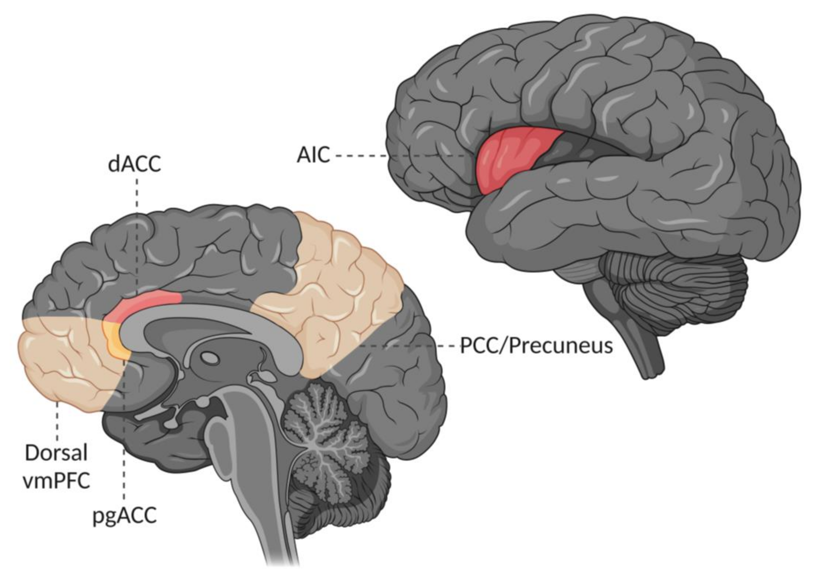
Abstract
:1. Introduction
2. Possible Causes of Altered Dorsal Ventromedial Prefrontal Cortex Behaviour in People with Chronic Pain
2.1. Pain Versus No Pain during Scanning
2.2. Inter-Individual Differences in Pain Processing
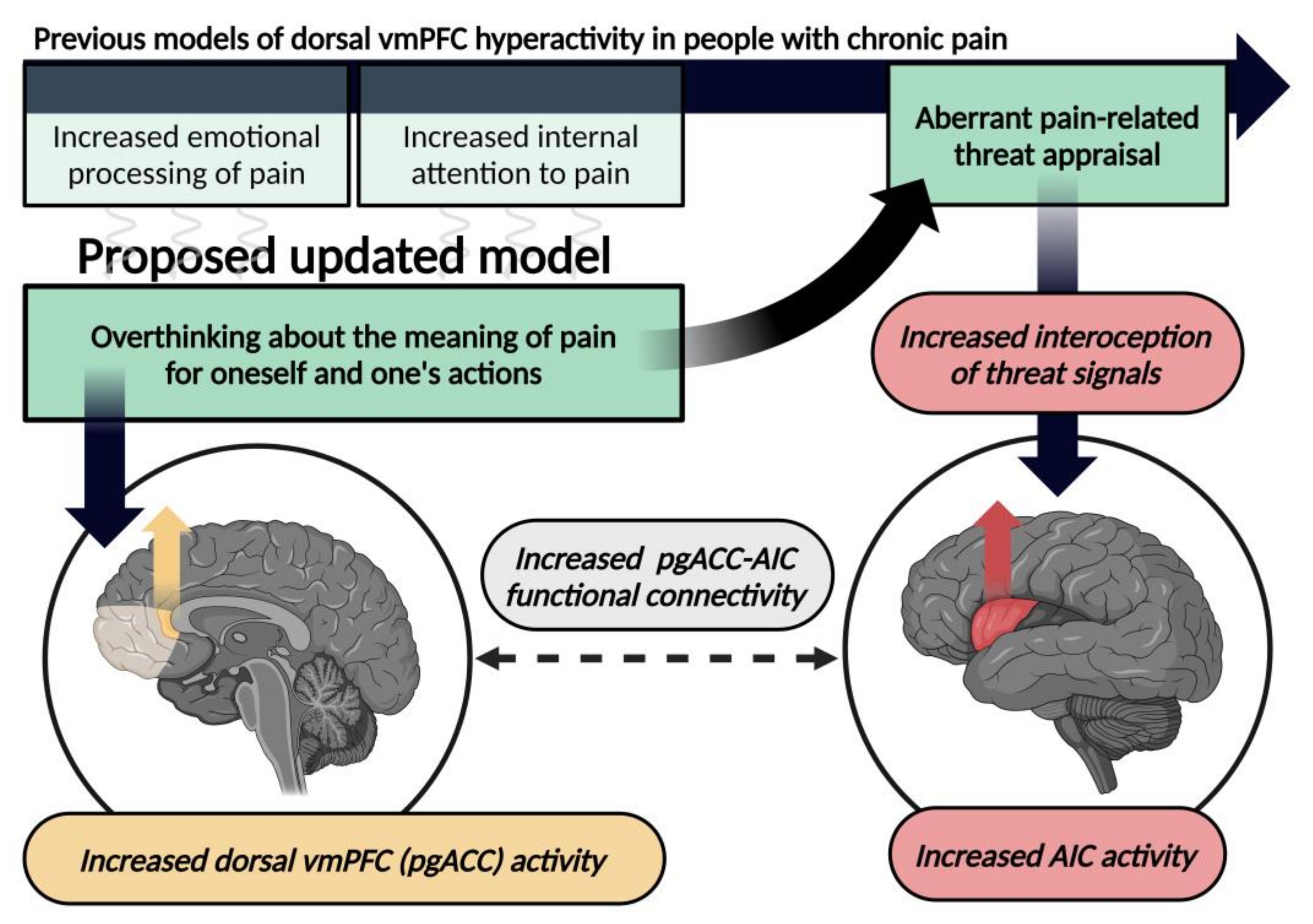
3. Revisiting Previous Models of Altered Dorsal Ventromedial Prefrontal Cortex Behaviour in People with Chronic Pain
3.1. Increased Emotional Processing of Pain
3.2. Aberrant Appraisal of Threat in the Context of Pain
3.3. Increased Internal Attention to Pain
4. An Updated Perspective on the Meaning of Altered Dorsal Ventromedial Prefrontal Cortex Behaviour in People with Chronic Pain
4.1. Shared Mechanisms with Placebo Analgesia
4.2. The Possible Role of the Pregenual Anterior Cingulate Cortex and Independence of Afferent Nociception
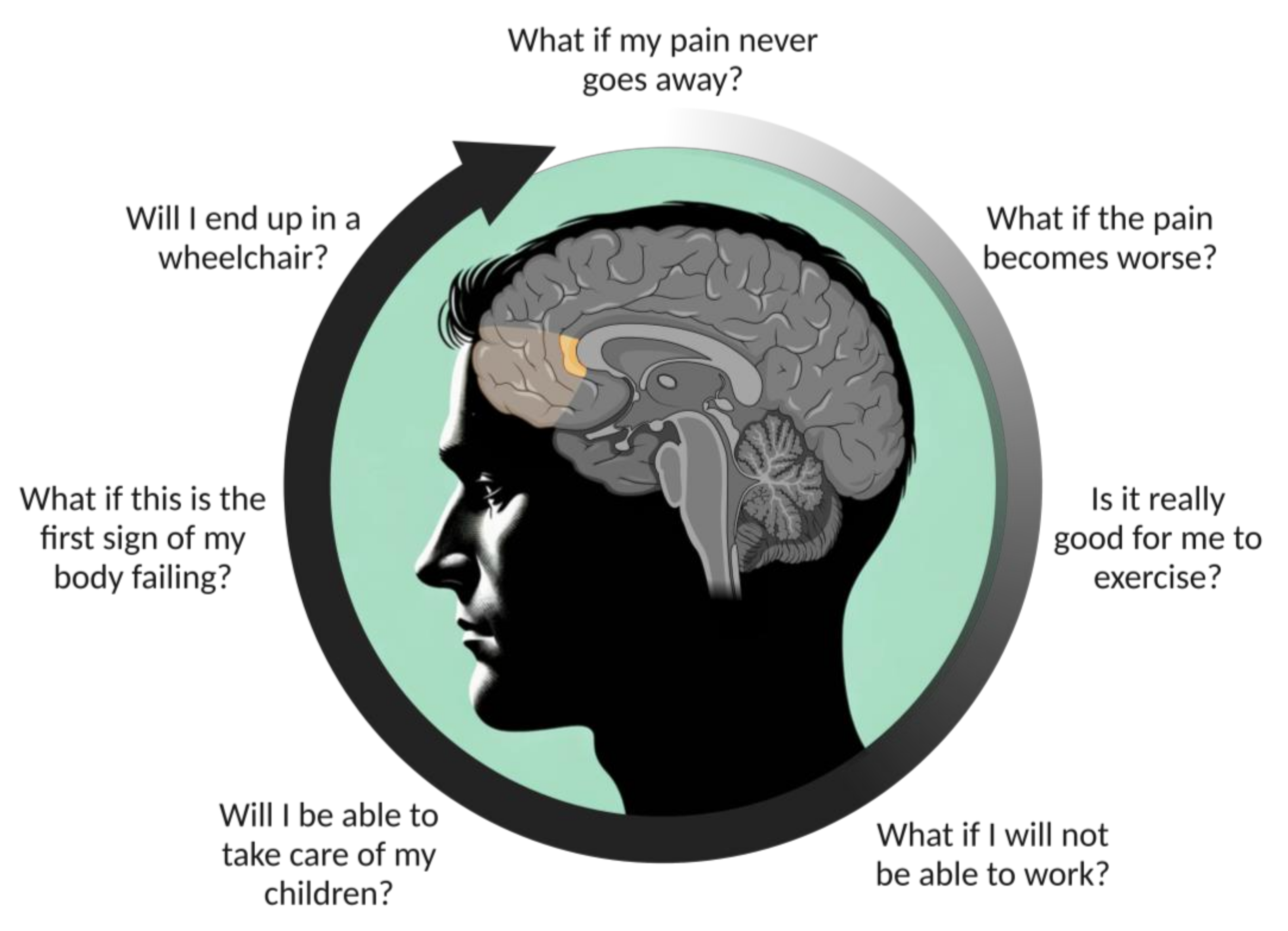
4.3. Overthinking about the Meaning of Pain for Oneself and One’s Actions
5. Anterior Insula Hyper-Connectivity to the Dorsal Ventromedial Prefrontal Cortex
6. Conclusions
Author Contributions
Funding
Institutional Review Board Statement
Informed Consent Statement
Data Availability Statement
Conflicts of Interest
References
- Tracey, I. Why pain hurts. Trends Cogn. Sci. 2022, 26, 1070–1072. [Google Scholar] [CrossRef]
- Cohen, S.P.; Vase, L.; Hooten, W.M. Chronic pain: An update on burden, best practices, and new advances. Lancet 2021, 397, 2082–2097. [Google Scholar] [CrossRef]
- Reddan, M.C.; Wager, T.D. Brain systems at the intersection of chronic pain and self-regulation. Neurosci. Lett. 2019, 702, 24–33. [Google Scholar] [CrossRef]
- van der Miesen, M.M.; Lindquist, M.A.; Wager, T.D. Neuroimaging-based biomarkers for pain: State of the field and current directions. Pain Rep. 2019, 4, e751. [Google Scholar] [CrossRef]
- Kucyi, A.; Davis, K.D. The dynamic pain connectome. Trends Neurosci. 2015, 38, 86–95. [Google Scholar] [CrossRef]
- Apkarian, V.A.; Hashmi, J.A.; Baliki, M.N. Pain and the brain: Specificity and plasticity of the brain in clinical chronic pain. Pain 2011, 152 (Suppl. S3), S49–S64. [Google Scholar] [CrossRef] [PubMed]
- Baliki, M.N.; Apkarian, A.V. Nociception, Pain, Negative Moods, and Behavior Selection. Neuron 2015, 87, 474–491. [Google Scholar] [CrossRef] [PubMed]
- Koban, L.; Gianaros, P.J.; Kober, H.; Wager, T.D. The self in context: Brain systems linking mental and physical health. Nat. Rev. Neurosci. 2021, 22, 309–322. [Google Scholar] [CrossRef] [PubMed]
- Pinto, A.M.; Geenen, R.; Wager, T.D.; Lumley, M.A.; Hauser, W.; Kosek, E.; Ablin, J.N.; Amris, K.; Branco, J.; Buskila, D.; et al. Emotion regulation and the salience network: A hypothetical integrative model of fibromyalgia. Nat. Rev. Rheumatol. 2023, 19, 44–60. [Google Scholar] [CrossRef] [PubMed]
- Johansson, E.; Coppieters, I.; Nijs, J. The default mode of chronic pain: What does it mean and how should we frame it to our patients? J. Spine Pract. 2023, 2, 32–42. [Google Scholar] [CrossRef]
- Seeley, W.W. The Salience Network: A Neural System for Perceiving and Responding to Homeostatic Demands. J. Neurosci. 2019, 39, 9878–9882. [Google Scholar] [CrossRef]
- Seeley, W.W.; Menon, V.; Schatzberg, A.F.; Keller, J.; Glover, G.H.; Kenna, H.; Reiss, A.L.; Greicius, M.D. Dissociable intrinsic connectivity networks for salience processing and executive control. J. Neurosci. 2007, 27, 2349–2356. [Google Scholar] [CrossRef] [PubMed]
- Bartra, O.; McGuire, J.T.; Kable, J.W. The valuation system: A coordinate-based meta-analysis of BOLD fMRI experiments examining neural correlates of subjective value. Neuroimage 2013, 76, 412–427. [Google Scholar] [CrossRef]
- Lindquist, K.A.; Satpute, A.B.; Wager, T.D.; Weber, J.; Barrett, L.F. The Brain Basis of Positive and Negative Affect: Evidence from a Meta-Analysis of the Human Neuroimaging Literature. Cereb. Cortex 2016, 26, 1910–1922. [Google Scholar] [CrossRef] [PubMed]
- Fouragnan, E.; Retzler, C.; Philiastides, M.G. Separate neural representations of prediction error valence and surprise: Evidence from an fMRI meta-analysis. Hum. Brain Mapp. 2018, 39, 2887–2906. [Google Scholar] [CrossRef]
- Lee, S.A.; Jae-Joong, L.; Han, J.; Choi, M.; Wager, T.D.; Woo, C.W. Brain representations of affective valence and intensity of in sustained pleasure and pain. bioRxiv 2023. [CrossRef]
- Uddin, L.Q. Salience processing and insular cortical function and dysfunction. Nat. Rev. Neurosci. 2015, 16, 55–61. [Google Scholar] [CrossRef]
- Eccleston, C.; Crombez, G. Pain demands attention: A cognitive-affective model of the interruptive function of pain. Psychol. Bull. 1999, 125, 356–366. [Google Scholar] [CrossRef]
- Wager, T.D.; Atlas, L.Y.; Lindquist, M.A.; Roy, M.; Woo, C.W.; Kross, E. An fMRI-based neurologic signature of physical pain. N. Engl. J. Med. 2013, 368, 1388–1397. [Google Scholar] [CrossRef]
- Beissner, F.; Meissner, K.; Bar, K.J.; Napadow, V. The autonomic brain: An activation likelihood estimation meta-analysis for central processing of autonomic function. J. Neurosci. 2013, 33, 10503–10511. [Google Scholar] [CrossRef] [PubMed]
- Ferraro, S.; Klugah-Brown, B.; Tench, C.R.; Bazinet, V.; Bore, M.C.; Nigri, A.; Demichelis, G.; Bruzzone, M.G.; Palermo, S.; Zhao, W.; et al. The central autonomic system revisited—Convergent evidence for a regulatory role of the insular and midcingulate cortex from neuroimaging meta-analyses. Neurosci. Biobehav. Rev. 2022, 142, 104915. [Google Scholar] [CrossRef] [PubMed]
- Kragel, P.A.; LaBar, K.S. Multivariate neural biomarkers of emotional states are categorically distinct. Soc. Cogn. Affect. Neurosci. 2015, 10, 1437–1448. [Google Scholar] [CrossRef] [PubMed]
- Wager, T.D.; Kang, J.; Johnson, T.D.; Nichols, T.E.; Satpute, A.B.; Barrett, L.F. A Bayesian model of category-specific emotional brain responses. PLoS Comput. Biol. 2015, 11, e1004066. [Google Scholar] [CrossRef]
- Ceko, M.; Kragel, P.A.; Woo, C.W.; Lopez-Sola, M.; Wager, T.D. Common and stimulus-type-specific brain representations of negative affect. Nat. Neurosci. 2022, 25, 760–770. [Google Scholar] [CrossRef]
- Yeo, B.T.; Krienen, F.M.; Sepulcre, J.; Sabuncu, M.R.; Lashkari, D.; Hollinshead, M.; Roffman, J.L.; Smoller, J.W.; Zöllei, L.; Polimeni, J.R.; et al. The organization of the human cerebral cortex estimated by intrinsic functional connectivity. J. Neurophysiol. 2011, 106, 1125–1165. [Google Scholar] [PubMed]
- Corbetta, M.; Patel, G.; Shulman, G.L. The reorienting system of the human brain: From environment to theory of mind. Neuron 2008, 58, 306–324. [Google Scholar] [CrossRef]
- Fox, M.D.; Snyder, A.Z.; Vincent, J.L.; Corbetta, M.; Van Essen, D.C.; Raichle, M.E. The human brain is intrinsically organized into dynamic, anticorrelated functional networks. Proc. Natl. Acad. Sci. USA 2005, 102, 9673–9678. [Google Scholar] [CrossRef]
- Raichle, M.E.; MacLeod, A.M.; Snyder, A.Z.; Powers, W.J.; Gusnard, D.A.; Shulman, G.L. A default mode of brain function. Proc. Natl. Acad. Sci. USA 2001, 98, 676–682. [Google Scholar] [CrossRef]
- Menon, V. 20 years of the default mode network: A review and synthesis. Neuron 2023, 111, 2469–2487. [Google Scholar] [CrossRef]
- Raichle, M.E. The brain’s default mode network. Annu. Rev. Neurosci. 2015, 38, 433–447. [Google Scholar] [CrossRef]
- Spreng, R.N.; Mar, R.A.; Kim, A.S. The common neural basis of autobiographical memory, prospection, navigation, theory of mind, and the default mode: A quantitative meta-analysis. J. Cogn. Neurosci. 2009, 21, 489–510. [Google Scholar] [CrossRef]
- Schacter, D.L.; Addis, D.R.; Buckner, R.L. Remembering the past to imagine the future: The prospective brain. Nat. Rev. Neurosci. 2007, 8, 657–661. [Google Scholar] [CrossRef]
- Warren, K.N.; Hermiller, M.S.; Nilakantan, A.S.; O’Neil, J.; Palumbo, R.T.; Voss, J.L. Increased fMRI activity correlations in autobiographical memory versus resting states. Hum. Brain Mapp. 2018, 39, 4312–4321. [Google Scholar] [CrossRef]
- Fox, K.C.; Spreng, R.N.; Ellamil, M.; Andrews-Hanna, J.R.; Christoff, K. The wandering brain: Meta-analysis of functional neuroimaging studies of mind-wandering and related spontaneous thought processes. Neuroimage 2015, 111, 611–621. [Google Scholar] [CrossRef]
- Kucyi, A.; Davis, K.D. Dynamic functional connectivity of the default mode network tracks daydreaming. Neuroimage 2014, 100, 471–480. [Google Scholar] [CrossRef] [PubMed]
- Christoff, K.; Irving, Z.C.; Fox, K.C.; Spreng, R.N.; Andrews-Hanna, J.R. Mind-wandering as spontaneous thought: A dynamic framework. Nat. Rev. Neurosci. 2016, 17, 718–731. [Google Scholar] [CrossRef] [PubMed]
- Andrews-Hanna, J.R.; Reidler, J.S.; Sepulcre, J.; Poulin, R.; Buckner, R.L. Functional-anatomic fractionation of the brain’s default network. Neuron 2010, 65, 550–562. [Google Scholar] [CrossRef] [PubMed]
- Ferraro, S.; Klugah-Brown, B.; Tench, C.R.; Yao, S.; Nigri, A.; Demichelis, G.; Pinardi, C.; Bruzzone, M.G.; Becker, B. Dysregulated anterior insula reactivity as robust functional biomarker for chronic pain-Meta-analytic evidence from neuroimaging studies. Hum. Brain Mapp. 2022, 43, 998–1010. [Google Scholar] [CrossRef] [PubMed]
- Jensen, K.B.; Regenbogen, C.; Ohse, M.C.; Frasnelli, J.; Freiherr, J.; Lundstrom, J.N. Brain activations during pain: A neuroimaging meta-analysis of patients with pain and healthy controls. Pain 2016, 157, 1279–1286. [Google Scholar] [CrossRef] [PubMed]
- Xu, A.; Larsen, B.; Henn, A.; Baller, E.B.; Scott, J.C.; Sharma, V.; Adebimpe, A.; Basbaum, A.I.; Corder, G.; Dworkin, R.H. Brain Responses to Noxious Stimuli in Patients With Chronic Pain: A Systematic Review and Meta-analysis. JAMA Netw. Open 2021, 4, e2032236. [Google Scholar] [CrossRef]
- Baliki, M.N.; Mansour, A.R.; Baria, A.T.; Apkarian, A.V. Functional reorganization of the default mode network across chronic pain conditions. PLoS ONE 2014, 9, e106133. [Google Scholar] [CrossRef]
- Baliki, M.N.; Baria, A.T.; Apkarian, A.V. The cortical rhythms of chronic back pain. J. Neurosci. 2011, 31, 13981–13990. [Google Scholar] [CrossRef]
- Napadow, V.; LaCount, L.; Park, K.; As-Sanie, S.; Clauw, D.J.; Harris, R.E. Intrinsic brain connectivity in fibromyalgia is associated with chronic pain intensity. Arthritis Rheum. 2010, 62, 2545–2555. [Google Scholar] [CrossRef]
- Tu, Y.; Jung, M.; Gollub, R.L.; Napadow, V.; Gerber, J.; Ortiz, A.; Lang, C.; Mawla, I.; Shen, W.; Chan, S.-T.; et al. Abnormal medial prefrontal cortex functional connectivity and its association with clinical symptoms in chronic low back pain. Pain 2019, 160, 1308–1318. [Google Scholar] [CrossRef]
- Loggia, M.L.; Kim, J.; Gollub, R.L.; Vangel, M.G.; Kirsch, I.; Kong, J.; Wasan, A.D.; Napadow, V. Default mode network connectivity encodes clinical pain: An arterial spin labeling study. Pain 2013, 154, 24–33. [Google Scholar] [CrossRef]
- Kim, J.; Mawla, I.; Kong, J.; Lee, J.; Gerber, J.; Ortiz, A.; Kim, H.; Chan, S.-T.; Loggia, M.L.; Wasan, A.D.; et al. Somatotopically specific primary somatosensory connectivity to salience and default mode networks encodes clinical pain. Pain 2019, 160, 1594–1605. [Google Scholar] [CrossRef]
- Ichesco, E.; Puiu, T.; Hampson, J.P.; Kairys, A.E.; Clauw, D.J.; Harte, S.E.; Peltier, S.; Harris, R.; Schmidt-Wilcke, T. Altered fMRI resting-state connectivity in individuals with fibromyalgia on acute pain stimulation. Eur. J. Pain 2016, 20, 1079–1089. [Google Scholar] [CrossRef]
- Ceko, M.; Frangos, E.; Gracely, J.; Richards, E.; Wang, B.; Schweinhardt, P.; Bushnell, M.C. Default mode network changes in fibromyalgia patients are largely dependent on current clinical pain. Neuroimage 2020, 216, 116877. [Google Scholar] [CrossRef] [PubMed]
- Bolwerk, A.; Seifert, F.; Maihofner, C. Altered resting-state functional connectivity in complex regional pain syndrome. J. Pain 2013, 14, 1107–1115.e8. [Google Scholar] [CrossRef] [PubMed]
- Alshelh, Z.; Marciszewski, K.K.; Akhter, R.; Di Pietro, F.; Mills, E.P.; Vickers, E.R.; Peck, C.; Murray, G.; Henderson, L. Disruption of default mode network dynamics in acute and chronic pain states. Neuroimage Clin. 2018, 17, 222–231. [Google Scholar] [CrossRef] [PubMed]
- Otti, A.; Guendel, H.; Wohlschlager, A.; Zimmer, C.; Noll-Hussong, M. Frequency shifts in the anterior default mode network and the salience network in chronic pain disorder. BMC Psychiatry 2013, 13, 84. [Google Scholar] [CrossRef]
- Hashmi, J.A.; Baliki, M.N.; Huang, L.; Baria, A.T.; Torbey, S.; Hermann, K.M.; Schnitzer, T.J.; Apkarian, A.V. Shape shifting pain: Chronification of back pain shifts brain representation from nociceptive to emotional circuits. Brain 2013, 136 Pt 9, 2751–2768. [Google Scholar] [CrossRef]
- Baliki, M.N.; Chialvo, D.R.; Geha, P.Y.; Levy, R.M.; Harden, R.N.; Parrish, T.B.; Apkarian, A.V. Chronic pain and the emotional brain: Specific brain activity associated with spontaneous fluctuations of intensity of chronic back pain. J. Neurosci. 2006, 26, 12165–12173. [Google Scholar] [CrossRef]
- Baliki, M.N.; Geha, P.Y.; Apkarian, A.V.; Chialvo, D.R. Beyond feeling: Chronic pain hurts the brain, disrupting the default-mode network dynamics. J. Neurosci. 2008, 28, 1398–1403. [Google Scholar] [CrossRef] [PubMed]
- Ceko, M.; Gracely, J.L.; Fitzcharles, M.A.; Seminowicz, D.A.; Schweinhardt, P.; Bushnell, M.C. Is a Responsive Default Mode Network Required for Successful Working Memory Task Performance? J. Neurosci. 2015, 35, 11595–11605. [Google Scholar] [CrossRef]
- Weissman-Fogel, I.; Moayedi, M.; Tenenbaum, H.C.; Goldberg, M.B.; Freeman, B.V.; Davis, K.D. Abnormal cortical activity in patients with temporomandibular disorder evoked by cognitive and emotional tasks. Pain 2011, 152, 384–396. [Google Scholar] [CrossRef] [PubMed]
- Alhajri, N.; Boudreau, S.A.; Graven-Nielsen, T. Decreased Default Mode Network Connectivity Following 24 Hours of Capsaicin-induced Pain Persists During Immediate Pain Relief and Facilitation. J. Pain 2023, 24, 796–811. [Google Scholar] [CrossRef]
- Makovac, E.; Dipasquale, O.; Jackson, J.B.; Medina, S.; O’Daly, O.; O’Muircheartaigh, J.; Rubio, A.d.L.; Williams, S.C.R.; McMahon, S.B.; Howard, M.A. Sustained perturbation in functional connectivity induced by cold pain. Eur. J. Pain 2020, 24, 1850–1861. [Google Scholar] [CrossRef] [PubMed]
- Kohoutova, L.; Atlas, L.Y.; Buchel, C.; Buhle, J.T.; Geuter, S.; Jepma, M.; Koban, L.; Krishnan, A.; Lee, D.H.; Lee, S.; et al. Individual variability in brain representations of pain. Nat. Neurosci. 2022, 25, 749–759. [Google Scholar] [CrossRef]
- Geuter, S.; Reynolds Losin, E.A.; Roy, M.; Atlas, L.Y.; Schmidt, L.; Krishnan, A.; Koban, L.; Wager, T.D.; Lindquist, M.A. Multiple Brain Networks Mediating Stimulus-Pain Relationships in Humans. Cereb. Cortex 2020, 30, 4204–4219. [Google Scholar] [CrossRef]
- Mayr, A.; Jahn, P.; Stankewitz, A.; Deak, B.; Winkler, A.; Witkovsky, V.; Eren, O.; Straube, A.; Schulz, E. Patients with chronic pain exhibit individually unique cortical signatures of pain encoding. Hum. Brain Mapp. 2022, 43, 1676–1693. [Google Scholar] [CrossRef]
- Mayr, A.; Jahn, P.; Deak, B.; Stankewitz, A.; Devulapally, V.; Witkovsky, V.; Dietrich, O.; Schulz, E. Individually unique dynamics of cortical connectivity reflect the ongoing intensity of chronic pain. Pain 2022, 163, 1987–1998. [Google Scholar] [CrossRef]
- Baliki, M.N.; Petre, B.; Torbey, S.; Herrmann, K.M.; Huang, L.; Schnitzer, T.J.; Fields, H.L.; Apkarian, A.V. Corticostriatal functional connectivity predicts transition to chronic back pain. Nat. Neurosci. 2012, 15, 1117–1119. [Google Scholar] [CrossRef] [PubMed]
- Loffler, M.; Levine, S.M.; Usai, K.; Desch, S.; Kandic, M.; Nees, F.; Flor, H. Corticostriatal circuits in the transition to chronic back pain: The predictive role of reward learning. Cell Rep. Med. 2022, 3, 100677. [Google Scholar] [CrossRef] [PubMed]
- de la Vega, A.; Chang, L.J.; Banich, M.T.; Wager, T.D.; Yarkoni, T. Large-Scale Meta-Analysis of Human Medial Frontal Cortex Reveals Tripartite Functional Organization. J. Neurosci. 2016, 36, 6553–6562. [Google Scholar] [CrossRef] [PubMed]
- Kragel, P.A.; Kano, M.; Van Oudenhove, L.; Ly, H.G.; Dupont, P.; Rubio, A.; Delon-Martin, C.; Bonaz, B.L.; Manuck, S.B.; Gianaros, P.J.; et al. Generalizable representations of pain, cognitive control, and negative emotion in medial frontal cortex. Nat. Neurosci. 2018, 21, 283–289. [Google Scholar] [CrossRef] [PubMed]
- Schneider, B.; Koenigs, M. Human lesion studies of ventromedial prefrontal cortex. Neuropsychologia 2017, 107, 84–93. [Google Scholar] [CrossRef] [PubMed]
- Gilam, G.; Gross, J.J.; Wager, T.D.; Keefe, F.J.; Mackey, S.C. What Is the Relationship between Pain and Emotion? Bridging Constructs and Communities. Neuron 2020, 107, 17–21. [Google Scholar] [CrossRef] [PubMed]
- Roy, M.; Piche, M.; Chen, J.I.; Peretz, I.; Rainville, P. Cerebral and spinal modulation of pain by emotions. Proc. Natl. Acad. Sci. USA 2009, 106, 20900–20905. [Google Scholar] [CrossRef]
- Morelli, S.A.; Sacchet, M.D.; Zaki, J. Common and distinct neural correlates of personal and vicarious reward: A quantitative meta-analysis. Neuroimage 2015, 112, 244–253. [Google Scholar] [CrossRef]
- Smith, D.V.; Hayden, B.Y.; Truong, T.K.; Song, A.W.; Platt, M.L.; Huettel, S.A. Distinct value signals in anterior and posterior ventromedial prefrontal cortex. J. Neurosci. 2010, 30, 2490–2495. [Google Scholar] [CrossRef]
- Koban, L.; Lee, S.; Schelski, D.S.; Simon, M.C.; Lerman, C.; Weber, B.; Kable, J.W.; Plassmann, H. An fMRI-Based Brain Marker of Individual Differences in Delay Discounting. J. Neurosci. 2023, 43, 1600–1613. [Google Scholar] [CrossRef] [PubMed]
- Kolling, N.; Scholl, J.; Chekroud, A.; Trier, H.A.; Rushworth, M.F.S. Prospection, Perseverance, and Insight in Sequential Behavior. Neuron 2018, 99, 1069–1082.e7. [Google Scholar] [CrossRef] [PubMed]
- Kolling, N.; Wittmann, M.; Rushworth, M.F.S. Multiple neural mechanisms of decision making and their competition under changing risk pressure. Neuron 2014, 81, 1190–1202. [Google Scholar] [CrossRef]
- Wittmann, M.K.; Kolling, N.; Akaishi, R.; Chau, B.K.; Brown, J.W.; Nelissen, N.; Rushworth, M.F.S. Predictive decision making driven by multiple time-linked reward representations in the anterior cingulate cortex. Nat. Commun. 2016, 7, 12327. [Google Scholar] [CrossRef]
- Denny, B.T.; Kober, H.; Wager, T.D.; Ochsner, K.N. A meta-analysis of functional neuroimaging studies of self- and other judgments reveals a spatial gradient for mentalizing in medial prefrontal cortex. J. Cogn. Neurosci. 2012, 24, 1742–1752. [Google Scholar] [CrossRef]
- Murray, R.J.; Schaer, M.; Debbane, M. Degrees of separation: A quantitative neuroimaging meta-analysis investigating self-specificity and shared neural activation between self- and other-reflection. Neurosci. Biobehav. Rev. 2012, 36, 1043–1059. [Google Scholar] [CrossRef] [PubMed]
- Qin, P.; Northoff, G. How is our self related to midline regions and the default-mode network? Neuroimage 2011, 57, 1221–1233. [Google Scholar] [CrossRef]
- Battaglia, S.; Garofalo, S.; di Pellegrino, G.; Starita, F. Revaluing the Role of vmPFC in the Acquisition of Pavlovian Threat Conditioning in Humans. J. Neurosci. 2020, 40, 8491–8500. [Google Scholar] [CrossRef]
- Motzkin, J.C.; Hiser, J.; Carroll, I.; Wolf, R.; Baskaya, M.K.; Koenigs, M.; Atlas, L.Y. Human ventromedial prefrontal cortex lesions enhance the effect of expectations on pain perception. Cortex 2023, 166, 188–206. [Google Scholar] [CrossRef]
- Raja, S.N.; Carr, D.B.; Cohen, M.; Finnerup, N.B.; Flor, H.; Gibson, S.; Keefe, F.J.; Mogil, J.S.; Ringkamp, M.; Sluka, K.A.; et al. The revised International Association for the Study of Pain definition of pain: Concepts, challenges, and compromises. Pain 2020, 161, 1976–1982. [Google Scholar] [CrossRef] [PubMed]
- Zhou, F.; Zhao, W.; Qi, Z.; Geng, Y.; Yao, S.; Kendrick, K.M.; Wager, T.D.; Becker, B. A distributed fMRI-based signature for the subjective experience of fear. Nat. Commun. 2021, 12, 6643. [Google Scholar] [CrossRef]
- Savage, H.S.; Davey, C.G.; Wager, T.D.; Garfinkel, S.N.; Moffat, B.A.; Glarin, R.K.; Harrison, B.J. Neural mediators of subjective and autonomic responding during threat learning and regulation. Neuroimage 2021, 245, 118643. [Google Scholar] [CrossRef]
- Fullana, M.A.; Harrison, B.J.; Soriano-Mas, C.; Vervliet, B.; Cardoner, N.; Avila-Parcet, A.; Radua, J. Neural signatures of human fear conditioning: An updated and extended meta-analysis of fMRI studies. Mol. Psychiatry 2016, 21, 500–508. [Google Scholar] [CrossRef] [PubMed]
- Savage, H.S.; Davey, C.G.; Fullana, M.A.; Harrison, B.J. Clarifying the neural substrates of threat and safety reversal learning in humans. Neuroimage 2020, 207, 116427. [Google Scholar] [CrossRef]
- Marstaller, L.; Burianova, H.; Reutens, D.C. Adaptive contextualization: A new role for the default mode network in affective learning. Hum. Brain Mapp. 2017, 38, 1082–1091. [Google Scholar] [CrossRef]
- Harrison, B.J.; Fullana, M.A.; Via, E.; Soriano-Mas, C.; Vervliet, B.; Martinez-Zalacain, I.; Pujol, J.; Davey, C.G.; Kircher, T.; Straube, B.; et al. Human ventromedial prefrontal cortex and the positive affective processing of safety signals. Neuroimage 2017, 152, 12–18. [Google Scholar] [CrossRef]
- Milad, M.R.; Wright, C.I.; Orr, S.P.; Pitman, R.K.; Quirk, G.J.; Rauch, S.L. Recall of fear extinction in humans activates the ventromedial prefrontal cortex and hippocampus in concert. Biol. Psychiatry 2007, 62, 446–454. [Google Scholar] [CrossRef]
- Tashjian, S.M.; Zbozinek, T.D.; Mobbs, D. A Decision Architecture for Safety Computations. Trends Cogn. Sci. 2021, 25, 342–354. [Google Scholar] [CrossRef] [PubMed]
- Wallis, C.U.; Cardinal, R.N.; Alexander, L.; Roberts, A.C.; Clarke, H.F. Opposing roles of primate areas 25 and 32 and their putative rodent homologs in the regulation of negative emotion. Proc. Natl. Acad. Sci. USA 2017, 114, E4075–E4084. [Google Scholar] [CrossRef] [PubMed]
- Kucyi, A.; Moayedi, M.; Weissman-Fogel, I.; Goldberg, M.B.; Freeman, B.V.; Tenenbaum, H.C.; Davis, K.D. Enhanced medial prefrontal-default mode network functional connectivity in chronic pain and its association with pain rumination. J. Neurosci. 2014, 34, 3969–3975. [Google Scholar] [CrossRef]
- Kucyi, A.; Salomons, T.V.; Davis, K.D. Mind wandering away from pain dynamically engages antinociceptive and default mode brain networks. Proc. Natl. Acad. Sci. USA 2013, 110, 18692–18697. [Google Scholar] [CrossRef] [PubMed]
- Chen, Q.; Heinricher, M.M. Shifting the Balance: How Top-Down and Bottom-Up Input Modulate Pain via the Rostral Ventromedial Medulla. Front. Pain Res. 2022, 3, 932476. [Google Scholar] [CrossRef] [PubMed]
- Fields, H. State-dependent opioid control of pain. Nat. Rev. Neurosci. 2004, 5, 565–575. [Google Scholar] [CrossRef]
- Leknes, S.; Berna, C.; Lee, M.C.; Snyder, G.D.; Biele, G.; Tracey, I. The importance of context: When relative relief renders pain pleasant. Pain 2013, 154, 402–410. [Google Scholar] [CrossRef] [PubMed]
- Leknes, S.; Lee, M.; Berna, C.; Andersson, J.; Tracey, I. Relief as a reward: Hedonic and neural responses to safety from pain. PLoS ONE 2011, 6, e17870. [Google Scholar] [CrossRef] [PubMed]
- Woo, C.W.; Roy, M.; Buhle, J.T.; Wager, T.D. Distinct brain systems mediate the effects of nociceptive input and self-regulation on pain. PLoS Biol. 2015, 13, e1002036. [Google Scholar] [CrossRef] [PubMed]
- Schulz, E.; Stankewitz, A.; Witkovsky, V.; Winkler, A.M.; Tracey, I. Strategy-dependent modulation of cortical pain circuits for the attenuation of pain. Cortex 2019, 113, 255–266. [Google Scholar] [CrossRef] [PubMed]
- Valet, M.; Sprenger, T.; Boecker, H.; Willoch, F.; Rummeny, E.; Conrad, B.; Erhard, P.; Tolle, T.R. Distraction modulates connectivity of the cingulo-frontal cortex and the midbrain during pain—An fMRI analysis. Pain 2004, 109, 399–408. [Google Scholar] [CrossRef]
- Bantick, S.J.; Wise, R.G.; Ploghaus, A.; Clare, S.; Smith, S.M.; Tracey, I. Imaging how attention modulates pain in humans using functional MRI. Brain 2002, 125 Pt 2, 310–319. [Google Scholar] [CrossRef]
- Atlas, L.Y.; Bolger, N.; Lindquist, M.A.; Wager, T.D. Brain mediators of predictive cue effects on perceived pain. J. Neurosci. 2010, 30, 12964–12977. [Google Scholar] [CrossRef]
- Atlas, L.Y.; Dildine, T.C.; Palacios-Barrios, E.E.; Yu, Q.; Reynolds, R.C.; Banker, L.A.; Grant, S.S.; Pine, D.S. Instructions and experiential learning have similar impacts on pain and pain-related brain responses but produce dissociations in value-based reversal learning. eLife 2022, 11, e73353. [Google Scholar] [CrossRef]
- Atlas, L.Y.; Wager, T.D. A meta-analysis of brain mechanisms of placebo analgesia: Consistent findings and unanswered questions. Handb. Exp. Pharmacol. 2014, 225, 37–69. [Google Scholar]
- Amanzio, M.; Benedetti, F.; Porro, C.A.; Palermo, S.; Cauda, F. Activation likelihood estimation meta-analysis of brain correlates of placebo analgesia in human experimental pain. Hum. Brain Mapp. 2013, 34, 738–752. [Google Scholar] [CrossRef]
- Jepma, M.; Koban, L.; van Doorn, J.; Jones, M.; Wager, T.D. Behavioural and neural evidence for self-reinforcing expectancy effects on pain. Nat. Hum. Behav. 2018, 2, 838–855. [Google Scholar] [CrossRef] [PubMed]
- Roy, M.; Shohamy, D.; Daw, N.; Jepma, M.; Wimmer, G.E.; Wager, T.D. Representation of aversive prediction errors in the human periaqueductal gray. Nat. Neurosci. 2014, 17, 1607–1612. [Google Scholar] [CrossRef] [PubMed]
- Lopez-Sola, M.; Koban, L.; Wager, T.D. Transforming Pain With Prosocial Meaning: A Functional Magnetic Resonance Imaging Study. Psychosom. Med. 2018, 80, 814–825. [Google Scholar] [CrossRef] [PubMed]
- Bingel, U.; Rose, M.; Glascher, J.; Buchel, C. fMRI reveals how pain modulates visual object processing in the ventral visual stream. Neuron 2007, 55, 157–167. [Google Scholar] [CrossRef]
- Eippert, F.; Bingel, U.; Schoell, E.D.; Yacubian, J.; Klinger, R.; Lorenz, J.; Büchel, C. Activation of the opioidergic descending pain control system underlies placebo analgesia. Neuron 2009, 63, 533–543. [Google Scholar] [CrossRef]
- Wager, T.D.; Atlas, L.Y.; Leotti, L.A.; Rilling, J.K. Predicting individual differences in placebo analgesia: Contributions of brain activity during anticipation and pain experience. J. Neurosci. 2011, 31, 439–452. [Google Scholar] [CrossRef]
- Salomons, T.V.; Johnstone, T.; Backonja, M.M.; Davidson, R.J. Perceived controllability modulates the neural response to pain. J. Neurosci. 2004, 24, 7199–7203. [Google Scholar] [CrossRef] [PubMed]
- Salomons, T.V.; Johnstone, T.; Backonja, M.M.; Shackman, A.J.; Davidson, R.J. Individual differences in the effects of perceived controllability on pain perception: Critical role of the prefrontal cortex. J. Cogn. Neurosci. 2007, 19, 993–1003. [Google Scholar] [CrossRef]
- Lopez-Sola, M.; Geuter, S.; Koban, L.; Coan, J.A.; Wager, T.D. Brain mechanisms of social touch-induced analgesia in females. Pain 2019, 160, 2072–2085. [Google Scholar] [CrossRef] [PubMed]
- Zunhammer, M.; Spisak, T.; Wager, T.D.; Bingel, U.; Placebo Imaging, C. Meta-analysis of neural systems underlying placebo analgesia from individual participant fMRI data. Nat. Commun. 2021, 12, 1391. [Google Scholar] [CrossRef] [PubMed]
- Kober, H.; Wager, T.D. Meta-analysis of neuroimaging data. Wiley Interdiscip. Rev. Cogn. Sci. 2010, 1, 293–300. [Google Scholar] [CrossRef] [PubMed]
- Kong, J.; Tu, P.C.; Zyloney, C.; Su, T.P. Intrinsic functional connectivity of the periaqueductal gray, a resting fMRI study. Behav. Brain Res. 2010, 211, 215–219. [Google Scholar] [CrossRef]
- Baliki, M.N.; Geha, P.Y.; Fields, H.L.; Apkarian, A.V. Predicting value of pain and analgesia: Nucleus accumbens response to noxious stimuli changes in the presence of chronic pain. Neuron 2010, 66, 149–160. [Google Scholar] [CrossRef]
- Woo, C.W.; Schmidt, L.; Krishnan, A.; Jepma, M.; Roy, M.; Lindquist, M.A.; Atlas, L.Y.; Wager, T.D. Quantifying cerebral contributions to pain beyond nociception. Nat. Commun. 2017, 8, 14211. [Google Scholar] [CrossRef]
- Gold, M.S.; Gebhart, G.F. Nociceptor sensitization in pain pathogenesis. Nat. Med. 2010, 16, 1248–1257. [Google Scholar] [CrossRef]
- Basbaum, A.I.; Bautista, D.M.; Scherrer, G.; Julius, D. Cellular and molecular mechanisms of pain. Cell 2009, 139, 267–284. [Google Scholar] [CrossRef]
- Nijs, J.; George, J.; Clauw, D.; Fernández-de-las-Peñas, C.; Kosek, E.; Ickmans, K.; Fernández-Carnero, J.; Polli, A.; Kapreli, E.; Huysmans, E.; et al. Central sensitisation in chronic pain conditions: Latest discoveries and their potential for precision medicine. Lancet Rheumatol. 2021, 3, E383–E392. [Google Scholar] [CrossRef] [PubMed]
- Tinnermann, A.; Geuter, S.; Sprenger, C.; Finsterbusch, J.; Buchel, C. Interactions between brain and spinal cord mediate value effects in nocebo hyperalgesia. Science 2017, 358, 105–108. [Google Scholar] [CrossRef] [PubMed]
- Atlas, L.Y. How Instructions, Learning, and Expectations Shape Pain and Neurobiological Responses. Annu. Rev. Neurosci. 2023, 46, 167–189. [Google Scholar] [CrossRef] [PubMed]
- de Lange, F.P.; Heilbron, M.; Kok, P. How Do Expectations Shape Perception? Trends Cogn. Sci. 2018, 22, 764–779. [Google Scholar] [CrossRef]
- Wager, T.D.; Atlas, L.Y. The neuroscience of placebo effects: Connecting context, learning and health. Nat. Rev. Neurosci. 2015, 16, 403–418. [Google Scholar] [CrossRef]
- Atlas, L.Y. A social affective neuroscience lens on placebo analgesia. Trends Cogn. Sci. 2021, 25, 992–1005. [Google Scholar] [CrossRef]
- Geuter, S.; Koban, L.; Wager, T.D. The Cognitive Neuroscience of Placebo Effects: Concepts, Predictions, and Physiology. Annu. Rev. Neurosci. 2017, 40, 167–188. [Google Scholar] [CrossRef]
- Zhang, S.; Mano, H.; Lee, M.; Yoshida, W.; Kawato, M.; Robbins, T.W.; Seymour, B. The control of tonic pain by active relief learning. eLife 2018, 7, e31949. [Google Scholar] [CrossRef]
- Yoshida, W.; Seymour, B.; Koltzenburg, M.; Dolan, R.J. Uncertainty increases pain: Evidence for a novel mechanism of pain modulation involving the periaqueductal gray. J. Neurosci. 2013, 33, 5638–5646. [Google Scholar] [CrossRef]
- Seidel, E.M.; Pfabigan, D.M.; Hahn, A.; Sladky, R.; Grahl, A.; Paul, K.; Kraus, C.; Küblböck, M.; Kranz, G.S.; Hummer, A.; et al. Uncertainty during pain anticipation: The adaptive value of preparatory processes. Hum. Brain Mapp. 2015, 36, 744–755. [Google Scholar] [CrossRef]
- Willems, A.L.; Van Oudenhove, L.; Vervliet, B. Omissions of threat trigger subjective relief and reward prediction error-like signaling in the human reward system. bioRxiv 2023. [Google Scholar] [CrossRef]
- Kolling, N.; Behrens, T.E.; Mars, R.B.; Rushworth, M.F. Neural mechanisms of foraging. Science 2012, 336, 95–98. [Google Scholar] [CrossRef] [PubMed]
- Craig, A.D. How do you feel? Interoception: The sense of the physiological condition of the body. Nat. Rev. Neurosci. 2002, 3, 655–666. [Google Scholar] [CrossRef] [PubMed]
- Craig, A.D. Interoception: The sense of the physiological condition of the body. Curr. Opin. Neurobiol. 2003, 13, 500–505. [Google Scholar] [CrossRef] [PubMed]
- Petre, B.; Kragel, P.; Atlas, L.Y.; Geuter, S.; Jepma, M.; Koban, L.; Krishnan, A.; Lopez-Sola, M.; Losin, E.A.R.; Roy, M.; et al. A multistudy analysis reveals that evoked pain intensity representation is distributed across brain systems. PLoS Biol. 2022, 20, e3001620. [Google Scholar] [CrossRef]
- Fazeli, S.; Buchel, C. Pain-Related Expectation and Prediction Error Signals in the Anterior Insula Are Not Related to Aversiveness. J. Neurosci. 2018, 38, 6461–6474. [Google Scholar] [CrossRef]
- Palermo, S.; Benedetti, F.; Costa, T.; Amanzio, M. Pain anticipation: An activation likelihood estimation meta-analysis of brain imaging studies. Hum. Brain Mapp. 2015, 36, 1648–1661. [Google Scholar] [CrossRef]


Disclaimer/Publisher’s Note: The statements, opinions and data contained in all publications are solely those of the individual author(s) and contributor(s) and not of MDPI and/or the editor(s). MDPI and/or the editor(s) disclaim responsibility for any injury to people or property resulting from any ideas, methods, instructions or products referred to in the content. |
© 2024 by the authors. Licensee MDPI, Basel, Switzerland. This article is an open access article distributed under the terms and conditions of the Creative Commons Attribution (CC BY) license (https://creativecommons.org/licenses/by/4.0/).
Share and Cite
Johansson, E.; Xiong, H.-Y.; Polli, A.; Coppieters, I.; Nijs, J. Towards a Real-Life Understanding of the Altered Functional Behaviour of the Default Mode and Salience Network in Chronic Pain: Are People with Chronic Pain Overthinking the Meaning of Their Pain? J. Clin. Med. 2024, 13, 1645. https://doi.org/10.3390/jcm13061645
Johansson E, Xiong H-Y, Polli A, Coppieters I, Nijs J. Towards a Real-Life Understanding of the Altered Functional Behaviour of the Default Mode and Salience Network in Chronic Pain: Are People with Chronic Pain Overthinking the Meaning of Their Pain? Journal of Clinical Medicine. 2024; 13(6):1645. https://doi.org/10.3390/jcm13061645
Chicago/Turabian StyleJohansson, Elin, Huan-Yu Xiong, Andrea Polli, Iris Coppieters, and Jo Nijs. 2024. "Towards a Real-Life Understanding of the Altered Functional Behaviour of the Default Mode and Salience Network in Chronic Pain: Are People with Chronic Pain Overthinking the Meaning of Their Pain?" Journal of Clinical Medicine 13, no. 6: 1645. https://doi.org/10.3390/jcm13061645