Article

Pulmonary Vascular Resistance to Predict Right Heart Failure in Patients Undergoing Left Ventricular Assist Device Implantation

1
Clinic for Thoracic and Cardiovascular Surgery, Herz- und Diabeteszentrum NRW, Ruhr-Universität Bochum, Georgstraße 11, 32545 Bad Oeynhausen, Germany
2
Clinic for General and Interventional Cardiology/Angiology, Herz- und Diabeteszentrum NRW, Ruhr-Universität Bochum, Georgstraße 11, 32545 Bad Oeynhausen, Germany
*
Author to whom correspondence should be addressed.
These authors contributed equally to this work.
J. Clin. Med. 2024, 13(2), 462; https://doi.org/10.3390/jcm13020462
Submission received: 13 December 2023 / Revised: 4 January 2024 / Accepted: 12 January 2024 / Published: 14 January 2024

Abstract

:
Right heart failure (RHF) is associated with poor outcomes, especially in patients undergoing left ventricular assist device (LVAD) implantation. The aim of this study was to identify predictors of RHF after LVAD implantation. Of 129 consecutive patients (mean age 56 ± 11 years, 89% male) undergoing LVAD implantation, 34 developed RHF. Compared to patients without RHF, those with RHF required longer invasive mechanical ventilation and had longer intensive care unit and hospital stays (p < 0.01). One-year all-cause mortality was significantly higher in patients with versus without RHF after LVAD implantation (29.4% vs. 1.2%; hazard ratio 35.4; 95% confidence interval 4.5–277; p < 0.001). Mortality was highest in patients with delayed RHF after initial LVAD-only implantation (66.7%). Patients who did versus did not develop RHF had significantly higher baseline pulmonary vascular resistance (PVR; 404 ± 375 vs. 234 ± 162 dyn/s/cm5; p = 0.01). PVR > 250 dyn/s/cm5 was a significant predictor of survival in patients with RHF after LVAD implantation. These data confirm the negative impact of RHF on morbidity and mortality after LVAD implantation. Preoperative PVR > 250 dyn/s/cm5 determined using invasive right heart catheterization was an independent predictor of developing RHF after LVAD implantation, and of subsequent mortality, and could be used for risk stratification in the setting for deciding between single or biventricular support strategy.

Graphical Abstract

1. Introduction

The prevalence of heart failure (HF) is increasing globally due to a number of factors, including the aging population demographic and more effective treatments for ischemic heart disease [1]. In addition, therapeutic advances mean that individuals with HF now live longer [1,2]. However, end-stage HF remains a therapeutic challenge, often requiring mechanical circulatory support [3]. Implantation of a left ventricular assist device (LVAD) is the most established approach for permanent mechanical circulatory support, and the use of an LVAD has been shown to be safe and effective, including for a bridge to heart transplantation [3].
Despite this, right HF (RHF) remains a challenge because available therapeutic options are limited, especially in patients ineligible for heart transplantation [3]. RHF is a known driver of both morbidity and mortality, in particular for patients who have an LVAD [4,5]. RHF can sometimes be improved by LVAD implantation because it not only unloads the left ventricle but also relieves pulmonary congestion. However, RHF persists after LVAD implantation in a significant proportion of individuals, and there is a lack of validated predictors of RHF development or resolution after LVAD implantation [3].
It has been suggested that poor right ventricular function might predict postoperative RHF after LVAD implantation [6]. Currently, the most widely used parameters for evaluating postoperative RHF are a qualitative assessment of the right ventricular size and a visual function using two-dimensional transthoracic echocardiography (2DE) [6,7]. However, there is significant inter-observer variability in 2DE measurements, and 2DE does not provide information about the complexities of right heart anatomy and the underlying disease impact [8]. Three-dimensional transthoracic echocardiography is not widely available and also often lacks sufficient image quality for detailed three-dimensional and performance measurements in the right heart [8]. Cardiac computed tomography (CT) and cardiac magnetic resonance imaging (CMR) provide high-definition three-dimensional images of the right heart, but in particular, CT has some limitations regarding functional analysis because images are conducted in a single cardiac cycle meaning that quantities such as cardiac volume and performance parameters may be misinterpreted. Another limitation of CT imaging is poor reproducibility between cardiac cycles [9,10,11]. Furthermore, right ventricular geometry is highly dependent on preload conditions [12], as well as cardiac rhythm, heart rate, blood pressure and numerous other influencing factors.
Right heart catheterization (RHC) is an established investigation to assess cardiac output and it provides detailed assessment of circulatory components. In this context, pulmonary vascular resistance (PVR) is a parameter of particular interest because it has been shown to be a predictor of outcomes in various cardiac diseases (calculation formula: 80 x (mean pulmonary arterial pressure—mean pulmonary artery wedge pressure) / cardiac output) [13]. PVR reflects right heart strain derived from vascular resistance, representing a measure of right ventricular workload. PVR is based on the hydraulic version of Ohm’s law [14], and PVR calculations require pulmonary artery pressure, pulmonary post capillary wedge pressure and cardiac output [14]. In this setting, PVR is not only a RHF parameter but also incorporates pressure-dependent aspects of the pulmonary circulation.
PVR measurements are established in the context of pulmonary hypertension [15], and studies have shown that high PVR is associated with unfavorable right ventricular remodeling and outcome in patients with tricuspid valve regurgitation [16,17]. As a marker of right heart function and congestion [18], PVR has been used as an endpoint in numerous clinical trials [19,20].
Little is known about PVR and RHF in the setting of LVAD, and published RHF risk scores, such as the Michigan risk score, Fitzpatrick risk score or European Registry for Patients with Mechanical Circulatory Support risk score, do not include PVR in their calculations and have failed to predict RHF in LVAD [21,22,23,24,25].
The aim of this study was to investigate invasive measures of RHC in patients undergoing LVAD implantation to identify predictors of RHF after LVAD implantation and their impact on survival.

2. Methods

2.1. Study Design and Population

This single-center, retrospective study included consecutive patients who underwent de novo LVAD implantation with Medtronic Heart Ware and Abbott Heart Mate3 devices, with and without a right ventricular assist device (RVAD), between 2011 and 2020. The study was approved by the institutional ethics committee.

2.2. Assessments

All patients were examined with transthoracic echocardiography prior to LVAD implantation and at regular intervals after LVAD implantation following current European Association of Cardiovascular Imaging (EACVI) guideline recommendations [26]. RHC was performed one week before and after LVAD implantation in all patients via Swan-Ganz catheterization using thermodilution measurement. Parameters recorded included pulmonary artery pressures, pulmonary capillary wedge pressure, right atrial and right ventricular pressures, systemic vascular resistance, PVR, and pulmonary artery oxygen saturation, plus cardiac output and cardiac index in thermodilution following the European Society of Cardiology recommendations. Clinical data collected included inotropic support, total intensive care unit (ICU) stay, total in-hospital stay, invasive mechanical ventilation and survival.

2.3. Right Heart Failure

RHF was defined as per the 2021 European Society of Cardiology heart failure guidelines [27]. Preoperative hemodynamics were classified using the Interagency Registry for Mechanically Assisted Circulatory Support (INTERMACS) definition (Adverse event definitions: adult and pediatric patients 2013). No standardized thresholds for RVAD indication are available. Therefore, the indication for RVAD implantation was determined on a case-by-case basis by cardiac surgeons in close consultation with the HF cardiologist and intensivist, considering hemodynamic parameters and echocardiographic measurements.

2.4. Endpoints

Study endpoints included survival, clinical parameters and time to hospital discharge and were compared between patients with or without RHF.

2.5. Statistical Analysis

Statistical analysis was performed using SPSS Statistics 27 provided by IBM (Armonk, NY, USA). Continuous and normally distributed variables are expressed as mean ± standard deviation. Receiver operating characteristic (ROC) curves were used to determine cut-off values for outcome analysis. Kaplan–Meier analysis was performed for clinical outcomes using time to first event. The study population was divided into three subgroups: one included individuals with LVAD only who developed RHF postoperatively and required secondary RVAD support, another included LVAD patients without RHF, and the third included LVAD patients with RHF who received intraoperative immediate RVAD support. Subgroups were also defined based on PVR (≥250 dyn/s/cm5 and <250 dyn/s/cm5). Differences in time-to-event distributions were evaluated by means of the log-rank test. Hazard ratio (HR) values were calculated using Cox regression. Univariate analyses were performed first and all parameters with p < 0.1 were then included in the multivariate analysis. A p-value of <0.05 was considered statistically significant.

3. Results

3.1. Study Population

A total of 129 patients were included (mean age 56 years, 89% male) (Table 1). Nearly three-quarters of participants had isolated left HF and received an LVAD only, and thirty-four also had RHF (biventricular HF) (Table 1). Of those with RHF, 28 had immediate temporary RVAD implantation intraoperatively, and 6 developed RHF postoperatively and required secondary RVAD support after initial LVAD-only implantation. Mean follow-up was 222 ± 59 days.

3.2. Left Ventricular Function and Invasive Hemodynamics
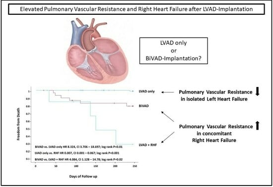
All transthoracic echocardiography values were indicative of severely impaired cardiac function (Table 2). Mean SVR and PVR were significantly higher in individuals with both right and left HF compared to those with left-sided HF only (Table 2). In the LVAD group, 66/95 patients (68%) required inotropic therapy, most often milrinone or dobutamine, while 28/34 (82%) of those with both left and right HF needed inotropes, also usually milrinone or dobutamine; requirement for inotropic therapy did not differ statistically significantly between groups, with numerically more dobutamine support in the LVAD-only group (Table 3).

3.3. Outcomes

Death occurred in 11/129 (8.5%) patients overall, 1 (1.1%) in the LVAD-only group and 10 (29.4%) in the group that also had RHF (HR 35.4; 95% confidence interval [CI] 4.5–277; log rank p < 0.001) (Table 4, Figure 1A). The duration of mechanical ventilation and time spent in the ICU were significantly longer in the RHF versus LVAD-only group (Table 4). LVEF did not change significantly from baseline in either group (Table 4).

3.4. Subgroups and Predictors

Individuals who developed RHF postoperatively and required secondary RVAD support had the worst overall survival (HR LVAD only vs. secondary RVAD 18.96; 95% CI 5.36–66.99; p < 0.01; and HR LVAD only vs. LVAD with intraoperative RVAD 4.41; 95% CI 1.13–14.78; p = 0.02). Survival was best in the group that had LVAD but did not develop RHF and intermediate in those who had LVAD implantation with RHF and intraoperative immediate RVAD support (HR LVAD without RHF vs. LVAD only 8.3; 95% CI 3.71–18.70; p < 0.001) (Figure 1B). The device manufacturer (HeartWare versus HeartMate3) did not influence the results or RHF occurrence in the sensitivity analysis.
Pre-LVAD implantation PVR > 250 dyn/s/cm5 was the only significant independent predictor of mortality on multivariate Cox regression analysis (HR [95% CI] value for risk of death in individuals with a preimplantation PVR of >250 vs. <250 dyn/s/cm5 10.89 [1.31–90.50]; log rank p = 0.006) (Table 5).

4. Discussion

This is the first and largest study to use RHC to evaluate the impact of RHF in individuals undergoing LVAD implantation. We identified preoperative PVR > 250 dyn/s/cm5 as being associated with the development of RHF and as a significant independent predictor of survival, after LVAD implantation. Patients who developed RHF after LVAD implantation required significantly more hours of invasive mechanical ventilation, spent longer in the ICU and in hospital, and had the worst survival compared with patients who did not develop RHF or had a RVAD implanted at the same time as an LVAD. This is clinically relevant because there is currently no satisfactory therapy for RHF in patients on an LVAD.
RHF has previously been shown to be associated with a higher risk of mortality after LVAD implantation [28]. However, available studies have not reported consistent results and used differing definitions of RHF. In general, RHF is diagnosed when patients undergoing LVAD require mechanical right ventricular support, show elevated vena cava pressures, show signs of liver congestion and/or ascites, or require continuous inotropic support for ≥14 days. However, all of these parameters represent postoperative clinical findings that cannot provide a preoperative indication of the risk of developing postoperative RHF, which would be useful to know because patients developing RHF in the postoperative course and requiring secondary subsequent RVAD support had by far the worst outcomes in our study. However, excessive or prophylactic RVAD supply during LVAD implantation prolongs the surgical procedure and carries additional risks, such as infection, bleeding, vessel injury and wound healing complications. Therefore, RVAD supply should only be used in patients undergoing LVAD who definitely need right ventricular support. However, there is currently no information about how to determine the requirement for RVAD during LVAD implantation, so our study adds important information to the field based on RHC findings.
Several calculation scores for RHF prediction after LVAD implantation have been developed over recent years. One of these is the Fitzpatrick score, which combines clinical parameters (e.g., previous cardiac surgery) with laboratory findings (e.g., creatinine) [21]. The Fitzpatrick score also included echocardiographic parameters indicating right ventricular dysfunction and the need for treatments such as inhaled nitric oxide or intravenous catecholamines. Similar parameters are included in the EUROMACS score [25]. However, all currently available scores have failed to reliably predict the postoperative development of RHF after LVAD implantation.
It was previously unclear whether immediate or belated RVAD support implantation had the ability to reduce mortality in patients at risk of RHF after LVAD implantation. This is relevant because observational studies have reported that RVAD support within the first 6–12 months after LVAD implantation is associated with significantly higher rates of bleeding, infection and death [29,30]. Therefore, to reduce the occurrence of these adverse events, interdisciplinary heart team decisions tend to favor LVAD-only strategies, even in patients with suspected RHF, with the potential for improvement in right ventricular function after LVAD implantation. However, there are not any currently validated predictors of right ventricular function recovery after LVAD implantation, due at least in part to the fact that studies have used heterogenous strategies even within LVAD centers and differing patient pathways [31,32].
The validity of the LVAD-only approach was confirmed by our data because this group had the best outcome at 250 days after LVAD implantation. However, participants who had immediate RVAD support implantation had much better outcomes than those who had later RVAD implantation, suggesting a beneficial impact of early RVAD support in patients with RHF. Given the lack of clear definitions relating to the need for RVAD, decisions about RVAD implantation in our study were made on a case-by-case basis. The risks of simultaneous RVAD and LVAD implantation but the better outcomes seen with this approach, as compared to an initial LVAD-only strategy followed by secondary RVAD addition, in patients with right ventricular heart failure who really need right ventricular support, means that there is a need for preoperative parameters that can be used to identify the best candidates for the addition of an RVAD during LVAD implantation.
Although the novelty of this study is an important strength, some limitations need to be considered when interpreting the findings of this study. First, it had a retrospective, single-center design, and decisions about RVAD implantation were made on a case-by-case basis by an interdisciplinary HF board rather than based on standardized criteria (which were not available during the study period). Second, we prospectively included de novo LVAD implantations, but device exchange procedures and revisions were excluded. This could have introduced a selection bias and means that the findings are only generalizable to de novo LVAD implantation procedures. Third, it is important to be aware that the findings can only be generalized to, and compared with, situations using the same definition of RHF. Moreover, only centrifugal LVADs have been used and allow conclusions from our findings, and the group of delayed right heart failure patients is rather small, allowing limited conclusions only. Finally, most variables in the limited set of parameters evaluated in this study are dynamic and might change over time based on volume management strategies.

5. Conclusions

This study makes an important contribution to the unresolved topic of RHF on an LVAD. The results showed a significant increase in the mortality risk in patients with RHF and LVAD, highlighting the need for parameters that identify which patients in this setting need early RVAD support. Our data showed that a preoperative PVR dyn/s/cm5 could be a useful marker of the need for right ventricular support. This now needs to be investigated in prospective, controlled trials because RHF remains a significant challenge in all patients undergoing LVAD implantation.

Author Contributions

Conceptualization, H.F.; Validation, R.S. and J.F.G.; Formal analysis, J.K. and J.F.G.; Investigation, M.I.; Resources, V.R. and H.F.; Writing—original draft, H.F.; Writing—review & editing, S.V.R., M.M., V.R., J.F.G. and H.F.; Visualization, V.R.; Supervision, M.M. and J.F.G.; Project administration, V.R. and H.F. All authors have read and agreed to the published version of the manuscript.

Funding

This study did not receive any external funding.

Institutional Review Board Statement

Approved by Ruhr-University institutional Ethics Committee, Reg: 2020-617.

Informed Consent Statement

All patients gave written informed consent prior to study participation.

Data Availability Statement

All data included in this manuscript.

Conflicts of Interest

The authors declare no conflicts of interest.

Abbreviations

2DEtwo-dimensional transthoracic echocardiography
CIconfidence interval
CTcomputed tomography
EACVIEuropean Association of Cardiovascular Imaging
HFheart failure
HRhazard ratio
LVADleft ventricular assist device
LVEFleft ventricular ejection fraction
PVRpulmonary vascular resistance
RHCright heart catheterization
RHFright heart failure
ROCreceiver operating characteristic
RVADright ventricular assist device

References

  1. Savarese, G.; Becher, P.M.; Lund, L.H.; Seferovic, P.; Rosano, G.M.C.; Coats, A.J.S. Global burden of heart failure: A comprehensive and updated review of epidemiology. Cardiovasc. Res. 2023, 118, 3272–3287. [Google Scholar] [CrossRef] [PubMed]
  2. Sohns, C.; Fox, H.; Marrouche, N.F.; Crijns, H.; Costard-Jaeckle, A.; Bergau, L.; Hindricks, G.; Dagres, N.; Sossalla, S.; Schramm, R.; et al. Catheter Ablation in End-Stage Heart Failure with Atrial Fibrillation. N. Engl. J. Med. 2023, 389, 1380–1389. [Google Scholar] [CrossRef] [PubMed]
  3. Morshuis, M.; Fox, H.; Lauenroth, V.; Schramm, R. Long-term assist device patients admitted to ICU: Tips and pitfalls. J. Intensive Med. 2023, 3, 81–88. [Google Scholar] [CrossRef] [PubMed]
  4. Alkhunaizi, F.A.; Azih, N.I.; Read, J.M.; Goldberg, R.L.; Gulati, A.A.; Scheel, P.J., 3rd; Muslem, R.; Gilotra, N.A.; Sharma, K.; Kilic, A.; et al. Characteristics and Predictors of Late Right Heart Failure after Left Ventricular Assist Device Implantation. ASAIO J. 2023, 69, 315–323. [Google Scholar] [CrossRef] [PubMed]
  5. Dang, N.C.; Topkara, V.K.; Mercando, M.; Kay, J.; Kruger, K.H.; Aboodi, M.S.; Oz, M.C.; Naka, Y. Right heart failure after left ventricular assist device implantation in patients with chronic congestive heart failure. J. Heart Lung Transplant. 2006, 25, 1–6. [Google Scholar] [CrossRef]
  6. Kormos, R.L.; Teuteberg, J.J.; Pagani, F.D.; Russell, S.D.; John, R.; Miller, L.W.; Massey, T.; Milano, C.A.; Moazami, N.; Sundareswaran, K.S.; et al. Right ventricular failure in patients with the HeartMate II continuous-flow left ventricular assist device: Incidence, risk factors, and effect on outcomes. J. Thorac. Cardiovasc. Surg. 2010, 139, 1316–1324. [Google Scholar] [CrossRef]
  7. Rich, J.D.; Gosev, I.; Patel, C.B.; Joseph, S.; Katz, J.N.; Eckman, P.M.; Lee, S.; Sundareswaran, K.; Kilic, A.; Bethea, B.; et al. The incidence, risk factors, and outcomes associated with late right-sided heart failure in patients supported with an axial-flow left ventricular assist device. J. Heart Lung Transplant. 2017, 36, 50–58. [Google Scholar] [CrossRef]
  8. Hahn, R.T.; Lerakis, S.; Delgado, V.; Addetia, K.; Burkhoff, D.; Muraru, D.; Pinney, S.; Friedberg, M.K. Multimodality Imaging of Right Heart Function: JACC Scientific Statement. J. Am. Coll. Cardiol. 2023, 81, 1954–1973. [Google Scholar] [CrossRef]
  9. Pickett, C.A.; Cheezum, M.K.; Kassop, D.; Villines, T.C.; Hulten, E.A. Accuracy of cardiac CT, radionucleotide and invasive ventriculography, two- and three-dimensional echocardiography, and SPECT for left and right ventricular ejection fraction compared with cardiac MRI: A meta-analysis. Eur. Heart J. Cardiovasc. Imaging 2015, 16, 848–852. [Google Scholar] [CrossRef]
  10. Rizvi, A.; Deaño, R.C.; Bachman, D.P.; Xiong, G.; Min, J.K.; Truong, Q.A. Analysis of ventricular function by CT. J. Cardiovasc. Comput. Tomogr. 2015, 9, 1–12. [Google Scholar] [CrossRef]
  11. Sugeng, L.; Mor-Avi, V.; Weinert, L.; Niel, J.; Ebner, C.; Steringer-Mascherbauer, R.; Bartolles, R.; Baumann, R.; Schummers, G.; Lang, R.M.; et al. Multimodality comparison of quantitative volumetric analysis of the right ventricle. JACC Cardiovasc. Imaging 2010, 3, 10–18. [Google Scholar] [CrossRef] [PubMed]
  12. Dandel, M.; Hetzer, R. Echocardiographic assessment of the right ventricle: Impact of the distinctly load dependency of its size, geometry and performance. Int. J. Cardiol. 2016, 221, 1132–1142. [Google Scholar] [CrossRef]
  13. Gyoten, T.; Rojas, S.V.; Fox, H.; Hata, M.; Deutsch, M.A.; Schramm, R.; Gummert, J.F.; Morshuis, M. Cardiac recovery following left ventricular assist device therapy: Experience of complete device explantation including ventricular patch plasty. Eur. J. Cardiothorac. Surg. 2021, 59, 855–862. [Google Scholar] [CrossRef] [PubMed]
  14. Newman, J.H. Pulmonary Hypertension by the Method of Paul Wood. Chest 2020, 158, 1164–1171. [Google Scholar] [CrossRef]
  15. Maron, B.A.; Brittain, E.L.; Hess, E.; Waldo, S.W.; Barón, A.E.; Huang, S.; Goldstein, R.H.; Assad, T.; Wertheim, B.M.; Alba, G.A.; et al. Pulmonary vascular resistance and clinical outcomes in patients with pulmonary hypertension: A retrospective cohort study. Lancet Respir. Med. 2020, 8, 873–884. [Google Scholar] [CrossRef] [PubMed]
  16. Gual-Capllonch, F.; Teis, A.; Ferrer, E.; Núñez, J.; Vallejo, N.; Juncà, G.; López-Ayerbe, J.; Lupón, J.; Bayes-Genis, A. Pulmonary vascular resistance versus pulmonary artery pressure for predicting right ventricular remodeling and functional tricuspid regurgitation. Echocardiography 2018, 35, 1736–1745. [Google Scholar] [CrossRef] [PubMed]
  17. Stocker, T.J.; Hertell, H.; Orban, M.; Braun, D.; Rommel, K.P.; Ruf, T.; Ong, G.; Nabauer, M.; Deseive, S.; Fam, N.; et al. Cardiopulmonary Hemodynamic Profile Predicts Mortality After Transcatheter Tricuspid Valve Repair in Chronic Heart Failure. JACC Cardiovasc. Interv. 2021, 14, 29–38. [Google Scholar] [CrossRef]
  18. Park, J.; Lee, S.H.; Kim, J.; Park, S.J.; Park, M.S.; Choi, G.S.; Lee, S.K.; Kim, G.S. Predictive Value of Intraoperative Pulmonary Vascular Resistance in Liver Transplantation. Liver Transpl. 2018, 24, 1680–1689. [Google Scholar] [CrossRef]
  19. Barst, R.J.; Rubin, L.J.; Long, W.A.; McGoon, M.D.; Rich, S.; Badesch, D.B.; Groves, B.M.; Tapson, V.F.; Bourge, R.C.; Brundage, B.H.; et al. A comparison of continuous intravenous epoprostenol (prostacyclin) with conventional therapy for primary pulmonary hypertension. N. Engl. J. Med. 1996, 334, 296–301. [Google Scholar] [CrossRef]
  20. Hoeper, M.M.; Badesch, D.B.; Ghofrani, H.A.; Gibbs, J.S.R.; Gomberg-Maitland, M.; McLaughlin, V.V.; Preston, I.R.; Souza, R.; Waxman, A.B.; Grünig, E.; et al. Phase 3 Trial of Sotatercept for Treatment of Pulmonary Arterial Hypertension. N. Engl. J. Med. 2023, 388, 1478–1490. [Google Scholar] [CrossRef]
  21. Fitzpatrick, J.R., 3rd; Frederick, J.R.; Hsu, V.M.; Kozin, E.D.; O’Hara, M.L.; Howell, E.; Dougherty, D.; McCormick, R.C.; Laporte, C.A.; Cohen, J.E.; et al. Risk score derived from pre-operative data analysis predicts the need for biventricular mechanical circulatory support. J. Heart Lung Transplant. 2008, 27, 1286–1292. [Google Scholar] [CrossRef] [PubMed]
  22. Matthews, J.C.; Koelling, T.M.; Pagani, F.D.; Aaronson, K.D. The right ventricular failure risk score a pre-operative tool for assessing the risk of right ventricular failure in left ventricular assist device candidates. J. Am. Coll. Cardiol. 2008, 51, 2163–2172. [Google Scholar] [CrossRef] [PubMed]
  23. McMurray, J.J.; Packer, M.; Desai, A.S.; Gong, J.; Lefkowitz, M.P.; Rizkala, A.R.; Rouleau, J.L.; Shi, V.C.; Solomon, S.D.; Swedberg, K.; et al. Angiotensin-neprilysin inhibition versus enalapril in heart failure. N. Engl. J. Med. 2014, 371, 993–1004. [Google Scholar] [CrossRef]
  24. McMurray, J.J.V.; Solomon, S.D.; Inzucchi, S.E.; Køber, L.; Kosiborod, M.N.; Martinez, F.A.; Ponikowski, P.; Sabatine, M.S.; Anand, I.S.; Bělohlávek, J.; et al. Dapagliflozin in Patients with Heart Failure and Reduced Ejection Fraction. N. Engl. J. Med. 2019, 381, 1995–2008. [Google Scholar] [CrossRef] [PubMed]
  25. Soliman, O.I.I.; Akin, S.; Muslem, R.; Boersma, E.; Manintveld, O.C.; Krabatsch, T.; Gummert, J.F.; de By, T.; Bogers, A.; Zijlstra, F.; et al. Derivation and Validation of a Novel Right-Sided Heart Failure Model After Implantation of Continuous Flow Left Ventricular Assist Devices: The EUROMACS (European Registry for Patients with Mechanical Circulatory Support) Right-Sided Heart Failure Risk Score. Circulation 2018, 137, 891–906. [Google Scholar] [CrossRef] [PubMed]
  26. Agricola, E.; Ancona, F.; Brochet, E.; Donal, E.; Dweck, M.; Faletra, F.; Lancellotti, P.; Mahmoud-Elsayed, H.; Marsan, N.A.; Maurovich-Hovart, P.; et al. The structural heart disease interventional imager rationale, skills and training: A position paper of the European Association of Cardiovascular Imaging. Eur. Heart J. Cardiovasc. Imaging 2021, 22, 471–479. [Google Scholar] [CrossRef]
  27. McDonagh, T.A.; Metra, M.; Adamo, M.; Gardner, R.S.; Baumbach, A.; Böhm, M.; Burri, H.; Butler, J.; Čelutkienė, J.; Chioncel, O.; et al. 2021 ESC Guidelines for the diagnosis and treatment of acute and chronic heart failure. Eur. Heart J. 2021, 42, 3599–3726. [Google Scholar] [CrossRef]
  28. Potapov, E.V.; Antonides, C.; Crespo-Leiro, M.G.; Combes, A.; Farber, G.; Hannan, M.M.; Kukucka, M.; de Jonge, N.; Loforte, A.; Lund, L.H.; et al. 2019 EACTS Expert Consensus on long-term mechanical circulatory support. Eur. J. Cardiothorac. Surg. 2019, 56, 230–270. [Google Scholar] [CrossRef]
  29. Cleveland, J.C., Jr.; Naftel, D.C.; Reece, T.B.; Murray, M.; Antaki, J.; Pagani, F.D.; Kirklin, J.K. Survival after biventricular assist device implantation: An analysis of the Interagency Registry for Mechanically Assisted Circulatory Support database. J. Heart Lung Transplant. 2011, 30, 862–869. [Google Scholar] [CrossRef]
  30. Rojas, S.V.; Junghans, S.; Fox, H.; Lazouski, K.; Schramm, R.; Morshuis, M.; Gummert, J.F.; Gross, J. Bacteriophage-Enriched Galenic for Intrapericardial Ventricular Assist Device Infection. Antibiotics 2022, 11, 602. [Google Scholar] [CrossRef]
  31. Mullan, C.; Caraballo, C.; Ravindra, N.G.; Miller, P.E.; Mori, M.; McCullough, M.; Clarke, J.D.; Anwer, M.; Velazquez, E.J.; Geirsson, A.; et al. Clinical impact of concomitant tricuspid valve procedures during left ventricular assist device implantation. J. Heart Lung Transplant. 2020, 39, 926–933. [Google Scholar] [CrossRef] [PubMed]
  32. Potapov, E.V.; Schweiger, M.; Stepanenko, A.; Dandel, M.; Kukucka, M.; Vierecke, J.; Hetzer, R.; Krabatsch, T. Tricuspid valve repair in patients supported with left ventricular assist devices. Asaio J. 2011, 57, 363–367. [Google Scholar] [CrossRef] [PubMed]
Figure 1. Kaplan–Meier survival curves. (A) Patients with left heart failure (LsHF) or left and right (bilateral ventricular) heart failure (BiVHF); (B) left ventricular assist device (LVAD) implantation, simultaneous LVAD and right ventricular assist device implantation (BiVAD); (C) LVAD implantation with subsequent development of right heart failure (LVAD + RHF). CI, confidence interval; HR, hazard ratio.
Jcm 13 00462 g001aJcm 13 00462 g001b
Table 1. Baseline characteristics (at the time for decision for LVAD) of the study population.
CharacteristicsTotal
(n = 129)
LVAD Only
(n = 95)
LVAD + RHF
(n = 34)
p-Value
Age, years56 ± 1157 ± 1054 ± 130.194
Male sex, n (%)116 (89%)84 (88%)32 (94%)0.512
Urea, mg/dL70 ± 4369 ± 42.671.8 ± 460.794
Creatinine, mg/dL1.6 ± 11.5 ± 11.7 ± 10.845
Bilirubin, mg/dL1.5 ± 1.61.4 ± 1.22 ± 2.50.196
ALT, U/L70 ± 15871 ± 17965 ± 790.422
AST, U/L51 ± 10553 ± 12047 ± 400.784
CRP, mg/dL3.4 ± 4.93.0 ± 4.44.4 ± 60.154
Values are mean ± standard deviation or number of participants (%). CRP, C-reactive protein; LVAD, left ventricular assist device; RHF, right heart failure; ALT, alanine aminotransferase; AST, aspartate aminotransferase.
Table 2. Transthoracic echocardiography data for left ventricular function and hemodynamics at baseline.
ParameterTotal
(n = 129)
LVAD Only
(n = 95)
LVAD + RHF
(n = 34)
p-Value
LVEF, %21.5 ± 6.221.5 ± 6.221.5 ± 6.50.961
LVEDD, mm72 ± 1171 ± 1173 ± 130.51
LVESD, mm66 ± 1266 ± 1165 ± 150.732
Heart rate, beats/min87 ± 1886 ± 1789 ± 200.414
Mean PAP, mmHg33 ± 1132 ± 733 ± 90.983
PCWP, mmHg22 ± 1022 ± 1122 ± 70.94
CO, L/min5.1 ± 7.94.7 ± 4.96.2 ± 13.30.339
CI, L/min/m22.1 ± 0.642.1 ± 0.672.02 ± 0.590.515
SVR, dyn/s/cm54152 ± 14831354 ± 6201904 ± 9420.008
PVR, dyn/s/cm5269 ± 231234 ± 162404 ± 3750.01
Values are mean ± standard deviation. CI, cardiac index; CO, cardiac output; LVAD, left ventricular assist device; LVEDD, left ventricular end-diastolic diameter; LVEF, left ventricular ejection fraction; LVESD, left ventricular end-systolic diameter; PAP, pulmonary artery pressure; PCWP, pulmonary capillary wedge pressure; PVR, peripheral vascular resistance; RHF, right heart failure; SVR, systemic vascular resistance.
Table 3. Inotropic therapy.
InotropesTotal
(n = 129)
LVAD Only
(n = 95)
LVAD + RHF
(n = 34)
p-Value
Milrinone67 (52%)49 (52%)18 (53%)0.99
Dobutamine64 (50%)49 (52%)15 (44%)0.55
Levosimendan2 (2%)2 (2%)0 (0%)0.99
Epinephrine2 (2%)1 (1%)1 (3%)0.459
Dopamine14 (11%)5 (5%)9 (26%)0.002
Norepinephrine3 (2%)3 (3%)0 (0%)0.566
Values are number of patients. LVAD, left ventricular assist device; RHF, right heart failure.
Table 4. Outcomes and follow-up.
Total
(n = 129)
LVAD Only
(n = 95)
LVAD + RHF
(n = 34)
p-Value
Death, n (%)11 (8.5)1 (1.1)10 (29.4)<0.01
Duration of invasive mechanical ventilation, days227 ± 535101 ± 200602 ± 922<0.001
Duration of ICU stay, days28.4 ± 39.515.5 ± 19.764.4 ± 56<0.001
LVEF, %24.6 ± 823.9 ± 6.526.8 ± 110.068
LVEDD, mm59 ± 1359 ± 1358 ± 130.516
LVESD, mm55 ± 1355 ± 1354 ± 140.729
Urea, mg/dL52 ± 3349 ± 3161 ± 380.094
Creatinine, mg/dL1.3 ± 1.11.3 1.71.4 ± 0.80.697
Bilirubin, mg/dL1 ± 1.90.68 ± 0.251.9 ± 0.250.06
ALT, U/L24 ± 2619 ± 1337 ± 420.045
AST, U/L33 ± 3427 ± 1047 ± 630.104
CRP, mg/dL4.1 ± 4.23.4 ± 3.15.9 ± 6.10.96
Values are mean ± standard deviation or number of participants (%). CRP, C-reactive protein; LVAD, left ventricular assist device; LVEDD, left ventricular end-diastolic diameter; LVEF, left ventricular ejection fraction; LVESD, left ventricular end-systolic diameter; RHF, right heart failure; ALT, alanine aminotransferase; AST, aspartate aminotransferase.
Table 5. Predictors of survival after left ventricular assist device implantation.
Univariate Cox RegressionMultivariate Cox Regression
HR (95% CI)p-ValueHR (95% CI)p-Value
Age1.00 (0.95–1.06)0.916-
Sex0.88 (0.11–6.90)0.905-
LVEF, %1.09 (0.99–1.20)0.0791.07 (0.95–1.19)0.272
Bilirubin, mg/dL1.21 (1.04–1.40)0.0120.53 (1.57–1.79)0.307
Urea, mg/dL1.00 (0.98–1.01)0.775-
ALT, U/L1.00 (0.99–1.01)0.914-
AST, U/L0.99 (0.98–1.01)0.437-
Creatinine, mg/dL1.20 (0.77–1.86)0.419-
Mean PAP, mmHg1.01 (0.95–1.06)0.817-
PVR >250 vs. <250 dyn/s/cm510.89 (1.31–90.50)0.02710.38 (1.21–89.04)0.033
SVR, dyn/s/cm51 (1.00–1.00)0.773-
PCWP, mmHg0.99 (0.93–1.07)0.867-
Cardiac index0.93 (0.37–2.33)0.872-
CI, confidence interval; HR, hazard ratio; LVEF, left ventricular ejection fraction; PAP, pulmonary artery pressure; PCWP, pulmonary capillary wedge pressure; PVR, peripheral vascular resistance; ALT, alanine aminotransferase; ALT, aspartate aminotransferase.
Disclaimer/Publisher’s Note: The statements, opinions and data contained in all publications are solely those of the individual author(s) and contributor(s) and not of MDPI and/or the editor(s). MDPI and/or the editor(s) disclaim responsibility for any injury to people or property resulting from any ideas, methods, instructions or products referred to in the content.

