
Dependence of Left Ventricular Rotational Mechanics on Left Atrial Volumes in Non-Smoker Healthy Adults: Analysis Based on the Three-Dimensional Speckle-Tracking Echocardiographic MAGYAR-Healthy Study

Abstract
:1. Introduction
2. Subjects and Methods
2.1. Subjects
2.2. Two-Dimensional Doppler Echocardiography
2.3. Three-Dimensional Speckle-Tracking Echocardiography
2.4. 3DSTE-Derived Determination of LV Twist
- Clockwise basal LV rotation (in degrees);
- Counterclockwise apical LV rotation (in degrees);
- LV twist (net difference between LV apical and basal rotations in degrees);
- Time-to-peak LV twist (in milliseconds).
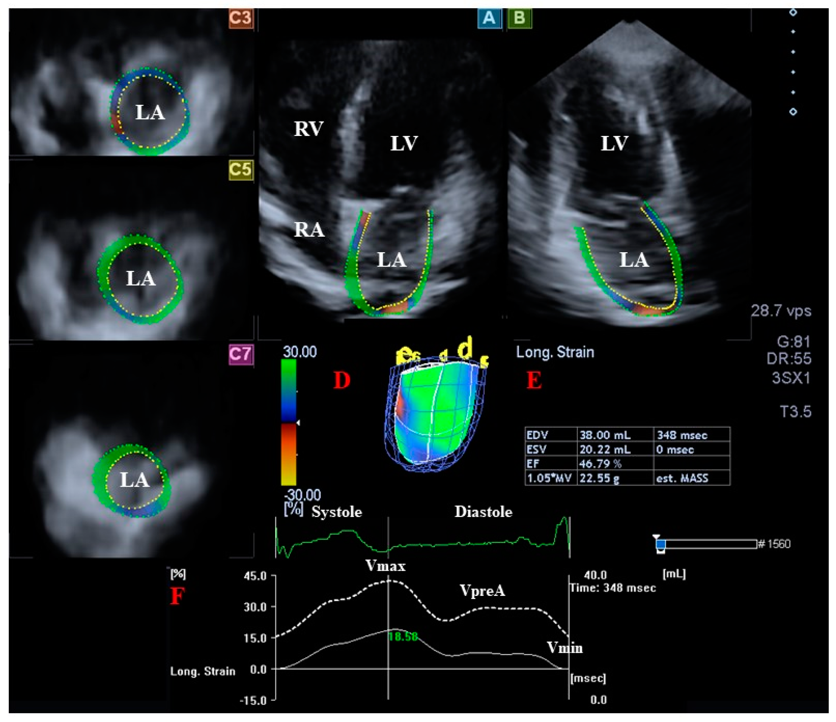
2.5. 3DSTE-Derived Determination of LA Volumes
- Vmax—end-systolic maximum LA volume, measured just before mitral valve opening (largest LA volume);
- VpreA—LA volume before atrial contraction in early diastole, at the time of P wave on ECG;
- Vmin—late diastolic minimum LA volume, measured just before mitral valve closure (smallest LA volume).
- TASV—LA total stroke volume, calculated by Vmax − Vmin;
- TAEF—LA total emptying fraction, calculated by total SV/Vmax.
- PASV—LA passive stroke volume, calculated by Vmax − VpreA;
- PAEF—LA passive emptying fraction, calculated by passive SV/Vmax.
- AASV—LA active stroke volume, calculated by VpreA − Vmin;
- AAEF—LA active emptying fraction, calculated by active SV/VpreA.
2.6. Statistical Analysis
3. Results
3.1. Clinical and Two-Dimensional Doppler Echocardiographic Data
3.2. Classification of Subjects
3.3. 3DSTE-Derived LA Volumes and LV Rotational Parameters
3.4. Correlations
4. Discussion
5. Limitations
- Although 3DSTE is suitable for simultaneous LV strain assessments using the same 3D echocardiographic dataset, a detailed analysis of these parameters (LV strains and LA volumes) would have surpassed the limits of this publication and is a topic for another paper demonstrating the dependence of LV deformation on LA volumetric changes [1,2,3,4].
- This study had a retrospective study design, and so associations were only established between risk factors and outcomes, and not between causes and effects.
6. Conclusions
Author Contributions
Funding
Institutional Review Board Statement
Informed Consent Statement
Data Availability Statement
Conflicts of Interest
References
- Nemes, A.; Kalapos, A.; Domsik, P.; Forster, T. Three-dimensional speckle-tracking echocardiography—A further step in non-invasive three-dimensional cardiac imaging. Orv. Hetil. 2012, 153, 1570–1577. [Google Scholar] [CrossRef]
- Ammar, K.A.; Paterick, T.E.; Khandheria, B.K.; Jan, M.F.; Kramer, C.; Umland, M.M.; Tercius, A.J.; Baratta, L.; Tajik, A.J. Myocardial mechanics: Understanding and applying three-dimensional speckle tracking echocardiography in clinical practice. Echocardiography 2012, 97, 861–872. [Google Scholar] [CrossRef] [PubMed]
- Urbano-Moral, J.A.; Patel, A.R.; Maron, M.S.; Arias-Godinez, J.A.; Pandian, N.G. Three-dimensional speckle-tracking echocardiography: Methodological aspects and clinical potential. Echocardiography 2012, 29, 997–1010. [Google Scholar] [CrossRef] [PubMed]
- Muraru, D.; Niero, A.; Rodriguez-Zanella, H.; Cherata, D.; Badano, L. Three-dimensional speckle-tracking echocardiography: Benefits and limitations of integrating myocardial mechanics with three-dimensional imaging. Cardiovasc. Diagn. Ther. 2018, 8, 101–117. [Google Scholar] [CrossRef]
- Nakatani, S. Left ventricular rotation and twist: Why should we learn? J. Cardiovasc. Ultrasound 2011, 19, 1–6. [Google Scholar] [CrossRef] [PubMed]
- Nemes, A.; Kalapos, A.; Domsik, P.; Forster, T. Left ventricular rotation and twist of the heart. Clarification of some concepts. Orv. Hetil. 2012, 153, 1547–1551. [Google Scholar] [CrossRef]
- Jacob, R.; Dierberger, B.; Kissling, G. Functional significance of the Frank-Starling mechanism under physiological and pathophysiological conditions. Eur. Heart J. 1992, 13 (Suppl. E), 7–14. [Google Scholar] [CrossRef]
- LaCombe, P.; Tariq, M.A.; Lappin, S.L. Physiology, Afterload Reduction; StatPearls Publishing: Treasure Island, FL, USA, 2022. [Google Scholar]
- Lang, R.M.; Badano, L.P.; Mor-Avi, V.; Afilalo, J.; Armstrong, A.; Ernande, L.; Flachskampf, F.A.; Foster, E.; Goldstein, S.A.; Kuznetsova, T.; et al. Recommendations for cardiac chamber quantification by echocardiography in adults: An update from the American Society of Echocardiography and the European Association of Cardiovascular Imaging. Eur. Heart J. Cardiovasc. Imaging 2015, 16, 233–270. [Google Scholar] [CrossRef]
- Kormányos, Á.; Kalapos, A.; Domsik, P.; Lengyel, C.; Forster, T.; Nemes, A. Normal values of left ventricular rotational parameters in healthy adults—Insights from the three-dimensional speckle tracking echocardiographic MAGYAR-Healthy Study. Echocardiography 2019, 36, 714–721. [Google Scholar] [CrossRef]
- Kormányos, Á.; Gyenes, N.; Horváth, Á.; Ambrus, N.; Lengyel, C.; Valkusz, Z.; Nemes, A. Left Ventricular Rotational Abnormalities in Treated Hypopituitarism: Insights from the Three-Dimensional Speckle-Tracking Echocardiographic MAGYAR-Path Study. Front. Cardiovasc. Med. 2021, 8, 703146. [Google Scholar] [CrossRef]
- Nemes, A.; Kormányos, Á. Prevalence of left ventricular ‘rigid body rotation’, the near absence of left ventricular twist (insights from the MAGYAR studies). Rev. Cardiovasc. Med. 2022, 23, 5. [Google Scholar] [CrossRef]
- Nemes, A.; Kormányos, Á.; Domsik, P.; Kalapos, A.; Ambrus, N.; Lengyel, C. Normal reference values of left atrial volumes and volume-based functional properties using three-dimensional speckle-tracking echocardiography in healthy adults (Insights from the MAGYAR-Healthy Study). J. Clin. Ultrasound 2021, 49, 49–55. [Google Scholar] [CrossRef]
- Földeák, D.; Kormányos, Á.; Domsik, P.; Kalapos, A.; Piros, G.Á.; Ambrus, N.; Ajtay, Z.; Sepp, R.; Borbényi, Z.; Forster, T.; et al. Left atrial dysfunction in light-chain cardiac amyloidosis and hypertrophic cardiomyopathy—A comparative three-dimensional speckle-tracking echocardiographic analysis from the MAGYAR-Path Study. Rev. Port. Cardiol. 2017, 36, 905–913. [Google Scholar] [CrossRef]
- Ashraf, M.; Myronenko, A.; Nguyen, T.; Inage, A.; Smith, W.; Lowe, R.I.; Thiele, K.; Gibbons-Kroeker, C.A.; Tyberg, J.V.; Smallhorn, J.E.; et al. Defining left ventricular apex-to-base twist mechanics computed from high-resolution 3D echocardiography: Validation against sonomicrometry. JACC Cardiovasc. Imaging 2010, 3, 227–234. [Google Scholar] [CrossRef]
- Zhou, Z.; Ashraf, M.; Hu, D.; Dai, X.; Xu, Y.; Kenny, B.; Cameron, B.; Nguyen, T.; Xiong, L.; Sahn, J.D. Three-dimensional speckle-tracking imaging for left ventricular rotation measurement: An in vitro validation study. J. Ultrasound Med. 2010, 29, 903–909. [Google Scholar] [CrossRef]
- Andrade, J.; Cortez, L.D.; Campos, O.; Arruda, A.L.; Pinheiro, J.; Vulcanis, L.; Shiratsuchi, T.S.; Kalil-Filho, R.; Cerri, G.G. Left ventricular twist: Comparison between two- and three-dimensional speckle-tracking echocardiography in healthy volunteers. Eur. J. Echocardiogr. 2011, 12, 76–79. [Google Scholar] [CrossRef]
- Aurigemma, G.P.; Zile, M.R.; Gaasch, W.H. Contractile behavior of the left ventricle in diastolic heart failure: With emphasis on regional systolic function. Circulation 2006, 113, 296–304. [Google Scholar] [CrossRef]
- Anwar, A.M.; Gelejinse, M.L.; Soliman, O.I.I.; Nemes, A.; ten Cate, F.J. Left atrial Frank-Starling law assessed by real-time three-dimensional echocardiographic left atrial volume changes. Heart 2007, 93, 1393–1397. [Google Scholar] [CrossRef]
- Nemes, A.; Kormányos, Á.; Domsik, P.; Kalapos, A.; Gyenes, N.; Lengyel, C. Correlations between left atrial volumes and strains in healthy adults: Detailed analysis from the three-dimensional speckle tracking echocardiographic MAGYAR-Healthy Study. J. Clin. Ultrasound 2021, 49, 650–658. [Google Scholar] [CrossRef]
- Nemes, A.; Kormányos, Á. Right atrial volumes and strains in healthy adults: Is the Frank-Starling mechanism working?—Detailed analysis from a three-dimensional speckle-tracking echocardiographic MAGYAR-Healthy Study. Quant. Imaging Med. Surg. 2023; in press. [Google Scholar]
- Kleijn, S.A.; Aly, M.F.; Terwee, C.B.; van Rossum, A.C.; Kamp, O. Comparison between direct volumetric and speckle tracking methodologies for left ventricular and left atrial chamber quantification by three-dimensional echocardiography. Am. J. Cardiol. 2011, 108, 1038–1044. [Google Scholar] [CrossRef]
- Nagaya, M.; Kawasaki, M.; Tanaka, R.; Onishi, N.; Sato, N.; Ono, K.; Watanabe, T.; Minatoguchi, S.; Miwa, H.; Goto, Y.; et al. Quantitative validation of left atrial structure and function by two-dimensional and three-dimensional speckle tracking echocardiography: A comparative study with three-dimensional computed tomography. J. Cardiol. 2013, 62, 188–194. [Google Scholar] [CrossRef] [PubMed]
- Nemes, A.; Domsik, P.; Kalapos, A.; Lengyel, C.; Orosz, A.; Forster, T. Comparison of three-dimensional speckle tracking echocardiography and two-dimensional echocardiography for evaluation of left atrial size and function in healthy volunteers (results from the MAGYAR-Healthy Study). Echocardiography 2014, 31, 865–871. [Google Scholar] [CrossRef] [PubMed] [Green Version]


| Parameters | Data |
|---|---|
| Left atrial volumes | |
| maximum left atrial volume (Vmax, mL) | 40.9 ± 13.1 |
| pre-atrial-contraction left atrial volume (VpreA, mL) | 27.7 ± 11.8 |
| minimum left atrial volume (Vmin, mL) | 19.4 ± 8.1 |
| total atrial stroke volume (TASV, mL) | 21.5 ± 8.2 |
| total atrial emptying fraction (TAEF, %) | 52.6 ± 11.9 |
| passive atrial stroke volume (PASV, mL) | 13.1 ± 5.7 |
| passive atrial emptying fraction (PAEF, %) | 33.1 ± 12.7 |
| active atrial stroke volume (AASV, mL) | 8.3 ± 5.8 |
| active atrial emptying fraction (AAEF, %) | 29.0 ± 11.9 |
| Left ventricular rotational mechanics | |
| basal left ventricular rotation (basal LVrot, °) | −4.1 ± 2.1 |
| apical left ventricular rotation (apical LVrot, °) | 9.2 ± 3.8 |
| left ventricular twist (LVtwist, °) | 13.3 ± 4.1 |
| time-to-LVtwist (ms) | 346 ± 132 |
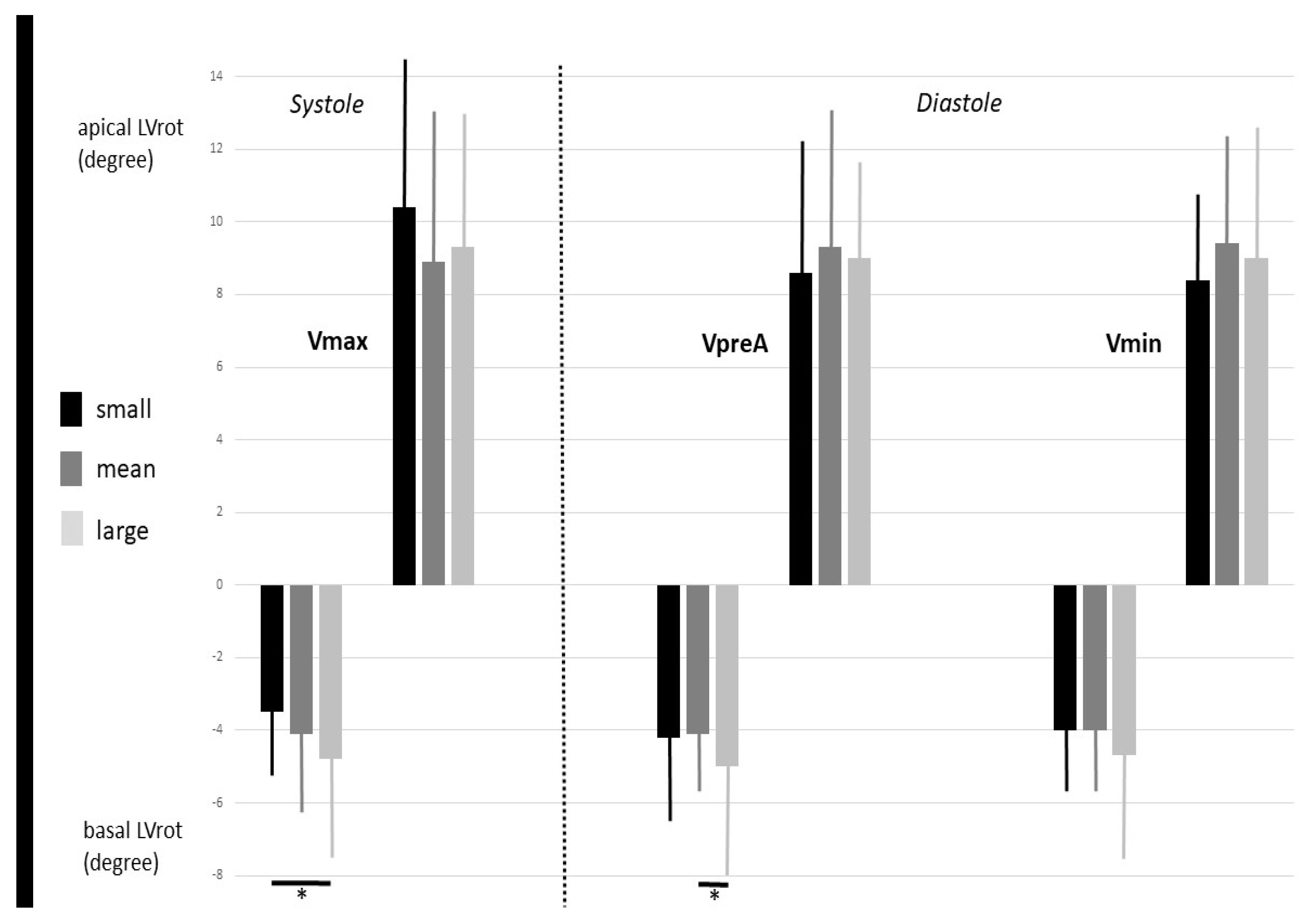
| Vmax < 27.8 mL (n = 25) | 27.8 mL ≤ Vmax ≤ 54.0 mL (n = 116) | 54 mL < Vmax (n = 26) | VpreA < 15.9 mL (n = 18) | 15.9 mL ≤ VpreA ≤ 39.5 mL (n = 125) | 39.5 mL < VpreA (n = 24) | Vmin < 11.3 mL (n = 23) | 11.2 mL ≤ Vmin ≤ 27.5 mL (n = 123) | 27.5 mL < Vmin (n = 21) | |
|---|---|---|---|---|---|---|---|---|---|
| Vmax (mL) | 23.9 ± 3.6 | 38.8 ± 6.4 ‡ | 64.8 ± 7.0 ‡,‡‡ | 25.6 ± 5.8 | 38.7 ± 8.5 * | 63.2 ± 9.6 *,** | 26.4 ± 5.4 | 39.7 ± 9.2 † | 62.7 ± 10.8 †,†† |
| VpreA (mL) | 16.0 ± 4.2 | 25.6 ± 7.0 ‡ | 47.5 ± 11.2 ‡,‡‡ | 12.9 ± 2.5 | 25.4 ± 6.0 * | 50.5 ± 8.3 *,** | 14.5 ± 3.5 | 26.5 ± 7.7 † | 49.3 ± 9.0 †,†† |
| Vmin (mL) | 11.9 ± 3.7 | 18.2 ± 5.5 ‡ | 31.3 ± 8.7 ‡,‡‡ | 9.1 ± 1.8 | 18.2 ± 4.9 * | 33.1 ± 7.8 *,** | 9.1 ± 1.7 | 18.5 ± 4.2 † | 35.6 ± 5.5 †,†† |
| TASV (mL) | 12.0 ± 3.1 | 20.6 ± 5.5 ‡ | 33.4 ± 7.6 ‡,‡‡ | 16.5 ± 5.0 | 20.6 ± 7.0 * | 30.0 ± 9.9 *,** | 17.3 ± 4.6 | 21.2 ± 8.0 † | 27.1 ± 9.1 †,†† |
| TAEF (%) | 50.5 ± 12.6 | 53.1 ± 11.8 | 51.8 ± 11.6 | 63.3 ± 7.9 | 52.4 ± 11.6 * | 46.9 ± 12.1 *,** | 64.7 ± 7.2 | 52.2 ± 11.1 † | 42.3 ± 9.4 †,†† |
| PASV (mL) | 7.9 ± 2.9 | 13.2 ± 4.9 ‡ | 17.2 ± 7.1 ‡,‡‡ | 12.7 ± 4.4 | 13.3 ± 5.9 | 12.7 ± 5.8 | 12.0 ± 4.4 | 13.2 ± 5.8 | 13.3 ± 6.6 |
| PAEF (%) | 33.5 ± 12.8 | 34.5 ± 12.4 | 27.2 ± 12.4 ‡‡ | 48.4 ± 8.7 | 33.7 ± 11.4 * | 19.7 ± 7.9 *,** | 44.5 ± 11.4 | 33.0 ± 11.6 † | 20.8 ± 9.1 †,†† |
| AASV (mL) | 4.1 ± 2.9 | 7.3 ± 3.6 ‡ | 16.2 ± 8.6 ‡,‡‡ | 3.8 ± 2.0 | 7.3 ± 3.4 * | 17.4 ± 8.4 *,** | 5.4 ± 3.2 | 8.0 ± 5.6 † | 13.8 ± 6.1 †,†† |
| AAEF (%) | 24.9 ± 14.6 | 28.5 ± 10.9 | 33.3 ± 13.3 ‡,‡‡ | 28.3 ± 12.9 | 28.3 ± 11.3 | 33.8 ± 13.9 ** | 34.7 ± 14.3 | 28.5 ± 11.7 † | 27.1 ± 8.4 † |
| basal LVrot (°) | −3.5 ± 1.8 | −4.1 ± 2.0 | −4.8 ± 2.7 ‡ | −4.2 ± 2.0 | −4.0 ± 1.9 | −5.0 ± 3.0 ** | −4.0 ± 1.9 | −4.0 ± 1.9 | −4.7 ± 3.0 |
| apical LVrot (°) | 10.4 ± 4.1 | 8.9 ± 3.7 | 9.3 ± 3.7 | 8.6 ± 3.5 | 9.3 ± 3.9 | 9.0 ± 3.7 | 8.4 ± 3.0 | 9.4 ± 3.9 | 9.0 ± 3.6 |
| LVtwist (°) | 13.9 ± 3.6 | 13.1 ± 4.2 | 14.1 ± 3.9 | 12.8 ± 3.5 | 13.2 ± 4.2 | 14.0 ± 4.3 | 12.4 ± 3.1 | 13.3 ± 4.1 | 13.7 ± 4.2 |
| time-to-LVtwist (ms) | 371 ± 146 | 340 ± 133 | 349 ± 108 | 372 ± 146 | 339 ± 132 | 355 ± 116 | 334 ± 131 | 342 ± 134 | 366 ± 115 |
| Basal LVrot < 2° (n = 21) | 2° ≤ Basal LVrot ≤ 6.2° (n = 121) | 6.2° < Basal LVrot (n = 25) | Apical LVrot < 5.4° (n = 24) | 5.4° ≤ Apical LVrot ≤ 13.0° (n = 116) | 13.0° < Apical LVrot (n = 27) | |
|---|---|---|---|---|---|---|
| Vmax (mL) | 39.8 ± 14.4 | 40.1 ± 12.3 | 43.5 ± 13.6 | 45.5 ± 11.4 | 40.4 ± 13.4 | 40.2 ± 12.9 |
| VpreA (mL) | 27.2 ± 14.8 | 26.6 ± 10.2 | 32.0 ± 13.3 † | 32.5 ± 13.0 | 27.3 ± 12.1 # | 27.2 ± 10.4 |
| Vmin (mL) | 19.9 ± 10.7 | 18.5 ± 7.3 | 22.4 ± 8.2 † | 22.4 ± 8.6 | 18.9 ± 8.1 # | 20.4 ± 8.4 |
| TASV (mL) | 19.9 ± 6.4 | 21.6 ± 8.4 | 21.2 ± 7.9 | 23.1 ± 6.2 | 21.5 ± 8.3 | 19.8 ± 8.7 |
| TAEF (%) | 51.3 ± 11.4 | 53.8 ± 12.1 | 48.5 ± 11.2 † | 51.6 ± 11.0 | 53.5 ± 11.4 | 48.6 ± 13.9 ‡ |
| PASV (mL) | 12.5 ± 5.0 | 13.6 ± 6.0 | 11.5 ± 4.8 | 13.0 ± 5.1 | 13.1 ± 5.7 | 13.0 ± 6.4 |
| PAEF (%) | 33.8 ± 14.2 | 34.3 ± 12.3 | 28.0 ± 12.4 † | 30.2 ± 13.7 | 33.4 ± 12.5 | 32.4 ± 13.9 |
| AASV (mL) | 7.4 ± 5.1 | 8.1 ± 5.5 | 9.6 ± 6.8 | 10.1 ± 6.4 | 8.5 ± 6.0 | 6.8 ± 4.4 # |
| AAEF (%) | 26.0 ± 10.7 | 29.6 ± 12.3 | 28.3 ± 10.6 | 30.3 ± 10.8 | 29.9 ± 11.8 | 24.0 ± 11.7 #‡ |
| basal LVrot (°) | −1.29 ± 0.65 | −3.74 ± 1.12 * | −7.97 ± 1.27 *† | −4.29 ± 2.65 | −4.22± 2.05 | −3.60 ± 1.80 |
| apical LVrot (°) | 9.46 ± 4.21 | 9.29 ± 3.79 | 8.26 ± 3.67 | 3.55 ± 1.52 | 8.73 ± 2.05 # | 15.37 ± 1.93 #‡ |
| LVtwist (°) | 10.76 ± 4.37 | 13.04 ± 3.81 * | 16.23 ± 3.97 *† | 7.84 ± 3.12 | 12.95 ± 2.70 # | 18.98 ± 2.54 #‡ |
| time-to-LVtwist (ms) | 289 ± 107 | 356 ± 142 * | 342 ± 86 | 326 ± 148 | 349 ± 131 | 345 ± 108 |
Disclaimer/Publisher’s Note: The statements, opinions and data contained in all publications are solely those of the individual author(s) and contributor(s) and not of MDPI and/or the editor(s). MDPI and/or the editor(s) disclaim responsibility for any injury to people or property resulting from any ideas, methods, instructions or products referred to in the content. |
© 2023 by the authors. Licensee MDPI, Basel, Switzerland. This article is an open access article distributed under the terms and conditions of the Creative Commons Attribution (CC BY) license (https://creativecommons.org/licenses/by/4.0/).
Share and Cite
Nemes, A.; Kormányos, Á.; Ruzsa, Z.; Achim, A.; Ambrus, N.; Lengyel, C. Dependence of Left Ventricular Rotational Mechanics on Left Atrial Volumes in Non-Smoker Healthy Adults: Analysis Based on the Three-Dimensional Speckle-Tracking Echocardiographic MAGYAR-Healthy Study. J. Clin. Med. 2023, 12, 1235. https://doi.org/10.3390/jcm12031235
Nemes A, Kormányos Á, Ruzsa Z, Achim A, Ambrus N, Lengyel C. Dependence of Left Ventricular Rotational Mechanics on Left Atrial Volumes in Non-Smoker Healthy Adults: Analysis Based on the Three-Dimensional Speckle-Tracking Echocardiographic MAGYAR-Healthy Study. Journal of Clinical Medicine. 2023; 12(3):1235. https://doi.org/10.3390/jcm12031235
Chicago/Turabian StyleNemes, Attila, Árpád Kormányos, Zoltán Ruzsa, Alexandru Achim, Nóra Ambrus, and Csaba Lengyel. 2023. "Dependence of Left Ventricular Rotational Mechanics on Left Atrial Volumes in Non-Smoker Healthy Adults: Analysis Based on the Three-Dimensional Speckle-Tracking Echocardiographic MAGYAR-Healthy Study" Journal of Clinical Medicine 12, no. 3: 1235. https://doi.org/10.3390/jcm12031235