
Outcomes of Deep Sclerectomy for Glaucoma Secondary to Sturge–Weber Syndrome

Abstract
:1. Introduction
2. Materials and Methods
2.1. Patients
2.2. Surgical Methods
2.3. Data Analysis
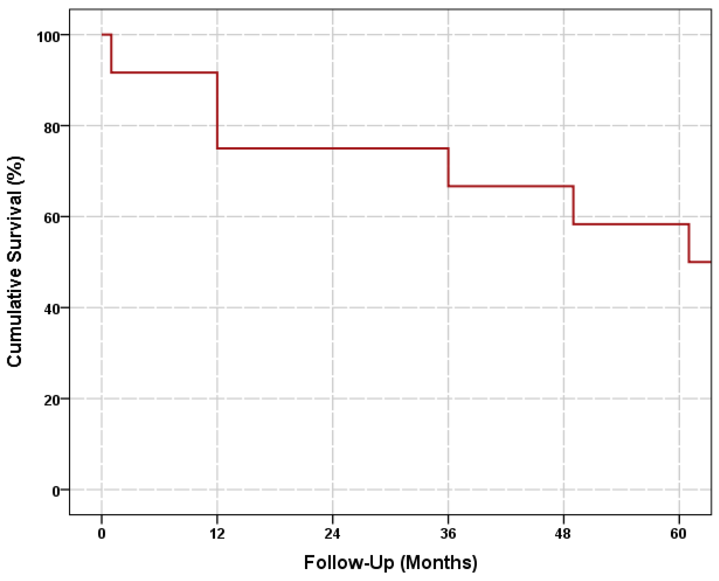
3. Results
3.1. Patient Characteristics
3.2. Efficacy
3.3. Safety
4. Discussion
5. Conclusions
Author Contributions
Funding
Institutional Review Board Statement
Informed Consent Statement
Data Availability Statement
Conflicts of Interest
References
- Comi, A.M. Update on Sturge-Weber syndrome: Diagnosis, treatment, quantitative measures, and controversies. Lymphat. Res. Biol. 2007, 5, 257–264. [Google Scholar] [CrossRef] [PubMed]
- Shirley, M.D.; Tang, H.; Gallione, C.J.; Baugher, J.D.; Frelin, L.P.; Cohen, B.; Pevsner, J. Sturge-Weber syndrome and port-wine stains caused by somatic mutation in GNAQ. N. Engl. J. Med. 2013, 368, 1971–1979. [Google Scholar] [CrossRef] [PubMed] [Green Version]
- Hassanpour, K.; Nourinia, R.; Gerami, E.; Mahmoudi, G.; Esfandiari, H. Ocular Manifestations of the Sturge-Weber Syndrome. J. Ophthalmic. Vis. Res. 2021, 16, 415–431. [Google Scholar] [CrossRef] [PubMed]
- Sullivan, T.J.; Clarke, M.P.; Morin, J.D. The ocular manifestations of the Sturge-Weber syndrome. J. Pediatr. Ophthalmol. Strabismus 1992, 29, 349–356. [Google Scholar] [CrossRef] [PubMed]
- Cibis, G.W.; Tripathi, R.C.; Tripathi, B.J. Glaucoma in Sturge-Weber syndrome. Ophthalmology 1984, 91, 1061–1071. [Google Scholar] [CrossRef] [PubMed]
- Mantelli, F.; Bruscolini, A.; La Cava, M.; Abdolrahimzadeh, S.; Lambiase, A. Ocular manifestations of Sturge-Weber syndrome: Pathogenesis, diagnosis, and management. Clin. Ophthalmol. 2016, 10, 871–878. [Google Scholar] [PubMed] [Green Version]
- Basler, L.; Sowka, J. Sturge-Weber syndrome and glaucoma. Optometry 2011, 82, 306–309. [Google Scholar] [CrossRef] [PubMed]
- Board, R.J.; Shields, M.B. Combined trabeculotomy-trabeculectomy for the management of glaucoma associated wih Sturge-Weber syndrome. Ophthalmic. Surg. 1981, 12, 813–817. [Google Scholar]
- Keverline, P.O.; Hiles, D.A. Trabeculectomy for adolescent onset glaucoma in the Sturge-Weber syndrome. J. Pediatr. Ophthalmol. 1976, 13, 144–148. [Google Scholar] [CrossRef]
- Iwach, A.G.; Hoskins, H.D., Jr.; Hetherington, J., Jr.; Shaffer, R.N. Analysis of surgical and medical management of glaucoma in Sturge-Weber syndrome. Ophthalmology 1990, 97, 904–909. [Google Scholar] [CrossRef]
- Al-Obeidan, S.A.; Osman Eel, D.; Dewedar, A.S.; Kestelyn, P.; Mousa, A. Efficacy and safety of deep sclerectomy in childhood glaucoma in Saudi Arabia. Acta Ophthalmol. 2014, 92, 65–70. [Google Scholar] [CrossRef]
- Audren, F.; Abitbol, O.; Dureau, P.; Hakiki, S.; Orssaud, C.; Bourgeois, M.; Dufier, J.L. Non-penetrating deep sclerectomy for glaucoma associated with Sturge-Weber syndrome. Acta Ophthalmol. Scand. 2006, 84, 656–660. [Google Scholar] [CrossRef]
- Phelps, C.D. The pathogenesis of glaucoma in Sturge-Weber syndrome. Ophthalmology 1978, 85, 276–286. [Google Scholar] [CrossRef] [PubMed]
- Arora, K.S.; Quigley, H.A.; Comi, A.M.; Miller, R.B.; Jampel, H.D. Increased choroidal thickness in patients with Sturge-Weber syndrome. JAMA Ophthalmol. 2013, 131, 1216–1219. [Google Scholar] [CrossRef] [PubMed]
- Wu, Y.; Peng, C.; Ding, X.; Zeng, C.; Cui, C.; Xu, L.; Guo, W. Episcleral hemangioma distribution patterns could be an indicator of trabeculotomy prognosis in young SWS patients. Acta Ophthalmol. 2020, 98, e685–e690. [Google Scholar] [CrossRef] [PubMed]
- Kaushik, J.; Parihar, J.K.S.; Jain, V.K.; Mathur, V. Ahmed valve implantation in childhood glaucoma associated with Sturge-Weber syndrome: Our experience. Eye 2019, 33, 464–468. [Google Scholar] [CrossRef] [PubMed] [Green Version]
- Almobarak, F.A.; Alharbi, A.H.; Morales, J.; Aljadaan, I. The influence of mitomycin C concentration on the outcome of trabeculectomy in uveitic glaucoma. Int. Ophthalmol. 2018, 38, 2371–2379. [Google Scholar] [CrossRef] [PubMed]
- Almobarak, F.A.; Alharbi, A.H.; Morales, J.; Aljadaan, I. Intermediate and Long-term Outcomes of Mitomycin C-enhanced Trabeculectomy as a First Glaucoma Procedure in Uveitic Glaucoma. J. Glaucoma 2017, 26, 478–485. [Google Scholar] [CrossRef]
- Olsen, K.E.; Huang, A.S.; Wright, M.M. The efficacy of goniotomy/trabeculotomy in early-onset glaucoma associated with the Sturge-Weber syndrome. J. Aapos. 1998, 2, 365–368. [Google Scholar] [CrossRef] [PubMed]
- Bichsel, C.A.; Goss, J.; Alomari, M.; Alexandrescu, S.; Robb, R.; Smith, L.E.; Bischoff, J. Association of Somatic GNAQ Mutation with Capillary Malformations in a Case of Choroidal Hemangioma. JAMA Ophthalmol. 2019, 137, 91–95. [Google Scholar] [CrossRef] [PubMed]
- Kaushik, S.; Kaur, S.; Pandav, S.S.; Gupta, A. Intractable choroidal effusion with exudative retinal detachment in Sturge-Weber syndrome. JAMA Ophthalmol. 2014, 132, 1143–1144. [Google Scholar] [CrossRef] [PubMed]
- Almobarak, F.A. Aqueous misdirection after Nd:YAG goniopuncture in deep sclerectomy treated with Nd:YAG irido-zonulo-hyaloidotomy. Eur. J. Ophthalmol. 2022, 32, NP28–NP31. [Google Scholar] [CrossRef] [PubMed]


| Patient | Eye | Age at Surgery | Preoperative IOP | No. of Preoperative Medications | Intraoperative Complications | Postoperative Complications | Follow-Up Duration | Status | Final IOP | No. of Final Medications |
|---|---|---|---|---|---|---|---|---|---|---|
| 1 | OD | 2 months | 43 | 4 | 7.8 years | Complete success | 13 | 0 | ||
| OS | 2 months | 39 | 4 | 7.8 years | Complete success | 13 | 0 | |||
| 2 | OS | 10 months | 30 | 2 | TDW perforation with iris prolapse | 15.6 years | Failed | 22 | 3 | |
| 3 | OD | 8.5 years | 28 | 4 | 1 month | Failed | 37 | 2 | ||
| 4 | OS | 9.5 years | 33 | 4 | TDW perforation with iris prolapse | Choroidal detachment | 7.8 years | Qualified success | 16 | 4 |
| 5 | OD | 3 months | 32 | 2 | TDW perforation | Choroidal effusion | 5.1 years | Failed | 30 | 3 |
| 6 | OS | 1.3 years | 25 | 3 | 4.1 years | Failed | 32 | 3 | ||
| 7 | OS | 2 years | 26 | 2 | TDW perforation with iris prolapse | 20.9 years | Qualified success | 14 | 4 | |
| 8 | OD | 8.3 years | 20 | 3 | 8.8 years | Qualified success | 18 | 1 | ||
| 9 | OD | 12.4 years | 30 | 3 | 3.7 years | Complete success | 12 | 0 | ||
| 10 | OD | 13.3 years | 20 | 3 | 1 year | Complete success | 10 | 0 | ||
| 11 | OS | 14.2 years | 19 | 4 | 1 year | Complete success | 9 | 0 |
Disclaimer/Publisher’s Note: The statements, opinions and data contained in all publications are solely those of the individual author(s) and contributor(s) and not of MDPI and/or the editor(s). MDPI and/or the editor(s) disclaim responsibility for any injury to people or property resulting from any ideas, methods, instructions or products referred to in the content. |
© 2023 by the authors. Licensee MDPI, Basel, Switzerland. This article is an open access article distributed under the terms and conditions of the Creative Commons Attribution (CC BY) license (https://creativecommons.org/licenses/by/4.0/).
Share and Cite
Almobarak, F.A.; Alobaidan, A.S.; Alobrah, M.A. Outcomes of Deep Sclerectomy for Glaucoma Secondary to Sturge–Weber Syndrome. J. Clin. Med. 2023, 12, 516. https://doi.org/10.3390/jcm12020516
Almobarak FA, Alobaidan AS, Alobrah MA. Outcomes of Deep Sclerectomy for Glaucoma Secondary to Sturge–Weber Syndrome. Journal of Clinical Medicine. 2023; 12(2):516. https://doi.org/10.3390/jcm12020516
Chicago/Turabian StyleAlmobarak, Faisal A., Abdullah S. Alobaidan, and Mansour A. Alobrah. 2023. "Outcomes of Deep Sclerectomy for Glaucoma Secondary to Sturge–Weber Syndrome" Journal of Clinical Medicine 12, no. 2: 516. https://doi.org/10.3390/jcm12020516