
Potential Diagnostic and Monitoring Biomarkers of Obstructive Sleep Apnea–Umbrella Review of Meta-Analyses

Abstract
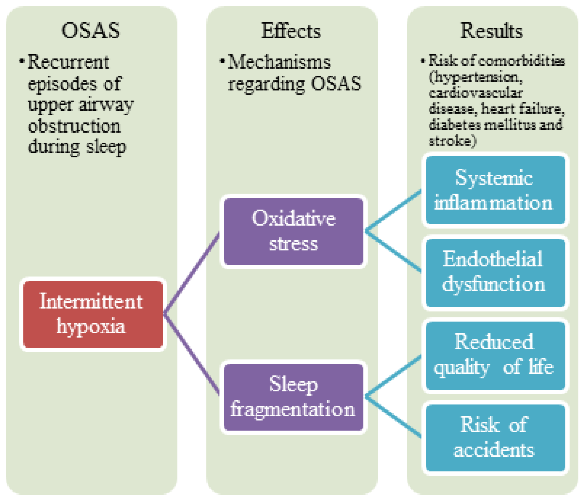
:1. Introduction
Aim of the Study
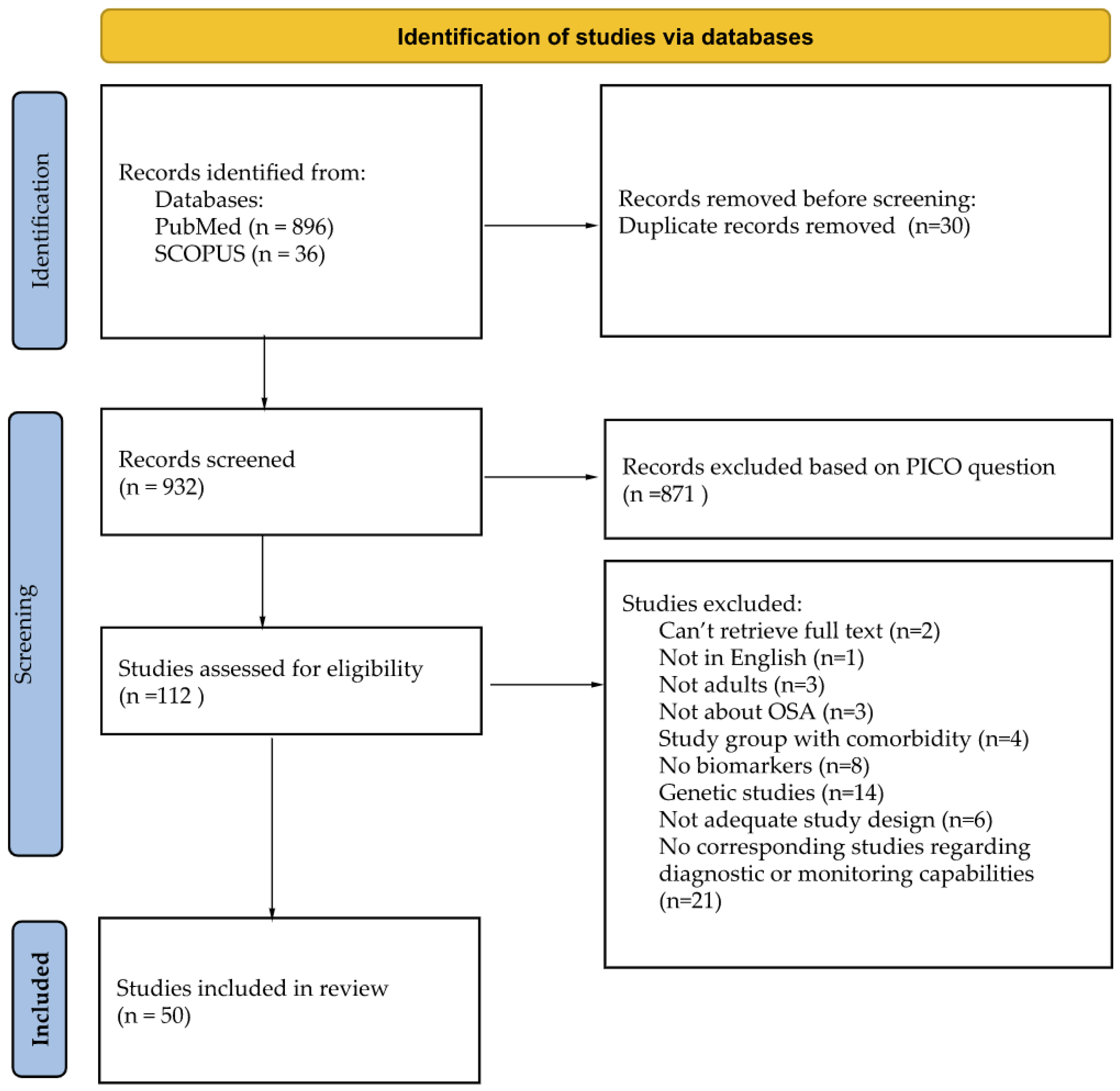
2. Methods
2.1. Potential Biomarkers of OSA in Adults
2.1.1. CRP
2.1.2. Interleukin-6
2.1.3. Tumor Necrosis Factor-Alpha
2.1.4. Interleukin-8
2.1.5. ICAM-1 and VCAM-1
2.1.6. VEGF
2.1.7. Homocysteine
2.1.8. Malondialdehyde
2.1.9. Leptin
2.1.10. IGF-1
2.1.11. ALT and AST
2.1.12. Lipid Profile
2.1.13. Adiponectin
2.1.14. Cortisol
2.2. Quality of Studies
3. Discussion
4. Conclusions
Supplementary Materials
Author Contributions
Funding
Institutional Review Board Statement
Informed Consent Statement
Data Availability Statement
Conflicts of Interest
References
- Olszewska, E.; Panek, J.; O’Day, J.; Rogowski, M. Usefulness of snoreplasty in the treatment of simple snoring and mild obstructive sleep apnea/hypopnea syndrome—Preliminary report. Otolaryngol. Pol. 2014, 68, 184–188. [Google Scholar] [CrossRef] [PubMed]
- Olszewska, E.; Sieskiewicz, A.; Rozycki, J.; Rogalewski, M.; Tarasow, E.; Rogowski, M.; Kulikowska, J. A comparison of cephalometric analysis using radiographs and craniofacial computed tomography in patients with obstructive sleep apnea syndrome: Preliminary report. Eur. Arch. Oto-Rhino-Laryngol. 2009, 266, 535–542. [Google Scholar] [CrossRef] [PubMed]
- Olszewska, E.; Rutkowska, J.; Czajkowska, A.; Rogowski, M. Selected surgical management in snoring and obstructive sleep apnea patients. Med. Sci. Monit. 2012, 18, 13–18. [Google Scholar] [CrossRef] [PubMed] [Green Version]
- Olszewska, E.; Woodson, B.T. Palatal anatomy for sleep apnea surgery. Laryngoscope Investig. Otolaryngol. 2019, 4, 181–187. [Google Scholar] [CrossRef] [PubMed] [Green Version]
- Pang, K.P.; Baptista, P.M.; Olszewska, E.; Braverman, I.; Carrasco-Llatas, M.; Kishore, S.; Chandra, S.; Yang, H.C.; Wang, C.M.Z.; Chan, Y.H.; et al. Does drug-induced sleep endoscopy affect surgical outcome? A multicenter study of 326 obstructive sleep apnea patients. Laryngoscope 2020, 130, 551–555. [Google Scholar] [CrossRef] [PubMed]
- Senaratna, C.V.; Perret, J.L.; Lodge, C.J.; Lowea, A.J.; Campbella, B.E.; Mathesona, M.C.; Hamiltonde, G.S.; Dharmagea, S.C. Prevalence of obstructive sleep apnea in the general population: A systematic review. Sleep Med. Rev. 2017, 34, 70–81. [Google Scholar] [CrossRef]
- McNicholas, W.T.; Bonsignore, M.R. Sleep apnoea as an independent risk for cardiovascular disease: Current evidence, basic mechanisms and research priorities. Eur. Respir. J. 2007, 29, 156–178. [Google Scholar] [CrossRef]
- Wang, J.; Yu, W.; Gao, M.; Zhang, F.; Gu, C.; Yu, Y.; Wei, Y. Impact of obstructive sleep apnea syndrome on endothelial function, arterial stiffening, and serum inflammatory markers: An updated meta-analysis and metaregression of 18 studies. J. Am. Heart Assoc. 2015, 4, e002454. [Google Scholar] [CrossRef] [Green Version]
- Chiang, C.L.; Chen, Y.T.; Wang, K.L.; Su, V.Y.-F.; Wu, L.-A.; Perng, D.-W.; Chang, S.-C.; Chen, Y.-M.; Chen, T.-J.; Chou, K.-T. Comorbidities and risk of mortality in patients with sleep apnea. Ann. Med. 2017, 49, 377–383. [Google Scholar] [CrossRef]
- Karimi, M.; Hedner, J.; Häbel, H.; Nerman, O.; Grote, L. Sleep apnea-related risk of motor vehicle accidents is reduced by continuous positive airway pressure: Swedish traffic accident registry data. Sleep 2015, 38, 341–349. [Google Scholar] [CrossRef] [Green Version]
- Teran-Santos, J.; Jimenez-Gomez, A.; Cordero-Guevara, J. The association between sleep apnea and the risk of traffic accidents. N. Engl. J. Med. 1999, 340, 847–851. [Google Scholar] [CrossRef] [PubMed]
- Young, T.; Blustein, J.; Finn, L.; Palta, M. Sleepiness, Driving, and Accidents Sleep-Disordered Breathing and Motor Vehicle Accidents in a Population-Based Sample of Employed Adults. Sleep 1997, 20, 608–613. [Google Scholar] [CrossRef] [PubMed] [Green Version]
- Lacasse, Y.; Godbout, C.; Sériès, F. Health-related quality of life in obstructive sleep apnoea. Eur. Respir. J. 2002, 19, 499–503. [Google Scholar] [CrossRef] [Green Version]
- Cofta, S.; Winiarska, H.M.; Płóciniczak, A.; Bielawska, L.; Brożek, A.; Piorunek, T.; Kostrzewska, T.M.; Wysocka, E. Oxidative Stress Markers and Severity of Obstructive Sleep Apnea. In Advances in Experimental Medicine and Biology; Springer: New York, NY, USA, 2019; Volume 1222, pp. 27–35. [Google Scholar] [CrossRef]
- Bonetti, P.O.; Lerman, L.O.; Lerman, A. Endothelial dysfunction: A marker of atherosclerotic risk. Arter. Thromb. Vasc. Biol. 2003, 23, 168–175. [Google Scholar] [CrossRef] [PubMed]
- Davignon, J.; Ganz, P. Role of endothelial dysfunction in atherosclerosis. Circulation 2004, 109 (Suppl. S23), III-27. [Google Scholar] [CrossRef] [PubMed]
- Mo, L.; Gupta, V.; Modi, R.; Munnur, K.; Cameron, J.D.; Seneviratne, S.; Edwards, B.A.; Landry, S.A.; Joosten, S.A.; Hamilton, G.S.; et al. Severe obstructive sleep apnea is associated with significant coronary artery plaque burden independent of traditional cardiovascular risk factors. Int. J. Cardiovasc. Imaging 2020, 36, 347–355. [Google Scholar] [CrossRef]
- Zamarrón, C.; Valdés Cuadrado, L.; Álvarez-Sala, R. Pathophysiologic mechanisms of cardiovascular disease in obstructive sleep apnea syndrome. Pulm Med. 2013, 2013, 521087. [Google Scholar] [CrossRef] [Green Version]
- Morsy, N.E.; Farrag, N.S.; Zaki, N.F.W.; Badawy, A.Y.; Abdelhafez, S.A.; El-Gilany, A.-H.; Shafey, M.M.E.; Pandi-Perumal, S.R.; Spence, D.W.; BaHammam, A.S. Obstructive sleep apnea: Personal, societal, public health, and legal implications. Rev. Environ. Health 2019, 34, 153–169. [Google Scholar] [CrossRef]
- Fiedorczuk, P.; Stróżyński, A.; Olszewska, E. Is the oxidative stress in obstructive sleep apnea associated with cardiovascular complications? Systematic review. J. Clin. Med. 2020, 9, 3734. [Google Scholar] [CrossRef]
- Racanelli, A.C.; Kikkers, S.A.; Choi, A.M.K.; Cloonan, S.M. Autophagy and inflammation in chronic respiratory disease. Autophagy 2018, 14, 221–232. [Google Scholar] [CrossRef]
- Lu, D.; Abulimiti, A.; Wu, T.; Abudureyim, A.; Li, N. Pulmonary surfactant-associated proteins and inflammatory factors in obstructive sleep apnea. Sleep Breath. 2018, 22, 99–107. [Google Scholar] [CrossRef]
- Destors, M.; Tamisier, R.; Baguet, J.P.; Levy, P.; Pepin, J.L. Cardiovascular morbidity associated with obstructive sleep apnea syndrome. Rev. Mal. Respir. 2014, 31, 375–385. [Google Scholar] [CrossRef] [PubMed]
- Mano, Y.; Anzai, T.; Kaneko, H.; Nagatomo, Y.; Nagai, T.; Anzai, A.; Maekawa, Y.; Takahashi, T.; Meguro, T.; Yoshikawa, T.; et al. Overexpression of human C-reactive protein exacerbatesleft ventricular remodeling in diabetic cardiomyopathy. Circ. J. 2011, 75, 1717–1727. [Google Scholar] [CrossRef] [PubMed] [Green Version]
- Tanaka, T.; Narazaki, M.; Kishimoto, T. Il-6 in inflammation, Immunity, and Disease. Cold Spring Harb. Perspect. Biol. 2014, 6, a016295. [Google Scholar] [CrossRef] [PubMed]
- Idriss, H.T.; Naismith, J.H. TNFα and the TNF receptor superfamily: Structure-function relationship(s). Microsc. Res. Tech. 2000, 50, 184–195. [Google Scholar] [CrossRef]
- Rockstrom, M.D.; Chen, L.; Taishi, P.; Nguyen, J.T.; Gibbons, C.M.; Veasey, S.C.; Krueger, J.M. Tumor necrosis factor alpha in sleep regulation. Sleep Med. Rev. 2018, 40, 69–78. [Google Scholar] [CrossRef]
- Jiang, W.G.; Sanders, A.J.; Ruge, F.; Harding, K.G. Influence of interleukin-8 (IL-8) and IL-8 receptors on the migration of human keratinocytes, the role of plc-γ and potential clinical implications. Exp. Med. 2012, 3, 231–236. [Google Scholar] [CrossRef] [Green Version]
- Azagra-Calero, E.; Espinar-Escalona, E.; Barrera-Mora, J.M.; Llamas-Carreras, J.M.; Solano-Reina, E. Obstructive sleep apnea syndrome (OSAS). Review of the literature. Med. Oral. Patol. Oral. Cir. Bucal 2012, 17, e925. [Google Scholar] [CrossRef]
- Nadeem, R.; Molnar, J.; Madbouly, E.M.; Nida, M.; Aggarwal, S.; Sajid, H.; Naseem, J.; Loomba, R. Serum inflammatory markers in obstructive sleep apnea: A meta-analysis. J. Clin. Sleep Med. 2013, 9, 1003–1012. [Google Scholar] [CrossRef] [Green Version]
- Li, A.M.; Lam, H.S.; Chan, M.H.M.; So, H.K.; Ng, S.K.; Chan, I.H.S.; Lam, C.W.K.; Wing, Y.K. Inflammatory Cytokines and Childhood Obstructive Sleep Apnoea. Ann. Acad. Med. Singap. 2008, 37, 649–654. [Google Scholar]
- Yang, H.; Engeland, C.G.; King, T.S.; Sawyer, A.M. The relationship between diurnal variation of cytokines and symptom expression in mild obstructive sleep apnea. J. Clin. Sleep Med. 2020, 16, 715–723. [Google Scholar] [CrossRef] [PubMed]
- Arnould, T.; Michiels, C.; Remacle, J. Increased PMN adherence on endothelial cells after hypoxia: Involvement of PAF, CDlt3/CDIlb, and ICAM. Am. J. Physiol. 1993, 264 Pt 1, C1102–C1110. [Google Scholar] [CrossRef] [PubMed]
- Springer, T.A. Adhesion receptors of the immune system. Nature 1990, 346, 425–434. [Google Scholar] [CrossRef] [PubMed]
- Li, H.; Cybulsky, M.I.; Gimbrone, M.A.; Libby, P. An atherogenic diet rapidly induces VCAM-1, a cytokine-regulatable mononuclear leukocyte adhesion molecule, in rabbit aortic endothelium. Arter. Thromb. 1993, 13, 197–204. [Google Scholar] [CrossRef] [Green Version]
- Amberger, A.; Maczek, C.; Jürgens, G.; Michaelis, D.; Schett, G.; Trieb, K.; Eberl, T.; Jindal, S.; Xu, Q.; Wick, G. Co-expression of ICAM-1, VCAM-1, ELAM-1 and Hsp60 in human arterial and venous endothelial cells in response to cytokines and oxidized low-density lipoproteins. Cell Stress Chaperones 1997, 2, 94–103. [Google Scholar] [CrossRef]
- Alberti, A.; Sarchielli, P.; Gallinella, E.; Floridi, A.; Floridi, A.; Mazzotta, G.; Gallai, V. Plasma cytokine levels in patients with obstructive sleep apnea syndrome: A preliminary study. J. Sleep Res. 2003, 12, 305–311. [Google Scholar] [CrossRef] [Green Version]
- Ciftci, T.U.; Kokturk, O.; Bukan, N.; Bilgihan, A. The relationship between serum cytokine levels with obesity and obstructive sleep apnea syndrome. Cytokine 2004, 28, 87–91. [Google Scholar] [CrossRef]
- Blankenberg, S.; Rupprecht, H.J.; Bickel, C.; Peetz, D.; Hafner, G.; Tiret, L.; Meyer, J.; the AtheroGene Investigators. Circulating cell adhesion molecules and death in patients with coronary artery disease. Circulation 2001, 104, 1336–1342. [Google Scholar] [CrossRef] [Green Version]
- van der Meer, I.M.; de Maat, M.P.; Bots, M.L.; Breteler, M.M.; Meijer, J.; Kiliaan, A.J.; Hofman, A.; Witteman, J.C. Inflammatory mediators and cell adhesion molecules as indicators of severity of atherosclerosis: The Rotterdam Study. Arter. Thromb. Vasc. Biol. 2002, 22, 838–842. [Google Scholar] [CrossRef] [Green Version]
- Blann, A.D.; Lip, G.Y.; McCollum, C.N. Changes in von Willebrand factor and soluble ICAM, but not soluble VCAM, soluble E selectin or soluble thrombomodulin, reflect the natural history of the progression of atherosclerosis. Atherosclerosis 2002, 165, 389–391. [Google Scholar] [CrossRef]
- Jenny, N.S.; Arnold, A.M.; Kuller, L.H.; Sharrett, A.R.; Fried, L.P.; Psaty, B.M.; Tracy, R.P. Soluble intracellular adhesion molecule-1 is associated with cardiovascular disease risk and mortality in older adults. J. Thromb. Haemost. 2006, 4, 107–113. [Google Scholar] [CrossRef] [PubMed]
- Leung, D.W.; Cachianes, G.; Kuang, W.J.; Goeddel, D.V.; Ferrara, N. Vascular endothelial growth factor is a secreted angiogenic mitogen. Science 1989, 246, 1306–1309. [Google Scholar] [CrossRef] [PubMed]
- Salven, P.; Mänpää, H.; Orpana, A.; Alitalo, K.; Joensuu, H. Serum vascular endothelial growth factor is often elevated in disseminated cancer. Clin. Cancer Res. 1997, 3, 647–651. [Google Scholar] [PubMed]
- Lin, T.-H.; Su, H.-M.; Wang, C.-L.; Voon, W.-C.; Shin, S.-J.; Lai, W.-T.; Sheu, S.-H. Vascular endothelial growth factor polymorphisms and extent of coronary atherosclerosis in Chinese population with advanced coronary artery disease. Am. J. Hypertens. 2010, 23, 960–966. [Google Scholar] [CrossRef] [PubMed]
- Inoue, M.; Itoh, H.; Ueda, M.; Naruko, T.; Kojima, A.; Komatsu, R.; Doi, K.; Ogawa, Y.; Tamura, N.; Takaya, K.; et al. Vascular endothelial growth factor (VEGF) expression in human coronary atherosclerotic lesions: Possible pathophysiological significance of VEGF in progression of atherosclerosis. Circulation 1998, 98, 2108–2116. [Google Scholar] [CrossRef] [Green Version]
- Shweiki, D.; Itin, A.; Soffer, D.; Keshet, E. Vascular endothelial growth factor induced by hypoxia may mediate hypoxia-initiated angiogenesis. Nature 1992, 359, 843–845. [Google Scholar] [CrossRef]
- Philippe, C.; Boussadia, Y.; Prulière-Escabasse, V.; Papon, J.F.; Clerici, C.; Isabey, D.; Coste, A.; Escudier, E.; D’Ortho, M.P. Airway cell involvement in intermittent hypoxia-induced airway inflammation. Sleep Breath. 2015, 19, 297–306. [Google Scholar] [CrossRef]
- Tekin, D.; Dursun, A.D.; Baştuǧ, M.; Karaorman, G.; Fiçicilar, H. The effects of acute and intermittent hypoxia on the expressions of HIF-1α and VEGF in the left and right ventricles of the rabbit heart. Anadolu Kardiyol. Derg. 2011, 11, 379–385. [Google Scholar] [CrossRef] [Green Version]
- Selhub, J. Homocysteine metabolism. Annu. Rev. Nutr. 1999, 19, 217–246. [Google Scholar] [CrossRef] [Green Version]
- Seshadri, S.; Beiser, A.; Selhub, J.; Jacques, P.F.; Rosenberg, I.H.; D’Agostino, R.B.; Wilson, P.W.; Wolf, P.A. Plasma Homocysteine as a Risk Factor for Dementia and Alzheimer’s Disease. N. Engl. J. Med. 2002, 346, 476–483. [Google Scholar] [CrossRef]
- Danesh, J.; Lewington, S. Plasma homocysteine and coronary heart disease: Systematic review of published epidemiological studies. J. Cardiovasc. Risk 1998, 5, 229–232. [Google Scholar] [CrossRef]
- Chambers, J.C.; McGregor, A.; Jean-Marie, J.; Obeid, O.A.; Kooner, J.S. Demonstration of rapid onset vascular endothelial dysfunction after hyperhomocysteinemia: An effect reversible with vitamin C therapy. Circulation 1999, 99, 1156–1160. [Google Scholar] [CrossRef] [Green Version]
- Bostom, A.G.; Silbershatz, H.; Rosenberg, I.H.; Selhub, J.; D’Agostino, R.B.; Wolf, P.A.; Jacques, P.F.; Wilson, P.W.F. Nonfasting Plasma Total Homocysteine Levels and All-Cause and Cardiovascular Disease Mortality in Elderly Framingham Men and Women. Arch. Intern. Med. 1999, 159, 1077–1080. [Google Scholar] [CrossRef] [Green Version]
- Mcdowell, I.F.W.; Lang, D. Homocysteine and endothelial dysfunction: A link with cardiovascular disease. J. Nutr. 2000, 130, 369–372. [Google Scholar] [CrossRef] [Green Version]
- Jacobsen, D.W. Hyperhomocysteinemia and oxidative stress time for a reality check? Arter. Thromb. Vasc. Biol. 2000, 20, 1182–1184. [Google Scholar] [CrossRef] [Green Version]
- Katsoulis, K.; Kontakiotis, T.; Spanogiannis, D.; Vlachogiannis, E.; Kougioulis, M.; Gerou, S.; Daskalopoulou, E. Total antioxidant status in patients with obstructive sleep apnea without comorbidities: The role of the severity of the disease. Sleep Breath. 2011, 15, 861–866. [Google Scholar] [CrossRef]
- Esterbauer, H.; Schaur, R.J.; Zollner, H. Chemistry and biochemistry of 4-hydroxynonenal, malonaldehyde and related aldehydes. Free Radic. Biol. Med. 1991, 11, 81–128. [Google Scholar] [CrossRef]
- Negre-Salvayre, A.; Coatrieux, C.; Ingueneau, C.; Salvayre, R. Advanced lipid peroxidation end products in oxidative damage to proteins. Potential role in diseases and therapeutic prospects for the inhibitors. Br. J. Pharm. 2008, 153, 6–20. [Google Scholar] [CrossRef] [Green Version]
- Epizzimenti, S.; Ciamporcero, E.S.; Edaga, M.; Epettazzoni, P.; Earcaro, A.; Ecetrangolo, G.; Eminelli, R.; Edianzani, C.; Elepore, A.; Egentile, F.; et al. Interaction of aldehydes derived from lipid peroxidation and membrane proteins. Front. Physiol. 2013, 4, 242. [Google Scholar] [CrossRef] [Green Version]
- Slatter, D.A.; Avery, N.C.; Bailey, A.J. Identification of a New Cross-link and Unique Histidine Adduct from Bovine Serum Albumin Incubated with Malondialdehyde. J. Biol. Chem. 2004, 279, 61–69. [Google Scholar] [CrossRef]
- Jordan, W.; Cohrs, S.; Degner, D.; Meier, A.; Rodenbeck, A.; Mayer, G.; Pilz, J.; Rüther, E.; Kornhuber, J.; Bleich, S. Evaluation of oxidative stress measurements in obstructive sleep apnea syndrome. J. Neural. Transm. 2006, 113, 239–254. [Google Scholar] [CrossRef] [PubMed]
- Cofta, S.; Wysocka, E.; Piorunek, T.; Rzymkowska, M.; Batura-Gabryel, H.; Torlinski, L. Oxidative stress markers in the blood of persons with different stages of obstructive sleep apnea syndrome. J. Physiol. Pharm. 2008, 59, 183–190. [Google Scholar]
- Triantafyllou, G.A.; Paschou, S.A.; Mantzoros, C.S. Leptin and Hormones: Energy Homeostasis. Endocrinol. Metab. Clin. North Am. 2016, 45, 633–645. [Google Scholar] [CrossRef] [PubMed]
- Cohen, P.; Ocrant, I.; Fielder, P.J.; Neely, E.; Gargosky, S.E.; Deal, C.I.; Ceda, G.; Youngman, O.; Pham, H.; Lamson, G.; et al. Insulin-like growth factors (IGFs): Implications for aging. Psychoneuroendocrinology 1992, 17, 335–342. [Google Scholar] [CrossRef]
- Le Roith, D. Seminars in medicine of the Beth Israel Deaconess Medical Center. Insulin-like growth factors. N. Engl. J. Med. 1997, 336, 633–640. [Google Scholar] [CrossRef]
- Bailes, J.; Soloviev, M. Insulin-like growth factor-1 (IGF-1) and its monitoring in medical diagnostic and in sports. Biomolecules 2021, 11, 217. [Google Scholar] [CrossRef]
- Kalra, A.; Yetiskul, E.; Wehrle, C.J.; Tuma, F. Physiology, Liver. In StatPearls; StatPearls Publishing: Treasure Island, FL, USA, 2022. Available online: https://www.ncbi.nlm.nih.gov/books/NBK535438 (accessed on 30 September 2022).
- Knell, A.J. Liver function and failure: The evolution of liver physiology. J. R. Coll. Physicians Lond. 1980, 14, 205–208. [Google Scholar]
- Lala, V.; Zubair, M.; Minter, D.A. Liver Function Tests; StatPearls Publishing: Treasure Island, FL, USA, 2022. Available online: https://www.ncbi.nlm.nih.gov/books/NBK482489 (accessed on 30 September 2022).
- Oh, R.C.; Hustead, T.R.; Ali, S.M.; Pantsari, M.W. Mildly Elevated Liver Transaminase Levels: Causes and Evaluation. Am. Fam. Physician 2017, 96, 709–715. [Google Scholar]
- Strollo, P.J.; Rogers, R. Obstructive sleep apnea. N. Engl. J. Med. 1996, 334, 99–104. [Google Scholar] [CrossRef]
- Henrion, J.; Colin, L.; Schapira, M.; Heller, F.R. Hypoxic hepatitis caused by severe hypoxemia from obstructive sleep apnea. J. Clin. Gastroenterol. 1997, 24, 245–249. [Google Scholar] [CrossRef]
- Stoohs, R.A.; Facchini, F.; Gullleminault, C. Insulin resistance and sleep-disordered breathing in healthy humans. Am. J. Respir. Crit. Care Med. 1996, 154, 170–174. [Google Scholar] [CrossRef] [PubMed]
- Wilcox, I.; Mcnamara, S.G.; Collins, F.L.; Grunstein, R.R.; Sullivan, C.E. “Syndrome Z”: The interaction of sleep apnoea, vascular risk factors and heart disease. Thorax 1998, 53 (Suppl. S3), S25–S28. [Google Scholar]
- Vgontzas, A.N.; Papanicolaou, D.A.; Bixler, E.O.; Hopper, K.; Lotsikas, A.; Lin, H.-M.; Kales, A.; Chrousos, G.P. Sleep Apnea and Daytime Sleepiness and Fatigue: Relation to Visceral Obesity, Insulin Resistance, and Hypercytokinemia. J. Clin. Endocrinol. Metab. 2000, 85, 1151–1158. [Google Scholar] [CrossRef] [PubMed]
- Califf, R.M. Biomarker definitions and their applications. Exp. Biol. Med. 2018, 243, 213–221. [Google Scholar] [CrossRef] [PubMed]
- Farrell, P.M.; Rosenstein, B.J.; White, T.B. Guidelines for Diagnosis of Cystic Fibrosis in Newborns through Older Adults: Cystic Fibrosis Foundation Consensus Report. J. Pediatr. 2008, 153, S4–S14. [Google Scholar] [CrossRef] [Green Version]
- Holbrook, A.; Schulman, S.; Witt, D.M.; Vandvik, P.O.; Fish, J.; Kovacs, M.J.; Svensson, P.J.; Veenstra, D.L.; Crowther, M.; Guyatt, G.H. Evidence-based management of anticoagulant therapy. Antithrombotic therapy and prevention of thrombosis, 9th ed: American College of Chest Physicians evidence-based clinical practice guidelines. Chest 2012, 141 (Suppl. S2), e152S–e184S. [Google Scholar] [CrossRef] [Green Version]
- Kapur, V.K.; Auckley, D.H.; Chowdhuri, S.; Kuhlmann, D.C.; Mehra, R.; Ramar, K.; Harrod, C.G. Clinical practice guideline for diagnostic testing for adult obstructive sleep apnea: An American academy of sleep medicine clinical practice guideline. J. Clin. Sleep Med. 2017, 13, 479–504. [Google Scholar] [CrossRef]
- Howick, J.; Chalmers Iain (James Lind Library); Glasziou, P. OCEBM Table of Evidence Working Group “The Oxford 2011 Levels of Evidence”. Oxford Centre for Evidence-Based Medicine. Available online: http://www.cebm.net/index.aspx?o=5653 (accessed on 30 September 2022).
- Moher, D.; Liberati, A.; Tetzlaff, J.; Altman, D.G.; PRISMA Group. Preferred reporting items for systematic reviews and meta-analyses: The PRISMA statement. PLoS Med. 2009, 6, e1000097. [Google Scholar] [CrossRef] [Green Version]
- Shea, B.J.; Grimshaw, J.M.; A Wells, G.; Boers, M.; Andersson, N.; Hamel, C.; Porter, A.C.; Tugwell, P.; Moher, D.; Bouter, L.M. Development of AMSTAR: A measurement tool to assess the methodological quality of systematic reviews. BMC Med. Res. Methodol. 2007, 7, 10. [Google Scholar] [CrossRef] [Green Version]
- Shamsuzzaman, A.S.; Winnicki, M.; Lanfranchi, P.; Wolk, R.; Kara, T.; Accurso, V.; Somers, V.K. Elevated C-reactive protein in patients with obstructive sleep apnea. Circulation 2002, 105, 2462–2464. [Google Scholar] [CrossRef] [Green Version]
- Imani, M.; Sadeghi, M.; Farokhzadeh, F.; Khazaie, H.; Brand, S.; Dürsteler, K.; Brühl, A.; Sadeghi-Bahmani, D. Evaluation of blood levels of C-reactive protein marker in obstructive sleep apnea: A systematic review, meta-analysis and meta-regression. Life 2021, 11, 362. [Google Scholar] [CrossRef] [PubMed]
- Van der Touw, T.; Andronicos, N.M.; Smart, N. Is C-reactive protein elevated in obstructive sleep apnea? A systematic review and meta-analysis. Biomarkers 2019, 24, 429–435. [Google Scholar] [CrossRef] [PubMed]
- Li, K.; Wei, P.; Qin, Y.; Wei, Y. Is C-reactive protein a marker of obstructive sleep apnea? Medicine 2017, 96, e6850. [Google Scholar] [CrossRef] [PubMed]
- Yeo, B.S.Y.; Koh, J.H.; Tan, B.K.J.; Ding, Y.; Teo, Y.H.; Alkan, U.; See, A.; Loh, S.; Toh, S.T. Improved Inflammatory and Cardiometabolic Profile after Soft-Tissue Sleep Surgery for Obstructive Sleep Apnea: A Systematic Review and Meta-analysis. JAMA Otolaryngol. Head Neck Surg. 2022, 148, 862–869. [Google Scholar] [CrossRef]
- Yi, M.; Zhao, W.; Tan, Y.; Fei, Q.; Liu, K.; Chen, Z.; Zhang, Y. The causal relationships between obstructive sleep apnea and elevated CRP and TNF-α protein levels. Ann. Med. 2022, 54, 1578–1589. [Google Scholar] [CrossRef]
- Wang, Y.; Ni Lin, Y.; Zhang, L.Y.; Li, C.X.; Li, S.Q.; Li, H.P.; Zhang, L.; Li, N.; Yan, Y.R.; Li, Q.Y. Changes of circulating biomarkers of inflammation and glycolipid metabolism by CPAP in OSA patients: A meta-analysis of time-dependent profiles. Ther. Adv. Chronic Dis. 2022, 13, 20406223211070919. [Google Scholar] [CrossRef]
- Kang, K.; Yeh, T.; Hsu, Y.; Ko, J.; Lee, C.; Lin, M.; Hsu, W. Effect of Sleep Surgery on C-Reactive Protein Levels in Adults with Obstructive Sleep Apnea: A Meta-Analysis. Laryngoscope 2021, 131, 1180–1187. [Google Scholar] [CrossRef]
- Ning, Y.; Zhang, T.-S.; Wen, W.-W.; Li, K.; Yang, Y.-X.; Qin, Y.-W.; Zhang, H.-N.; Du, Y.-H.; Li, L.-Y.; Yang, S.; et al. Effects of continuous positive airway pressure on cardiovascular biomarkers in patients with obstructive sleep apnea: A meta-analysis of randomized controlled trials. Sleep Breath. 2019, 23, 77–86. [Google Scholar] [CrossRef]
- Guo, Y.; Pan, L.; Ren, D.; Xie, X. Impact of continuous positive airway pressure on C-reactive protein in patients with obstructive sleep apnea: A meta-analysis. Sleep Breath. 2013, 17, 495–503. [Google Scholar] [CrossRef]
- Baessler, A.; Nadeem, R.; Harvey, M.; Madbouly, E.; Younus, A.; Sajid, H.; Naseem, J.; Asif, A.; Bawaadam, H. Treatment for sleep apnea by continuous positive airway pressure improves levels of inflammatory markers-a meta-analysis. J. Inflamm. 2013, 10, 13. [Google Scholar] [CrossRef] [Green Version]
- Xie, X.M.; Pan, L.; Ren, D.Q.; Du, C.J.; Guo, Y.Z. Effects of continuous positive airway pressure therapy on systemic inflammation in obstructive sleep apnea: A meta-analysis. Sleep Med. 2013, 14, 1139–1150. [Google Scholar] [CrossRef] [PubMed]
- Friedman, M.; Samuelson, C.G.; Hamilton, C.; Fisher, M.; Kelley, K.; Joseph, N.J.; Wang, P.-C.; Lin, H.-C. Effect of continuous positive airway pressure on c-reactive protein levels in sleep apnea: A meta-analysis. Otolaryngol. Head Neck Surg. 2012, 147, 423–433. [Google Scholar] [CrossRef] [PubMed] [Green Version]
- de Luca Canto, G.; Pachêco-Pereira, C.; Aydinoz, S.; Major, P.W.; Flores-Mir, C.; Gozal, D. Diagnostic capability of biological markers in assessment of obstructive sleep apnea: A systematic review and meta-analysis. J. Clin. Sleep Med. 2015, 11, 27–36. [Google Scholar] [CrossRef] [PubMed]
- Gaspar, L.S.; Santos-Carvalho, A.; Santos, B.; Carvalhas-Almeida, C.; Barros-Viegas, A.T.; Oliveiros, B.; Donato, H.; Santos, C.; Moita, J.; Cavadas, C.; et al. Peripheral biomarkers to diagnose obstructive sleep apnea in adults: A systematic review and meta-analysis. Sleep Med. Rev. 2022, 64, 101659. [Google Scholar] [CrossRef] [PubMed]
- Yi, M.; Zhao, W.; Fei, Q.; Tan, Y.; Liu, K.; Chen, Z.; Zhang, Y. Causal analysis between altered levels of interleukins and obstructive sleep apnea. Front. Immunol. 2022, 13, 3937. [Google Scholar] [CrossRef]
- Imani, M.M.; Sadeghi, M.; Khazaie, H.; Emami, M.; Sadeghi Bahmani, D.; Brand, S. Evaluation of Serum and Plasma Interleukin-6 Levels in Obstructive Sleep Apnea Syndrome: A Meta-Analysis and Meta-Regression. Front. Immunol. 2020, 11, 1343. [Google Scholar] [CrossRef]
- Zhong, A.; Xiong, X.; Shi, M.; Xu, H. Roles of interleukin (IL)-6 gene polymorphisms, serum IL-6 levels, and treatment in obstructive sleep apnea: A meta-analysis. Sleep Breath. 2016, 20, 719–731. [Google Scholar] [CrossRef]
- Lee, C.H.; Hsu, W.C.; Yeh, T.H.; Ko, J.Y.; Lin, M.T.; Kang, K.T. Effect of Sleep Surgery on Inflammatory Cytokines in Adult Obstructive Sleep Apnea: A Systematic Review and Meta-Analysis. Laryngoscope 2022, 132, 2275–2284. [Google Scholar] [CrossRef]
- Imani, M.M.; Sadeghi, M.; Khazaie, H.; Emami, M.; Bahmani, D.S.; Brand, S. Serum and plasma tumor necrosis factor alpha levels in individuals with obstructive sleep apnea syndrome: A meta-analysis and meta-regression. Life 2020, 10, 87. [Google Scholar] [CrossRef] [PubMed]
- Cao, Y.; Song, Y.; Ning, P.; Zhang, L.; Wu, S.; Quan, J.; Li, Q. Association between tumor necrosis factor alpha and obstructive sleep apnea in adults: A meta-analysis update. BMC Pulm. Med. 2020, 20, 215. [Google Scholar] [CrossRef]
- Li, Q.; Zheng, X. Tumor necrosis factor alpha is a promising circulating biomarker for the development of obstructive sleep apnea syndrome: A meta-analysis. Oncotarget 2017, 8, 27616–27626. [Google Scholar] [CrossRef] [PubMed] [Green Version]
- Li, X.; Hu, R.; Ren, X.; He, J. Interleukin-8 concentrations in obstructive sleep apnea syndrome: A systematic review and meta-analysis. Bioengineered 2021, 12, 10666–10681. [Google Scholar] [CrossRef]
- Tian, Z.; Xiao, J.; Kang, J.; Sun, H.; Mu, Z.; Tong, D.; Li, M. Effects of Continuous Positive Airway Pressure on Cell Adhesion Molecules in Patients with Obstructive Sleep Apnea: A Meta-Analysis. Lung 2021, 199, 639–651. [Google Scholar] [CrossRef] [PubMed]
- Qiu, Y.; Li, X.; Zhang, X.; Wang, W.; Chen, J.; Liu, Y.; Fang, X.; Ni, X.; Zhang, J.; Wang, S.; et al. Prothrombotic Factors in Obstructive Sleep Apnea: A Systematic Review with Meta-Analysis. Ear Nose Throat J. 2020, 101, NP412–NP421. [Google Scholar] [CrossRef] [PubMed]
- Zhang X bin Jiang, X.T.; Cai, F.R.; Zeng, H.Q.; Du, Y.P. Vascular endothelial growth factor levels in patients with obstructive sleep apnea: A meta-analysis. Eur. Arch. Oto-Rhino-Laryngol. 2017, 274, 661–670. [Google Scholar] [CrossRef] [PubMed]
- Qi, J.-C.; Zhang, L.; Li, H.; Zeng, H.; Ye, Y.; Wang, T.; Wu, Q.; Chen, L.; Xu, Q.; Zheng, Y.; et al. Impact of continuous positive airway pressure on vascular endothelial growth factor in patients with obstructive sleep apnea: A meta-analysis. Sleep Breath. 2019, 23, 5–12. [Google Scholar] [CrossRef] [PubMed]
- Li, K.; Zhang, J.; Qin, Y.; Wei, Y.X. Association between Serum Homocysteine Level and Obstructive Sleep Apnea: A Meta-Analysis. Biomed Res Int 2017, 2017, 7234528. [Google Scholar] [CrossRef] [PubMed] [Green Version]
- Niu, X.; Chen, X.; Xiao, Y.; Dong, J.; Zhang, R.; Lü, M.; Kong, W. The differences in homocysteine level between obstructive sleep apnea patients and controls: A meta-analysis. PLoS ONE 2014, 9, e95794. [Google Scholar] [CrossRef] [PubMed]
- Chen, X.; Niu, X.; Xiao, Y.; Dong, J.; Zhang, R.; Lu, M.; Kong, W. Effect of continuous positive airway pressure on homocysteine levels in patients with obstructive sleep apnea: A meta-analysis. Sleep Breath. 2014, 18, 687–694. [Google Scholar] [CrossRef] [PubMed]
- Fadaei, R.; Safari-Faramani, R.; Hosseini, H.; Koushki, M.; Ahmadi, R.; Rostampour, M.; Khazaie, H. Increased the circulating levels of malondialdehyde in patients with obstructive sleep apnea: A systematic review and meta-analysis. Sleep Breath. 2021, 25, 1753–1760. [Google Scholar] [CrossRef]
- Chen, Q.; Chen, L.-D.; Chen, M.-X.; Wu, Y.-H.; Zeng, H.-X.; Hu, M.-F.; Zhang, W.-L.; Zheng, Y.-F.; Lin, Q.-C. The effect of continuous positive airway pressure on circulating malondialdehyde among obstructive sleep apnea patients: A meta-analysis. Sleep Breath. 2020, 24, 1407–1415. [Google Scholar] [CrossRef]
- Fadaei, R.; Koushki, M.; Sharafkhaneh, A.; Moradi, N.; Ahmadi, R.; Rostampour, M.; Khazaie, H. The impact of continuous positive airway pressure therapy on circulating levels of malondialdehyde: A systematic review and meta-analysis. Sleep Med. 2020, 75, 27–36. [Google Scholar] [CrossRef] [PubMed]
- Li, X.; He, J. The Association Between Serum/Plasma Leptin Levels and Obstructive Sleep Apnea Syndrome: A Meta-Analysis and Meta-Regression. Front. Endocrinol. 2021, 12, 696418. [Google Scholar] [CrossRef] [PubMed]
- Zhang, P.; Liu, J.; Long, S.; Xie, X.; Guo, Y. Association between continuous positive airway pressure and changes in serum leptin in patients with obstructive sleep apnoea: A meta-analysis. Sleep Breath. 2014, 18, 695–702. [Google Scholar] [CrossRef] [PubMed]
- Chen, X.; Niu, X.; Xiao, Y.; Dong, J.; Lu, M.; Kong, W. Effect of continuous positive airway pressure on leptin levels in patients with obstructive sleep apnea: A meta-analysis. Otolaryngol.-Head Neck Surg. 2015, 152, 610–618. [Google Scholar] [CrossRef] [PubMed]
- Chen L da Lin, L.; Huang, J.F.; Chen, X.; Xu, Q.Z.; Liu, J.N. Effect of continuous positive airway pressure on insulin growth factor-1 in patients with obstructive sleep apnea: A meta-analysis. Growth Horm. IGF Res. 2015, 25, 75–79. [Google Scholar] [CrossRef]
- He, J.; Li, X.; Yu, M. The correlation of serum/plasma IGF-1 concentrations with obstructive sleep apnea hypopnea syndrome: A meta-analysis and meta-regression. Front. Endocrinol. 2022, 13, 922229. [Google Scholar] [CrossRef]
- Sookoian, S.; Pirola, C.J. Obstructive sleep apnea is associated with fatty liver and abnormal liver enzymes: A meta-analysis. Obes. Surg. 2013, 23, 1815–1825. [Google Scholar] [CrossRef]
- Chen, L.-D.; Lin, L.; Zhang, L.-J.; Zeng, H.-X.; Wu, Q.-Y.; Hu, M.-F.; Xie, J.-J.; Liu, J.-N. Effect of continuous positive airway pressure on liver enzymes in obstructive sleep apnea: A meta-analysis. Clin. Respir. J. 2018, 12, 373–381. [Google Scholar] [CrossRef]
- Nadeem, R.M.; Singh, M.M.; Nida, M.; Waheed, I.; Khan, A.; Ahmed, S.; Naseem, J.; Champeau, D. Effect of obstructive sleep apnea hypopnea syndrome on lipid profile: A meta-regression analysis. J. Clin. Sleep Med. 2014, 10, 475–489. [Google Scholar] [CrossRef]
- Lee, C.H.; Hsu, W.C.; Yeh, T.H.; Ko, J.Y.; Lin, M.T.; Kang, K.T. Effect of sleep surgery on lipid profiles in adults with obstructive sleep apnea: A meta-analysis. Eur. Arch. Oto-Rhino-Laryngol. 2022, 279, 3811–3820. [Google Scholar] [CrossRef] [PubMed]
- Nadeem, R.; Singh, M.; Nida, M.; Kwon, S.; Sajid, H.; Witkowski, J.; Pahomov, E.; Shah, K.; Park, W.; Champeau, D. Effect of CPAP treatment for obstructive sleep apnea hypopnea syndrome on lipid profile: A meta-regression analysis. J. Clin. Sleep Med. 2014, 10, 1295–1302. [Google Scholar] [CrossRef] [PubMed] [Green Version]
- Xu, H.; Yi, H.; Guan, J.; Yin, S. Effect of continuous positive airway pressure on lipid profile in patients with obstructive sleep apnea syndrome: A meta-analysis of randomized controlled trials. Atherosclerosis 2014, 234, 446–453. [Google Scholar] [CrossRef] [PubMed] [Green Version]
- Lu, M.; Fang, F.; Wang, Z.; Wei, P.; Hu, C.; Wei, Y. Association between serum/plasma levels of adiponectin and obstructive sleep apnea hypopnea syndrome: A meta-analysis. Lipids Health Dis. 2019, 18, 30. [Google Scholar] [CrossRef] [PubMed]
- Chen, L.-D.; Liu, J.-N.; Lin, L.; Wu, Z.; Li, H.; Ye, Y.-M.; Xu, Q.-Z.; Lin, Q.-C. Effect of continuous positive airway pressure on adiponectin in patients with obstructive sleep apnea: A meta-analysis. PLoS ONE 2015, 10, e0136837. [Google Scholar] [CrossRef] [PubMed] [Green Version]
- Iftikhar, I.H.; Hoyos, C.M.; Phillips, C.L.; Magalang, U.J. Meta-analyses of the association of sleep apnea with insulin resistance, and the effects of CPAP on HOMA-IR, adiponectin, and visceral adipose fat. J. Clin. Sleep Med. 2015, 11, 475–485. [Google Scholar] [CrossRef] [PubMed] [Green Version]
- Hecht, L.; Möhler, R.; Meyer, G. Effects of CPAP-respiration on markers of glucose metabolism in patients with obstructive sleep apnoea syndrome: A systematic review and meta-analysis. Ger. Med. Sci. 2011, 9, Doc20. [Google Scholar] [CrossRef] [PubMed]
- Imani, M.M.; Sadeghi, M.; Khazaie, H.; Sanjabi, A.; Brand, S.; Brühl, A.; Bahmani, D.S. Associations Between Morning Salivary and Blood Cortisol Concentrations in Individuals with Obstructive Sleep Apnea Syndrome: A Meta-Analysis. Front. Endocrinol. 2021, 11, 568823. [Google Scholar] [CrossRef]
- Ken-Dror, G.; Fry, C.H.; Murray, P.; Fluck, D.; Han, T.S. Changes in cortisol levels by continuous positive airway pressure in patients with obstructive sleep apnoea: Meta-analysis of 637 individuals. Clin. Endocrinol. 2021, 95, 909–917. [Google Scholar] [CrossRef]


| Can Patients with Obstructive Sleep Apnea be Diagnosed and Monitored During Treatment Using Serum or Plasma Biomarkers? | |
|---|---|
| The population | Patients with OSA defined as AHI > 5 in polysomnographic sleep study (PSG). |
| The indicator | Serum or plasma biomarkers. |
| The control | Groups of patients without OSA (AHI < 5 in PSG), other OSA patients, same patients after treatment. |
| The outcome | The difference in serum or plasma biomarker levels. |
| The study design | Peer-reviewed English articles. Adult (>18 years) human subjects. Meta-analysis of case–control/observational studies, randomized control trials. Meta-analyses regarding differences in serum or plasma potential biomarker levels between obstructive sleep apnea subjects and controls. OR Meta-analyses regarding changes in serum or plasma potential biomarker levels in obstructive sleep apnea subjects after treatment. |
| Potential Serum or Plasma Biomarker | The Number of Meta-Analyses Selected | |
|---|---|---|
| Comparison to the Control Group | Change after Treatment | |
| CRP | 5 | 9 |
| IL-6 | 6 | 6 |
| TNF-α | 5 | 7 |
| Il-8 | 3 | 1 |
| HCY | 2 | 1 |
| ICAM-1, VCAM-1 | 2 | 2 |
| VEGF | 2 | 1 |
| TC, LDLc, HDLc, TG | 1 | 4 |
| Leptin | 1 | 3 |
| Adiponectin | 1 | 3 |
| MDA | 1 | 2 |
| ALT, AST | 1 | 1 |
| IGF-1 | 1 | 1 |
| Cortisol | 1 | 1 |
| Authors | Material | Results and Conclusion |
|---|---|---|
| Comparison to the Control Group | ||
| Imani et al., 2021 [85] | 96 studies: 11 studies, 2994 subjects (plasma hs-CRP); 44 studies, 5097 subjects (serum hs-CRP); 9 studies, 938 subjects (plasma CRP); 32 studies, 3877 subjects (serum CRP) | Adults with OSA had considerably higher plasma and serum levels of hs-CRP and CRP than controls, although there was no discernible difference in plasma CRP levels between adults with OSA and controls. Plasma hs-CRP levels pooled MD in adults with OSA—0.11 mg/dL (p < 0.00001). Serum hs-CRP levels with OSA in adult 0.09 mg/dL (p < 0.00001). Plasma CRP pooled MD 0.06 mg/dL (p = 0.72). Compared to controls, the pooled MD of serum CRP levels in adults with OSA was 0.36 mg/dL (p < 0.00001). |
| Van der Touw et al., 2019 [86] | 5 studies, 335 subjects | CRP levels in nonsmoking OSA participants without comorbidities were increased relative to levels in healthy matched nonsmoking control participants. SMD was 0.61mg/dL higher in OSA participants than in control group (CI 0.38–0.84, p < 0.00001). |
| Li et al., 2016 [87] | 15 studies, 1297 subjects | Serum CRP/hs-CRP levels higher in moderate–severe OSA patients compared with control subjects. Serum CRP levels in the OSA group were 1.98 mmol/L higher than those in control group (95% CI: 1.39–2.58, p < 0.01). Serum hs-CRP levels in the OSA group were 1.57 mmol/L higher than those in the control group (95% CI: 0.96–2.18, p < 0.01). |
| Wang et al., 2015 [8] | 18 studies, 1160 subjects | Serum levels of high-sensitivity C-reactive protein and C-reactive protein were significantly higher in patients with OSA than in controls (SMD 0.58, 95% CI 0.42–0.73, p < 0.0001). |
| Nadeem et al., 2013 [30] | 30 studies, 4283 subjects | Patients with OSA had a statistically significant higher level of CRP when compared to control individuals (SMD, 1.77). |
| Change after treatment | ||
| Yeo et al., 2022 [88] | 11 studies, 397 subjects | Evidence suggested that individuals with OSA who underwent soft-tissue sleep surgery saw a reduction in CRP; this reduction was significant (SMD, 0.377; 95% CI, 0.617 to 0.137). |
| Yi et al., 2022 [89] | 64 studies, 4285 OSA patients, and 3692 controls | Following CPAP intervention, CRP was marginally lower in the CPAP group compared to the non-CPAP group (WMD (95% CI) = 0.91 (1.65, 0.17) mg/L, p = 0.02). |
| Wang et al., 2022 [90] | 34 studies, 1385 patients | Subgroup analysis showed that short-term CPAP decreases CRP (SMD: 0.73, 95% CI: 0.15–1.31; p = 0.011). |
| Kang et al., 2022 [91] | 9 studies, 235 adults | Adult CRP levels significantly dropped after sleep surgery for OSA. Patients with OSA who underwent sleep surgery saw a considerable decline in their CRP levels (standardized mean difference (SMD) = −0.39, 95% CI, −0.67 to −0.11). Patients who have seen significant OSA improvement following sleep surgery (i.e., an AHI reduction of >20 events/h) experience a stronger positive effect of surgery on CRP levels. |
| Ning et al., 2018 [92] | 15 studies, 1090 patients | CPAP reduces the inflammatory marker hs-CRP in OSA patients. The serum hs-CRP levels may serve as a predictor of how well OSA patients respond to CPAP therapy (SMD 0.64; 95% (CI) −1.19 to −0.09; p= 0.02). |
| Guo et al., 2013 [93] | 14 studies, 1199 patients | In individuals with OSA, elevated CRP was found and was considerably decreased by successful CPAP treatment. Clinically, the utilization of CRP levels may be acknowledged as a useful predictor for OSA therapy monitoring (SMD 0.64; 95% (CI) 0.40 to 0.88; p = 0.000). |
| Baessler et al., 2013 [94] | 14 studies with 771 patients | The serum inflammatory marker CRP is considerably reduced in OSA patients who use CPAP (pooled MD 0.14; 95% (CI) 0.08 to 0.20, p < 0.00001). |
| Xie et al., 2013 [95] | 24 studies, 1597 patients | The combined data demonstrated that OSA patients’ CRP levels might be reduced by CPAP treatment (SMD 0.452; 95% (CI), 0.252–0.651). |
| Friedman et al., 2012 [96] | 10 studies, 325 patients | Patients with OSA treated with CPAP seem to have a considerable reduction in CRP levels. |
| Authors | Material | Conclusion |
|---|---|---|
| Comparison to the control group | ||
| Gaspar et al., 2022 [98] | 16 studies, 1369 cases, 787 controls | IL-6 levels in plasma revealed the most promising candidates to further explore in future studies, as single or clustered biomarkers. |
| Yi et al., 2022 [99] | 48 articles, 1974 patients, 1657 controls | OSA patients had greater IL-6 levels than controls—the weighted mean difference was 3.89. In comparison to the control, there were substantial differences for IL-6 with severe OSA severity; the causal relationship between IL-6 levels and OSA was insignificant, with an OR of 0.853 and p = 0.114. |
| Imani et al., 2020 [100] | 63 studies: 39 studies, 2558 patients, 1897 controls (serum IL-6); 22 studies, 974 patients, 692 controls (plasma IL-6); | Higher levels of IL-6 have been linked to OSA severity. There were no significant independent effects of the publication year, mean BMI, mean AHI, or the number of participants on serum (pooled MD 2.89 pg/mL; p < 0.00001) or plasma IL-6 levels (pooled MD 2.89 pg/mL, p < 0.00001). |
| Zhong et al., 2015 [101] | 31 studies, 1666 cases, 989 controls | Levels of IL-6 are higher in patients with OSA and are significantly correlated with OSA severity. IL-6 levels were higher in OSA compared to controls (SMD = 1.56, 95% CI = 1.18–1.95). |
| Nadeem et al., 2013 [30] | 19 studies, 1316 subjects | IL-6 levels were greater in OSA patients than in healthy controls. A significant association was found between IL-6 and AHI. SMD 2.16. |
| De Luca Canto et al., 2015 [97] | 1 study, 88 cases, 32 controls | Exhaled breath condensate IL-6 has the potential to predict the severity of OSA in nonsmoker OSA suspects. |
| Change after treatment | ||
| Lee et al., 2022 [102] | 6 studies, 140 subjects | Sleep surgery lowers IL-6 levels in adults with OSA (MD of 0.6 pg/mL and SMD of 0.66). |
| Yeo et al., 2022 [88] | 5 studies, 149 subjects | A significant reduction in IL-6 was seen following soft-tissue sleep surgery (SMD, 1.086; 95% CI, 1.952 to 0.221). |
| Yi et al., 2022 [99] | 21 articles, 1047 patients | Upon therapy, the level of IL-6 significantly decreased—WMD −3.21. |
| Ning et al., 2019 [92] | 6 RCTs, 546 subjects | The overall MD was 0.15, p = 0.23, and no discernible decrease in IL-6 levels was seen after CPAP therapy. Moderate heterogeneity was detected. |
| Zhong et al., 2015 [101] | 20 studies, 646 patients | No significant result was observed for IL-6 serum levels after CPAP treatment (SMD = −0.24, 95% CI = −0.73 to 0.26). |
| Xie et al., 2013 [95] | 16 studies, 491 subjects | According to several studies, IL-6 is the best predictor of changes in AHI and had an independent impact on OSA, although analysis produced inconclusive findings for IL-6 (SMD = 0.299 (95% CI, 0.001–0.596)). |
| Authors | Material | Conclusion |
|---|---|---|
| Comparison to the control group | ||
| Yi et al., 2022 [89] | 34 studies, 1981 OSA patients and 1112 controls in | TNF-α levels were raised in OSA, and these rising tendencies were severity-dependent. Mendelian randomization did not reveal a causal link between OSA and increased TNF-α (WMD (95% CI)= 5.86 (4.80–6.93) pg/mL, p < 0.00001). |
| Imani et al., 2020 [103] | 29 studies, 2718 OSA patients, 1893 controls (serum TNF-α) 17 studies, 1021 OSA patients, 601 controls (plasma TNF-α) | Adult patients with OSA had considerably higher plasma (pooled MD 5.90 pg/mL (95% CI = 4.00, 7.80; p < 0.00001) and serum (pooled MD 10.22 pg/mL (95% CI = 8.86, 11.58; p < 0.00001) TNF-α levels than controls. Increased TNF-α levels in OSA patients seemed to correlate with the severity of the condition. |
| Cao et al., 2020 [104] | 50 articles 3503 OSA patients and 3379 healthy controls | Patients with OSA had TNF-α levels that were 1.77 (95% CI, 1.37 to 2.17, I2 = 97.8%, p < 0.0001) times greater than those in the control group. The concentration of TNF-α positively correlated with the severity of OSA. Older age substantially predicted a larger impact size of TNF-α level in OSA patients, according to meta-regression. |
| Li et al., 2017 [105] | 59 studies, 2857 OSA patients and 2115 | Circulating TNF-αlpha was considerably greater in patients than in controls (WMD]: 9.66 pg/mL, 95% confidence interval (CI): 8.66 to 11.24, p < 0.001), suggesting that TNF-αlpha may be a viable circulating biomarker for OSA development. |
| Nadeem et al., 2013 [30] | 19 studies with 1316 subjects pooled for TNF-α | TNF-α was higher in patients with OSA than in control individuals (pooled SMD 1.03). The moderate trend of the age, BMI, and AHI’s substantial effect was also seen in meta-regression; the level of TNF-α was correlated with OSA severity. |
| Change after treatment | ||
| Wang et al., 2022 [90] | 15 studies, 454 patients | After using CPAP for less than three months, there was a slight drop in TNF-α. The total pooled SMD for TNF-α was 0.49. |
| Lee et al., 2022 [102] | 8 studies, 205 subjects | Elevated TNF-α can be decreased by successful sleep surgery (SMD −0.56 (95% CI, −0.85 to −0.27). |
| Yeo et al., 2022 [88] | 6 studies, 173 patients | TNF-α can be decreased by successful soft-tissue sleep surgery (SMD = −0.822). |
| Yi et al., 2022 [89] | 4 articles, 125 patients, 109 controls | TNF-α was considerably reduced following CPAP management as compared to the non-CPAP group (WMD = 4.44 pg/mL, p < 0.00001). |
| Ning et al., 2019 [92] | 3 RCTs, 311 patients | The treatment effect of CPAP for TNF-α was not statistically significant (MD 0.61; p = 0.35) with indications of considerable heterogeneity. |
| Xie et al., 2013 [95] | 12 studies, 403 patients | TNF-α levels were found to decrease significantly (SMD, 0.478, p = 0.000). Subgroup analysis revealed that TNF-α levels decreased after less than three months of PAP treatment. |
| Baessler et al., 2013 [94] | 8 studies, 165 patients | TNF-α and inflammatory marker levels in the blood are considerably reduced in OSA patients who use CPAP (pooled MD 1.14; CI (95%) 0.12 to 2.15, p = 0.03). |
| Authors | Material | Conclusion |
|---|---|---|
| Comparison to the control group | ||
| Li et al., 2021 [106] | 25 studies, 2301 patients, 1123 controls | In patients with OSA, levels of IL-8 are elevated. The severity of OSA has a beneficial impact on this effect; the higher the AHI, the higher the levels. |
| Nadeem et al., 2013 [30] | 16 studies (serum IL-8) 4 studies (plasma IL-8) | Patients with OSA had significantly elevated serum IL-8 concentrations compared with controls (SMD = 0.997, 95% CI = 0.437–1.517, p < 0.001). Individuals with OSA had significantly elevated plasma IL-8 concentrations compared with controls (pooled SMD 4.22). |
| Yi et al., 2022 [99] | 13 studies, 1267 patients, 574 controls | Patients with OSA had higher serum concentrations of IL-8 than controls, although the difference was not statistically significant. |
| Change after treatment | ||
| Xie et al., 2013 [95] | 3 studies, 101 patients | IL-8 levels decreased after OSA treatment (SMD, 0.645 (95% CI, 0.362–0.929); z = 4.46; p = 0.000). |
| Authors | Material | Conclusion |
|---|---|---|
| Comparison to the control group | ||
| Nadeem et al., 2013 [30] De Luca Canto et al., 2015 [97] | 51 studies, 2952 patients, 2784 controls 8 studies, 495 participants (serum ICAM-1) 6 studies, 269 participants (serum VCAM-1) 1 article, 39 patients, 34 controls—Diagnostic capability-focused study | Significantly higher serum ICAM-1 level in subjects with OSA (pooled SMD 2.93). Significantly higher serum VCAM-1 level in subjects with OSA (pooled SMD 2.08). Potential biomarkers to distinguish OSA from non-OSA adults include ICAM-1 and VCAM-1, although with low diagnostic values. ICAM-1′s sensitivity/specificity (%) was 69/82, whereas VCAM-1′s was 74/65. |
| Change after treatment | ||
| Tian et al., 2021 [107] | ICAM-1 was significantly reduced by CPAP therapy (SMD = 0.283, 95% CI 0.464 to 0.101, p = 0.002). However, after receiving CPAP therapy, VCAM-1 levels did not significantly change (SMD = 0.160, 95% CI: 0.641 to 0.320, p = 0.513). | |
| Authors | Material | Conclusion |
|---|---|---|
| Comparison to the control group | ||
| Qiu et al., 2020 [108] | 5 relevant studies involving 262 patients | Patients with OSA had higher levels of VEGF than controls (SMD = 0.37, 95% CI = 0.90 to 1.63, p = 0.000). |
| Zhang et al., 2016 [109] | 15 studies, 426 patients, 271 controls | The VEGF in the OSA group is relatively higher than that in the control group (SMD 1.89, 95% CI 0.92–2.87, p = 0.000). In subjects with age ≥50 years, higher VEGF concentrations were identified in the OSA group when compared with the control group, but they failed to find any difference in VEGF between the two groups in subjects aged <50 years. The increased VEGF levels were observed in OSA patients compared to controls when using serum to detect VEGF, whereas the VEGF levels did not differ between the two groups when using plasma for VEGF detection. |
| Change after treatment | ||
| Qi et al., 2018 [110] | 3 studies, 392 patients | Before and following CPAP therapy, VEGF levels in patients with OSA significantly decreased (SMD = −0.440, 95% (CI) = −0.684 to −0.196, z = 3.53, p = 0.000). |
| Authors | Material | Conclusion |
|---|---|---|
| Comparison to the control group | ||
| Li et al., 2013 [111] | 10 studies, 457 cases, 316 controls | Patients with OSA had a higher serum Hcy level than healthy controls (Hcy levels were 2.40 μmol/L higher in OSA patients than in control group (95% CI: 0.6 to 4.20, p < 0.01). In addition, this difference is more significant in moderate or severe OSA patients. |
| Niu et al., 2014 [112] | 10 studies, 413 patients, 344 controls | Homocysteine levels were found to be 3.11 mmol/L higher in OSA patients compared to control subjects (95% CI: 2.08 to 4.15, p < 0.01). |
| Change after treatment | ||
| Chen et al., 2014 [113] | 6 studies, 206 cases | CPAP caused a statistically significant decrease in Hcy levels (WMD −0.62 (95% (CI) −1.21 to −0.04, p < 0.05). |
| Authors | Material | Conclusion |
|---|---|---|
| Comparison to the control group | ||
| Fadaei et al., 2020 [114] | 14 studies, 867 cases, 429 controls | MDA serum concentration significantly increased in OSA patients compared to the controls (SMD (95% CI): 1.18 (0.68, 1.68), p < 0.0001). |
| Change after treatment | ||
| Chen et al., 2020 [115] Fadaei et al., 2020 [116] | 10 studies, 220 cases 13 studies, 606 cases | A significant decrease in serum or plasma MDA was observed after CPAP treatment (SMD = 1.164, 95% CI = 0.443 to 1.885, z = 3.16, p = 0.002). CPAP therapy significantly reduced MDA levels in OSA patients (SMD −1.51 (95% CI, −2.06 to 0.97), p < 0.05). |
| Authors | Material | Conclusion |
|---|---|---|
| Comparison to the control group | ||
| Li et al., 2021 [117] | 34 studies, 1485 cases, 1201 controls | Plasma leptin levels in adults with OSA were significantly higher than the corresponding levels in the control group (WMD = 3.80 ng/mL, 95% CI = 3.09–4.50, p < 0.00001). |
| Change after treatment | ||
| Zhang et al., 2014 [118] Chen et al., 2015 [119] | 15 studies, 427 cases 11 studies, 413 cases | A significant decrease in serum leptin levels was observed among patients with OSA after CPAP therapy (SMD = 0.137; 95% (CI) 0.002 to 0.272; p = 0.046). CPAP therapy significantly reduced leptin levels in OSA patients (WMD = 1.44; CI (95%): 1.11–1.77, p < 0.01). |
| Authors | Material | Conclusion |
|---|---|---|
| Comparison to the control group | ||
| He et al., 2022 [121] | 34 studies, 1407 cases, 1039 controls | IGF-1 levels in patients with OSA were significantly lower than those of healthy controls (SMD = −1.37, 95% CI = −1.78–0.96, p < 0.001). |
| Change after treatment | ||
| Chen et al., 2014 [120] | 9 studies, 168 cases | CPAP was associated with a statistically significant increase in IGF-1 in OSA patients (SMD = −0.436, 95% CI = −0.653 to −0.218, p = 0.000). |
| Authors | Material | Conclusion |
|---|---|---|
| Comparison to the control group | ||
| Sookoian et al., 2013 [122] | 11 studies, 668 cases, 404 controls | The SMD values of ALT levels were significantly different in OSA patients compared to controls. AST levels were significantly different in OSA patients compared to controls. There was an increase of 13.3% in ALT and 4.4% in AST levels in OSA patients. |
| Change after treatment | ||
| Chen et al., 2016 [123] | 5 studies, 192 cases | CPAP was associated with a statistically significant decrease in ALT in OSA patients (WMD = 8.036, 95% CI = 2.788 to 13.285, z = 3.00, p = 0.003). A significant difference in AST was observed before and after CPAP treatment (WMD = 4.612, 95% CI = 0.817 to 8.407, z = 2.38, p = 0.017). |
| Authors | Material | Conclusion |
|---|---|---|
| Comparison to the control group | ||
| Nadeem et al., 2014 [124] | 64 studies, 18,116 patients (TC: 63 studies, 18,111 subjects; LDL 50 studies, 13,894 subjects; HDL: 64 studies, 18,116 subjects; TG 62 studies, 17,831 subjects) | Patients with OSA have increased dyslipidemia (LDL, HDL, TG, TC). Pooled SDM for total cholesterol = 0.267 (p = 0.001); pooled SDM for LDL cholesterol = 0.296 (p = 0.001). Pooled SDM for HDL cholesterol = −0.433 (p = 0.001). Pooled SDM for the triglyceride = 0.603 (p = 0.001). Age has a considerable impact on TC, LDL, and HDL, according to meta-regression for age, BMI, and AHI. While AHI had a substantial impact on LDL and TG, BMI had a large impact on LDL and HDL. |
| Change after treatment | ||
| Lee et al., 2022 [125] | 13 studies, 710 adults | In subjects with OSA, sleep surgery may enhance the lipid profile. Triglycerides were reduced by 14.0 mg/dL (mean−14.0 mg/dL; 95% CI −22.2 to −5.8), LDL was reduced by 7.2 mg/dL (mean −7.2 mg/dL; 95% CI −11.0 to −3.3), and total cholesterol reduced by 7.7 mg/dL (mean −7.7 mg/dL; 95% CI −12.2 to −3.2). Improvements in lipid profile measures positively correlated with the degree of OSA improvement. |
| Wang et al., 2022 [90] | TC: 28 studies, 918 subjects; LDL 21 studies, 670 subjects; HDL: 26 studies, 864 subjects; TG 30 studies, 1021 subjects | PAP therapy lowers blood cholesterol levels in OSA patients. Short-term therapy might lower LDL levels in the blood (SMD: 0.40 (95% CI: 0.18–0.62); p = 0.000), but medium- or long-term CPAP treatment raised HDL levels (SMD: –0.20 (95% CI: –0.36 to –0.03); p = 0.018) and lowered TC levels (SMD: 0.20 (95% CI: 0.05–0.34); p = 0.007) in the blood. |
| Nadeem et al., 2015 [126] | 29 studies, 1958 subjects TC: 24 studies, 1929 subjects; LDL 18 studies, 676 subjects; HDL: 23 studies, 806 subjects; TG 25 studies, 1926 subjects | Dyslipidemia that results from OSA therapy with CPAP appears to be improved (lower total and LDL cholesterol and higher HDL cholesterol). It does not seem to have an impact on TG levels. SMD for TC was −41.5 to −0.077, and PMD was -5.660 (p < 0.001). In LDL, SMD −3.7 to 0; PMD −0.488 (p < 0.001). For HDL, SMD −0.498 to 1.94; PMD was 0.207 (p < 0.01). For TG, SMD −9.327 to 1.98; PMD was −0.054 (p < 0.129). |
| Xu et al., 2014 [127] | 6 RCTs, TC: 370 patients vs. 371 controls, LDL 276 patients vs. 274 controls, HDL 269 patients and 266 controls, TG 330 patients vs. 328 controls | The difference in mean TC levels between the study and control groups shows that CPAP therapy of OSA patients reduced metabolic dyslipidemia; TG, LDL, and HDL levels did not change. The difference in mean TC level between the CPAP and sham CPAP/control groups was statistically different according to the pooled estimate (0.15, p = 0.01). |
| Authors | Material | Conclusion |
|---|---|---|
| Comparison to the control group | ||
| Lu et al., 2019 [128] | 20 studies, 1356 cases | The serum/plasma adiponectin levels were considerably lower in OSA patients than that in control subjects. It suggests a possible role of adiponectin in OSAHS pathogenesis. (SMD = −0.71, 95% CI = −0.92 to −0.49, p < 0.001). |
| Change after treatment | ||
| Chen et al., 2015 [129] | 11 studies, 240 cases (10 observational studies, 1 randomized controlled study) | Adiponectin levels in OSA patients did not differ between those taken before and after CPAP therapy (SMD = 0.059, 95% confidence interval (CI) = −0.250 to 0.368, z = 0.37, p = 0.710). |
| Iftikhar et al., 2015 [130] | 3 RCTs with 200 participants (101 in CPAP and 99 in sham CPAP groups) | There was no significant change in adiponectin levels before and after CPAP treatment in OSA patients (SMD =−0.06 (95% CI: −0.28 to 0.15), p = 0.56). |
| Hecht et al. [131] | 2 studies, 144 subjects | A significant change in adiponectin levels before and after CPAP treatment could not be observed. |
| Authors | Material | Conclusion |
|---|---|---|
| Comparison to the control group | ||
| Imani et al., 2021 [132] | Serum (5 studies, 314 subjects)’ Plasma (6 studies, 253 subjects) | Cortisol concentrations in the serum and plasma did not differ between adults with OSA and healthy controls. |
| Change after treatment | ||
| Ken-Dror et al., 2021 [133] | Plasma (15 prospective control studies, 335 subjects; 3 randomized control trials, 195 subjects) | CPAP treatment reduced plasma cortisol levels in prospective cohort studies: (SMD = −0.28) and in randomized control trials (SMD = −0.39). |
Disclaimer/Publisher’s Note: The statements, opinions and data contained in all publications are solely those of the individual author(s) and contributor(s) and not of MDPI and/or the editor(s). MDPI and/or the editor(s) disclaim responsibility for any injury to people or property resulting from any ideas, methods, instructions or products referred to in the content. |
© 2022 by the authors. Licensee MDPI, Basel, Switzerland. This article is an open access article distributed under the terms and conditions of the Creative Commons Attribution (CC BY) license (https://creativecommons.org/licenses/by/4.0/).
Share and Cite
Fiedorczuk, P.; Polecka, A.; Walasek, M.; Olszewska, E. Potential Diagnostic and Monitoring Biomarkers of Obstructive Sleep Apnea–Umbrella Review of Meta-Analyses. J. Clin. Med. 2023, 12, 60. https://doi.org/10.3390/jcm12010060
Fiedorczuk P, Polecka A, Walasek M, Olszewska E. Potential Diagnostic and Monitoring Biomarkers of Obstructive Sleep Apnea–Umbrella Review of Meta-Analyses. Journal of Clinical Medicine. 2023; 12(1):60. https://doi.org/10.3390/jcm12010060
Chicago/Turabian StyleFiedorczuk, Piotr, Agnieszka Polecka, Marzena Walasek, and Ewa Olszewska. 2023. "Potential Diagnostic and Monitoring Biomarkers of Obstructive Sleep Apnea–Umbrella Review of Meta-Analyses" Journal of Clinical Medicine 12, no. 1: 60. https://doi.org/10.3390/jcm12010060