
Secondary Osteoporosis and Metabolic Bone Diseases

 ,
,  , ,
, , 
Abstract
:1. Introduction
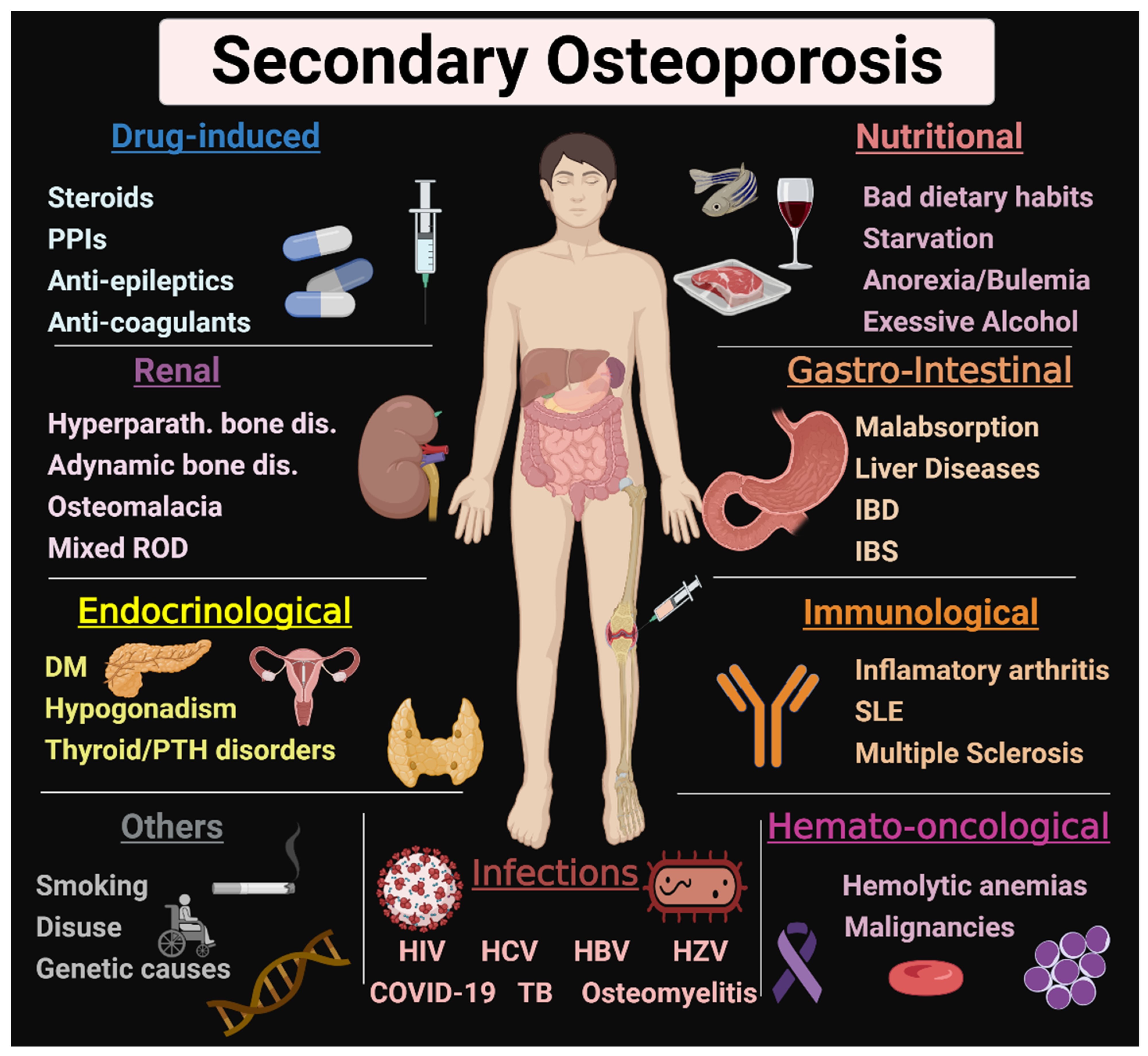
2. Renal Causes
3. Endocrinological Causes
3.1. Diabetes Mellitus
3.2. Gonadal Disorders
3.3. Parathyroid Disorders (Hypoparathyroidism and Primary Hyperparathyroidism)
3.4. Thyroid Disorders
3.5. Adrenal Disorders
3.6. Growth Hormone
4. Gastrointestinal Causes
4.1. Celiac Disease
4.2. Chronic Pancreatitis
4.3. Short Bowel Syndrome
4.4. Hepatic Osteodystrophy
4.5. Peptic Ulcer Disease
4.6. Inflammatory Bowel Disease (IBD)
4.7. Irritable Bowel Syndrome
4.8. Dysbiosis
5. Nutritional Causes
6. Drug-Induced
7. Infection
8. Hemato-Oncological Causes
9. Rheumatological-Immunological Causes
9.1. Inflammatory Arthritis
9.2. Systemic Lupus Erythematosus (SLE)
9.3. Multiple Sclerosis (MS)
10. Others
10.1. Smoking
10.2. Disuse Osteoporosis
10.3. Genetic Causes of Osteoporosis
11. Conclusions
Author Contributions
Funding
Institutional Review Board Statement
Informed Consent Statement
Data Availability Statement
Conflicts of Interest
References
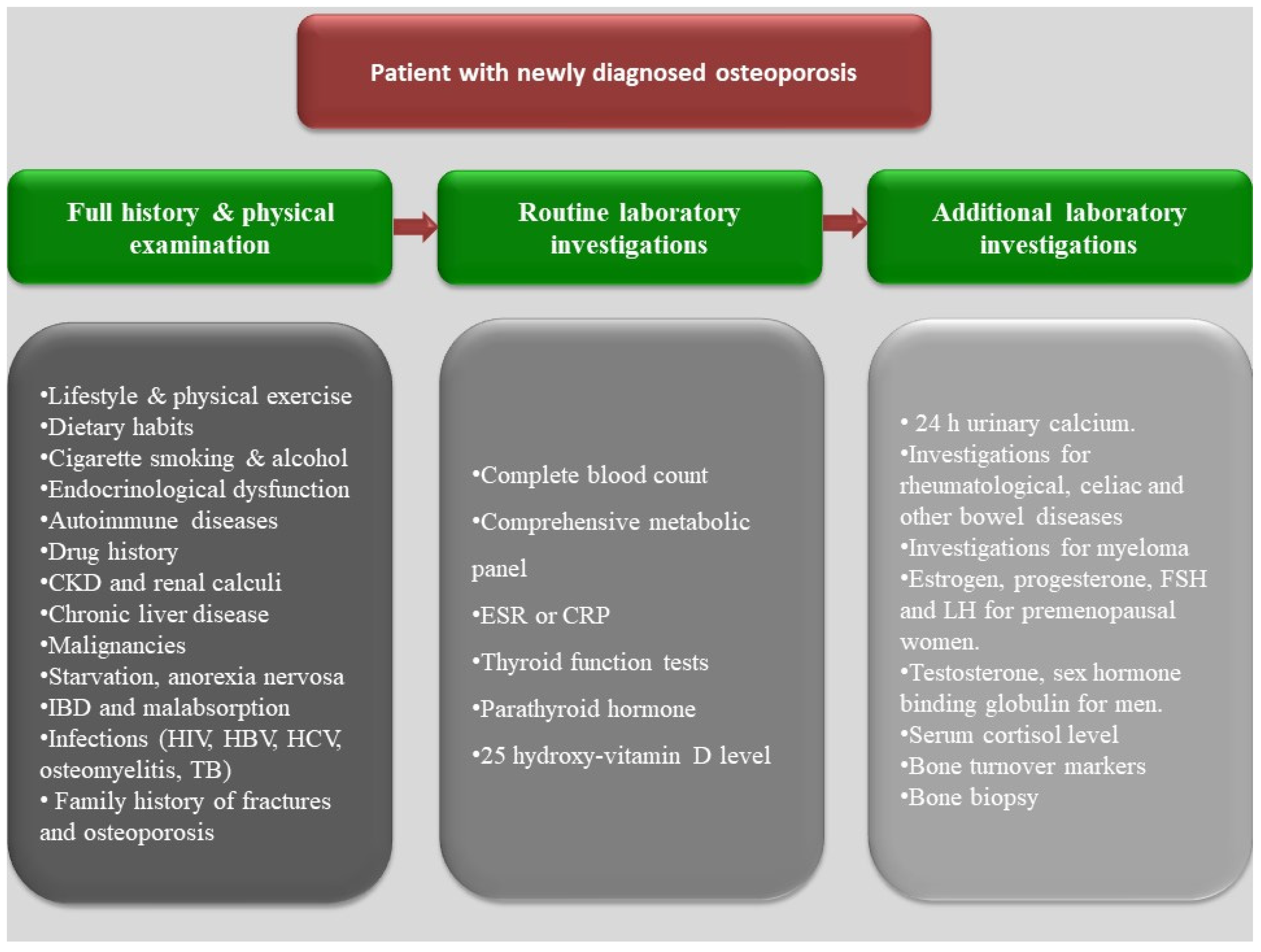
- Tannenbaum, C.; Clark, J.; Schwartzman, K.; Wallenstein, S.; Lapinski, R.; Meier, D.; Luckey, M. Yield of Laboratory Testing to Identify Secondary Contributors to Osteoporosis in Otherwise Healthy Women. J. Clin. Endocrinol. Metab. 2002, 87, 4431–4437. [Google Scholar] [CrossRef] [PubMed] [Green Version]
- Kanis, J.A.; Hans, D.; Cooper, C.; Baim, S.; Bilezikian, J.P.; Binkley, N.; Cauley, J.A.; Compston, J.E.; Dawson-Hughes, B.; El-Hajj Fuleihan, G.; et al. Interpretation and use of FRAX in clinical practice. Osteoporos. Int. 2011, 22, 2395. [Google Scholar] [CrossRef] [PubMed]
- Najar, M.S.; Mir, M.M.; Muzamil, M. Prevalence of osteoporosis in patients with chronic kidney disease (stages 3–5) in comparison with age-and sex-matched controls: A study from Kashmir Valley Tertiary Care Center. Saudi J. Kidney Dis. Transpl. 2017, 28, 538. [Google Scholar] [PubMed]
- Aggarwal, H.; Jain, D.; Yadav, S.; Kaverappa, V. Bone mineral density in patients with predialysis chronic kidney disease. Ren. Fail. 2013, 35, 1105–1111. [Google Scholar] [CrossRef] [PubMed]
- Fidan, N.; Inci, A.; Coban, M.; Ulman, C.; Kursat, S. Bone mineral density and biochemical markers of bone metabolism in predialysis patients with chronic kidney disease. J. Investig. Med. 2016, 64, 861–866. [Google Scholar] [CrossRef] [PubMed]
- Mazariegos, C.; Loaiza, J.; Sánchez-Polo, V. Pos-275 osteoporosis in ckd: Prevalence and associated factors in a guatemalan center. Kidney Int. Rep. 2021, 6, S116–S117. [Google Scholar] [CrossRef]
- Asadipooya, K.; Abdalbary, M.; Ahmad, Y.; Kakani, E.; Monier-Faugere, M.-C.; El-Husseini, A. Bone Quality in CKD Patients: Current Concepts and Future Directions–Part I. Kidney Dis. 2021, 7, 268–277. [Google Scholar] [CrossRef]
- Malluche, H.H.; Porter, D.S.; Monier-Faugere, M.C.; Mawad, H.; Pienkowski, D. Differences in bone quality in low- and high-turnover renal osteodystrophy. J. Am. Soc. Nephrol. 2012, 23, 525–532. [Google Scholar] [CrossRef] [Green Version]
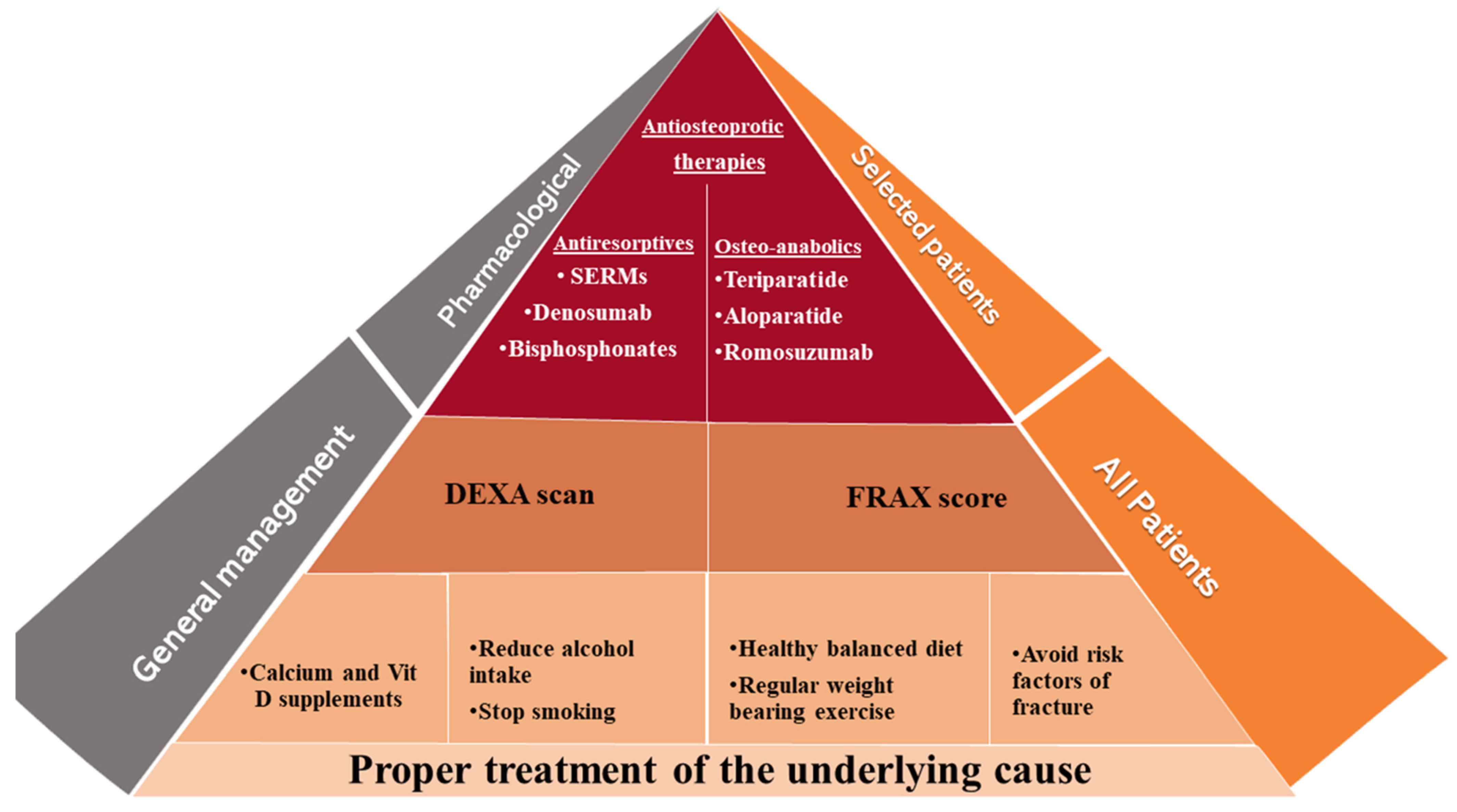
- Qaseem, A.; Forciea, M.A.; McLean, R.M.; Denberg, T.D. Treatment of Low Bone Density or Osteoporosis to Prevent Fractures in Men and Women: A Clinical Practice Guideline Update From the American College of Physicians. Ann. Intern. Med. 2017, 166, 818–839. [Google Scholar] [CrossRef]
- Camacho, P.M.; Petak, S.M.; Binkley, N.; Diab, D.L.; Eldeiry, L.S.; Farooki, A.; Harris, S.T.; Hurley, D.L.; Kelly, J.; Lewiecki, E.M.; et al. American association of clinical endocrinologists/american college of endocrinology clinical practice guidelines for the diagnosis and treatment of postmenopausal osteoporosis- 2020 update executive summary. Endocr. Pract. 2020, 26, 564–570. [Google Scholar] [CrossRef]
- Jadoul, M.; Albert, J.; Akiba, T.y.c.; Akizawa, T.; Arab, L.; Bragg-Gresham, J.; Mason, N.; Prutz, K.-G.; Young, E.; Pisoni, R. Incidence and risk factors for hip or other bone fractures among hemodialysis patients in the Dialysis Outcomes and Practice Patterns Study. Kidney Int. 2006, 70, 1358–1366. [Google Scholar] [CrossRef] [PubMed] [Green Version]
- Cenzer, I.S.; Tang, V.; Boscardin, W.J.; Smith, A.K.; Ritchie, C.; Wallhagen, M.I.; Espaldon, R.; Covinsky, K.E. One-Year Mortality After Hip Fracture: Development and Validation of a Prognostic Index. J. Am. Geriatr. Soc. 2016, 64, 1863–1868. [Google Scholar] [CrossRef] [PubMed] [Green Version]
- Morri, M.; Ambrosi, E.; Chiari, P.; Orlandi Magli, A.; Gazineo, D.; D’Alessandro, F.; Forni, C. One-year mortality after hip fracture surgery and prognostic factors: A prospective cohort study. Sci. Rep. 2019, 9, 18718. [Google Scholar] [CrossRef] [PubMed]
- Coco, M.; Rush, H. Increased incidence of hip fractures in dialysis patients with low serum parathyroid hormone. Am. J. Kidney Dis. 2000, 36, 1115–1121. [Google Scholar] [CrossRef] [PubMed]
- Mittalhenkle, A.; Gillen, D.L.; Stehman-Breen, C.O. Increased risk of mortality associated with hip fracture in the dialysis population. Am. J. Kidney Dis. 2004, 44, 672–679. [Google Scholar] [CrossRef]
- Moe, S.; Drüeke, T.; Cunningham, J.; Goodman, W.; Martin, K.; Olgaard, K.; Ott, S.; Sprague, S.; Lameire, N.; Eknoyan, G. Definition, evaluation, and classification of renal osteodystrophy: A position statement from Kidney Disease: Improving Global Outcomes (KDIGO). Kidney Int. 2006, 69, 1945–1953. [Google Scholar] [CrossRef] [Green Version]
- Farrow, E.G.; Davis, S.I.; Summers, L.J.; White, K.E. Initial FGF23-mediated signaling occurs in the distal convoluted tubule. J. Am. Soc. Nephrol. 2009, 20, 955–960. [Google Scholar] [CrossRef]
- Kolek, O.I.; Hines, E.R.; Jones, M.D.; LeSueur, L.K.; Lipko, M.A.; Kiela, P.R.; Collins, J.F.; Haussler, M.R.; Ghishan, F.K. 1α, 25-Dihydroxyvitamin D3 upregulates FGF23 gene expression in bone: The final link in a renal-gastrointestinal-skeletal axis that controls phosphate transport. Am. J. Physiol.-Gastrointest. Liver Physiol. 2005, 289, G1036–G1042. [Google Scholar] [CrossRef] [Green Version]
- Koh, N.; Fujimori, T.; Nishiguchi, S.; Tamori, A.; Shiomi, S.; Nakatani, T.; Sugimura, K.; Kishimoto, T.; Kinoshita, S.; Kuroki, T. Severely reduced production of klotho in human chronic renal failure kidney. Biochem. Biophys. Res. Commun. 2001, 280, 1015–1020. [Google Scholar] [CrossRef]
- Shimada, T.; Hasegawa, H.; Yamazaki, Y.; Muto, T.; Hino, R.; Takeuchi, Y.; Fujita, T.; Nakahara, K.; Fukumoto, S.; Yamashita, T. FGF-23 is a potent regulator of vitamin D metabolism and phosphate homeostasis. J. Bone Miner. Res. 2004, 19, 429–435. [Google Scholar] [CrossRef] [Green Version]
- Perwad, F.; Zhang, M.Y.; Tenenhouse, H.S.; Portale, A.A. Fibroblast growth factor 23 impairs phosphorus and vitamin D metabolism in vivo and suppresses 25-hydroxyvitamin D-1α-hydroxylase expression in vitro. Am. J. Physiol.-Ren. Physiol. 2007, 293, F1577–F1583. [Google Scholar] [CrossRef] [PubMed] [Green Version]
- Tomasello, S. Secondary hyperparathyroidism and chronic kidney disease. Diabetes Spectr. 2008, 21, 19–25. [Google Scholar] [CrossRef] [Green Version]
- Almaden, Y.; Canalejo, A.; Hernandez, A.; Ballesteros, E.; Garcia-Navarro, S.; Torres, A.; Rodriguez, M.; Rodriguez, M. Direct effect of phosphorus on PTH secretion from whole rat parathyroid glands in vitro. J. Bone Miner. Res. 1996, 11, 970–976. [Google Scholar] [CrossRef] [PubMed]
- Ritter, C.S.; Finch, J.L.; Slatopolsky, E.A.; Brown, A.J. Parathyroid hyperplasia in uremic rats precedes down-regulation of the calcium receptor. Kidney Int. 2001, 60, 1737–1744. [Google Scholar] [CrossRef] [Green Version]
- Fukuda, N.; Tanaka, H.; Tominaga, Y.; Fukagawa, M.; Kurokawa, K.; Seino, Y. Decreased 1, 25-dihydroxyvitamin D3 receptor density is associated with a more severe form of parathyroid hyperplasia in chronic uremic patients. J. Clin. Investig. 1993, 92, 1436–1443. [Google Scholar] [CrossRef] [PubMed]
- Hamada-Ode, K.; Taniguchi, Y.; Shimamura, Y.; Fujimoto, S.; Terada, Y. Serum dickkopf-related protein 1 and sclerostin may predict the progression of chronic kidney disease in Japanese patients. Nephrol. Dial. Transplant. 2019, 34, 1426–1427. [Google Scholar] [CrossRef]
- Neto, R.; Pereira, L.; Magalhães, J.; Quelhas-Santos, J.; Martins, S.; Carvalho, C.; Frazão, J.M. Sclerostin and DKK1 circulating levels associate with low bone turnover in patients with chronic kidney disease Stages 3 and 4. Clin. Kidney J. 2021, 14, 2401–2408. [Google Scholar] [CrossRef]
- Hofbauer, L.C.; Heufelder, A.E. Role of receptor activator of nuclear factor-κB ligand and osteoprotegerin in bone cell biology. J. Mol. Med. 2001, 79, 243–253. [Google Scholar] [CrossRef]
- Gal-Moscovici, A.; Sprague, S.M. Bone Health in Chronic Kidney Disease–Mineral and Bone Disease. Adv. Chronic Kidney Dis. 2007, 14, 27–36. [Google Scholar] [CrossRef]
- Taal, M.W.; Masud, T.; Green, D.; Cassidy, M.J. Risk factors for reduced bone density in haemodialysis patients. Nephrol. Dial. Transplant. 1999, 14, 1922–1928. [Google Scholar] [CrossRef] [Green Version]
- Evenepoel, P.; Claes, K.; Meijers, B.; Laurent, M.; Bammens, B.; Naesens, M.; Sprangers, B.; Pottel, H.; Cavalier, E.; Kuypers, D. Poor Vitamin K Status Is Associated with Low Bone Mineral Density and Increased Fracture Risk in End-Stage Renal Disease. J. Bone Miner. Res. 2019, 34, 262–269. [Google Scholar] [CrossRef] [PubMed] [Green Version]
- Kanis, J.A.; Cooper, C.; Rizzoli, R.; Reginster, J.Y. European guidance for the diagnosis and management of osteoporosis in postmenopausal women. Osteoporos. Int. 2019, 30, 3–44. [Google Scholar] [CrossRef] [PubMed] [Green Version]
- Nickolas, T.L.; Stein, E.; Cohen, A.; Thomas, V.; Staron, R.B.; McMahon, D.J.; Leonard, M.B.; Shane, E. Bone mass and microarchitecture in CKD patients with fracture. J. Am. Soc. Nephrol. 2010, 21, 1371–1380. [Google Scholar] [CrossRef] [PubMed]
- Delmas, P.D.; Confavreux, E.; Garnero, P.; Fardellone, P.; de Vernejoul, M.C.; Cormier, C.; Arce, J.C. A combination of low doses of 17 beta-estradiol and norethisterone acetate prevents bone loss and normalizes bone turnover in postmenopausal women. Osteoporos. Int. 2000, 11, 177–187. [Google Scholar] [CrossRef]
- Jørgensen, H.S.; Behets, G.; Viaene, L.; Bammens, B.; Claes, K.; Meijers, B.; Naesens, M.; Sprangers, B.; Kuypers, D.; Cavalier, E.; et al. Diagnostic Accuracy of Noninvasive Bone Turnover Markers in Renal Osteodystrophy. Am. J. Kidney Dis. 2022, 79, 667–676.e1. [Google Scholar] [CrossRef] [PubMed]
- Sprague, S.M.; Bellorin-Font, E.; Jorgetti, V.; Carvalho, A.B.; Malluche, H.H.; Ferreira, A.; D’Haese, P.C.; Drueke, T.B.; Du, H.; Manley, T.; et al. Diagnostic Accuracy of Bone Turnover Markers and Bone Histology in Patients with CKD Treated by Dialysis. Am. J. Kidney Dis. 2016, 67, 559–566. [Google Scholar] [CrossRef] [PubMed]
- Nickolas, T.L.; Cremers, S.; Zhang, A.; Thomas, V.; Stein, E.; Cohen, A.; Chauncey, R.; Nikkel, L.; Yin, M.T.; Liu, X.S.; et al. Discriminants of prevalent fractures in chronic kidney disease. J. Am. Soc. Nephrol. 2011, 22, 1560–1572. [Google Scholar] [CrossRef]
- Iimori, S.; Mori, Y.; Akita, W.; Kuyama, T.; Takada, S.; Asai, T.; Kuwahara, M.; Sasaki, S.; Tsukamoto, Y. Diagnostic usefulness of bone mineral density and biochemical markers of bone turnover in predicting fracture in CKD stage 5D patients--a single-center cohort study. Nephrol. Dial. Transplant. 2012, 27, 345–351. [Google Scholar] [CrossRef] [Green Version]
- Malluche, H.H.; Faugere, M.C. Atlas of Mineralized Bone Histology; Karger: New York, NY, USA, 1986. [Google Scholar]
- Malluche, H.H.; Monier-Faugere, M.C. Renal osteodystrophy: What’s in a name? Presentation of a clinically useful new model to interpret bone histologic findings. Clin. Nephrol. 2006, 65, 235–242. [Google Scholar] [CrossRef]
- Drüeke, T.B.; Massy, Z.A. Changing bone patterns with progression of chronic kidney disease. Kidney Int. 2016, 89, 289–302. [Google Scholar] [CrossRef] [Green Version]
- Asadipooya, K.; Abdalbary, M.; Ahmad, Y.; Kakani, E.; Monier-Faugere, M.C.; El-Husseini, A. Bone Quality in Chronic Kidney Disease Patients: Current Concepts and Future Directions—Part II. Kidney Dis. 2021, 7, 359–371. [Google Scholar] [CrossRef] [PubMed]
- Wheeler, D.C.; Winkelmayer, W.C. KDIGO 2017 Clinical Practice Guideline Update for the Diagnosis, Evaluation, Prevention, and Treatment of Chronic Kidney Disease–Mineral and Bone Disorder (CKD-MBD). Kidney Int. Suppl. 2017, 7, 1–59. [Google Scholar] [CrossRef] [Green Version]
- Sugimoto, T.; Inoue, D.; Maehara, M.; Oikawa, I.; Shigematsu, T.; Nishizawa, Y. Efficacy and safety of once-monthly risedronate in osteoporosis subjects with mild-to-moderate chronic kidney disease: A post hoc subgroup analysis of a phase III trial in Japan. J. Bone Miner. Metab. 2019, 37, 730–740. [Google Scholar] [CrossRef] [PubMed]
- Robinson, D.E.; Ali, M.S.; Pallares, N.; Tebé, C.; Elhussein, L.; Abrahamsen, B.; Arden, N.K.; Ben-Shlomo, Y.; Caskey, F.J.; Cooper, C.; et al. Safety of Oral Bisphosphonates in Moderate-to-Severe Chronic Kidney Disease: A Binational Cohort Analysis. J. Bone Miner. Res. 2021, 36, 820–832. [Google Scholar] [CrossRef] [PubMed]
- Muschitz, C.; Thaler, H.W.; Dimai, H.P.; Resch, H.; Kocijan, R.; Kostic, M.; Geiger, C.; Kaider, A.; Muschitz, G.K.; Szivak, M.; et al. Atypical Femoral Fractures-Ongoing and History of Bone-Specific Therapy, Concomitant Diseases, Medications, and Survival. J. Clin. Densitom. 2016, 19, 359–367. [Google Scholar] [CrossRef] [PubMed]
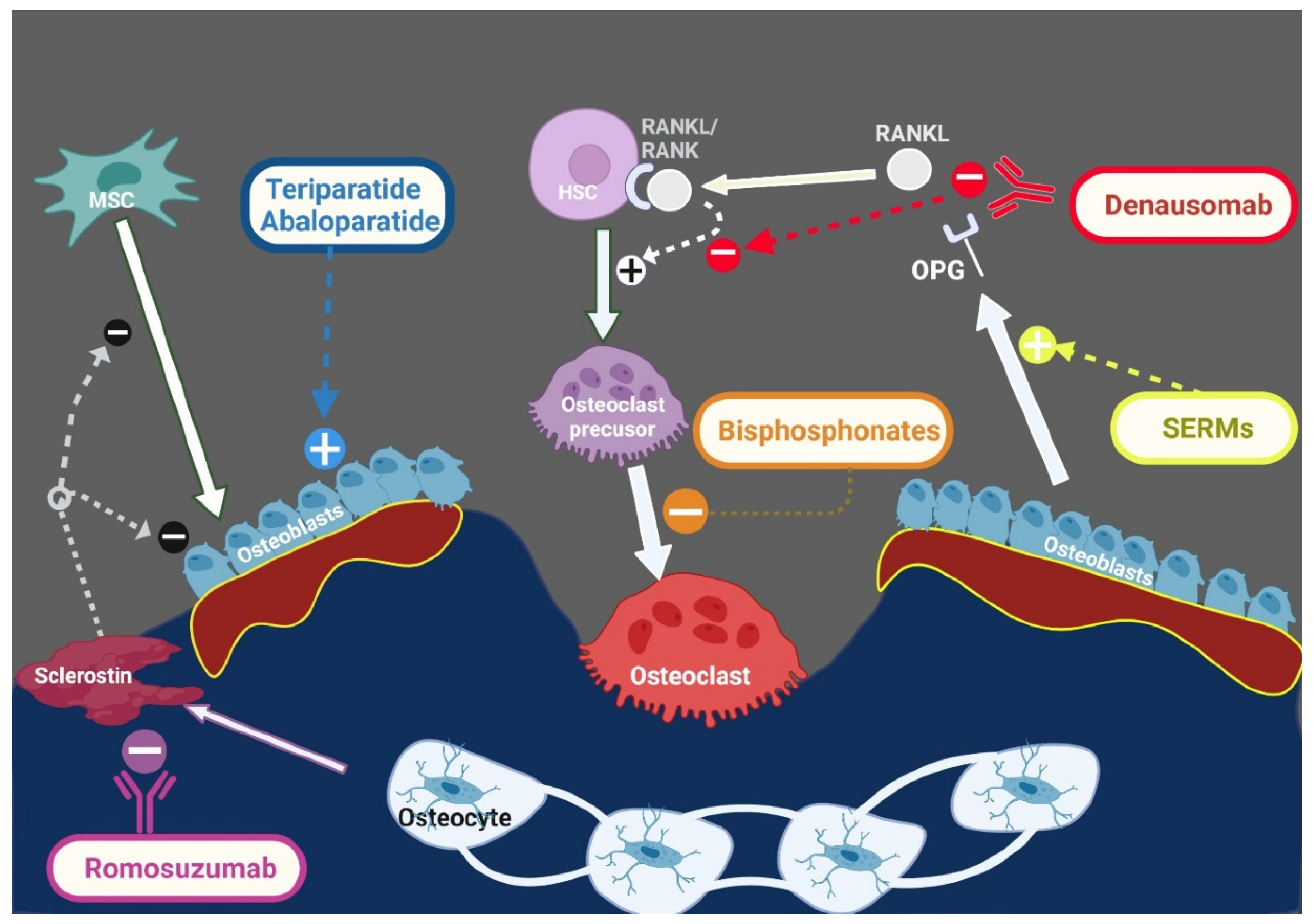
- Gopaul, A.; Kanagalingam, T.; Thain, J.; Khan, T.; Cowan, A.; Sultan, N.; Clemens, K.K. Denosumab in chronic kidney disease: A narrative review of treatment efficacy and safety. Arch. Osteoporos. 2021, 16, 116. [Google Scholar] [CrossRef]
- Iseri, K.; Watanabe, M.; Yoshikawa, H.; Mitsui, H.; Endo, T.; Yamamoto, Y.; Iyoda, M.; Ryu, K.; Inaba, T.; Shibata, T. Effects of Denosumab and Alendronate on Bone Health and Vascular Function in Hemodialysis Patients: A Randomized, Controlled Trial. J. Bone Miner. Res. 2019, 34, 1014–1024. [Google Scholar] [CrossRef]
- Cejka, D.; Kodras, K.; Bader, T.; Haas, M. Treatment of Hemodialysis-Associated Adynamic Bone Disease with Teriparatide (PTH1-34): A Pilot Study. Kidney Blood Press. Res. 2010, 33, 221–226. [Google Scholar] [CrossRef]
- Mitsopoulos, E.; Ginikopoulou, E.; Economidou, D.; Zanos, S.; Pateinakis, P.; Minasidis, E.; Memmos, D.; Thodis, E.; Vargemezis, V.; Tsakiris, D. Impact of long-term cinacalcet, ibandronate or teriparatide therapy on bone mineral density of hemodialysis patients: A pilot study. Am. J. Nephrol. 2012, 36, 238–244. [Google Scholar] [CrossRef]
- Sumida, K.; Ubara, Y.; Hoshino, J.; Mise, K.; Hayami, N.; Suwabe, T.; Kawada, M.; Imafuku, A.; Hiramatsu, R.; Hasegawa, E.; et al. Once-weekly teriparatide in hemodialysis patients with hypoparathyroidism and low bone mass: A prospective study. Osteoporos. Int. 2016, 27, 1441–1450. [Google Scholar] [CrossRef]
- Yamamoto, J.; Nakazawa, D.; Nishio, S.; Ishikawa, Y.; Makita, M.; Kusunoki, Y.; Nagai, S.; Fujieda, Y.; Takahata, M.; Yamada, K.; et al. Impact of Weekly Teriparatide on the Bone and Mineral Metabolism in Hemodialysis Patients With Relatively Low Serum Parathyroid Hormone: A Pilot Study. Ther. Apher. Dial. 2020, 24, 146–153. [Google Scholar] [CrossRef] [PubMed]
- Bilezikian, J.P.; Hattersley, G.; Mitlak, B.H.; Hu, M.Y.; Fitzpatrick, L.A.; Dabrowski, C.; Miller, P.D.; Papapoulos, S.E. Abaloparatide in patients with mild or moderate renal impairment: Results from the ACTIVE phase 3 trial. Curr. Med. Res. Opin. 2019, 35, 2097–2102. [Google Scholar] [CrossRef] [PubMed] [Green Version]
- Miller, P.; Adachi, J.; Albergaria, B.H.; Cheung, A.M.; Chines, A.; Gielen, E.; Langdahl, B.; Miyauchi, A.; Oates, M.; Reid, I.; et al. OP0297|Efficacy and safety of romosozumab among postmenopausal women with osteoporosis and mild-to-moderate chronic kidney disease. Ann. Rheum. Dis. 2020, 79, 185. [Google Scholar] [CrossRef]
- Sato, M.; Inaba, M.; Yamada, S.; Emoto, M.; Ohno, Y.; Tsujimoto, Y. Efficacy of romosozumab in patients with osteoporosis on maintenance hemodialysis in Japan; An observational study. J. Bone Miner. Metab. 2021, 39, 1082–1090. [Google Scholar] [CrossRef]
- Vilaca, T.; Schini, M.; Harnan, S.; Sutton, A.; Poku, E.; Allen, I.E.; Cummings, S.R.; Eastell, R. The risk of hip and non-vertebral fractures in type 1 and type 2 diabetes: A systematic review and meta-analysis update. Bone 2020, 137, 115457. [Google Scholar] [CrossRef]
- Ha, J.; Jeong, C.; Han, K.D.; Lim, Y.; Kim, M.K.; Kwon, H.S.; Song, K.H.; Kang, M.I.; Baek, K.H. Comparison of fracture risk between type 1 and type 2 diabetes: A comprehensive real-world data. Osteoporos. Int. 2021, 32, 2543–2553. [Google Scholar] [CrossRef]
- Koromani, F.; Oei, L.; Shevroja, E.; Trajanoska, K.; Schoufour, J.; Muka, T.; Franco, O.H.; Ikram, M.A.; Zillikens, M.C.; Uitterlinden, A.G.; et al. Vertebral Fractures in Individuals with Type 2 Diabetes: More Than Skeletal Complications Alone. Diabetes Care 2020, 43, 137–144. [Google Scholar] [CrossRef]
- Shanbhogue, V.V.; Mitchell, D.M.; Rosen, C.J.; Bouxsein, M.L. Type 2 diabetes and the skeleton: New insights into sweet bones. Lancet Diabetes Endocrinol. 2016, 4, 159–173. [Google Scholar] [CrossRef]
- Asadipooya, K.; Uy, E.M. Advanced Glycation End Products (AGEs), Receptor for AGEs, Diabetes, and Bone: Review of the Literature. J. Endocr. Soc. 2019, 3, 1799–1818. [Google Scholar] [CrossRef] [Green Version]
- Stumpf, U.; Hadji, P.; van den Boom, L.; Böcker, W.; Kostev, K. Incidence of fractures in patients with type 1 diabetes mellitus-a retrospective study with 4420 patients. Osteoporos. Int. 2020, 31, 1315–1322. [Google Scholar] [CrossRef]
- Halper-Stromberg, E.; Gallo, T.; Champakanath, A.; Taki, I.; Rewers, M.; Snell-Bergeon, J.; Frohnert, B.I.; Shah, V.N. Bone Mineral Density across the Lifespan in Patients with Type 1 Diabetes. J. Clin. Endocrinol. Metab. 2020, 105, 746–753. [Google Scholar] [CrossRef] [PubMed]
- Shah, V.N.; Harrall, K.K.; Shah, C.S.; Gallo, T.L.; Joshee, P.; Snell-Bergeon, J.K.; Kohrt, W.M. Bone mineral density at femoral neck and lumbar spine in adults with type 1 diabetes: A meta-analysis and review of the literature. Osteoporos. Int. 2017, 28, 2601–2610. [Google Scholar] [CrossRef] [PubMed]
- Ma, L.; Oei, L.; Jiang, L.; Estrada, K.; Chen, H.; Wang, Z.; Yu, Q.; Zillikens, M.C.; Gao, X.; Rivadeneira, F. Association between bone mineral density and type 2 diabetes mellitus: A meta-analysis of observational studies. Eur. J. Epidemiol. 2012, 27, 319–332. [Google Scholar] [CrossRef] [PubMed] [Green Version]
- Ferrari, S.L.; Abrahamsen, B.; Napoli, N.; Akesson, K.; Chandran, M.; Eastell, R.; El-Hajj Fuleihan, G.; Josse, R.; Kendler, D.L.; Kraenzlin, M.; et al. Diagnosis and management of bone fragility in diabetes: An emerging challenge. Osteoporos. Int. 2018, 29, 2585–2596. [Google Scholar] [CrossRef] [Green Version]
- Mitchell, A.; Fall, T.; Melhus, H.; Lind, L.; Michaëlsson, K.; Byberg, L. Type 2 diabetes and Change in Total Hip Bone Area and Bone Mineral Density in Elderly Swedish Men and Women. J. Clin. Endocrinol. Metab. 2021, 106, 2840–2854. [Google Scholar] [CrossRef]
- Khosla, S.; Samakkarnthai, P.; Monroe, D.G.; Farr, J.N. Update on the pathogenesis and treatment of skeletal fragility in type 2 diabetes mellitus. Nat. Rev. Endocrinol. 2021, 17, 685–697. [Google Scholar] [CrossRef]
- Shah, V.N.; Snell-Bergeon, J.K. Fracture risk in type 1 diabetes: Think beyond bone mineral density. J. Diabetes Complicat. 2019, 33, 107411. [Google Scholar] [CrossRef]
- Leslie, W.D.; Aubry-Rozier, B.; Lamy, O.; Hans, D. TBS (trabecular bone score) and diabetes-related fracture risk. J. Clin. Endocrinol. Metab. 2013, 98, 602–609. [Google Scholar] [CrossRef] [Green Version]
- Jiang, H.; Robinson, D.L.; Nankervis, A.; Garland, S.M.; Callegari, E.T.; Price, S.; Lee, P.V.S.; Wark, J.D. Bone Measures by Dual-Energy X-ray Absorptiometry and Peripheral Quantitative Computed Tomography in Young Women with Type 1 Diabetes Mellitus. J. Clin. Densitom. 2021, 24, 259–267. [Google Scholar] [CrossRef]
- Heilmeier, U.; Joseph, G.B.; Pasco, C.; Dinh, N.; Torabi, S.; Darakananda, K.; Youm, J.; Carballido-Gamio, J.; Burghardt, A.J.; Link, T.M.; et al. Longitudinal Evolution of Bone Microarchitecture and Bone Strength in Type 2 Diabetic Postmenopausal Women With and Without History of Fragility Fractures-A 5-Year Follow-Up Study Using High Resolution Peripheral Quantitative Computed Tomography. Front. Endocrinol. 2021, 12, 599316. [Google Scholar] [CrossRef]
- Jiang, N.; Xia, W. Assessment of bone quality in patients with diabetes mellitus. Osteoporos. Int. 2018, 29, 1721–1736. [Google Scholar] [CrossRef] [PubMed]
- Vestergaard, P.; Rejnmark, L.; Mosekilde, L.J.D. Relative fracture risk in patients with diabetes mellitus, and the impact of insulin and oral antidiabetic medication on relative fracture risk. Diabetologia 2005, 48, 1292–1299. [Google Scholar] [CrossRef] [PubMed]
- Oei, L.; Zillikens, M.C.; Dehghan, A.; Buitendijk, G.H.; Castaño-Betancourt, M.C.; Estrada, K.; Stolk, L.; Oei, E.H.; van Meurs, J.B.; Janssen, J.A.M.J.L. High bone mineral density and fracture risk in type 2 diabetes as skeletal complications of inadequate glucose control: The Rotterdam Study. Diabetes Care 2013, 36, 1619–1628. [Google Scholar] [CrossRef] [PubMed] [Green Version]
- Conway, B.N.; Long, D.M.; Figaro, M.K.; May, M.E. Glycemic control and fracture risk in elderly patients with diabetes. Diabetes Res. Clin. Pract. 2016, 115, 47–53. [Google Scholar] [CrossRef] [Green Version]
- Zhu, Z.-N.; Jiang, Y.-F.; Ding, T.J.B. Risk of fracture with thiazolidinediones: An updated meta-analysis of randomized clinical trials. Bone 2014, 68, 115–123. [Google Scholar] [CrossRef]
- Keegan, T.H.; Schwartz, A.V.; Bauer, D.C.; Sellmeyer, D.E.; Kelsey, J.L. Effect of alendronate on bone mineral density and biochemical markers of bone turnover in type 2 diabetic women: The fracture intervention trial. Diabetes Care 2004, 27, 1547–1553. [Google Scholar] [CrossRef] [Green Version]
- Anagnostis, P.; Paschou, S.A.; Gkekas, N.N.; Artzouchaltzi, A.-M.; Christou, K.; Stogiannou, D.; Vryonidou, A.; Potoupnis, M.; Goulis, D.G.J.E. Efficacy of anti-osteoporotic medications in patients with type 1 and 2 diabetes mellitus: A systematic review. Endocrine 2018, 60, 373–383. [Google Scholar] [CrossRef]
- Dhaliwal, R.; Hans, D.; Hattersley, G.; Mitlak, B.; Fitzpatrick, L.A.; Wang, Y.; Schwartz, A.V.; Miller, P.D.; Josse, R.G. Abaloparatide in postmenopausal women with osteoporosis and type 2 diabetes: A post hoc analysis of the ACTIVE study. JBMR Plus 2020, 4, e10346. [Google Scholar] [CrossRef] [Green Version]
- Golds, G.; Houdek, D.; Arnason, T. Male Hypogonadism and Osteoporosis: The Effects, Clinical Consequences, and Treatment of Testosterone Deficiency in Bone Health. Int. J. Endocrinol. 2017, 2017, 4602129. [Google Scholar] [CrossRef]
- Rochira, V.; Balestrieri, A.; Madeo, B.; Zirilli, L.; Granata, A.R.; Carani, C. Osteoporosis and male age-related hypogonadism: Role of sex steroids on bone (patho)physiology. Eur. J. Endocrinol. 2006, 154, 175–185. [Google Scholar] [CrossRef] [Green Version]
- Amory, J.K.; Watts, N.B.; Easley, K.A.; Sutton, P.R.; Anawalt, B.D.; Matsumoto, A.M.; Bremner, W.J.; Tenover, J.L. Exogenous testosterone or testosterone with finasteride increases bone mineral density in older men with low serum testosterone. J. Clin. Endocrinol. Metab. 2004, 89, 503–510. [Google Scholar] [CrossRef] [PubMed] [Green Version]
- Snyder, P.J.; Kopperdahl, D.L.; Stephens-Shields, A.J.; Ellenberg, S.S.; Cauley, J.A.; Ensrud, K.E.; Lewis, C.E.; Barrett-Connor, E.; Schwartz, A.V.; Lee, D.C.; et al. Effect of Testosterone Treatment on Volumetric Bone Density and Strength in Older Men With Low Testosterone: A Controlled Clinical Trial. JAMA Intern. Med. 2017, 177, 471–479. [Google Scholar] [CrossRef] [PubMed]
- Underbjerg, L.; Sikjaer, T.; Mosekilde, L.; Rejnmark, L. Postsurgical hypoparathyroidism--risk of fractures, psychiatric diseases, cancer, cataract, and infections. J. Bone Miner. Res. 2014, 29, 2504–2510. [Google Scholar] [CrossRef]
- Mendonça, M.L.; Pereira, F.A.; Nogueira-Barbosa, M.H.; Monsignore, L.M.; Teixeira, S.R.; Watanabe, P.C.; Maciel, L.M.; de Paula, F.J. Increased vertebral morphometric fracture in patients with postsurgical hypoparathyroidism despite normal bone mineral density. BMC Endocr. Disord. 2013, 13, 1. [Google Scholar] [CrossRef] [PubMed]
- Pal, R.; Bhadada, S.K.; Mukherjee, S.; Banerjee, M.; Kumar, A. Fracture risk in hypoparathyroidism: A systematic review and meta-analysis. Osteoporos. Int. 2021, 32, 2145–2153. [Google Scholar] [CrossRef] [PubMed]
- Chawla, H.; Saha, S.; Kandasamy, D.; Sharma, R.; Sreenivas, V.; Goswami, R. Vertebral Fractures and Bone Mineral Density in Patients With Idiopathic Hypoparathyroidism on Long-Term Follow-Up. J. Clin. Endocrinol. Metab. 2017, 102, 251–258. [Google Scholar] [CrossRef]
- Underbjerg, L.; Sikjaer, T.; Mosekilde, L.; Rejnmark, L. The Epidemiology of Nonsurgical Hypoparathyroidism in Denmark: A Nationwide Case Finding Study. J. Bone Miner. Res. 2015, 30, 1738–1744. [Google Scholar] [CrossRef]
- Silva, B.C.; Rubin, M.R.; Cusano, N.E.; Bilezikian, J.P. Bone imaging in hypoparathyroidism. Osteoporos. Int. 2017, 28, 463–471. [Google Scholar] [CrossRef]
- Cipriani, C.; Abraham, A.; Silva, B.C.; Cusano, N.E.; Rubin, M.R.; McMahon, D.J.; Zhang, C.; Hans, D.; Silverberg, S.J.; Bilezikian, J.P. Skeletal changes after restoration of the euparathyroid state in patients with hypoparathyroidism and primary hyperparathyroidism. Endocrine 2017, 55, 591–598. [Google Scholar] [CrossRef]
- Cipriani, C.; Minisola, S.; Bilezikian, J.P.; Diacinti, D.; Colangelo, L.; Piazzolla, V.; Angelozzi, M.; Nieddu, L.; Pepe, J.; Diacinti, D. Vertebral Fracture Assessment in Postmenopausal Women with Postsurgical Hypoparathyroidism. J. Clin. Endocrinol. Metab. 2021, 106, 1303–1311. [Google Scholar] [CrossRef]
- Sakane, E.N.; Vieira, M.C.C.; Lazaretti-Castro, M.; Maeda, S.S. Predictors of Poor Bone Microarchitecture Assessed by Trabecular Bone Score in Postsurgical Hypoparathyroidism. J. Clin. Endocrinol. Metab. 2019, 104, 5795–5803. [Google Scholar] [CrossRef] [PubMed]
- Chen, Q.; Kaji, H.; Iu, M.F.; Nomura, R.; Sowa, H.; Yamauchi, M.; Tsukamoto, T.; Sugimoto, T.; Chihara, K. Effects of an excess and a deficiency of endogenous parathyroid hormone on volumetric bone mineral density and bone geometry determined by peripheral quantitative computed tomography in female subjects. J. Clin. Endocrinol. Metab. 2003, 88, 4655–4658. [Google Scholar] [CrossRef] [PubMed] [Green Version]
- Cusano, N.E.; Nishiyama, K.K.; Zhang, C.; Rubin, M.R.; Boutroy, S.; McMahon, D.J.; Guo, X.E.; Bilezikian, J.P. Noninvasive Assessment of Skeletal Microstructure and Estimated Bone Strength in Hypoparathyroidism. J. Bone Miner. Res. 2016, 31, 308–316. [Google Scholar] [CrossRef]
- Cohen, A.; Dempster, D.W.; Müller, R.; Guo, X.E.; Nickolas, T.L.; Liu, X.S.; Zhang, X.H.; Wirth, A.J.; van Lenthe, G.H.; Kohler, T.; et al. Assessment of trabecular and cortical architecture and mechanical competence of bone by high-resolution peripheral computed tomography: Comparison with transiliac bone biopsy. Osteoporos. Int. 2010, 21, 263–273. [Google Scholar] [CrossRef] [PubMed] [Green Version]
- Starr, J.R.; Tabacco, G.; Majeed, R.; Omeragic, B.; Bandeira, L.; Rubin, M.R. PTH and bone material strength in hypoparathyroidism as measured by impact microindentation. Osteoporos. Int. 2020, 31, 327–333. [Google Scholar] [CrossRef] [PubMed]
- Tay, Y.D.; Tabacco, G.; Cusano, N.E.; Williams, J.; Omeragic, B.; Majeed, R.; Gomez Almonte, M.; Bilezikian, J.P.; Rubin, M.R. Therapy of Hypoparathyroidism With rhPTH(1-84): A Prospective, 8-Year Investigation of Efficacy and Safety. J. Clin. Endocrinol. Metab. 2019, 104, 5601–5610. [Google Scholar] [CrossRef] [PubMed]
- Mannstadt, M.; Clarke, B.L.; Vokes, T.; Brandi, M.L.; Ranganath, L.; Fraser, W.D.; Lakatos, P.; Bajnok, L.; Garceau, R.; Mosekilde, L.; et al. Efficacy and safety of recombinant human parathyroid hormone (1-84) in hypoparathyroidism (REPLACE): A double-blind, placebo-controlled, randomised, phase 3 study. Lancet Diabetes Endocrinol. 2013, 1, 275–283. [Google Scholar] [CrossRef]
- Cusano, N.E.; Rubin, M.R.; McMahon, D.J.; Irani, D.; Tulley, A.; Sliney, J., Jr.; Bilezikian, J.P. The effect of PTH(1-84) on quality of life in hypoparathyroidism. J. Clin. Endocrinol. Metab. 2013, 98, 2356–2361. [Google Scholar] [CrossRef]
- Orloff, L.A.; Wiseman, S.M.; Bernet, V.J.; Fahey, T.J., 3rd; Shaha, A.R.; Shindo, M.L.; Snyder, S.K.; Stack, B.C., Jr.; Sunwoo, J.B.; Wang, M.B. American Thyroid Association Statement on Postoperative Hypoparathyroidism: Diagnosis, Prevention, and Management in Adults. Thyroid 2018, 28, 830–841. [Google Scholar] [CrossRef]
- Palui, R.; Das, R.R.; Roy, A.; Kamalanathan, S.; Kar, S.S.; Sahoo, J.; Selvarajan, S.; Satapathy, A.K. Parathyroid Hormone Replacement versus Oral Calcium and Active Vitamin D Supplementation in Hypoparathyroidism: A Meta-analysis. Indian J. Endocrinol. Metab. 2020, 24, 206–214. [Google Scholar] [CrossRef]
- Jolette, J.; Wilker, C.E.; Smith, S.Y.; Doyle, N.; Hardisty, J.F.; Metcalfe, A.J.; Marriott, T.B.; Fox, J.; Wells, D.S. Defining a noncarcinogenic dose of recombinant human parathyroid hormone 1-84 in a 2-year study in Fischer 344 rats. Toxicol. Pathol. 2006, 34, 929–940. [Google Scholar] [CrossRef] [PubMed] [Green Version]
- Cipriani, C.; Irani, D.; Bilezikian, J.P. Safety of osteoanabolic therapy: A decade of experience. J. Bone Miner. Res. 2012, 27, 2419–2428. [Google Scholar] [CrossRef] [PubMed]
- Parameswaran, R.; Samuel, M.; Satish, R.L.; Kripesh, A.; Moorthy, V.; Vajjhala, R.; Ng, X.L.; Yip, G.W.; Voon, F.C.T.; Chandran, M. Parathyroid allotransplantation to treat post-thyroidectomy hypoparathyroidism: A review of case studies. Surgeon 2021, 19, 183–192. [Google Scholar] [CrossRef] [PubMed]
- Ejlsmark-Svensson, H.; Rolighed, L.; Harsløf, T.; Rejnmark, L. Risk of fractures in primary hyperparathyroidism: A systematic review and meta-analysis. Osteoporos. Int. 2021, 32, 1053–1060. [Google Scholar] [CrossRef] [PubMed]
- Narayanan, N.; Palui, R.; Merugu, C.; Kar, S.S.; Kamalanathan, S.; Sahoo, J.; Selvarajan, S.; Naik, D. The Risk of Fractures in Primary Hyperparathyroidism: A Meta-Analysis. JBMR Plus 2021, 5, e10482. [Google Scholar] [CrossRef]
- Ejlsmark-Svensson, H.; Bislev, L.S.; Lajlev, S.; Harsløf, T.; Rolighed, L.; Sikjaer, T.; Rejnmark, L. Prevalence and Risk of Vertebral Fractures in Primary Hyperparathyroidism: A Nested Case-Control Study. J. Bone Miner. Res. 2018, 33, 1657–1664. [Google Scholar] [CrossRef] [Green Version]
- Arboiro-Pinel, R.; Mahillo-Fernández, I.; Díaz-Curiel, M. Bone Analysis by the Trabecular Bone Score and 3D-DXA in Postmenopausal Women with Primary Hyperparathyroidism. Endocr. Pract. 2021, 28, 83–89. [Google Scholar] [CrossRef]
- Tabacco, G.; Naciu, A.M.; Messina, C.; Sanson, G.; Rinaudo, L.; Cesareo, R.; Falcone, S.; Manfrini, S.; Napoli, N.; Bilezikian, J.P.; et al. DXA-Based Bone Strain Index: A New Tool to Evaluate Bone Quality in Primary Hyperparathyroidism. J. Clin. Endocrinol. Metab. 2021, 106, 2304–2312. [Google Scholar] [CrossRef]
- Stein, E.M.; Silva, B.C.; Boutroy, S.; Zhou, B.; Wang, J.; Udesky, J.; Zhang, C.; McMahon, D.J.; Romano, M.; Dworakowski, E.; et al. Primary hyperparathyroidism is associated with abnormal cortical and trabecular microstructure and reduced bone stiffness in postmenopausal women. J. Bone Miner. Res. 2013, 28, 1029–1040. [Google Scholar] [CrossRef] [Green Version]
- Hansen, S.; Beck Jensen, J.E.; Rasmussen, L.; Hauge, E.M.; Brixen, K. Effects on bone geometry, density, and microarchitecture in the distal radius but not the tibia in women with primary hyperparathyroidism: A case-control study using HR-pQCT. J. Bone Miner. Res. 2010, 25, 1941–1947. [Google Scholar] [CrossRef]
- Eriksen, E.F. Primary hyperparathyroidism: Lessons from bone histomorphometry. J. Bone Miner. Res. 2002, 17 (Suppl. 2), N95–N97. [Google Scholar] [PubMed]
- Schoeb, M.; Winter, E.M.; Sleddering, M.A.; Lips, M.A.; Schepers, A.; Snel, M.; Appelman-Dijkstra, N.M. Bone Material Strength Index as Measured by Impact Microindentation is Low in Patients with Primary Hyperparathyroidism. J. Clin. Endocrinol. Metab. 2021, 106, e2527–e2534. [Google Scholar] [CrossRef] [PubMed]
- Zhang, L.; Liu, X.; Li, H. Long-Term Skeletal Outcomes of Primary Hyperparathyroidism Patients After Treatment with Parathyroidectomy: A Systematic Review and Meta-Analysis. Horm. Metab. Res. 2018, 50, 242–249. [Google Scholar] [CrossRef] [PubMed]
- Anagnostis, P.; Vaitsi, K.; Veneti, S.; Potoupni, V.; Kenanidis, E.; Tsiridis, E.; Papavramidis, T.S.; Goulis, D.G. Efficacy of parathyroidectomy compared with active surveillance in patients with mild asymptomatic primary hyperparathyroidism: A systematic review and meta-analysis of randomized-controlled studies. J. Endocrinol. Investig. 2021, 44, 1127–1137. [Google Scholar] [CrossRef]
- Cusano, N.E.; Rubin, M.R.; Silva, B.C.; Tay, Y.D.; Williams, J.M.; Agarwal, S.; Omeragic, B.; Guo, X.E.; Bilezikian, J.P. Skeletal Microstructure and Estimated Bone Strength Improve Following Parathyroidectomy in Primary Hyperparathyroidism. J. Clin. Endocrinol. Metab. 2018, 103, 196–205. [Google Scholar] [CrossRef] [Green Version]
- Marcocci, C.; Bollerslev, J.; Khan, A.A.; Shoback, D.M. Medical management of primary hyperparathyroidism: Proceedings of the fourth International Workshop on the Management of Asymptomatic Primary Hyperparathyroidism. J. Clin. Endocrinol. Metab. 2014, 99, 3607–3618. [Google Scholar] [CrossRef] [Green Version]
- Jorde, R.; Szumlas, K.; Haug, E.; Sundsfjord, J. The effects of calcium supplementation to patients with primary hyperparathyroidism and a low calcium intake. Eur. J. Nutr. 2002, 41, 258–263. [Google Scholar] [CrossRef]
- Nicolaisen, P.; Obling, M.L.; Winther, K.H.; Hansen, S.; Hermann, A.P.; Hegedüs, L.; Bonnema, S.J.; Brix, T.H. Consequences of Hyperthyroidism and Its Treatment for Bone Microarchitecture Assessed by High-Resolution Peripheral Quantitative Computed Tomography. Thyroid 2021, 31, 208–216. [Google Scholar] [CrossRef]
- Zhu, H.; Zhang, J.; Wang, J.; Zhao, X.; Gu, M. Association of subclinical thyroid dysfunction with bone mineral density and fracture: A meta-analysis of prospective cohort studies. Endocrine 2020, 67, 685–698. [Google Scholar] [CrossRef]
- Ku, E.J.; Yoo, W.S.; Lee, E.K.; Ahn, H.Y.; Woo, S.H.; Hong, J.H.; Chung, H.K.; Park, J.W. Effect of TSH Suppression Therapy on Bone Mineral Density in Differentiated Thyroid Cancer: A Systematic Review and Meta-analysis. J. Clin. Endocrinol. Metab. 2021, 106, 3655–3667. [Google Scholar] [CrossRef]
- Delitala, A.P.; Scuteri, A.; Doria, C. Thyroid Hormone Diseases and Osteoporosis. J. Clin. Med. 2020, 9, 1034. [Google Scholar] [CrossRef] [PubMed] [Green Version]
- Ohmori, N.; Nomura, K.; Ohmori, K.; Kato, Y.; Itoh, T.; Takano, K. Osteoporosis is more prevalent in adrenal than in pituitary Cushing’s syndrome. Endocr. J. 2003, 50, 1–7. [Google Scholar] [CrossRef] [PubMed] [Green Version]
- Pecori Giraldi, F.; Moro, M.; Cavagnini, F. Gender-related differences in the presentation and course of Cushing’s disease. J. Clin. Endocrinol. Metab. 2003, 88, 1554–1558. [Google Scholar] [CrossRef] [PubMed] [Green Version]
- dos Santos, C.V.; Vieira Neto, L.; Madeira, M.; Alves Coelho, M.C.; de Mendonça, L.M.; Paranhos-Neto Fde, P.; Lima, I.C.; Gadelha, M.R.; Farias, M.L. Bone density and microarchitecture in endogenous hypercortisolism. Clin. Endocrinol. 2015, 83, 468–474. [Google Scholar] [CrossRef]
- Fleseriu, M.; Auchus, R.; Bancos, I.; Ben-Shlomo, A.; Bertherat, J.; Biermasz, N.R.; Boguszewski, C.L.; Bronstein, M.D.; Buchfelder, M.; Carmichael, J.D.; et al. Consensus on diagnosis and management of Cushing’s disease: A guideline update. Lancet Diabetes Endocrinol. 2021, 9, 847–875. [Google Scholar] [CrossRef]
- Tauchmanovà, L.; Pivonello, R.; Di Somma, C.; Rossi, R.; De Martino, M.C.; Camera, L.; Klain, M.; Salvatore, M.; Lombardi, G.; Colao, A. Bone demineralization and vertebral fractures in endogenous cortisol excess: Role of disease etiology and gonadal status. J. Clin. Endocrinol. Metab. 2006, 91, 1779–1784. [Google Scholar] [CrossRef] [Green Version]
- Moraes, A.B.; de Paula, M.P.; de Paula Paranhos-Neto, F.; Cavalari, E.M.R.; de Morais, F.F.C.; Curi, D.S.C.; Lima, L.F.C.; de Mendonça, L.M.C.; Farias, M.L.F.; Madeira, M.; et al. Bone Evaluation by High-Resolution Peripheral Quantitative Computed Tomography in Patients with Adrenal Incidentaloma. J. Clin. Endocrinol. Metab. 2020, 105, e2726–e2737. [Google Scholar] [CrossRef]
- Kim, B.J.; Kwak, M.K.; Ahn, S.H.; Kim, H.; Lee, S.H.; Koh, J.M. Lower Trabecular Bone Score in Patients with Primary Aldosteronism: Human Skeletal Deterioration by Aldosterone Excess. J. Clin. Endocrinol. Metab. 2018, 103, 615–621. [Google Scholar] [CrossRef] [Green Version]
- Kim, B.J.; Kwak, M.K.; Ahn, S.H.; Kim, H.; Lee, S.H.; Song, K.H.; Suh, S.; Kim, J.H.; Koh, J.M. Lower Bone Mass and Higher Bone Resorption in Pheochromocytoma: Importance of Sympathetic Activity on Human Bone. J. Clin. Endocrinol. Metab. 2017, 102, 2711–2718. [Google Scholar] [CrossRef] [Green Version]
- Yokomoto-Umakoshi, M.; Umakoshi, H.; Sakamoto, R.; Fukumoto, T.; Ogata, M.; Nakano, Y.; Iwahashi, N.; Kaneko, H.; Mizoguchi, N.; Hattori, A.; et al. Role of deteriorated bone quality in the development of osteoporosis in pheochromocytoma and paraganglioma. Bone 2021, 142, 115607. [Google Scholar] [CrossRef]
- Falhammar, H.; Frisén, L.; Hirschberg, A.L.; Nordenskjöld, A.; Almqvist, C.; Nordenström, A. Increased Prevalence of Fractures in Congenital Adrenal Hyperplasia: A Swedish Population-Based National Cohort Study. J. Clin. Endocrinol. Metab. 2021, 107, e475–e486. [Google Scholar] [CrossRef] [PubMed]
- Mazziotti, G.; Biagioli, E.; Maffezzoni, F.; Spinello, M.; Serra, V.; Maroldi, R.; Floriani, I.; Giustina, A. Bone turnover, bone mineral density, and fracture risk in acromegaly: A meta-analysis. J. Clin. Endocrinol. Metab. 2015, 100, 384–394. [Google Scholar] [CrossRef] [Green Version]
- Silva, P.P.B.; Pereira, R.M.R.; Takayama, L.; Borba, C.G.; Duarte, F.H.; Trarbach, E.B.; Martin, R.M.; Bronstein, M.D.; Tritos, N.A.; Jallad, R.S. Impaired Bone Microarchitecture in Premenopausal Women with Acromegaly: The Possible Role of Wnt Signaling. J. Clin. Endocrinol. Metab. 2021, 106, 2690–2706. [Google Scholar] [CrossRef] [PubMed]
- Giustina, A.; Mazziotti, G.; Canalis, E. Growth hormone, insulin-like growth factors, and the skeleton. Endocr. Rev. 2008, 29, 535–559. [Google Scholar] [CrossRef] [PubMed] [Green Version]
- Jørgensen, J.O.L.; Johannsson, G.; Barkan, A. Should patients with adult GH deficiency receive GH replacement? Eur. J. Endocrinol. 2021, 186, D1–D15. [Google Scholar] [CrossRef] [PubMed]
- Ueland, T.; Olarescu, N.C.; Jørgensen, A.P.; Otterdal, K.; Aukrust, P.; Godang, K.; Lekva, T.; Bollerslev, J. Increased serum and bone matrix levels of the secreted Wnt antagonist DKK-1 in patients with growth hormone deficiency in response to growth hormone treatment. J. Clin. Endocrinol. Metab. 2015, 100, 736–743. [Google Scholar] [CrossRef] [Green Version]
- Fedewa, M.V.; Bentley, J.L.; Higgins, S.; Kindler, J.M.; Esco, M.R.; MacDonald, H.V. Celiac Disease and Bone Health in Children and Adolescents: A Systematic Review and Meta-Analysis. J. Clin. Densitom. 2020, 23, 200–211. [Google Scholar] [CrossRef]
- Duggan, S.N.; Smyth, N.D.; Murphy, A.; MacNaughton, D.; O’Keefe, S.J.D.; Conlon, K.C. High prevalence of osteoporosis in patients with chronic pancreatitis: A systematic review and meta-analysis. Clin. Gastroenterol. Hepatol. 2014, 12, 219–228. [Google Scholar] [CrossRef]
- Fan, J.; Wang, Q.; Sun, L. Association between primary biliary cholangitis and osteoporosis: Meta-analysis. Clin. Rheumatol. 2017, 36, 2565–2571. [Google Scholar] [CrossRef]
- Nygaard, L.; Skallerup, A.; Olesen, S.S.; Kohler, M.; Vinter-Jensen, L.; Kruse, C.; Vestergaard, P.; Rasmussen, H.H. Osteoporosis in patients with intestinal insufficiency and intestinal failure: Prevalence and clinical risk factors. Clin. Nutr. 2018, 37, 1654–1660. [Google Scholar] [CrossRef]
- Yamanashi, Y.; Takada, T.; Kurauchi, R.; Tanaka, Y.; Komine, T.; Suzuki, H. Transporters for the intestinal absorption of cholesterol, vitamin E, and vitamin K. J. Atheroscler. Thromb. 2017, 24, 347–359. [Google Scholar] [CrossRef] [PubMed] [Green Version]
- Maurya, V.K.; Aggarwal, M. Factors influencing the absorption of vitamin D in GIT: An overview. J. Food Sci. Technol. 2017, 54, 3753–3765. [Google Scholar] [CrossRef] [PubMed]
- Ferraz, R.R.; Tiselius, H.G.; Heilberg, I.P. Fat malabsorption induced by gastrointestinal lipase inhibitor leads to an increase in urinary oxalate excretion. Kidney Int. 2004, 66, 676–682. [Google Scholar] [CrossRef] [PubMed] [Green Version]
- Ganji, R.; Moghbeli, M.; Sadeghi, R.; Bayat, G.; Ganji, A. Prevalence of osteoporosis and osteopenia in men and premenopausal women with celiac disease: A systematic review. Nutr. J. 2019, 18, 9. [Google Scholar] [CrossRef] [PubMed] [Green Version]
- Mustalahti, K.; Collin, P.; Sievanen, H.; Salmi, J.; Maki, M. Osteopenia in patients with clinically silent coeliac disease warrants screening. Lancet 1999, 354, 744–745. [Google Scholar] [CrossRef]
- Karakan, T.; Ozyemisci-Taskiran, O.; Gunendi, Z.; Atalay, F.; Tuncer, C. Prevalence of IgA-antiendomysial antibody in a patient cohort with idiopathic low bone mineral density. World J. Gastroenterol. 2007, 13, 2978–2982. [Google Scholar] [CrossRef]
- Siddh, L.; Sengar, G.; Nagraj, N.; Shyam, R.; Garg, P. Body mass index in celiac disease and effect of a gluten-free diet on body mass index. Int. J. Adv. Med. 2016, 3, 813–815. [Google Scholar] [CrossRef]
- Kemppainen, T.; Kroger, H.; Janatuinen, E.; Arnala, I.; Lamberg-Allardt, C.; Karkkainen, M.; Kosma, V.M.; Julkunen, R.; Jurvelin, J.; Alhava, E.; et al. Bone recovery after a gluten-free diet: A 5-year follow-up study. Bone 1999, 25, 355–360. [Google Scholar] [CrossRef]
- Di Stefano, M.; Bergonzi, M.; Benedetti, I.; De Amici, M.; Torre, C.; Brondino, N.; Miceli, E.; Pagani, E.; Marseglia, G.L.; Corazza, G.R.; et al. Alterations of Inflammatory and Matrix Production Indices in Celiac Disease With Low Bone Mass on Long-term Gluten-free Diet. J. Clin. Gastroenterol. 2019, 53, e221–e226. [Google Scholar] [CrossRef]
- Vujasinovic, M.; Nezirevic Dobrijevic, L.; Asplund, E.; Rutkowski, W.; Dugic, A.; Kahn, M.; Dahlman, I.; Saaf, M.; Hagstrom, H.; Lohr, J.M. Low Bone Mineral Density and Risk for Osteoporotic Fractures in Patients with Chronic Pancreatitis. Nutrients 2021, 13, 2386. [Google Scholar] [CrossRef]
- Haworth, C.S. Impact of cystic fibrosis on bone health. Curr. Opin. Pulm. Med. 2010, 16, 616–622. [Google Scholar] [CrossRef] [PubMed]
- Johnson, E.; Vu, L.; Matarese, L.E. Bacteria, Bones, and Stones: Managing Complications of Short Bowel Syndrome. Nutr. Clin. Pract. 2018, 33, 454–466. [Google Scholar] [CrossRef] [PubMed]
- Yadav, A.; Carey, E.J. Osteoporosis in chronic liver disease. Nutr. Clin. Pract. 2013, 28, 52–64. [Google Scholar] [CrossRef] [PubMed]
- Wariaghli, G.; Allali, F.; Maghraoui, A.E.; Hajjaj-Hassouni, N. Osteoporosis in patients with primary biliary cirrhosis. Eur. J. Gastroenterol. Hepatol. 2010, 22, 1397–1401. [Google Scholar] [CrossRef]
- Guanabens, N.; Pares, A.; Ros, I.; Caballeria, L.; Pons, F.; Vidal, S.; Monegal, A.; Peris, P.; Rodes, J. Severity of cholestasis and advanced histological stage but not menopausal status are the major risk factors for osteoporosis in primary biliary cirrhosis. J. Hepatol. 2005, 42, 573–577. [Google Scholar] [CrossRef]
- Chappard, D.; Plantard, B.; Fraisse, H.; Palle, S.; Alexandre, C.; Riffat, G. Bone changes in alcoholic cirrhosis of the liver. A histomorphometric study. Pathol. Res. Pract. 1989, 184, 480–485. [Google Scholar] [CrossRef]
- Chen, C.H.; Lin, C.L.; Kao, C.H. Association Between Chronic Hepatitis B Virus Infection and Risk of Osteoporosis: A Nationwide Population-Based Study. Medicine (Baltimore) 2015, 94, e2276. [Google Scholar] [CrossRef]
- Chen, C.H.; Lin, C.L.; Kao, C.H. Relation Between Hepatitis C Virus Exposure and Risk of Osteoporosis: A Nationwide Population-Based Study. Medicine (Baltimore) 2015, 94, e2086. [Google Scholar] [CrossRef]
- Schmidt, T.; Schmidt, C.; Strahl, A.; Mussawy, H.; Rolvien, T.; Jandl, N.M.; Casar, C.; Oheim, R.; Schinke, T.; Lohse, A.W.; et al. A System to Determine Risk of Osteoporosis in Patients with Autoimmune Hepatitis. Clin. Gastroenterol. Hepatol. 2020, 18, 226–233.e3. [Google Scholar] [CrossRef]
- Valenti, L.; Varenna, M.; Fracanzani, A.L.; Rossi, V.; Fargion, S.; Sinigaglia, L. Association between iron overload and osteoporosis in patients with hereditary hemochromatosis. Osteoporos. Int. 2009, 20, 549–555. [Google Scholar] [CrossRef]
- Ehnert, S.; Aspera-Werz, R.H.; Ruoß, M.; Dooley, S.; Hengstler, J.G.; Nadalin, S.; Relja, B.; Badke, A.; Nussler, A.K. Hepatic Osteodystrophy-Molecular Mechanisms Proposed to Favor Its Development. Int. J. Mol. Sci. 2019, 20, 2555. [Google Scholar] [CrossRef] [PubMed] [Green Version]
- Barbu, E.C.; Chitu-Tisu, C.E.; Lazar, M.; Olariu, C.; Bojinca, M.; Ionescu, R.A.; Ion, D.A.; Badarau, I.A. Hepatic Osteodystrophy: A Global (Re)View of the Problem. Acta Clin. Croat. 2017, 56, 512–525. [Google Scholar] [CrossRef] [PubMed] [Green Version]
- Zhao, S.; Ding, L.; Xie, Q.; Zhang, J.; Yang, S.; Xu, W.; Yang, J.; Xu, Y.; Zheng, C. Is there an association between peptic ulcer disease and osteoporosis: A systematic review and cumulative analysis. Eur. J. Gastroenterol. Hepatol. 2021, 33, 9–16. [Google Scholar] [CrossRef] [PubMed]
- Gennari, L.; Merlotti, D.; Figura, N.; Mingiano, C.; Franci, M.B.; Lucani, B.; Picchioni, T.; Alessandri, M.; Campagna, M.S.; Gonnelli, S.; et al. Infection by CagA-Positive Helicobacter pylori Strains and Bone Fragility: A Prospective Cohort Study. J. Bone Miner. Res. 2021, 36, 80–89. [Google Scholar] [CrossRef] [PubMed]
- Miranda-Bautista, J.; Verdejo, C.; Diaz-Redondo, A.; Breton, I.; Bellon, J.M.; Perez-Valderas, M.D.; Caballero-Marcos, A.; de Dios-Lascuevas, M.; Gonzalez-Rio, E.; Garcia-Sanchez, C.; et al. Metabolic bone disease in patients diagnosed with inflammatory bowel disease from Spain. Therap. Adv. Gastroenterol. 2019, 12, 1756284819862152. [Google Scholar] [CrossRef] [Green Version]
- Soare, I.; Sirbu, A.; Martin, S.; Diculescu, M.; Mateescu, B.; Tieranu, C.; Fica, S. Assessment of bone quality with trabecular bone score in patients with inflammatory bowel disease. Sci. Rep. 2021, 11, 20345. [Google Scholar] [CrossRef]
- Haschka, J.; Kraus, D.A.; Behanova, M.; Huber, S.; Bartko, J.; Schanda, J.E.; Meier, P.; Bahrami, A.; Zandieh, S.; Zwerina, J.; et al. Fractal-Based Analysis of Bone Microstructure in Crohn’s Disease: A Pilot Study. J. Clin. Med. 2020, 9, 4116. [Google Scholar] [CrossRef]
- Maldonado-Perez, M.B.; Castro-Laria, L.; Caunedo-Alvarez, A.; Montoya-Garcia, M.J.; Giner-Garcia, M.; Arguelles-Arias, F.; Romero-Gomez, M.; Vazquez-Gamez, M.A. Does the Antitumor Necrosis Factor-alpha Therapy Decrease the Vertebral Fractures Occurrence in Inflammatory Bowel Disease? J. Clin. Densitom. 2019, 22, 195–202. [Google Scholar] [CrossRef]
- Bernstein, C.N.; Blanchard, J.F.; Leslie, W.; Wajda, A.; Yu, B.N. The incidence of fracture among patients with inflammatory bowel disease. A population-based cohort study. Ann. Intern. Med. 2000, 133, 795–799. [Google Scholar] [CrossRef]
- Komaki, Y.; Komaki, F.; Micic, D.; Ido, A.; Sakuraba, A. Risk of Fractures in Inflammatory Bowel Diseases: A Systematic Review and Meta-Analysis. J. Clin. Gastroenterol. 2019, 53, 441–448. [Google Scholar] [CrossRef]
- Hidalgo, D.F.; Boonpheng, B.; Phemister, J.; Hidalgo, J.; Young, M. Inflammatory Bowel Disease and Risk of Osteoporotic Fractures: A Meta-Analysis. Cureus 2019, 11, e5810. [Google Scholar] [CrossRef] [PubMed] [Green Version]
- Bravenboer, N.; Oostlander, A.E.; Van Bodegraven, A.A. Bone loss in patients with inflammatory bowel disease: Cause, detection and treatment. Curr. Opin. Gastroenterol. 2021, 37, 128–134. [Google Scholar] [CrossRef] [PubMed]
- Morgan, S.; Hooper, K.M.; Milne, E.M.; Farquharson, C.; Stevens, C.; Staines, K.A. Azathioprine Has a Deleterious Effect on the Bone Health of Mice with DSS-Induced Inflammatory Bowel Disease. Int. J. Mol. Sci. 2019, 20, 6085. [Google Scholar] [CrossRef] [Green Version]
- Misof, B.M.; Roschger, P.; Klaushofer, K.; Rauch, F.; Ma, J.; Mack, D.R.; Ward, L.M. Increased bone matrix mineralization in treatment-naive children with inflammatory bowel disease. Bone 2017, 105, 50–56. [Google Scholar] [CrossRef] [PubMed]
- Oostlander, A.E.; Bravenboer, N.; Sohl, E.; Holzmann, P.J.; van der Woude, C.J.; Dijkstra, G.; Stokkers, P.C.; Oldenburg, B.; Netelenbos, J.C.; Hommes, D.W.; et al. Histomorphometric analysis reveals reduced bone mass and bone formation in patients with quiescent Crohn’s disease. Gastroenterology 2011, 140, 116–123. [Google Scholar] [CrossRef] [Green Version]
- Farraye, F.A.; Melmed, G.Y.; Lichtenstein, G.R.; Kane, S.V. ACG Clinical Guideline: Preventive Care in Inflammatory Bowel Disease. Am. J. Gastroenterol. 2017, 112, 241–258. [Google Scholar] [CrossRef] [PubMed]
- IBD Checklists—Cornerstone Health. Available online: https://www.cornerstoneshealth.org/ibd-checklists (accessed on 1 December 2021).
- Maldonado, F.J.; Al Bawardy, B.F.; Nehra, A.K.; Lee, Y.S.; Bruining, D.H.; Adkins, M.C.; Keaveny, T.M.; Johnson, M.P.; Fidler, J.L.; McCollough, C.H.; et al. Findings of CT-Derived Bone Strength Assessment in Inflammatory Bowel Disease Patients Undergoing CT Enterography in Clinical Practice. Inflamm. Bowel Dis. 2019, 25, 1072–1079. [Google Scholar] [CrossRef]
- Frei, R.; Fournier, N.; Zeitz, J.; Scharl, M.; Morell, B.; Greuter, T.; Schreiner, P.; Misselwitz, B.; Safroneeva, E.; Schoepfer, A.M.; et al. Early Initiation of Anti-TNF is Associated with Favourable Long-term Outcome in Crohn’s Disease: 10-Year-Follow-up Data from the Swiss IBD Cohort Study. J. Crohns Colitis 2019, 13, 1292–1301. [Google Scholar] [CrossRef]
- Yao, L.; Wang, H.; Dong, W.; Liu, Z.; Mao, H. Efficacy and safety of bisphosphonates in management of low bone density in inflammatory bowel disease: A meta-analysis. Medicine 2017, 96, e5861. [Google Scholar] [CrossRef]
- Luo, J.S.; Zhao, X.; Yang, Y. Effects of emodin on inflammatory bowel disease-related osteoporosis. Biosci. Rep. 2020, 40, BSR20192317. [Google Scholar] [CrossRef]
- Lee, S.Y.; Hwang, H.R.; Yi, Y.H.; Kim, J.M.; Kim, Y.J.; Lee, J.G.; Cho, Y.H.; Tak, Y.J.; Lee, S.H.; Park, E.J.; et al. Association between Irritable Bowel Syndrome and Risk of Osteoporosis in Korean Premenopausal Women. Med. Princ. Pract. 2021, 30, 527–534. [Google Scholar] [CrossRef] [PubMed]
- Wongtrakul, W.; Charoenngam, N.; Ungprasert, P. The association between irritable bowel syndrome and osteoporosis: A systematic review and meta-analysis. Osteoporos. Int. 2020, 31, 1049–1057. [Google Scholar] [CrossRef] [PubMed]
- Cao, R.R.; He, P.; Lei, S.F. Novel microbiota-related gene set enrichment analysis identified osteoporosis associated gut microbiota from autoimmune diseases. J. Bone Miner. Metab. 2021, 39, 984–996. [Google Scholar] [CrossRef] [PubMed]
- Tu, Y.; Yang, R.; Xu, X.; Zhou, X. The microbiota-gut-bone axis and bone health. J. Leukoc. Biol. 2021, 110, 525–537. [Google Scholar] [CrossRef] [PubMed]
- Takimoto, T.; Hatanaka, M.; Hoshino, T.; Takara, T.; Tanaka, K.; Shimizu, A.; Morita, H.; Nakamura, T. Effect of Bacillus subtilis C-3102 on bone mineral density in healthy postmenopausal Japanese women: A randomized, placebo-controlled, double-blind clinical trial. Biosci. Microbiota Food Health 2018, 37, 87–96. [Google Scholar] [CrossRef] [Green Version]
- Yoo, S.H.; Lee, J.A.; Kang, S.Y.; Kim, Y.S.; Sunwoo, S.; Kim, B.S.; Yook, J.H. Risk of osteoporosis after gastrectomy in long-term gastric cancer survivors. Gastric Cancer 2018, 21, 720–727. [Google Scholar] [CrossRef] [Green Version]
- Kim, H.W.; Kim, Y.H.; Han, K.; Nam, G.E.; Kim, G.S.; Han, B.D.; Lee, A.; Ahn, J.Y.; Ko, B.J. Atrophic gastritis: A related factor for osteoporosis in elderly women. PLoS ONE 2014, 9, e101852. [Google Scholar] [CrossRef]
- Aasarod, K.M.; Mosti, M.P.; Stunes, A.K.; Reseland, J.E.; Basso, T.; Syversen, U.; Fossmark, R. Impaired skeletal health in patients with chronic atrophic gastritis. Scand. J. Gastroenterol. 2016, 51, 774–781. [Google Scholar] [CrossRef]
- Paccou, J.; Caiazzo, R.; Lespessailles, E.; Cortet, B. Bariatric Surgery and Osteoporosis. Calcif. Tissue Res. 2021, 110, 576–591. [Google Scholar] [CrossRef]
- Hejazi, J.; Davoodi, A.; Khosravi, M.; Sedaghat, M.; Abedi, V.; Hosseinverdi, S.; Ehrampoush, E.; Homayounfar, R.; Shojaie, L. Nutrition and osteoporosis prevention and treatment. Biomed. Res. Ther. 2020, 7, 3709–3720. [Google Scholar] [CrossRef]
- Ross, A.C.; Manson, J.E.; Abrams, S.A.; Aloia, J.F.; Brannon, P.M.; Clinton, S.K.; Durazo-Arvizu, R.A.; Gallagher, J.C.; Gallo, R.L.; Jones, G. The 2011 report on dietary reference intakes for calcium and vitamin D from the Institute of Medicine: What clinicians need to know. J. Clin. Endocrinol. Metab. 2011, 96, 53–58. [Google Scholar] [CrossRef] [PubMed]
- Institute of Medicine (US) Standing Committee on the Scientific Evaluation of Dietary Reference IntakesIntakes. Dietary reference intakes. In Dietary Reference Intakes for Calcium, Phosphorus, Magnesium, Vitamin D, and Fluoride; National Academies Press (US): Washington, DC, USA, 1997. [Google Scholar]
- Bauer, J.; Biolo, G.; Cederholm, T.; Cesari, M.; Cruz-Jentoft, A.J.; Morley, J.E.; Phillips, S.; Sieber, C.; Stehle, P.; Teta, D. Evidence-based recommendations for optimal dietary protein intake in older people: A position paper from the PROT-AGE Study Group. J. Am. Med. Dir. Assoc. 2013, 14, 542–559. [Google Scholar] [CrossRef] [PubMed]
- Rizzoli, R.; Stevenson, J.C.; Bauer, J.M.; van Loon, L.J.; Walrand, S.; Kanis, J.A.; Cooper, C.; Brandi, M.-L.; Diez-Perez, A.; Reginster, J.-Y. The role of dietary protein and vitamin D in maintaining musculoskeletal health in postmenopausal women: A consensus statement from the European Society for Clinical and Economic Aspects of Osteoporosis and Osteoarthritis (ESCEO). Maturitas 2014, 79, 122–132. [Google Scholar] [CrossRef] [PubMed]
- Rizzoli, R. Vitamin D supplementation: Upper limit for safety revisited? Aging Clin. Exp. Res. 2021, 33, 19–24. [Google Scholar] [CrossRef] [PubMed]
- Saunders, J.; Smith, T. Malnutrition: Causes and consequences. Clin. Med. 2010, 10, 624. [Google Scholar] [CrossRef] [PubMed]
- Colangelo, L.; Biamonte, F.; Pepe, J.; Cipriani, C.; Minisola, S. Understanding and managing secondary osteoporosis. Expert Rev. Endocrinol. Metab. 2019, 14, 111–122. [Google Scholar] [CrossRef] [PubMed]
- Rizzoli, R.; Biver, E.; Brennan-Speranza, T.C. Nutritional intake and bone health. Lancet Diabetes Endocrinol. 2021, 9, 606–621. [Google Scholar] [CrossRef]
- Kueper, J.; Beyth, S.; Liebergall, M.; Kaplan, L.; Schroeder, J.E. Evidence for the adverse effect of starvation on bone quality: A review of the literature. Int. J. Endocrinol. 2015, 2015, 628740. [Google Scholar] [CrossRef] [Green Version]
- Dulloo, A.G.; Jacquet, J. Adaptive reduction in basal metabolic rate in response to food deprivation in humans: A role for feedback signals from fat stores. Am. J. Clin. Nutr. 1998, 68, 599–606. [Google Scholar] [CrossRef]
- Zong, L.; Cai, L.; Liang, J.; Lin, W.; Yao, J.; Huang, H.; Tang, K.; Chen, L.; Li, L.; Lin, L. Exposure to famine in early life and the risk of osteoporosis in adulthood: A prospective study. Endocr. Pract. 2019, 25, 299–305. [Google Scholar] [CrossRef]
- Hercshlag-Elkayam, O.; Even, L.; Shasha, S.M. Clinical manifestations of “Hunger Disease” among children in the ghettos during the Holocaust. Harefuah 2003, 142, 345–349. [Google Scholar] [PubMed]
- Weisz, G.M.; Albury, W.R. Hunger whilst “in utero” programming adult osteoporosis. Rambam Maimonides Med. J. 2014, 5, e0004. [Google Scholar] [CrossRef] [PubMed]
- Marcus, E.-L.; Menczel, J. Higher prevalence of osteoporosis among female Holocaust survivors. Osteoporos. Int. 2007, 18, 1501–1506. [Google Scholar] [CrossRef] [PubMed]
- Kin, C.F.W.; Shan, W.S.Y.; Shun, L.J.C.; Chung, L.P.; Jean, W. Experience of famine and bone health in post-menopausal women. Int. J. Epidemiol. 2007, 36, 1143–1150. [Google Scholar] [CrossRef] [Green Version]
- Lips, P. Vitamin D deficiency and secondary hyperparathyroidism in the elderly: Consequences for bone loss and fractures and therapeutic implications. Endocr. Rev. 2001, 22, 477–501. [Google Scholar] [CrossRef]
- Macdonald, H.M.; Reid, I.R.; Gamble, G.D.; Fraser, W.D.; Tang, J.C.; Wood, A.D. 25-hydroxyvitamin D threshold for the effects of vitamin D supplements on bone density: Secondary analysis of a randomized controlled trial. J. Bone Miner. Res. 2018, 33, 1464–1469. [Google Scholar] [CrossRef] [Green Version]
- Reid, I.; Horne, A.; Mihov, B.; Gamble, G.; Al-Abuwsi, F.; Singh, M.; Taylor, L.; Fenwick, S.; Camargo, C.; Stewart, A. Effect of monthly high-dose vitamin D on bone density in community-dwelling older adults substudy of a randomized controlled trial. J. Intern. Med. 2017, 282, 452–460. [Google Scholar] [CrossRef] [Green Version]
- Bolland, M.J.; Grey, A.; Avenell, A. Effects of vitamin D supplementation on musculoskeletal health: A systematic review, meta-analysis, and trial sequential analysis. Lancet Diabetes Endocrinol. 2018, 6, 847–858. [Google Scholar] [CrossRef] [Green Version]
- Reid, I.R.; Bolland, M.J.; Grey, A. Effects of vitamin D supplements on bone mineral density: A systematic review and meta-analysis. Lancet 2014, 383, 146–155. [Google Scholar] [CrossRef]
- Zhao, J.-G.; Zeng, X.-T.; Wang, J.; Liu, L. Association between calcium or vitamin D supplementation and fracture incidence in community-dwelling older adults: A systematic review and meta-analysis. JAMA 2017, 318, 2466–2482. [Google Scholar] [CrossRef]
- Burt, L.A.; Billington, E.O.; Rose, M.S.; Raymond, D.A.; Hanley, D.A.; Boyd, S.K. Effect of high-dose vitamin D supplementation on volumetric bone density and bone strength: A randomized clinical trial. JAMA 2019, 322, 736–745. [Google Scholar] [CrossRef] [PubMed]
- Liu, C.; Kuang, X.; Li, K.; Guo, X.; Deng, Q.; Li, D. Effects of combined calcium and vitamin D supplementation on osteoporosis in postmenopausal women: A systematic review and meta-analysis of randomized controlled trials. Food Funct. 2020, 11, 10817–10827. [Google Scholar] [CrossRef] [PubMed]
- Weaver, C.; Alexander, D.; Boushey, C.; Dawson-Hughes, B.; Lappe, J.; LeBoff, M.; Liu, S.; Looker, A.; Wallace, T.; Wang, D. Calcium plus vitamin D supplementation and risk of fractures: An updated meta-analysis from the National Osteoporosis Foundation. Osteoporos. Int. 2016, 27, 367–376. [Google Scholar] [CrossRef] [PubMed] [Green Version]
- Premaor, M.O.; Pilbrow, L.; Tonkin, C.; Parker, R.A.; Compston, J. Obesity and fractures in postmenopausal women. J. Bone Miner. Res. 2010, 25, 292–297. [Google Scholar] [CrossRef] [PubMed]
- Schafer, A.L. Decline in bone mass during weight loss: A cause for concern? J. Bone Miner. Res. 2016, 31, 36–39. [Google Scholar] [CrossRef] [PubMed]
- Jiang, B.C.; Villareal, D.T. Weight loss-induced reduction of bone mineral density in older adults with obesity. J. Nutr. Gerontol. Geriatr. 2019, 38, 100–114. [Google Scholar] [CrossRef]
- Compston, J.E.; Watts, N.B.; Chapurlat, R.; Cooper, C.; Boonen, S.; Greenspan, S.; Pfeilschifter, J.; Silverman, S.; Díez-Pérez, A.; Lindsay, R. Obesity is not protective against fracture in postmenopausal women: GLOW. Am. J. Med. 2011, 124, 1043–1050. [Google Scholar] [CrossRef] [Green Version]
- Schwartz, A.V.; Johnson, K.C.; Kahn, S.E.; Shepherd, J.A.; Nevitt, M.C.; Peters, A.L.; Walkup, M.P.; Hodges, A.; Williams, C.C.; Bray, G.A. Effect of 1 year of an intentional weight loss intervention on bone mineral density in type 2 diabetes: Results from the Look AHEAD randomized trial. J. Bone Miner. Res. 2012, 27, 619–627. [Google Scholar] [CrossRef]
- Lipkin, E.W.; Schwartz, A.V.; Anderson, A.M.; Davis, C.; Johnson, K.C.; Gregg, E.W.; Bray, G.A.; Berkowitz, R.; Peters, A.L.; Hodges, A. The Look AHEAD Trial: Bone loss at 4-year follow-up in type 2 diabetes. Diabetes Care 2014, 37, 2822–2829. [Google Scholar] [CrossRef] [Green Version]
- Viégas, M.; Vasconcelos, R.S.d.; Neves, A.P.; Diniz, E.T.; Bandeira, F. Bariatric surgery and bone metabolism: A systematic review. Arq. Bras. Endocrinol. Metabol. 2010, 54, 158–163. [Google Scholar] [CrossRef] [Green Version]
- Carrasco, F.; Ruz, M.; Rojas, P.; Csendes, A.; Rebolledo, A.; Codoceo, J.; Inostroza, J.; Basfi-Fer, K.; Papapietro, K.; Rojas, J. Changes in bone mineral density, body composition and adiponectin levels in morbidly obese patients after bariatric surgery. Obes. Surg. 2009, 19, 41–46. [Google Scholar] [CrossRef] [PubMed]
- Shaker, J.; Norton, A.; Woods, M.; Fallon, M.; Findling, J. Secondary hyperparathyroidism and osteopenia in women following gastric exclusion surgery for obesity. Osteoporos. Int. 1991, 1, 177–181. [Google Scholar] [CrossRef] [PubMed]
- Gagnon, C.; Schafer, A.L. Bone health after bariatric surgery. JBMR Plus 2018, 2, 121–133. [Google Scholar] [CrossRef] [PubMed]
- American Psychiatric Association, A. Diagnostic and Statistical Manual of Mental Disorders; American Psychiatric Association: Washington, DC, USA, 1980; Volume 3. [Google Scholar]
- Westmoreland, P.; Krantz, M.J.; Mehler, P.S. Medical complications of anorexia nervosa and bulimia. Am. J. Med. 2016, 129, 30–37. [Google Scholar] [CrossRef] [PubMed] [Green Version]
- Vestergaard, P.; Emborg, C.; Støving, R.K.; Hagen, C.; Mosekilde, L.; Brixen, K. Fractures in patients with anorexia nervosa, bulimia nervosa, and other eating disorders—a nationwide register study. Int. J. Eat. Disord. 2002, 32, 301–308. [Google Scholar] [CrossRef] [PubMed]
- Rigotti, N.A.; Neer, R.M.; Skates, S.J.; Herzog, D.B.; Nussbaum, S.R. The clinical course of osteoporosis in anorexia nervosa: A longitudinal study of cortical bone mass. JAMA 1991, 265, 1133–1138. [Google Scholar] [CrossRef]
- Steinman, J.; Shibli-Rahhal, A. Anorexia nervosa and osteoporosis: Pathophysiology and treatment. J. Bone Metab. 2019, 26, 133–143. [Google Scholar] [CrossRef]
- Garnero, P. Bone markers in osteoporosis. Curr. Osteoporos. Rep. 2009, 7, 84–90. [Google Scholar] [CrossRef]
- Robinson, L.; Aldridge, V.; Clark, E.M.; Misra, M.; Micali, N. Pharmacological treatment options for low bone mineral density and secondary osteoporosis in anorexia nervosa: A systematic review of the literature. J. Psychosom. Res. 2017, 98, 87–97. [Google Scholar] [CrossRef]
- Miller, K.K.; Meenaghan, E.; Lawson, E.A.; Misra, M.; Gleysteen, S.; Schoenfeld, D.; Herzog, D.; Klibanski, A. Effects of risedronate and low-dose transdermal testosterone on bone mineral density in women with anorexia nervosa: A randomized, placebo-controlled study. J. Clin. Endocrinol. Metab. 2011, 96, 2081–2088. [Google Scholar] [CrossRef]
- Miller, K.K.; Grieco, K.A.; Mulder, J.; Grinspoon, S.; Mickley, D.; Yehezkel, R.; Herzog, D.B.; Klibanski, A. Effects of risedronate on bone density in anorexia nervosa. J. Clin. Endocrinol. Metab. 2004, 89, 3903–3906. [Google Scholar] [CrossRef] [PubMed] [Green Version]
- Misra, M.; Katzman, D.; Miller, K.K.; Mendes, N.; Snelgrove, D.; Russell, M.; Goldstein, M.A.; Ebrahimi, S.; Clauss, L.; Weigel, T. Physiologic estrogen replacement increases bone density in adolescent girls with anorexia nervosa. J. Bone Miner. Res. 2011, 26, 2430–2438. [Google Scholar] [CrossRef] [PubMed] [Green Version]
- Haines, M.S.; Kimball, A.; Meenaghan, E.; Bachmann, K.N.; Santoso, K.; Eddy, K.T.; Singhal, V.; Ebrahimi, S.; Dechant, E.; Weigel, T.; et al. Sequential Therapy with Recombinant Human IGF-1 Followed by Risedronate Increases Spine Bone Mineral Density in Women With Anorexia Nervosa: A Randomized, Placebo-Controlled Trial. J. Bone Miner. Res. 2021, 36, 2116–2126. [Google Scholar] [CrossRef] [PubMed]
- Fazeli, P.K.; Wang, I.S.; Miller, K.K.; Herzog, D.B.; Misra, M.; Lee, H.; Finkelstein, J.S.; Bouxsein, M.L.; Klibanski, A. Teriparatide increases bone formation and bone mineral density in adult women with anorexia nervosa. J. Clin. Endocrinol. Metab. 2014, 99, 1322–1329. [Google Scholar] [CrossRef] [PubMed] [Green Version]
- Napartivaumnuay, N.; Gramlich, L. The prevalence of vitamin D insufficiency and deficiency and their relationship with bone mineral density and fracture risk in adults receiving long-term home parenteral nutrition. Nutrients 2017, 9, 481. [Google Scholar] [CrossRef] [Green Version]
- Saitta, J.C.; Ott, S.M.; Sherrard, D.J.; Walden, C.E.; Lipkin, E.W. Metabolic bone disease in adults receiving long-term parenteral nutrition: Longitudinal study with regional densitometry and bone biopsy. J. Parenter. Enter. Nutr. 1993, 17, 214–219. [Google Scholar] [CrossRef]
- Verhage, A.H.; Cheong, W.K.; Allard, J.P.; Jeejeebhoy, K.N. Increase in lumbar spine bone mineral content in patients on long-term parenteral nutrition without vitamin D supplementation. J. Parenter. Enter. Nutr. 1995, 19, 431–436. [Google Scholar] [CrossRef]
- Demehri, F.R.; Barrett, M.; Teitelbaum, D.H. Changes to the intestinal microbiome with parenteral nutrition: Review of a murine model and potential clinical implications. Nutr. Clin. Pract. 2015, 30, 798–806. [Google Scholar] [CrossRef]
- Bengoa, J.M.; Sitrin, M.D.; Wood, R.J.; Rosenberg, I.H. Amino acid-induced hypercalciuria in patients on total parenteral nutrition. Am. J. Clin. Nutr. 1983, 38, 264–269. [Google Scholar] [CrossRef] [Green Version]
- Nishikawa, R.; Siepler, S.; Siepler, J.; Diamantidis, T.; Okamoto, R. Intravenous pamidronate improves bone mineral density in home parenteral nutrition patients. Clin. Nutr. 2003, 22, S88. [Google Scholar] [CrossRef]
- Haderslev, K.V.; Tjellesen, L.; Sorensen, H.A.; Staun, M. Effect of cyclical intravenous clodronate therapy on bone mineral density and markers of bone turnover in patients receiving home parenteral nutrition. Am. J. Clin. Nutr. 2002, 76, 482–488. [Google Scholar] [CrossRef] [PubMed] [Green Version]
- Wyshak, G.; Frisch, R.E. Carbonated beverages, dietary calcium, the dietary calcium/phosphorus ratio, and bone fractures in girls and boys. J. Adolesc. Health 1994, 15, 210–215. [Google Scholar] [CrossRef]
- Lindeman, R.D. Influence of various nutrients and hormones on urinary divalent cation excretion. Ann. N. Y. Acad. Sci. 1969, 162, 802–809. [Google Scholar] [CrossRef] [PubMed]
- Lemann, J.; Lennon, E.; Piering, W.; Prien, E.; Ricanati, E. Evidence that glucose ingestion inhibits net renal tubular reabsorption of calcium and magnesium in man. J. Lab. Clin. Med. 1970, 75, 578–585. [Google Scholar] [PubMed]
- Douard, V.; Sabbagh, Y.; Lee, J.; Patel, C.; Kemp, F.W.; Bogden, J.D.; Lin, S.; Ferraris, R.P. Excessive fructose intake causes 1, 25-(OH) 2D3-dependent inhibition of intestinal and renal calcium transport in growing rats. Am. J. Physiol.-Endocrinol. Metab. 2013, 304, E1303–E1313. [Google Scholar] [CrossRef] [PubMed] [Green Version]
- Baron, R.; Neff, L.; Louvard, D.; Courtoy, P.J. Cell-mediated extracellular acidification and bone resorption: Evidence for a low pH in resorbing lacunae and localization of a 100-kD lysosomal membrane protein at the osteoclast ruffled border. J. Cell Biol. 1985, 101, 2210–2222. [Google Scholar] [CrossRef] [Green Version]
- Reginster, J.-Y.; Burlet, N. Osteoporosis: A still increasing prevalence. Bone 2006, 38, 4–9. [Google Scholar] [CrossRef]
- DiNicolantonio, J.J.; Mehta, V.; Zaman, S.B.; O’Keefe, J.H. Not salt but sugar as aetiological in osteoporosis: A review. Mo. Med. 2018, 115, 247. [Google Scholar]
- Ganry, O.; Baudoin, C.; Fardellone, P. Effect of alcohol intake on bone mineral density in elderly women: The EPIDOS study. Am. J. Epidemiol. 2000, 151, 773–780. [Google Scholar] [CrossRef] [Green Version]
- Maurel, D.; Boisseau, N.; Benhamou, C.; Jaffre, C. Alcohol and bone: Review of dose effects and mechanisms. Osteoporos. Int. 2012, 23, 1–16. [Google Scholar] [CrossRef]
- Maurel, D.; Jaffré, C.; Rochefort, G.Y.; Aveline, P.; Boisseau, N.; Uzbekov, R.; Gosset, D.; Pichon, C.; Fazzalari, N.; Pallu, S. Low bone accrual is associated with osteocyte apoptosis in alcohol-induced osteopenia. Bone 2011, 49, 543–552. [Google Scholar] [CrossRef] [PubMed]
- Maurel, D.B.; Boisseau, N.; Benhamou, C.-L.; Jaffré, C. Cortical bone is more sensitive to alcohol dose effects than trabecular bone in the rat. Jt. Bone Spine 2012, 79, 492–499. [Google Scholar] [CrossRef] [PubMed]
- Reid, I. Relationships between fat and bone. Osteoporos. Int. 2008, 19, 595–606. [Google Scholar] [CrossRef] [PubMed]
- Gaddini, G.W.; Turner, R.T.; Grant, K.A.; Iwaniec, U.T. Alcohol: A simple nutrient with complex actions on bone in the adult skeleton. Alcohol. Clin. Exp. Res. 2016, 40, 657–671. [Google Scholar] [CrossRef] [Green Version]
- Song, T.-H.; Shim, J.-C.; Jung, D.-U.; Moon, J.-J.; Jeon, D.-W.; Kim, S.-J.; Oh, M.-K. Increased Bone Mineral Density after Abstinence in Male Patients with Alcohol Dependence. Clin. Psychopharmacol. Neurosci. 2018, 16, 282. [Google Scholar] [CrossRef]
- Malik, P.; von Gleissenthall, G.; Gasser, R.W.; Moncayo, R.; Giesinger, J.M.; Mechtcheriakov, S. Osteoprotegerin Levels Decrease in Abstinent Alcohol-Dependent Patients. Alcohol. Clin. Exp. Res. 2016, 40, 1235–1240. [Google Scholar] [CrossRef]
- Overman, R.A.; Yeh, J.Y.; Deal, C.L. Prevalence of oral glucocorticoid usage in the United States: A general population perspective. Arthritis Care Res. (Hoboken) 2013, 65, 294–298. [Google Scholar] [CrossRef]
- Van Staa, T.P.; Leufkens, H.G.; Abenhaim, L.; Zhang, B.; Cooper, C. Use of oral corticosteroids and risk of fractures. J. Bone Miner. Res. 2000, 15, 993–1000. [Google Scholar] [CrossRef] [Green Version]
- Van Staa, T.P.; Laan, R.F.; Barton, I.P.; Cohen, S.; Reid, D.M.; Cooper, C. Bone density threshold and other predictors of vertebral fracture in patients receiving oral glucocorticoid therapy. Arthritis Rheum. 2003, 48, 3224–3229. [Google Scholar] [CrossRef]
- Kanis, J.A.; Johnell, O.; Oden, A.; Dawson, A.; De Laet, C.; Jonsson, B. Ten year probabilities of osteoporotic fractures according to BMD and diagnostic thresholds. Osteoporos. Int. 2001, 12, 989–995. [Google Scholar] [CrossRef]
- De Vries, F.; Bracke, M.; Leufkens, H.G.; Lammers, J.W.; Cooper, C.; Van Staa, T.P. Fracture risk with intermittent high-dose oral glucocorticoid therapy. Arthritis Rheum. 2007, 56, 208–214. [Google Scholar] [CrossRef] [PubMed]
- Sutter, S.A.; Stein, E.M. The Skeletal Effects of Inhaled Glucocorticoids. Curr. Osteoporos. Rep. 2016, 14, 106–113. [Google Scholar] [CrossRef] [PubMed] [Green Version]
- Israel, E.; Banerjee, T.R.; Fitzmaurice, G.M.; Kotlov, T.V.; LaHive, K.; LeBoff, M.S. Effects of inhaled glucocorticoids on bone density in premenopausal women. N. Engl. J. Med. 2001, 345, 941–947. [Google Scholar] [CrossRef] [PubMed] [Green Version]
- Schoon, E.J.; Bollani, S.; Mills, P.R.; Israeli, E.; Felsenberg, D.; Ljunghall, S.; Persson, T.; Hapten-White, L.; Graffner, H.; Bianchi Porro, G.; et al. Bone mineral density in relation to efficacy and side effects of budesonide and prednisolone in Crohn’s disease. Clin. Gastroenterol. Hepatol. 2005, 3, 113–121. [Google Scholar] [CrossRef]
- Egeberg, A.; Schwarz, P.; Harslof, T.; Andersen, Y.M.F.; Pottegard, A.; Hallas, J.; Thyssen, J.P. Association of Potent and Very Potent Topical Corticosteroids and the Risk of Osteoporosis and Major Osteoporotic Fractures. JAMA Derm. 2021, 157, 275–282. [Google Scholar] [CrossRef]
- Kondo, T.; Kitazawa, R.; Yamaguchi, A.; Kitazawa, S. Dexamethasone promotes osteoclastogenesis by inhibiting osteoprotegerin through multiple levels. J. Cell. Biochem. 2008, 103, 335–345. [Google Scholar] [CrossRef]
- Gennari, C. Differential effect of glucocorticoids on calcium absorption and bone mass. Br. J. Rheumatol. 1993, 32 (Suppl. 2), 11–14. [Google Scholar] [CrossRef]
- Ton, F.N.; Gunawardene, S.C.; Lee, H.; Neer, R.M. Effects of low-dose prednisone on bone metabolism. J. Bone Miner. Res. 2005, 20, 464–470. [Google Scholar] [CrossRef]
- Davidson, Z.E.; Walker, K.Z.; Truby, H. Clinical review: Do glucocorticosteroids alter vitamin D status? A systematic review with meta-analyses of observational studies. J. Clin. Endocrinol. Metab. 2012, 97, 738–744. [Google Scholar] [CrossRef] [Green Version]
- Weinstein, R.S. Glucocorticoid-induced osteoporosis and osteonecrosis. Endocrinol. Metab. Clin. N. Am. 2012, 41, 595–611. [Google Scholar] [CrossRef] [Green Version]
- Buckley, L.; Guyatt, G.; Fink, H.A.; Cannon, M.; Grossman, J.; Hansen, K.E.; Humphrey, M.B.; Lane, N.E.; Magrey, M.; Miller, M.; et al. 2017 American College of Rheumatology Guideline for the Prevention and Treatment of Glucocorticoid-Induced Osteoporosis. Arthritis Rheumatol. 2017, 69, 1521–1537. [Google Scholar] [CrossRef]
- Ak, E.; Bulut, S.D.; Bulut, S.; Akdag, H.A.; Oter, G.B.; Kaya, H.; Kaya, O.B.; Sengul, C.B.; Kisa, C. Evaluation of the effect of selective serotonin reuptake inhibitors on bone mineral density: An observational cross-sectional study. Osteoporos. Int. 2015, 26, 273–279. [Google Scholar] [CrossRef] [PubMed] [Green Version]
- Saag, K.G.; Zanchetta, J.R.; Devogelaer, J.P.; Adler, R.A.; Eastell, R.; See, K.; Krege, J.H.; Krohn, K.; Warner, M.R. Effects of teriparatide versus alendronate for treating glucocorticoid-induced osteoporosis: Thirty-six-month results of a randomized, double-blind, controlled trial. Arthritis Rheum. 2009, 60, 3346–3355. [Google Scholar] [CrossRef] [PubMed]
- Hung, S.C.; Lin, C.H.; Hung, H.C.; Lin, C.L.; Lai, S.W. Use of Selective Serotonin Reuptake Inhibitors and Risk of Hip Fracture in the Elderly: A Case-Control Study in Taiwan. J. Am. Med. Dir. Assoc. 2017, 18, 350–354. [Google Scholar] [CrossRef] [PubMed] [Green Version]
- Bolton, J.M.; Morin, S.N.; Majumdar, S.R.; Sareen, J.; Lix, L.M.; Johansson, H.; Oden, A.; McCloskey, E.V.; Kanis, J.A.; Leslie, W.D. Association of Mental Disorders and Related Medication Use With Risk for Major Osteoporotic Fractures. JAMA Psychiatry 2017, 74, 641–648. [Google Scholar] [CrossRef] [PubMed]
- Kang, S.; Han, M.; Park, C.I.; Jung, I.; Kim, E.H.; Boo, Y.J.; Kang, J.I.; Kim, S.J. Use of serotonin reuptake inhibitors and risk of subsequent bone loss in a nationwide population-based cohort study. Sci. Rep. 2021, 11, 13461. [Google Scholar] [CrossRef] [PubMed]
- Ortuno, M.J.; Robinson, S.T.; Subramanyam, P.; Paone, R.; Huang, Y.Y.; Guo, X.E.; Colecraft, H.M.; Mann, J.J.; Ducy, P. Serotonin-reuptake inhibitors act centrally to cause bone loss in mice by counteracting a local anti-resorptive effect. Nat. Med. 2016, 22, 1170–1179. [Google Scholar] [CrossRef] [PubMed] [Green Version]
- Kruse, R. Osteopathies in antiepileptic long-term therapy (preliminary report). Monatsschr. Kinderheilkd. 1968, 116, 378–381. [Google Scholar]
- Beerhorst, K.; van der Kruijs, S.J.; Verschuure, P.; Tan, I.Y.; Aldenkamp, A.P. Bone disease during chronic antiepileptic drug therapy: General versus specific risk factors. J. Neurol. Sci. 2013, 331, 19–25. [Google Scholar] [CrossRef]
- Pack, A.M.; Olarte, L.S.; Morrell, M.J.; Flaster, E.; Resor, S.R.; Shane, E. Bone mineral density in an outpatient population receiving enzyme-inducing antiepileptic drugs. Epilepsy Behav. 2003, 4, 169–174. [Google Scholar] [CrossRef]
- Verrotti, A.; Greco, R.; Morgese, G.; Chiarelli, F. Increased bone turnover in epileptic patients treated with carbamazepine. Ann. Neurol. 2000, 47, 385–388. [Google Scholar] [CrossRef]
- Verrotti, A.; Greco, R.; Latini, G.; Morgese, G.; Chiarelli, F. Increased bone turnover in prepubertal, pubertal, and postpubertal patients receiving carbamazepine. Epilepsia 2002, 43, 1488–1492. [Google Scholar] [CrossRef] [PubMed] [Green Version]
- Pascussi, J.M.; Robert, A.; Nguyen, M.; Walrant-Debray, O.; Garabedian, M.; Martin, P.; Pineau, T.; Saric, J.; Navarro, F.; Maurel, P.; et al. Possible involvement of pregnane X receptor-enhanced CYP24 expression in drug-induced osteomalacia. J. Clin. Investig. 2005, 115, 177–186. [Google Scholar] [CrossRef] [Green Version]
- Pack, A.M.; Morrell, M.J.; Randall, A.; McMahon, D.J.; Shane, E. Bone health in young women with epilepsy after one year of antiepileptic drug monotherapy. Neurology 2008, 70, 1586–1593. [Google Scholar] [CrossRef] [PubMed]
- Richens, A.; Rowe, D.J. Disturbance of calcium metabolism by anticonvulsant drugs. Br. Med. J. 1970, 4, 73–76. [Google Scholar] [CrossRef] [PubMed] [Green Version]
- Mosekilde, L.; Melsen, F. Dynamic differences in trabecular bone remodeling between patients after jejuno-ileal bypass for obesity and epileptic patients receiving anticonvulsant therapy. Metab. Bone Dis. Relat. Res. 1980, 2, 77–82. [Google Scholar] [CrossRef]
- Weinstein, R.S.; Bryce, G.F.; Sappington, L.J.; King, D.W.; Gallagher, B.B. Decreased serum ionized calcium and normal vitamin D metabolite levels with anticonvulsant drug treatment. J. Clin. Endocrinol. Metab. 1984, 58, 1003–1009. [Google Scholar] [CrossRef]
- Coleman, R. Effect of anastrozole on bone mineral density: 5-year results from the ‘Arimidex’, Tamoxifen, Alone or in Combination (ATAC) trial. J. Clin. Oncol. 2006, 24, 511. [Google Scholar] [CrossRef]
- Perez, E.A.; Josse, R.G.; Pritchard, K.I.; Ingle, J.N.; Martino, S.; Findlay, B.P.; Shenkier, T.N.; Tozer, R.G.; Palmer, M.J.; Shepherd, L.E.; et al. Effect of letrozole versus placebo on bone mineral density in women with primary breast cancer completing 5 or more years of adjuvant tamoxifen: A companion study to NCIC CTG MA.17. J. Clin. Oncol. 2006, 24, 3629–3635. [Google Scholar] [CrossRef]
- Walsh, P.C. Immediate versus deferred treatment for advanced prostatic cancer: Initial results of the Medical Research Council trial. The Medical Research Council Prostate Cancer Working Party Investigators Group. J. Urol. 1997, 158, 1623–1624. [Google Scholar]
- Smith, M.R.; Lee, W.C.; Brandman, J.; Wang, Q.; Botteman, M.; Pashos, C.L. Gonadotropin-releasing hormone agonists and fracture risk: A claims-based cohort study of men with nonmetastatic prostate cancer. J. Clin. Oncol. 2005, 23, 7897–7903. [Google Scholar] [CrossRef] [PubMed]
- Kokorovic, A.; So, A.I.; Serag, H.; French, C.; Hamilton, R.J.; Izard, J.P.; Nayak, J.G.; Pouliot, F.; Saad, F.; Shayegan, B.; et al. Canadian Urological Association guideline on androgen deprivation therapy: Adverse events and management strategies. Can Urol. Assoc. J. 2021, 15, E307–E322. [Google Scholar] [CrossRef] [PubMed]
- Napoli, N.; Strollo, R.; Paladini, A.; Briganti, S.I.; Pozzilli, P.; Epstein, S. The alliance of mesenchymal stem cells, bone, and diabetes. Int. J. Endocrinol. 2014, 2014, 690783. [Google Scholar] [CrossRef] [PubMed]
- Napoli, N.; Conte, C.; Pedone, C.; Strotmeyer, E.S.; Barbour, K.E.; Black, D.M.; Samelson, E.J.; Schwartz, A.V. Effect of Insulin Resistance on BMD and Fracture Risk in Older Adults. J. Clin. Endocrinol. Metab. 2019, 104, 3303–3310. [Google Scholar] [CrossRef]
- Napoli, N.; Chandran, M.; Pierroz, D.D.; Abrahamsen, B.; Schwartz, A.V.; Ferrari, S.L.; Bone, I.O.F.; Diabetes Working, G. Mechanisms of diabetes mellitus-induced bone fragility. Nat. Rev. Endocrinol. 2017, 13, 208–219. [Google Scholar] [CrossRef]
- Yaturu, S.; Bryant, B.; Jain, S.K. Thiazolidinedione treatment decreases bone mineral density in type 2 diabetic men. Diabetes Care 2007, 30, 1574–1576. [Google Scholar] [CrossRef] [Green Version]
- Yang, B.R.; Cha, S.H.; Lee, K.E.; Kim, J.W.; Lee, J.; Shin, K.H. Effect of dipeptidyl peptidase IV inhibitors, thiazolidinedione, and sulfonylurea on osteoporosis in patients with type 2 diabetes: Population-based cohort study. Osteoporos. Int. 2021, 32, 1705–1712. [Google Scholar] [CrossRef]
- Bilezikian, J.P.; Watts, N.B.; Usiskin, K.; Polidori, D.; Fung, A.; Sullivan, D.; Rosenthal, N. Evaluation of Bone Mineral Density and Bone Biomarkers in Patients With Type 2 Diabetes Treated With Canagliflozin. J. Clin. Endocrinol. Metab. 2016, 101, 44–51. [Google Scholar] [CrossRef] [Green Version]
- Koshizaka, M.; Ishikawa, K.; Ishibashi, R.; Maezawa, Y.; Sakamoto, K.; Uchida, D.; Nakamura, S.; Yamaga, M.; Yokoh, H.; Kobayashi, A.; et al. Effects of ipragliflozin versus metformin in combination with sitagliptin on bone and muscle in Japanese patients with type 2 diabetes mellitus: Subanalysis of a prospective, randomized, controlled study (PRIME-V study). J. Diabetes Investig. 2021, 12, 200–206. [Google Scholar] [CrossRef]
- U.S. Food & Drug Administration. FDA Drug Safety Communication: Possible Increased Risk of Fractures of the Hip, Wrist, and Spine with the Use of Proton Pump Inhibitors. Available online: https://www.fda.gov/drugs/postmarket-drug-safety-information-patients-and-providers/fda-drug-safety-communication-possible-increased-risk-fractures-hip-wrist-and-spine-use-proton-pump#TableofEpidemiologicalStudiesevaluatingfractureriskwithprotonpumpinhibitors (accessed on 15 November 2021).
- Yokota, T.; Matsui, H.; Matsuura, B.; Maeyama, K.; Onji, M. Direct effects of proton pump inhibitors on histamine release from rat enterochromaffin-like cells. Eur. J. Pharmacol. 2003, 481, 233–240. [Google Scholar] [CrossRef]
- Dammann, H.G.; Burkhardt, F.; Wolf, N. The effects of oral rabeprazole on endocrine and gastric secretory function in healthy volunteers. Aliment. Pharmacol. Ther. 1999, 13, 1195–1203. [Google Scholar] [CrossRef] [PubMed]
- Marcuard, S.P.; Albernaz, L.; Khazanie, P.G. Omeprazole therapy causes malabsorption of cyanocobalamin (vitamin B12). Ann. Intern. Med. 1994, 120, 211–215. [Google Scholar] [CrossRef] [PubMed]
- Signorelli, S.S.; Scuto, S.; Marino, E.; Giusti, M.; Xourafa, A.; Gaudio, A. Anticoagulants and Osteoporosis. Int. J. Mol. Sci. 2019, 20, 5275. [Google Scholar] [CrossRef] [PubMed] [Green Version]
- Monov, S.V.; Monova, D.; Shumnalieva, R. THU0500|Effects of long-term use of unfractionated heparin (UFH) or low-molecular-weight heparin (LMWH) on bone mineral density (BMD) in patients with nephrotic syndrome. Ann. Rheum. Dis. 2018, 77, 456. [Google Scholar] [CrossRef]
- Yang, S.; Niu, Q.; Gan, L.; Zhang, X.; Tu, L.; Zuo, L. Effect of long-term use of unfractionated or low-molecular-weight heparin on bone mineral density in maintenance hemodialysis patients. Hemodial. Int. 2020, 24, 374–382. [Google Scholar] [CrossRef]
- Yokoyama, S.; Ieda, S.; Nagano, M.; Nakagawa, C.; Iwase, M.; Hosomi, K.; Takada, M. Association between oral anticoagulants and osteoporosis: Real-world data mining using a multi-methodological approach. Int. J. Med. Sci. 2020, 17, 471–479. [Google Scholar] [CrossRef] [Green Version]
- Marques, J.V.O.; Nalevaiko, J.Z.; Oliveira, M.F.; Raetsch, A.W.P.; Marques, G.L.; Petterle, R.R.; Moreira, C.A.; Borba, V.Z.C. Trabecular bone score (TBS) and bone mineral density in patients with long-term therapy with warfarin. Arch. Osteoporos. 2020, 15, 102. [Google Scholar] [CrossRef]
- Nalevaiko, J.Z.; Marques, J.V.O.; Oliveira, M.F.; Raetsch, A.W.P.; Marques, G.L.; Petterle, R.R.; Moreira, C.A.; Borba, V.Z.C. Bone density and quality in patients treated with direct-acting oral anticoagulants versus warfarin. Bone 2021, 150, 116000. [Google Scholar] [CrossRef]
- Brown, T.T.; Qaqish, R.B. Antiretroviral therapy and the prevalence of osteopenia and osteoporosis: A meta-analytic review. AIDS 2006, 20, 2165–2174. [Google Scholar] [CrossRef]
- Starup-Linde, J.; Rosendahl, S.B.; Storgaard, M.; Langdahl, B. Management of osteoporosis in patients living with HIV—A systematic review and meta-analysis. J. Acquir. Immune Defic. Syndr. 2020, 83, 1–8. [Google Scholar] [CrossRef]
- McComsey, G.A.; Tebas, P.; Shane, E.; Yin, M.T.; Overton, E.T.; Huang, J.S.; Aldrovandi, G.M.; Cardoso, S.W.; Santana, J.L.; Brown, T.T. Bone disease in HIV infection: A practical review and recommendations for HIV care providers. Clin. Infect. Dis. 2010, 51, 937–946. [Google Scholar] [CrossRef] [PubMed] [Green Version]
- Grant, P.M.; Kitch, D.; McComsey, G.A.; Dube, M.P.; Haubrich, R.; Huang, J.; Riddler, S.; Tebas, P.; Zolopa, A.R.; Collier, A.C. Low baseline CD4+ count is associated with greater bone mineral density loss after antiretroviral therapy initiation. Clin. Infect. Dis. 2013, 57, 1483–1488. [Google Scholar] [CrossRef] [PubMed]
- Rochira, V.; Zirilli, L.; Orlando, G.; Santi, D.; Brigante, G.; Diazzi, C.; Carli, F.; Carani, C.; Guaraldi, G. Premature decline of serum total testosterone in HIV-infected men in the HAART-era. PLoS ONE 2011, 6, e28512. [Google Scholar] [CrossRef] [PubMed]
- Maalouf, N.M.; Zhang, S.; Drechsler, H.; Brown, G.R.; Tebas, P.; Bedimo, R. Hepatitis C co-infection and severity of liver disease as risk factors for osteoporotic fractures among HIV-infected patients. J. Bone Miner. Res. 2013, 28, 2577–2583. [Google Scholar] [CrossRef] [PubMed]
- Eckard, A.R.; McComsey, G.A. Vitamin D deficiency and altered bone mineral metabolism in HIV-infected individuals. Curr. HIV/AIDS Rep. 2014, 11, 263–270. [Google Scholar] [CrossRef]
- Gibellini, D.; De Crignis, E.; Ponti, C.; Cimatti, L.; Borderi, M.; Tschon, M.; Giardino, R.; Re, M.C. HIV-1 triggers apoptosis in primary osteoblasts and HOBIT cells through TNFα activation. J. Med. Virol. 2008, 80, 1507–1514. [Google Scholar] [CrossRef]
- Butler, J.S.; Dunning, E.C.; Murray, D.W.; Doran, P.P.; O’Byrne, J.M. HIV-1 protein induced modulation of primary human osteoblast differentiation and function via a Wnt/β-catenin-dependent mechanism. J. Orthop. Res. 2013, 31, 218–226. [Google Scholar] [CrossRef] [Green Version]
- Fakruddin, J.M.; Laurence, J. HIV envelope gp120-mediated regulation of osteoclastogenesis via receptor activator of nuclear factor κB ligand (RANKL) secretion and its modulation by certain HIV protease inhibitors through interferon-γ/RANKL cross-talk. J. Biol. Chem. 2003, 278, 48251–48258. [Google Scholar] [CrossRef] [Green Version]
- Cotter, E.J.; Malizia, A.P.; Chew, N.; Powderly, W.G.; Doran, P.P. HIV proteins regulate bone marker secretion and transcription factor activity in cultured human osteoblasts with consequent potential implications for osteoblast function and development. AIDS Res. Hum. Retrovir. 2007, 23, 1521–1530. [Google Scholar] [CrossRef]
- Gohda, J.; Ma, Y.; Huang, Y.; Zhang, Y.; Gu, L.; Han, Y.; Li, T.; Gao, B.; Gao, G.F.; Inoue, J.-i. HIV-1 replicates in human osteoclasts and enhances their differentiation in vitro. Retrovirology 2015, 12, 1–10. [Google Scholar] [CrossRef] [Green Version]
- Hileman, C.O.; Labbato, D.E.; Storer, N.J.; Tangpricha, V.; McComsey, G.A. Is Bone Loss Linked to Chronic Inflammation in Antiretroviral-Naïve HIV-Infected Adults? A 48 Week Matched Cohort Study. AIDS 2014, 28, 1759. [Google Scholar] [CrossRef] [Green Version]
- de Menezes, E.G.M.; Machado, A.A.; Barbosa, F.; de Paula, F.J.A.; Navarro, A.M. Bone metabolism dysfunction mediated by the increase of proinflammatory cytokines in chronic HIV infection. J. Bone Miner. Metab. 2017, 35, 234–242. [Google Scholar] [CrossRef] [PubMed] [Green Version]
- Vlot, M.C.; Grijsen, M.L.; Prins, J.M.; de Jongh, R.T.; de Jonge, R.; den Heijer, M.; Heijboer, A.C. Effect of antiretroviral therapy on bone turnover and bone mineral density in men with primary HIV-1 infection. PLoS ONE 2018, 13, e0193679. [Google Scholar] [CrossRef] [PubMed] [Green Version]
- Stellbrink, H.-J.; Orkin, C.; Arribas, J.R.; Compston, J.; Gerstoft, J.; Van Wijngaerden, E.; Lazzarin, A.; Rizzardini, G.; Sprenger, H.G.; Lambert, J. Comparison of changes in bone density and turnover with abacavir-lamivudine versus tenofovir-emtricitabine in HIV-infected adults: 48-week results from the ASSERT study. Clin. Infect. Dis. 2010, 51, 963–972. [Google Scholar] [CrossRef] [PubMed] [Green Version]
- McComsey, G.A.; Lupo, S.; Parks, D.; Poggio, M.C.; De Wet, J.; Kahl, L.P.; Angelis, K.; Wynne, B.; Vandermeulen, K.; Gartland, M. Switch from tenofovir disoproxil fumarate combination to dolutegravir with rilpivirine improves parameters of bone health. AIDS 2018, 32, 477. [Google Scholar] [CrossRef] [PubMed] [Green Version]
- Negredo, E.; Domingo, P.; Pérez-Álvarez, N.; Gutiérrez, M.; Mateo, G.; Puig, J.; Escrig, R.; Echeverría, P.; Bonjoch, A.; Clotet, B. Improvement in bone mineral density after switching from tenofovir to abacavir in HIV-1-infected patients with low bone mineral density: Two-centre randomized pilot study (OsteoTDF study). J. Antimicrob. Chemother. 2014, 69, 3368–3371. [Google Scholar] [CrossRef] [Green Version]
- Maggiolo, F.; Rizzardini, G.; Raffi, F.; Pulido, F.; Mateo-Garcia, M.G.; Molina, J.-M.; Ong, E.; Shao, Y.; Piontkowsky, D.; Das, M. Bone mineral density in virologically suppressed people aged 60 years or older with HIV-1 switching from a regimen containing tenofovir disoproxil fumarate to an elvitegravir, cobicistat, emtricitabine, and tenofovir alafenamide single-tablet regimen: A multicentre, open-label, phase 3b, randomised trial. Lancet HIV 2019, 6, e655–e666. [Google Scholar]
- Mateo, L.; Holgado, S.; Mariñoso, M.L.; Pérez-Andrés, R.; Bonjoch, A.; Romeu, J.; Olivé, A. Hypophosphatemic osteomalacia induced by tenofovir in HIV-infected patients. Clin. Rheumatol. 2016, 35, 1271–1279. [Google Scholar] [CrossRef]
- Carr, A.; Miller, J.; Eisman, J.A.; Cooper, D.A. Osteopenia in HIV-infected men: Association with asymptomatic lactic acidemia and lower weight pre-antiretroviral therapy. AIDS 2001, 15, 703–709. [Google Scholar] [CrossRef]
- EACS (European AIDS Clinical Society) Version 9.0. 2017. Available online: http://www.eacsociety.org/files/guidelines_9.0-english.pdf (accessed on 1 November 2021).
- Wang, H.; Lu, X.; Yang, X.; Xu, N. The efficacy and safety of tenofovir alafenamide versus tenofovir disoproxil fumarate in antiretroviral regimens for HIV-1 therapy: Meta-analysis. Medicine 2016, 95, e5146. [Google Scholar] [CrossRef]
- Hill, A.; Hughes, S.L.; Gotham, D.; Pozniak, A.L. Tenofovir alafenamide versus tenofovir disoproxil fumarate: Is there a true difference in efficacy and safety? J. Virus Erad. 2018, 4, 72–79. [Google Scholar] [CrossRef]
- Brown, T.T.; Hoy, J.; Borderi, M.; Guaraldi, G.; Renjifo, B.; Vescini, F.; Yin, M.T.; Powderly, W.G. Recommendations for evaluation and management of bone disease in HIV. Clin. Infect. Dis. 2015, 60, 1242–1251. [Google Scholar] [CrossRef] [PubMed] [Green Version]
- Lai, J.C.; Shoback, D.M.; Zipperstein, J.; Lizaola, B.; Tseng, S.; Terrault, N.A. Bone mineral density, bone turnover, and systemic inflammation in non-cirrhotics with chronic hepatitis C. Dig. Dis. Sci. 2015, 60, 1813–1819. [Google Scholar] [CrossRef] [PubMed] [Green Version]
- Orsini, L.G.; Pinheiro, M.M.; Castro, C.H.; Silva, A.E.; Szejnfeld, V.L. Bone mineral density measurements, bone markers and serum vitamin D concentrations in men with chronic non-cirrhotic untreated hepatitis C. PLoS ONE 2013, 8, e81652. [Google Scholar] [CrossRef] [Green Version]
- Schiefke, I.; Fach, A.; Wiedmann, M.; Aretin, A.V.; Schenker, E.; Borte, G.; Wiese, M.; Moessner, J. Reduced bone mineral density and altered bone turnover markers in patients with non-cirrhotic chronic hepatitis B or C infection. World J. Gastroenterol. 2005, 11, 1843. [Google Scholar] [CrossRef]
- Lin, M.-S.; Chen, P.-H.; Wang, P.-C.; Lin, H.-S.; Huang, T.-J.; Chang, S.-T.; Chiu, W.-N.; Chen, M.-Y. Association between hepatitis C virus infection and osteoporotic fracture risk among postmenopausal women: A cross-sectional investigation in Taiwan. BMJ Open 2019, 9, e021990. [Google Scholar] [CrossRef]
- Arase, Y.; Suzuki, F.; Suzuki, Y.; Akuta, N.; Kobayashi, M.; Sezaki, H.; Hosaka, T.; Kawamura, Y.; Yatsuji, H.; Hirakawa, M.; et al. Virus clearance reduces bone fracture in postmenopausal women with osteoporosis and chronic liver disease caused by hepatitis C virus. J. Med. Virol. 2010, 82, 390–395. [Google Scholar] [CrossRef]
- Wu, C.-H.; Chai, C.-Y.; Tung, Y.-C.; Lu, Y.-Y.; Su, Y.-F.; Tsai, T.-H.; Tzou, R.-D.; Lin, C.-L. Herpes zoster as a risk factor for osteoporosis: A 15-year nationwide population-based study. Medicine 2016, 95, e3943. [Google Scholar] [CrossRef]
- Min, C.; Bang, W.J.; Oh, D.J.; Sim, S.; Choi, H.G. Association between Herpes Zoster and Osteoporosis: A Nested Case-Control Study Using a National Sample Cohort. BioMed Res. Int. 2019, 2019, 4789679. [Google Scholar] [CrossRef] [Green Version]
- Lin, S.-M.; Wang, C.-Y.; Chen, Y.-Y.; Wang, J.-H.; Liang, C.-C.; Huang, H.-K. Herpes zoster and the risks of osteoporosis and fracture: A nationwide cohort study. Eur. J. Clin. Microbiol. Infect. Dis. 2019, 38, 365–372. [Google Scholar] [CrossRef]
- Shi, H.-J.; Cui, Z.-Q. Correlation of serum inflammatory cytokine and immunoglobulin content with post-herpetic neuralgia in patients with acute herpes zoster. J. Hainan Med. Univ. 2017, 23, 97–100. [Google Scholar]
- Hampson, G.; Stone, M.; Lindsay, J.; Crowley, R.; Ralston, S. Diagnosis and Management of Osteoporosis during COVID-19: Systematic Review and Practical Guidance. Calcif. Tissue Int. 2021, 109, 351–362. [Google Scholar] [CrossRef] [PubMed]
- Huang, C.; Wang, Y.; Li, X.; Ren, L.; Zhao, J.; Hu, Y.; Zhang, L.; Fan, G.; Xu, J.; Gu, X. Clinical features of patients infected with 2019 novel coronavirus in Wuhan, China. Lancet 2020, 395, 497–506. [Google Scholar] [CrossRef] [Green Version]
- Napoli, N.; Elderkin, A.L.; Kiel, D.P.; Khosla, S. Managing fragility fractures during the COVID-19 pandemic. Nat. Rev. Endocrinol. 2020, 16, 467–468. [Google Scholar] [CrossRef] [PubMed]
- Queiroz-Junior, C.M.; Santos, A.C.P.M.; Galvão, I.; Souto, G.R.; Mesquita, R.A.; Sá, M.A.; Ferreira, A.J. The angiotensin converting enzyme 2/angiotensin-(1-7)/Mas Receptor axis as a key player in alveolar bone remodeling. Bone 2019, 128, 115041. [Google Scholar] [CrossRef] [PubMed]
- Hsieh, E.; Shiau, S.; Nolan, O.; Gibert, C.L.; Bedimo, R.J.; Rodriguez-Barradas, M.C.; Justice, A.C.; Womack, J.A.; Yin, M.T. Increased fragility fracture rates in older men with osteomyelitis. Clin. Infect. Dis. 2019, 69, 1239–1242. [Google Scholar] [CrossRef]
- Jones, D.; Glimcher, L.H.; Aliprantis, A.O. Osteoimmunology at the nexus of arthritis, osteoporosis, cancer, and infection. J. Clin. Investig. 2011, 121, 2534–2542. [Google Scholar] [CrossRef]
- Claro, T.; Widaa, A.; O’Seaghdha, M.; Miajlovic, H.; Foster, T.J.; O’Brien, F.J.; Kerrigan, S.W. Staphylococcus aureus protein A binds to osteoblasts and triggers signals that weaken bone in osteomyelitis. PLoS ONE 2011, 6, e18748. [Google Scholar] [CrossRef] [Green Version]
- Choi, C.-J.; Choi, W.-S.; Kim, C.-M.; Lee, S.-Y.; Kim, K.-S. Risk of sarcopenia and osteoporosis in male tuberculosis survivors: Korea National Health and Nutrition Examination Survey. Sci. Rep. 2017, 7, 13127. [Google Scholar] [CrossRef] [Green Version]
- Nnoaham, K.E.; Clarke, A. Low serum vitamin D levels and tuberculosis: A systematic review and meta-analysis. Int. J. Epidemiol. 2008, 37, 113–119. [Google Scholar] [CrossRef]
- Kaufmann, S.H.; Dorhoi, A. Inflammation in tuberculosis: Interactions, imbalances and interventions. Curr. Opin. Immunol. 2013, 25, 441–449. [Google Scholar] [CrossRef] [PubMed]
- Sutherland, J.S.; Adetifa, I.M.; Hill, P.C.; Adegbola, R.A.; Ota, M.O. Pattern and diversity of cytokine production differentiates between Mycobacterium tuberculosis infection and disease. Eur. J. Immunol. 2009, 39, 723–729. [Google Scholar] [CrossRef] [PubMed]
- Valderrábano, R.J.; Wu, J.Y. Bone and blood interactions in human health and disease. Bone 2019, 119, 65–70. [Google Scholar] [CrossRef] [PubMed]
- Morabito, N.; Gaudio, A.; Lasco, A.; Atteritano, M.; Pizzoleo, M.A.; Cincotta, M.; La Rosa, M.; Guarino, R.; Meo, A.; Frisina, N. Osteoprotegerin and RANKL in the pathogenesis of thalassemia-induced osteoporosis: New pieces of the puzzle. J. Bone Miner. Res. 2004, 19, 722–727. [Google Scholar] [CrossRef] [PubMed]
- Giuliani, N.; Colla, S.; Sala, R.; Moroni, M.; Lazzaretti, M.; La Monica, S.; Bonomini, S.; Hojden, M.; Sammarelli, G.; Barille, S. Human myeloma cells stimulate the receptor activator of nuclear factor-κB ligand (RANKL) in T lymphocytes: A potential role in multiple myeloma bone disease. Blood J. Am. Soc. Hematol. 2002, 100, 4615–4621. [Google Scholar] [CrossRef] [PubMed] [Green Version]
- Kleber, M.; Ntanasis-Stathopoulos, I.; Dimopoulos, M.A.; Terpos, E. Monoclonal antibodies against RANKL and sclerostin for myeloma-related bone disease: Can they change the standard of care? Expert Rev. Hematol. 2019, 12, 651–663. [Google Scholar] [CrossRef]
- Rossini, M.; Viapiana, O.; Zanotti, R.; Tripi, G.; Perbellini, O.; Idolazzi, L.; Bonifacio, M.; Adami, S.; Gatti, D. Dickkopf-1 and sclerostin serum levels in patients with systemic mastocytosis. Calcif. Tissue Int. 2015, 96, 410–416. [Google Scholar] [CrossRef]
- Korkmaz, U.; Korkmaz, N.; Yazici, S.; Erkan, M.; Baki, A.E.; Yazici, M.; Ozhan, H.; Ataoğlu, S. Anemia as a risk factor for low bone mineral density in postmenopausal Turkish women. Eur. J. Intern. Med. 2012, 23, 154–158. [Google Scholar] [CrossRef]
- Valderrábano, R.J.; Lee, J.; Lui, L.-Y.; Hoffman, A.R.; Cummings, S.R.; Orwoll, E.S.; Wu, J.Y. Older men with anemia have increased fracture risk independent of bone mineral density. J. Clin. Endocrinol. Metab. 2017, 102, 2199–2206. [Google Scholar] [CrossRef]
- Toxqui, L.; Vaquero, M.P. Chronic iron deficiency as an emerging risk factor for osteoporosis: A hypothesis. Nutrients 2015, 7, 2324–2344. [Google Scholar] [CrossRef] [Green Version]
- Tunacı, M.; Tunacı, A.; Engin, G.; Özkorkmaz, B.; Dinçol, G.; Acunaş, G.; Acunaş, B. Imaging features of thalassemia. Eur. Radiol. 1999, 9, 1804–1809. [Google Scholar] [CrossRef] [PubMed]
- Scacchi, M.; Danesi, L.; Cattaneo, A.; Valassi, E.; Giraldi, F.P.; Argento, C.; D’Angelo, E.; Mirra, N.; Carnelli, V.; Zanaboni, L. Bone demineralization in adult thalassaemic patients: Contribution of GH and IGF-I at different skeletal sites. Clin. Endocrinol. 2008, 69, 202–207. [Google Scholar] [CrossRef]
- Soliman, A.T.; El Banna, N.; Fattah, M.A.; ElZalabani, M.M.; Ansari, B. Bone mineral density in prepubertal children with β-thalassemia: Correlation with growth and hormonal data. Metabolism 1998, 47, 541–548. [Google Scholar] [CrossRef]
- Bordat, C.; Constans, A.; Bouet, O.; Blanc, I.; Trubert, C.-L.; Girot, R.; Cournot, G. Iron distribution in thalassemic bone by energy-loss spectroscopy and electron spectroscopic imaging. Calcif. Tissue Int. 1993, 53, 29–37. [Google Scholar] [CrossRef] [PubMed]
- Dandona, P.; Menon, R.; Houlder, S.; Thomas, M.; Hoffbrand, A.; Flynn, D. Serum 1, 25 dihydroxyvitamin D and osteocalcin concentrations in thalassaemia major. Arch. Dis. Child. 1987, 62, 474–477. [Google Scholar] [CrossRef] [PubMed]
- Chan, Y.-L.; Pang, L.-M.; Chik, K.-W.; Cheng, J.C.; Li, C.-K. Patterns of bone diseases in transfusion-dependent homozygous thalassaemia major: Predominance of osteoporosis and desferrioxamine-induced bone dysplasia. Pediatr. Radiol. 2002, 32, 492–497. [Google Scholar] [CrossRef]
- Morabito, N.; Russo, G.T.; Gaudio, A.; Lasco, A.; Catalano, A.; Morini, E.; Franchina, F.; Maisano, D.; La Rosa, M.; Plota, M. The “lively” cytokines network in β-thalassemia major-related osteoporosis. Bone 2007, 40, 1588–1594. [Google Scholar] [CrossRef]
- Voskaridou, E.; Anagnostopoulos, A.; Konstantopoulos, K.; Stoupa, E.; Spyropoulou, E.; Kiamouris, C.; Terpos, E. Zoledronic acid for the treatment of osteoporosis in patients with beta-thalassemia: Results from a single-center, randomized, placebo-controlled trial. Haematologica 2006, 91, 1193–1202. [Google Scholar]
- Gaudio, A.; Xourafa, A.; Rapisarda, R.; Zanoli, L.; Signorelli, S.S.; Castellino, P. Hematological diseases and osteoporosis. Int. J. Mol. Sci. 2020, 21, 3538. [Google Scholar] [CrossRef]
- Tournis, S.; Dede, A.D.; Savvidis, C.; Triantafyllopoulos, I.K.; Kattamis, A.; Papaioannou, N. Effects of teriparatide retreatment in a patient with β-thalassemia major. Transfusion 2015, 55, 2905–2910. [Google Scholar] [CrossRef]
- Voskaridou, E.; Ntanasis-Stathopoulos, I.; Papaefstathiou, A.; Christoulas, D.; Dimopoulou, M.; Repa, K.; Papatheodorou, A.; Peppa, M.; Terpos, E. Denosumab in transfusion-dependent thalassemia osteoporosis: A randomized, placebo-controlled, double-blind phase 2b clinical trial. Blood Adv. 2018, 2, 2837–2847. [Google Scholar] [CrossRef] [PubMed]
- Anagnostis, P.; Vakalopoulou, S.; Slavakis, A.; Charizopoulou, M.; Kazantzidou, E.; Chrysopoulou, T.; Vyzantiadis, T.-A.; Moka, E.; Agapidou, A.; Garipidou, V. Reduced bone mineral density in patients with haemophilia A and B in Northern Greece. Thromb. Haemost. 2012, 107, 545–551. [Google Scholar] [PubMed] [Green Version]
- Forsyth, A.; Quon, D.; Konkle, B. Role of exercise and physical activity on haemophilic arthropathy, fall prevention and osteoporosis. Haemophilia 2011, 17, e870–e876. [Google Scholar] [CrossRef] [PubMed]
- Wallny, T.; Scholz, D.; Oldenburg, J.; Nicolay, C.; Ezziddin, S.; Pennekamp, P.; Stoffel-Wagner, B.; Kraft, C. Osteoporosis in haemophilia–an underestimated comorbidity? Haemophilia 2007, 13, 79–84. [Google Scholar] [CrossRef]
- Simpson, M.L.; Valentino, L.A. Vitamin D deficiency and Osteoporosis in Hemophilia: An Underappreciated Risk; American Society of Hematology: Washington, DC, USA, 2013. [Google Scholar]
- Anagnostis, P.; Vakalopoulou, S.; Christoulas, D.; Paschou, S.; Papatheodorou, A.; Garipidou, V.; Kokkoris, P.; Terpos, E. The role of sclerostin/dickkopf-1 and receptor activator of nuclear factor kB ligand/osteoprotegerin signalling pathways in the development of osteoporosis in patients with haemophilia A and B: A cross-sectional study. Haemophilia 2018, 24, 316–322. [Google Scholar] [CrossRef]
- Baud’Huin, M.; Duplomb, L.; Téletchéa, S.; Charrier, C.; Maillasson, M.; Fouassier, M.; Heymann, D. Factor VIII-von Willebrand factor complex inhibits osteoclastogenesis and controls cell survival. J. Biol. Chem. 2009, 284, 31704–31713. [Google Scholar] [CrossRef] [Green Version]
- Kempton, C.L.; Antun, A.; Antoniucci, D.M.; Carpenter, W.; Ribeiro, M.; Stein, S.; Slovensky, L.; Elon, L. Bone density in haemophilia: A single institutional cross-sectional study. Haemophilia 2014, 20, 121–128. [Google Scholar] [CrossRef] [Green Version]
- Khawaji, M.; Åkesson, K.; Berntorp, E. Long-term prophylaxis in severe haemophilia seems to preserve bone mineral density. Haemophilia 2009, 15, 261–266. [Google Scholar] [CrossRef]
- Melton, L.J., III; Rajkumar, S.V.; Khosla, S.; Achenbach, S.J.; Oberg, A.L.; Kyle, R.A. Fracture risk in monoclonal gammopathy of undetermined significance. J. Bone Miner. Res. 2004, 19, 25–30. [Google Scholar] [CrossRef]
- Melton, L.J., III; Kyle, R.A.; Achenbach, S.J.; Oberg, A.L.; Rajkumar, S.V. Fracture risk with multiple myeloma: A population-based study. J. Bone Miner. Res. 2005, 20, 487–493. [Google Scholar] [CrossRef]
- Kaiser, M.; Mieth, M.; Liebisch, P.; Oberländer, R.; Rademacher, J.; Jakob, C.; Kleeberg, L.; Fleissner, C.; Braendle, E.; Peters, M. Serum concentrations of DKK-1 correlate with the extent of bone disease in patients with multiple myeloma. Eur. J. Haematol. 2008, 80, 490–494. [Google Scholar] [CrossRef] [PubMed]
- Oshima, T.; Abe, M.; Asano, J.; Hara, T.; Kitazoe, K.; Sekimoto, E.; Tanaka, Y.; Shibata, H.; Hashimoto, T.; Ozaki, S. Myeloma cells suppress bone formation by secreting a soluble Wnt inhibitor, sFRP-2. Blood 2005, 106, 3160–3165. [Google Scholar] [CrossRef] [PubMed] [Green Version]
- Berenson, J.R.; Anderson, K.C.; Audell, R.A.; Boccia, R.V.; Coleman, M.; Dimopoulos, M.A.; Drake, M.T.; Fonseca, R.; Harousseau, J.L.; Joshua, D. Monoclonal gammopathy of undetermined significance: A consensus statement. Br. J. Haematol. 2010, 150, 28–38. [Google Scholar] [CrossRef] [PubMed]
- van de Donk, N.W.; Palumbo, A.; Johnsen, H.E.; Engelhardt, M.; Gay, F.; Gregersen, H.; Hajek, R.; Kleber, M.; Ludwig, H.; Morgan, G. The clinical relevance and management of monoclonal gammopathy of undetermined significance and related disorders: Recommendations from the European Myeloma Network. Haematologica 2014, 99, 984. [Google Scholar] [CrossRef] [Green Version]
- Derenne, S.; Amiot, M.; Barillé, S.; Collette, M.; Robillard, N.; Berthaud, P.; Harousseau, J.L.; Bataille, R. Zoledronate is a potent inhibitor of myeloma cell growth and secretion of IL-6 and MMP-1 by the tumoral environment. J. Bone Miner. Res. 1999, 14, 2048–2056. [Google Scholar] [CrossRef] [PubMed]
- Bellido, T.; Plotkin, L.I. Novel actions of bisphosphonates in bone: Preservation of osteoblast and osteocyte viability. Bone 2011, 49, 50–55. [Google Scholar] [CrossRef] [Green Version]
- Body, J.J.; Pfister, T.; Bauss, F. Preclinical perspectives on bisphosphonate renal safety. Oncologist 2005, 10, 3–7. [Google Scholar] [CrossRef] [Green Version]
- Block, G.A.; Bone, H.G.; Fang, L.; Lee, E.; Padhi, D. A single-dose study of denosumab in patients with various degrees of renal impairment. J. Bone Miner. Res. 2012, 27, 1471–1479. [Google Scholar] [CrossRef]
- Von Metzler, I.; Krebbel, H.; Hecht, M.; Manz, R.; Fleissner, C.; Mieth, M.; Kaiser, M.; Jakob, C.; Sterz, J.; Kleeberg, L. Bortezomib inhibits human osteoclastogenesis. Leukemia 2007, 21, 2025–2034. [Google Scholar] [CrossRef]
- Fulciniti, M.; Tassone, P.; Hideshima, T.; Vallet, S.; Nanjappa, P.; Ettenberg, S.A.; Shen, Z.; Patel, N.; Tai, Y.-t.; Chauhan, D. Anti-DKK1 mAb (BHQ880) as a potential therapeutic agent for multiple myeloma. Blood J. Am. Soc. Hematol. 2009, 114, 371–379. [Google Scholar]
- Delgado-Calle, J.; Anderson, J.; Cregor, M.D.; Condon, K.W.; Kuhstoss, S.A.; Plotkin, L.I.; Bellido, T.; Roodman, G.D. Genetic deletion of Sost or pharmacological inhibition of sclerostin prevent multiple myeloma-induced bone disease without affecting tumor growth. Leukemia 2017, 31, 2686–2694. [Google Scholar] [CrossRef] [PubMed]
- Van der Veer, E.; Van Der Goot, W.; De Monchy, J.; Kluin-Nelemans, H.; Van Doormaal, J. High prevalence of fractures and osteoporosis in patients with indolent systemic mastocytosis. Allergy 2012, 67, 431–438. [Google Scholar] [CrossRef] [PubMed]
- Degboé, Y.; Eischen, M.; Nigon, D.; Apoil, P.-A.; Mailhol, C.; Tournier, E.; Laurent, C.; Hanssens, K.; Hermine, O.; Paul, C. Prevalence and risk factors for fragility fracture in systemic mastocytosis. Bone 2017, 105, 219–225. [Google Scholar] [CrossRef] [PubMed]
- Escribano, L.; Álvarez-Twose, I.; Sánchez-Muñoz, L.; Garcia-Montero, A.; Núñez, R.; Almeida, J.; Jara-Acevedo, M.; Teodósio, C.; García-Cosío, M.; Bellas, C. Prognosis in adult indolent systemic mastocytosis: A long-term study of the Spanish Network on Mastocytosis in a series of 145 patients. J. Allergy Clin. Immunol. 2009, 124, 514–521. [Google Scholar] [CrossRef] [PubMed]
- Rabenhorst, A.; Christopeit, B.; Leja, S.; Gerbaulet, A.; Kleiner, S.; Förster, A.; Raap, U.; Wickenhauser, C.; Hartmann, K. Serum levels of bone cytokines are increased in indolent systemic mastocytosis associated with osteopenia or osteoporosis. J. Allergy Clin. Immunol. 2013, 132, 1234–1237.e7. [Google Scholar] [CrossRef]
- Rossini, M.; Zanotti, R.; Viapiana, O.; Tripi, G.; Orsolini, G.; Idolazzi, L.; Bonadonna, P.; Schena, D.; Escribano, L.; Adami, S.; et al. Bone involvement and osteoporosis in mastocytosis. Immunol. Allergy Clin. N. Am. 2014, 34, 383–396. [Google Scholar] [CrossRef]
- Barete, S.; Assous, N.; De Gennes, C.; Grandpeix, C.; Feger, F.; Palmerini, F.; Dubreuil, P.; Arock, M.; Roux, C.; Launay, J. Systemic mastocytosis and bone involvement in a cohort of 75 patients. Ann. Rheum. Dis. 2010, 69, 1838–1841. [Google Scholar] [CrossRef]
- Orsolini, G.; Gavioli, I.; Tripi, G.; Viapiana, O.; Gatti, D.; Idolazzi, L.; Zanotti, R.; Rossini, M. Denosumab for the treatment of mastocytosis-related osteoporosis: A case series. Calcif. Tissue Int. 2017, 100, 595. [Google Scholar] [CrossRef]
- Laroche, M.; Livideanu, C.; Paul, C.; Cantagrel, A. Interferon alpha and pamidronate in osteoporosis with fracture secondary to mastocytosis. Am. J. Med. 2011, 124, 776–778. [Google Scholar] [CrossRef]
- Ogawa, T.; Ohshika, S.; Yanagisawa, M.; Kurose, A.; Ishibashi, Y. Teriparatide may accelerate the growth of a pre-existing malignant tumor in an elderly patient with osteoporosis: A case report. Mol. Clin. Oncol. 2020, 12, 144–147. [Google Scholar] [CrossRef] [Green Version]
- Infante, M.; Fabi, A.; Cognetti, F.; Gorini, S.; Caprio, M.; Fabbri, A. RANKL/RANK/OPG system beyond bone remodeling: Involvement in breast cancer and clinical perspectives. J. Exp. Clin. Cancer Res. 2019, 38, 12. [Google Scholar] [CrossRef] [Green Version]
- Cabanillas, M.E.; Lu, H.; Fang, S.; Du, X.L. Elderly patients with non-Hodgkin lymphoma who receive chemotherapy are at higher risk for osteoporosis and fractures. Leuk. Lymphoma 2007, 48, 1514–1521. [Google Scholar] [CrossRef] [PubMed]
- Hamood, R.; Hamood, H.; Merhasin, I.; Keinan-Boker, L. Hormone therapy and osteoporosis in breast cancer survivors: Assessment of risk and adherence to screening recommendations. Osteoporos. Int. 2019, 30, 187–200. [Google Scholar] [CrossRef] [PubMed]
- Bruning, P.; Pit, M.; de Jong-Bakker, M.; Van den Ende, A.; Hart, A.; Van Enk, A. Bone mineral density after adjuvant chemotherapy for premenopausal breast cancer. Br. J. Cancer 1990, 61, 308–310. [Google Scholar] [CrossRef] [PubMed] [Green Version]
- Eastell, R.; Hannon, R.A.; Cuzick, J.; Dowsett, M.; Clack, G.; Adams, J.E. Effect of an aromatase inhibitor on BMD and bone turnover markers: 2-year results of the Anastrozole, Tamoxifen, Alone or in Combination (ATAC) trial (18233230). J. Bone Miner. Res. 2006, 21, 1215–1223. [Google Scholar] [CrossRef]
- Smith, M.R.; McGovern, F.J.; Zietman, A.L.; Fallon, M.A.; Hayden, D.L.; Schoenfeld, D.A.; Kantoff, P.W.; Finkelstein, J.S. Pamidronate to prevent bone loss during androgen-deprivation therapy for prostate cancer. N. Engl. J. Med. 2001, 345, 948–955. [Google Scholar] [CrossRef]
- Gibson, K.; O’Bryant, C.L. Screening and management of osteoporosis in breast cancer patients on aromatase inhibitors. J. Oncol. Pharm. Pract. 2008, 14, 139–145. [Google Scholar] [CrossRef]
- Van Tongeren, L.S.; Duncan, G.G.; Kendler, D.L.; Pai, H. Implementation of osteoporosis screening guidelines in prostate cancer patients on androgen ablation. J. Clin. Densitom. 2009, 12, 287–291. [Google Scholar] [CrossRef]
- Hadji, P.; Body, J.J.; Aapro, M.S.; Brufsky, A.; Coleman, R.E.; Guise, T.; Lipton, A.; Tubiana-Hulin, M. Practical guidance for the management of aromatase inhibitor-associated bone loss. Ann. Oncol. 2008, 19, 1407–1416. [Google Scholar] [CrossRef]
- Srivastava, R.K.; Dar, H.Y.; Mishra, P.K. Immunoporosis: Immunology of Osteoporosis-Role of T Cells. Front. Immunol. 2018, 9, 657. [Google Scholar] [CrossRef]
- Sato, K.; Suematsu, A.; Okamoto, K.; Yamaguchi, A.; Morishita, Y.; Kadono, Y.; Tanaka, S.; Kodama, T.; Akira, S.; Iwakura, Y.; et al. Th17 functions as an osteoclastogenic helper T cell subset that links T cell activation and bone destruction. J. Exp. Med. 2006, 203, 2673–2682. [Google Scholar] [CrossRef] [PubMed] [Green Version]
- Glorieux, F.H.; Devogelaer, J.P.; Durigova, M.; Goemaere, S.; Hemsley, S.; Jakob, F.; Junker, U.; Ruckle, J.; Seefried, L.; Winkle, P.J. BPS804 Anti-Sclerostin Antibody in Adults With Moderate Osteogenesis Imperfecta: Results of a Randomized Phase 2a Trial. J. Bone Miner. Res. 2017, 32, 1496–1504. [Google Scholar] [CrossRef] [PubMed]
- Rotta, D.; Fassio, A.; Rossini, M.; Giollo, A.; Viapiana, O.; Orsolini, G.; Bertoldo, E.; Gatti, D.; Adami, G. Osteoporosis in Inflammatory Arthritides: New Perspective on Pathogenesis and Treatment. Front. Med. 2020, 7, 613720. [Google Scholar] [CrossRef] [PubMed]
- Haugeberg, G.; Uhlig, T.; Falch, J.A.; Halse, J.I.; Kvien, T.K. Bone mineral density and frequency of osteoporosis in female patients with rheumatoid arthritis: Results from 394 patients in the Oslo County Rheumatoid Arthritis register. Arthritis Rheum. 2000, 43, 522–530. [Google Scholar] [CrossRef]
- Hauser, B.; Riches, P.L.; Wilson, J.F.; Horne, A.E.; Ralston, S.H. Prevalence and clinical prediction of osteoporosis in a contemporary cohort of patients with rheumatoid arthritis. Rheumatology 2014, 53, 1759–1766. [Google Scholar] [CrossRef] [PubMed] [Green Version]
- Jin, S.; Hsieh, E.; Peng, L.; Yu, C.; Wang, Y.; Wu, C.; Wang, Q.; Li, M.; Zeng, X. Incidence of fractures among patients with rheumatoid arthritis: A systematic review and meta-analysis. Osteoporos. Int. 2018, 29, 1263–1275. [Google Scholar] [CrossRef] [PubMed]
- Adami, G.; Saag, K.G. Osteoporosis Pathophysiology, Epidemiology, and Screening in Rheumatoid Arthritis. Curr. Rheumatol. Rep. 2019, 21, 34. [Google Scholar] [CrossRef]
- Stach, C.M.; Bäuerle, M.; Englbrecht, M.; Kronke, G.; Engelke, K.; Manger, B.; Schett, G. Periarticular bone structure in rheumatoid arthritis patients and healthy individuals assessed by high-resolution computed tomography. Arthritis Rheum. 2010, 62, 330–339. [Google Scholar] [CrossRef]
- Fessler, J.; Husic, R.; Schwetz, V.; Lerchbaum, E.; Aberer, F.; Fasching, P.; Ficjan, A.; Obermayer-Pietsch, B.; Duftner, C.; Graninger, W.; et al. Senescent T-Cells Promote Bone Loss in Rheumatoid Arthritis. Front. Immunol. 2018, 9, 95. [Google Scholar] [CrossRef] [Green Version]
- Castañeda, S.; Llorente, I.; García-Vicuña, R.; González-Álvaro, I. Anti-citrullinated protein antibodies and bone loss in patients with early arthritis: Comment on the article “Anti-citrullinated protein antibodies and high levels of rheumatoid factor are associated with systemic bone loss in patients with early untreated rheumatoid arthritis” by Bugatti et al. Arthritis Res. Ther. 2017, 19, 152. [Google Scholar] [CrossRef] [Green Version]
- Llorente, I.; Merino, L.; Ortiz, A.M.; Escolano, E.; González-Ortega, S.; García-Vicuña, R.; García-Vadillo, J.A.; Castañeda, S.; González-Álvaro, I. Anti-citrullinated protein antibodies are associated with decreased bone mineral density: Baseline data from a register of early arthritis patients. Rheumatol. Int. 2017, 37, 799–806. [Google Scholar] [CrossRef] [Green Version]
- Regueiro, C.; Ortiz, A.M.; Boveda, M.D.; Castañeda, S.; Gonzalez-Alvaro, I.; Gonzalez, A. Association of high titers of anti-carbamylated protein antibodies with decreased bone mineral density in early arthritis patients. PLoS ONE 2018, 13, e0202583. [Google Scholar] [CrossRef] [PubMed] [Green Version]
- Orsolini, G.; Fassio, A.; Rossini, M.; Adami, G.; Giollo, A.; Caimmi, C.; Idolazzi, L.; Viapiana, O.; Gatti, D. Effects of biological and targeted synthetic DMARDs on bone loss in rheumatoid arthritis. Pharmacol. Res. 2019, 147, 104354. [Google Scholar] [CrossRef] [PubMed]
- Kwon, O.C.; Oh, J.S.; Hong, S.; Lee, C.K.; Yoo, B.; Kim, Y.G. Conventional synthetic disease-modifying antirheumatic drugs and bone mineral density in rheumatoid arthritis patients with osteoporosis: Possible beneficial effect of leflunomide. Clin. Exp. Rheumatol. 2019, 37, 813–819. [Google Scholar] [PubMed]
- Fassio, A.; Adami, G.; Gatti, D.; Orsolini, G.; Giollo, A.; Idolazzi, L.; Benini, C.; Vantaggiato, E.; Rossini, M.; Viapiana, O. Inhibition of tumor necrosis factor-alpha (TNF-alpha) in patients with early rheumatoid arthritis results in acute changes of bone modulators. Int. Immunopharmacol. 2019, 67, 487–489. [Google Scholar] [CrossRef]
- Chen, Y.M.; Chen, H.H.; Huang, W.N.; Liao, T.L.; Chen, J.P.; Chao, W.C.; Lin, C.T.; Hung, W.T.; Hsieh, C.W.; Hsieh, T.Y.; et al. Tocilizumab potentially prevents bone loss in patients with anticitrullinated protein antibody-positive rheumatoid arthritis. PLoS ONE 2017, 12, e0188454. [Google Scholar] [CrossRef]
- Tada, M.; Inui, K.; Sugioka, Y.; Mamoto, K.; Okano, T.; Koike, T. Abatacept might increase bone mineral density at femoral neck for patients with rheumatoid arthritis in clinical practice: AIRTIGHT study. Rheumatol. Int. 2018, 38, 777–784. [Google Scholar] [CrossRef] [Green Version]
- Tascioglu, F.; Oner, C.; Armagan, O. The effect of low-dose methotrexate on bone mineral density in patients with early rheumatoid arthritis. Rheumatol. Int. 2003, 23, 231–235. [Google Scholar] [CrossRef]
- Xia, J.; Xie, S.Y.; Liu, K.Q.; Xu, L.; Zhao, P.P.; Gai, S.R.; Guan, P.L.; Zhao, J.Q.; Zhu, Y.P.; Tsoi, L.C.; et al. Systemic evaluation of the relationship between psoriasis, psoriatic arthritis and osteoporosis: Observational and Mendelian randomisation study. Ann. Rheum. Dis. 2020, 79, 1460–1467. [Google Scholar] [CrossRef]
- Kathuria, P.; Gordon, K.B.; Silverberg, J.I. Association of psoriasis and psoriatic arthritis with osteoporosis and pathological fractures. J. Am. Acad. Dermatol. 2017, 76, 1045–1053.e3. [Google Scholar] [CrossRef]
- Gulati, A.M.; Michelsen, B.; Diamantopoulos, A.; Grandaunet, B.; Salvesen, Ø.; Kavanaugh, A.; Hoff, M.; Haugeberg, G. Osteoporosis in psoriatic arthritis: A cross-sectional study of an outpatient clinic population. RMD Open 2018, 4, e000631. [Google Scholar] [CrossRef] [PubMed] [Green Version]
- Paine, A.; Ritchlin, C. Altered Bone Remodeling in Psoriatic Disease: New Insights and Future Directions. Calcif. Tissue Int. 2018, 102, 559–574. [Google Scholar] [CrossRef] [PubMed]
- van der Weijden, M.A.; Claushuis, T.A.; Nazari, T.; Lems, W.F.; Dijkmans, B.A.; van der Horst-Bruinsma, I.E. High prevalence of low bone mineral density in patients within 10 years of onset of ankylosing spondylitis: A systematic review. Clin. Rheumatol. 2012, 31, 1529–1535. [Google Scholar] [CrossRef] [PubMed] [Green Version]
- Muñoz-Ortego, J.; Vestergaard, P.; Rubio, J.B.; Wordsworth, P.; Judge, A.; Javaid, M.K.; Arden, N.K.; Cooper, C.; Díez-Pérez, A.; Prieto-Alhambra, D. Ankylosing spondylitis is associated with an increased risk of vertebral and nonvertebral clinical fractures: A population-based cohort study. J. Bone Miner. Res. 2014, 29, 1770–1776. [Google Scholar] [CrossRef]
- Beek, K.J.; Rusman, T.; van der Weijden, M.A.C.; Lems, W.F.; van Denderen, J.C.; Konsta, M.; Visman, I.; Nurmohamed, M.T.; van der Horst-Bruinsma, I.E. Long-Term Treatment With TNF-Alpha Inhibitors Improves Bone Mineral Density But Not Vertebral Fracture Progression in Ankylosing Spondylitis. J. Bone Miner. Res. 2019, 34, 1041–1048. [Google Scholar] [CrossRef]
- Gu, C.; Zhao, R.; Zhang, X.; Gu, Z.; Zhou, W.; Wang, Y.; Guo, J.; Bao, Y.; Sun, C.; Dong, C.; et al. A meta-analysis of secondary osteoporosis in systemic lupus erythematosus: Prevalence and risk factors. Arch. Osteoporos. 2019, 15, 1. [Google Scholar] [CrossRef]
- Dey, M.; Bukhari, M. Predictors of fracture risk in patients with systemic lupus erythematosus. Lupus 2018, 27, 1547–1551. [Google Scholar] [CrossRef]
- Le, B.; Waller, J.L.; Radhakrishnan, R.; Oh, S.J.; Kheda, M.F.; Nahman, N.S., Jr.; Carbone, L. Osteoporotic fractures in patients with systemic lupus erythematosus and end stage renal disease. Lupus 2018, 27, 17–24. [Google Scholar] [CrossRef]
- Bultink, I.E.; Lems, W.F. Lupus and fractures. Curr. Opin. Rheumatol. 2016, 28, 426–432. [Google Scholar] [CrossRef]
- Sarkissian, A.; Sivaraman, V.; Bout-Tabaku, S.; Ardoin, S.P.; Moore-Clingenpeel, M.; Mruk, V.; Steigelman, H.; Morris, K.; Bowden, S.A. Bone turnover markers in relation to vitamin D status and disease activity in adults with systemic lupus erythematosus. Lupus 2019, 28, 156–162. [Google Scholar] [CrossRef]
- Briot, K.; Geusens, P.; Em Bultink, I.; Lems, W.F.; Roux, C. Inflammatory diseases and bone fragility. Osteoporos. Int. 2017, 28, 3301–3314. [Google Scholar] [CrossRef] [PubMed]
- Balasubramanian, A.; Wade, S.W.; Adler, R.A.; Saag, K.; Pannacciulli, N.; Curtis, J.R. Glucocorticoid Exposure and Fracture Risk in a Cohort of US Patients With Selected Conditions. J. Bone Miner. Res. 2018, 33, 1881–1888. [Google Scholar] [CrossRef] [PubMed]
- Seguro, L.P.; Casella, C.B.; Caparbo, V.F.; Oliveira, R.M.; Bonfa, A.; Bonfa, E.; Pereira, R.M. Lower P1NP serum levels: A predictive marker of bone loss after 1 year follow-up in premenopausal systemic lupus erythematosus patients. Osteoporos. Int. 2015, 26, 459–467. [Google Scholar] [CrossRef] [PubMed]
- Coskun Benlidayi, I.; Basaran, S.; Evlice, A.; Erdem, M.; Demirkiran, M. Prevalence and risk factors of low bone mineral density in patients with multiple sclerosis. Acta Clin. Belg. 2015, 70, 188–192. [Google Scholar] [CrossRef] [PubMed]
- Bisson, E.J.; Finlayson, M.L.; Ekuma, O.; Leslie, W.D.; Marrie, R.A. Multiple sclerosis is associated with low bone mineral density and osteoporosis. Neurol. Clin. Pract. 2019, 9, 391–399. [Google Scholar] [CrossRef] [PubMed]
- Huang, Z.; Qi, Y.; Du, S.; Chen, G.; Yan, W. BMI levels with MS Bone mineral density levels in adults with multiple sclerosis: A meta-analysis. Int. J. Neurosci. 2015, 125, 904–912. [Google Scholar] [CrossRef]
- Murphy, O.; Zandi, M.S.; Lindenberg, N.; Murphy, E.; Chataway, J. Bone health in patients with multiple sclerosis relapses. Mult. Scler. Relat. Disord. 2016, 6, 75–80. [Google Scholar] [CrossRef]
- Wen, S.R.; Liu, G.J.; Feng, R.N.; Gong, F.C.; Zhong, H.; Duan, S.R.; Bi, S. Increased levels of IL-23 and osteopontin in serum and cerebrospinal fluid of multiple sclerosis patients. J. Neuroimmunol. 2012, 244, 94–96. [Google Scholar] [CrossRef]
- Kanis, J.A.; Harvey, N.C.; Johansson, H.; Odén, A.; Leslie, W.D.; McCloskey, E.V. FRAX update. J. Clin. Densitom. 2017, 20, 360–367. [Google Scholar] [CrossRef]
- Yoon, V.; Maalouf, N.; Sakhaee, K.J. The effects of smoking on bone metabolism. Osteoporos. Int. 2012, 23, 2081–2092. [Google Scholar] [CrossRef]
- Need, A.; Kemp, A.; Giles, N.; Morris, H.; Horowitz, M.; Nordin, B. Relationships between intestinal calcium absorption, serum vitamin D metabolites and smoking in postmenopausal women. Osteoporos. Int. 2002, 13, 83–88. [Google Scholar] [CrossRef] [PubMed]
- Jorde, R.; Saleh, F.; Figenschau, Y.; Kamycheva, E.; Haug, E.; Sundsfjord, J. Serum parathyroid hormone (PTH) levels in smokers and non-smokers. The fifth Tromsø study. Eur. J. Endocrinol. 2005, 152, 39–45. [Google Scholar] [CrossRef] [PubMed] [Green Version]
- Allen, A.M.; Oncken, C.; Hatsukami, D.J.C.A.R. Women and smoking: The effect of gender on the epidemiology, health effects, and cessation of smoking. Curr. Addict. Rep. 2014, 1, 53–60. [Google Scholar] [CrossRef] [PubMed] [Green Version]
- Oncken, C.; Prestwood, K.; Kleppinger, A.; Wang, Y.; Cooney, J.; Raisz, L. Impact of smoking cessation on bone mineral density in postmenopausal women. J. Women’s Health 2006, 15, 1141–1150. [Google Scholar] [CrossRef] [PubMed] [Green Version]
- Thorin, M.H.; Wihlborg, A.; Åkesson, K.; Gerdhem, P.J. Smoking, smoking cessation, and fracture risk in elderly women followed for 10 years. Osteoporos. Int. 2016, 27, 249–255. [Google Scholar] [CrossRef]
- Rolvien, T.; Amling, M. Disuse Osteoporosis: Clinical and Mechanistic Insights. Calcif. Tissue Int. 2021, 110, 592–604. [Google Scholar] [CrossRef]
- Cirnigliaro, C.M.; La Fountaine, M.F.; Parrott, J.S.; Kirshblum, S.C.; McKenna, C.; Sauer, S.J.; Shapses, S.A.; Hao, L.; McClure, I.A.; Hobson, J.C.; et al. Administration of Denosumab Preserves Bone Mineral Density at the Knee in Persons With Subacute Spinal Cord Injury: Findings From a Randomized Clinical Trial. JBMR Plus 2020, 4, e10375. [Google Scholar] [CrossRef]
- Qin, W.; Li, X.; Peng, Y.; Harlow, L.M.; Ren, Y.; Wu, Y.; Li, J.; Qin, Y.; Sun, J.; Zheng, S.; et al. Sclerostin Antibody Preserves the Morphology and Structure of Osteocytes and Blocks the Severe Skeletal Deterioration After Motor-Complete Spinal Cord Injury in Rats. J. Bone Miner. Res. 2015, 30, 1994–2004. [Google Scholar] [CrossRef]
- Hannan, F.M.; Newey, P.J.; Whyte, M.P.; Thakker, R.V. Genetic approaches to metabolic bone diseases. Br. J. Clin. Pharmacol. 2019, 85, 1147–1160. [Google Scholar] [CrossRef] [PubMed] [Green Version]
- Forlino, A.; Marini, J.C. Osteogenesis imperfecta. Lancet 2016, 387, 1657–1671. [Google Scholar] [CrossRef]
- Styrkarsdottir, U.; Thorleifsson, G.; Eiriksdottir, B.; Gudjonsson, S.A.; Ingvarsson, T.; Center, J.R.; Nguyen, T.V.; Eisman, J.A.; Christiansen, C.; Thorsteinsdottir, U.; et al. Two Rare Mutations in the COL1A2 Gene Associate With Low Bone Mineral Density and Fractures in Iceland. J. Bone Miner. Res. 2016, 31, 173–179. [Google Scholar] [CrossRef] [PubMed]
- Nijhuis, W.H.; Eastwood, D.M.; Allgrove, J.; Hvid, I.; Weinans, H.H.; Bank, R.A.; Sakkers, R.J. Current concepts in osteogenesis imperfecta: Bone structure, biomechanics and medical management. J. Child. Orthop. 2019, 13, 1–11. [Google Scholar] [CrossRef] [PubMed]
- Fratzl-Zelman, N.; Schmidt, I.; Roschger, P.; Glorieux, F.H.; Klaushofer, K.; Fratzl, P.; Rauch, F.; Wagermaier, W. Mineral particle size in children with osteogenesis imperfecta type I is not increased independently of specific collagen mutations. Bone 2014, 60, 122–128. [Google Scholar] [CrossRef] [PubMed]
- Kocijan, R.; Muschitz, C.; Haschka, J.; Hans, D.; Nia, A.; Geroldinger, A.; Ardelt, M.; Wakolbinger, R.; Resch, H. Bone structure assessed by HR-pQCT, TBS and DXL in adult patients with different types of osteogenesis imperfecta. Osteoporos. Int. 2015, 26, 2431–2440. [Google Scholar] [CrossRef]
- Vardakastani, V.; Saletti, D.; Skalli, W.; Marry, P.; Allain, J.M.; Adam, C. Increased intra-cortical porosity reduces bone stiffness and strength in pediatric patients with osteogenesis imperfecta. Bone 2014, 69, 61–67. [Google Scholar] [CrossRef] [Green Version]
- Albert, C.; Jameson, J.; Smith, P.; Harris, G. Reduced diaphyseal strength associated with high intracortical vascular porosity within long bones of children with osteogenesis imperfecta. Bone 2014, 66, 121–130. [Google Scholar] [CrossRef] [Green Version]
- Dwan, K.; Phillipi, C.A.; Steiner, R.D.; Basel, D. Bisphosphonate therapy for osteogenesis imperfecta. Cochrane Database Syst. Rev. 2016, 10, Cd005088. [Google Scholar] [CrossRef]
- Li, G.; Jin, Y.; Levine, M.A.H.; Hoyer-Kuhn, H.; Ward, L.; Adachi, J.D. Systematic review of the effect of denosumab on children with osteogenesis imperfecta showed inconsistent findings. Acta Paediatr. 2018, 107, 534–537. [Google Scholar] [CrossRef]
- Zheng, H.F.; Forgetta, V.; Hsu, Y.H.; Estrada, K.; Rosello-Diez, A.; Leo, P.J.; Dahia, C.L.; Park-Min, K.H.; Tobias, J.H.; Kooperberg, C.; et al. Whole-genome sequencing identifies EN1 as a determinant of bone density and fracture. Nature 2015, 526, 112–117. [Google Scholar] [CrossRef] [Green Version]
- Younes, N.; Syed, N.; Yadav, S.K.; Haris, M.; Abdallah, A.M.; Abu-Madi, M. A Whole-Genome Sequencing Association Study of Low Bone Mineral Density Identifies New Susceptibility Loci in the Phase I Qatar Biobank Cohort. J. Pers. Med. 2021, 11, 34. [Google Scholar] [CrossRef]
- Kanis, J.A.; McCloskey, E.V.; Johansson, H.; Cooper, C.; Rizzoli, R.; Reginster, J.-Y. European guidance for the diagnosis and management of osteoporosis in postmenopausal women. Osteoporos. Int. 2013, 24, 23–57. [Google Scholar] [CrossRef] [PubMed] [Green Version]
- Yaacobi, E.; Sanchez, D.; Maniar, H.; Horwitz, D.S.J.I. Surgical treatment of osteoporotic fractures: An update on the principles of management. Injury 2017, 48, S34–S40. [Google Scholar] [CrossRef] [PubMed]
- Ammarullah, M.I.; Afif, I.Y.; Maula, M.I.; Winarni, T.I.; Tauviqirrahman, M.; Akbar, I.; Basri, H.; van der Heide, E.; Jamari, J.J.M. Tresca Stress Simulation of Metal-on-Metal Total Hip Arthroplasty during Normal Walking Activity. Materials 2021, 14, 7554. [Google Scholar] [CrossRef] [PubMed]
- Jamari, J.; Ammarullah, M.I.; Saad, A.P.M.; Syahrom, A.; Uddin, M.; van der Heide, E.; Basri, H. The effect of bottom profile dimples on the femoral head on wear in metal-on-metal total hip arthroplasty. J. Funct. Biomater. 2021, 12, 38. [Google Scholar] [CrossRef] [PubMed]
- Gorter, E.; Reinders, C.; Krijnen, P.; Appelman-Dijkstra, N.; Schipper, I.J.B.R. The effect of osteoporosis and its treatment on fracture healing a systematic review of animal and clinical studies. Bone Rep. 2021, 15, 101117. [Google Scholar] [CrossRef]


Publisher’s Note: MDPI stays neutral with regard to jurisdictional claims in published maps and institutional affiliations. |
© 2022 by the authors. Licensee MDPI, Basel, Switzerland. This article is an open access article distributed under the terms and conditions of the Creative Commons Attribution (CC BY) license (https://creativecommons.org/licenses/by/4.0/).
Share and Cite
Sobh, M.M.; Abdalbary, M.; Elnagar, S.; Nagy, E.; Elshabrawy, N.; Abdelsalam, M.; Asadipooya, K.; El-Husseini, A. Secondary Osteoporosis and Metabolic Bone Diseases. J. Clin. Med. 2022, 11, 2382. https://doi.org/10.3390/jcm11092382
Sobh MM, Abdalbary M, Elnagar S, Nagy E, Elshabrawy N, Abdelsalam M, Asadipooya K, El-Husseini A. Secondary Osteoporosis and Metabolic Bone Diseases. Journal of Clinical Medicine. 2022; 11(9):2382. https://doi.org/10.3390/jcm11092382
Chicago/Turabian StyleSobh, Mahmoud M., Mohamed Abdalbary, Sherouk Elnagar, Eman Nagy, Nehal Elshabrawy, Mostafa Abdelsalam, Kamyar Asadipooya, and Amr El-Husseini. 2022. "Secondary Osteoporosis and Metabolic Bone Diseases" Journal of Clinical Medicine 11, no. 9: 2382. https://doi.org/10.3390/jcm11092382