
Involvement of Proinflammatory Arachidonic Acid (ARA) Derivatives in Crohn’s Disease (CD) and Ulcerative Colitis (UC)

 and
and 
Abstract
:1. Introduction
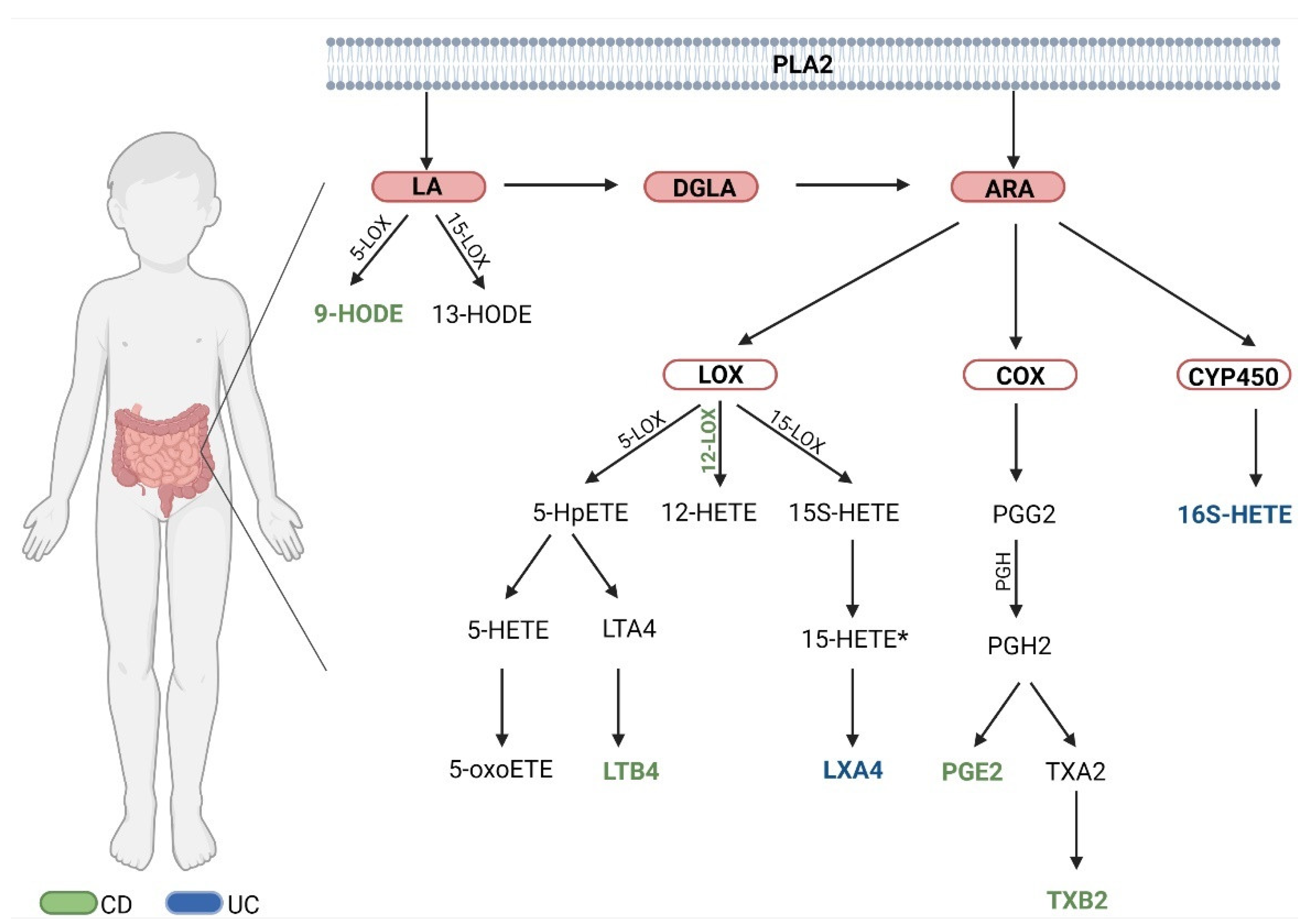
2. Results
2.1. Characteristics of the Study Group
2.2. Division of the Study Group
3. Discussion
3.1. Comparison of Both Diseases (CD vs. UC)

| References | IBD/UC/CD | Participants | Sample | Results |
|---|---|---|---|---|
| IBD | ||||
| Jupp et al., 2007 [34] | IBD | Study group (n = 23)
| Colonic biopsies | higher 5-LOX activity and higher LTB4 levels in biopsy material from patients with active disease relative to controls changes in eicosanoid concentrations more markedly observed in the UC group relative to controls |
| Ikehata et al., 1994 [35] | IBD | Study group (n = 17)
| Colonic mucosa | 5-LOX activity was higher in CD than in UC, especially in CD compared to mucosa of UC patients without inflammation |
| Sharon et al., 1984 [36] | IBD | Study group (n = 11 )
| Colonic mucosa | higher levels of TXB2, LTB4 and 5-HETE in the colonic mucosa of IBD patients |
| Lauritsen et al., 1988 [43] | IBD untreated patients | Study group (n = 37)
| Dialysis of the rectum | higher concentration of PGE2, LTB4, TXB2 in UC relative to CD |
| Baumeister et al., 1996 [44] | IBD | Study group (n = 7) Control group (n = 10) | Colonic mucosa | increased PGE2 production in IBD relative to controls PGE2 may be one of the major eicosanoids in less severe IBD |
| Shannon et al., 1993 [49] | IBD Active disease | Study group (n = 8)
| Colonic mucosa samples from inflamed and non-inflamed tissue | increased 12-lipoxygenase activity in inflamed regions of the colon in IBD compared to controls (12-HETE increased in IBD) |
| ACTIVE UC | ||||
| Zijlstra et al., 1992 [37] | UC | Study group of active UC (n = 11)
| Colonic tissue | the main eicosanoid identified in inflamed colon tissue was 15-HETE 12-HETE, PGF2, PGE2, TXB2 were present in much lower concentrations than 15-HETE |
| Zijlstra et al., 1992 [39] | UC | Study group of active UC (n = 11)
| Colonic tissue | 15-HETE as the predominant mediator was observed in biopsies from UC patients relative to healthy individuals |
| Masoodi et al., 2013 [50] | UC | Study group of active UC (n = 54)
| Colonic mucosa biopsy | levels of 5-, 11-, 12-, and 15-HETE were found in the study group relative to healthy controls higher levels of PGE2 and TXB2 were observed in the mucosa of patients compared to healthy controls 5-, 11-, 12-, 15-HEPE mediators were indeterminate in biopsy material from these patients |
| Zijlstra et al., 1991 [51] | UC |
| Mucus from morning stool | 15-HETE was identified in the highest amount in the patient’s mucus, followed by LTB4, TXB2 and PGE2 which were also identified in lower amounts |
| ACTIVE AND INACTIVE UC | ||||
| Hamabata et al., 2018 [48] | DSS-induced Colitis in mice | - | Colon tissue | production of lipid mediators changes significantly with the progression of active inflammatory bowel disease |
| Gewirtz et al., 2002 [52] | DSS-induced Colitis in mice | - | - | Oral administration of LXA(4) analog (10g per day) resulted in slowed weight loss and reduced mortality, and resolution of inflammation |
| Vong et al., 2012 [53] | UC | Study group (n = 24)
| Colonic mucosa biopsies | LXA(4) higher in biopsies from patients in remission only upregulation of AnxA1 protein expression in inactive group |
| Fiorucci et al., 2004 [54] | TNBS-induced Colitis in mice (CD model) | - | Plasma and colonic mucosa | 12-HETE-PE, which can be formed from 12-LOX is removed in the acute phase of peritonitis and reappears in the resolution of acute inflammation ZK-192 (oral pharmacokinetics) and related 3-oxa-ATL analog may have a therapeutic function in CD |
| Diab et al., 2019 [55] | UC | Study group (n = 20)
| Colon biopsies | higher levels of 15S-HETE in remission patients relative to healthy patients |
| CD | ||||
| Pochard et al., 2016 [56] | CD | Study group (n = 6)
Sprague Dawley rats | Cultures of human and adult rat enteric glial cells (EGC) | 15-HETE production in EGC CD patients was reduced compared to controls 15-HETE inhibition in rats increased intestinal epithelial barrier permeability |
3.2. Comparison of Both Phases in CD
3.3. Comparison of Both Phases in UC
4. Materials and Methods
4.1. Anthropometric Measurements
4.2. Sample Collection
4.3. Extraction of Eicosanoids
4.4. Statistical Analysis
5. Conclusions
Author Contributions
Funding
Institutional Review Board Statement
Informed Consent Statement
Data Availability Statement
Conflicts of Interest
References
- Abraham, B.P.; Ahmed, T.; Ali, T. Inflammatory Bowel Disease: Pathophysiology and Current Therapeutic Approaches. In Gastrointestinal Pharmacology; Greenwood-Van Meerveld, B., Ed.; Handbook of Experimental Pharmacology; Springer International Publishing: Cham, Switzerland, 2017; pp. 115–146. ISBN 978-3-319-56360-2. [Google Scholar]
- Cobrin, G.M.; Abreu, M.T. Defects in Mucosal Immunity Leading to Crohn’s Disease. Immunol. Rev. 2005, 206, 277–295. [Google Scholar] [CrossRef] [PubMed]
- Sheng, Q.; Li, F.; Chen, G.; Li, J.; Li, J.; Wang, Y.; Lu, Y.; Li, Q.; Li, M.; Chai, K. Ursolic Acid Regulates Intestinal Microbiota and Inflammatory Cell Infiltration to Prevent Ulcerative Colitis. J. Immunol. Res. 2021, 2021, 6679316. [Google Scholar] [CrossRef]
- de Mesquita, M.B.; Shouval, D.S. Evaluation of very early-onset inflammatory bowel disease. Curr. Opin. Gastroenterol. 2020, 36, 464–469. [Google Scholar] [CrossRef] [PubMed]
- Ng, S.C.; Shi, H.Y.; Hamidi, N.; Underwood, F.E.; Tang, W.; Benchimol, E.I.; Panaccione, R.; Ghosh, S.; Wu, J.C.Y.; Chan, F.K.L.; et al. Worldwide Incidence and Prevalence of Inflammatory Bowel Disease in the 21st Century: A Systematic Review of Population-Based Studies. Lancet 2017, 390, 2769–2778. [Google Scholar] [CrossRef]
- Ma, C.; Vasu, R.; Zhang, H. The Role of Long-Chain Fatty Acids in Inflammatory Bowel Disease. Mediat. Inflamm. 2019, 2019, 8495913. [Google Scholar] [CrossRef] [PubMed]
- Sýkora, J.; Pomahačová, R.; Kreslová, M.; Cvalínová, D.; Štych, P.; Schwarz, J. Current Global Trends in the Incidence of Pediatric-Onset Inflammatory Bowel Disease. World J. Gastroenterol. 2018, 24, 2741–2763. [Google Scholar] [CrossRef] [PubMed]
- Dmochowska, N.; Wardill, H.R.; Hughes, P.A. Advances in Imaging Specific Mediators of Inflammatory Bowel Disease. Int. J. Mol. Sci. 2018, 19, 2471. [Google Scholar] [CrossRef] [Green Version]
- Marafini, I.; Sedda, S.; Dinallo, V.; Monteleone, G. Inflammatory Cytokines: From Discoveries to Therapies in IBD. Expert Opin. Biol. Ther. 2019, 19, 1207–1217. [Google Scholar] [CrossRef] [PubMed]
- Chen, P.; Zhou, G.; Lin, J.; Li, L.; Zeng, Z.; Chen, M.; Zhang, S. Serum Biomarkers for Inflammatory Bowel Disease. Front. Med. 2020, 7, 123. [Google Scholar] [CrossRef] [PubMed] [Green Version]
- Sands, B.E. Biomarkers of Inflammation in Inflammatory Bowel Disease. Gastroenterology 2015, 149, 1275–1285.e2. [Google Scholar] [CrossRef] [PubMed]
- Manceau, H.; Chicha-Cattoir, V.; Puy, H.; Peoc’h, K. Fecal Calprotectin in Inflammatory Bowel Diseases: Update and Perspectives. Clin. Chem. Lab. Med. 2017, 55, 474–483. [Google Scholar] [CrossRef] [PubMed]
- Agrawal, M.; Spencer, E.A.; Colombel, J.-F.; Ungaro, R.C. Approach to the Management of Recently Diagnosed Inflammatory Bowel Disease Patients: A User’s Guide for Adult and Pediatric Gastroenterologists. Gastroenterology 2021, 161, 47–65. [Google Scholar] [CrossRef] [PubMed]
- Dragoni, G.; Innocenti, T.; Galli, A. Biomarkers of Inflammation in Inflammatory Bowel Disease: How Long before Abandoning Single-Marker Approaches? Dig. Dis. 2021, 39, 190–203. [Google Scholar] [CrossRef] [PubMed]
- Camba-Gómez, M.; Gualillo, O.; Conde-Aranda, J. New Perspectives in the Study of Intestinal Inflammation: Focus on the Resolution of Inflammation. Int. J. Mol. Sci. 2021, 22, 2605. [Google Scholar] [CrossRef] [PubMed]
- Graham, D.B.; Xavier, R.J. Pathway Paradigms Revealed from the Genetics of Inflammatory Bowel Disease. Nature 2020, 578, 527. [Google Scholar] [CrossRef] [PubMed]
- Ananthakrishnan, A.N.; Khalili, H.; Konijeti, G.G.; Higuchi, L.M.; de Silva, P.; Fuchs, C.S.; Willett, W.C.; Richter, J.M.; Chan, A.T. Long-Term Intake of Dietary Fat and Risk of Ulcerative Colitis and Crohn’s Disease. Gut 2014, 63, 776. [Google Scholar] [CrossRef] [PubMed] [Green Version]
- Pearl, D.S.; Masoodi, M.; Eiden, M.; Brümmer, J.; Gullick, D.; McKeever, T.M.; Whittaker, M.A.; Nitch-Smith, H.; Brown, J.F.; Shute, J.K.; et al. Altered Colonic Mucosal Availability of N-3 and n-6 Polyunsaturated Fatty Acids in Ulcerative Colitis and the Relationship to Disease Activity. J. Crohns Colitis 2014, 8, 70–79. [Google Scholar] [CrossRef]
- Das, U.N. Inflammatory Bowel Disease as a Disorder of an Imbalance between Pro-and Anti-Inflammatory Molecules and Deficiency of Resolution Bioactive Lipids. Lipids Health Dis. 2016, 15, 11. [Google Scholar] [CrossRef] [PubMed] [Green Version]
- Shores, D.R.; Binion, D.G.; Freeman, B.A.; Baker, P.R.S. New Insights into the Role of Fatty Acids in the Pathogenesis and Resolution of Inflammatory Bowel Disease. Inflamm. Bowel Dis. 2011, 17, 2192–2204. [Google Scholar] [CrossRef] [Green Version]
- Ahluwalia, B.; Moraes, L.; Magnusson, M.K.; Öhman, L. Immunopathogenesis of Inflammatory Bowel Disease and Mechanisms of Biological Therapies. Scand. J. Gastroenterol. 2018, 53, 379–389. [Google Scholar] [CrossRef]
- Funk, C.D. Prostaglandins and Leukotrienes: Advances in Eicosanoid Biology. Science 2001, 294, 1871–1875. [Google Scholar] [CrossRef] [Green Version]
- Scaioli, E.; Liverani, E.; Belluzzi, A. The Imbalance between N-6/n-3 Polyunsaturated Fatty Acids and Inflammatory Bowel Disease: A Comprehensive Review and Future Therapeutic Perspectives. Int. J. Mol. Sci. 2017, 18, 2619. [Google Scholar] [CrossRef] [PubMed] [Green Version]
- Klawitter, J.; Zafar, I.; Klawitter, J.; Pennington, A.T.; Klepacki, J.; Gitomer, B.Y.; Schrier, R.W.; Christians, U.; Edelstein, C.L. Effects of Lovastatin Treatment on the Metabolic Distributions in the Han:SPRD Rat Model of Polycystic Kidney Disease. BMC Nephrol. 2013, 14, 165. [Google Scholar] [CrossRef] [PubMed] [Green Version]
- Stenson, W.F. The universe of arachidonic acid metabolites in inflammatory bowel disease: Can we tell the good from the bad? Curr. Opin. Gastroenterol. 2014, 30, 347. [Google Scholar] [CrossRef]
- Scoville, E.A.; Allaman, M.M.; Adams, D.W.; Motley, A.K.; Peyton, S.C.; Ferguson, S.L.; Horst, S.N.; Williams, C.S.; Beaulieu, D.B.; Schwartz, D.A.; et al. Serum Polyunsaturated Fatty Acids Correlate with Serum Cytokines and Clinical Disease Activity in Crohn’s Disease. Sci. Rep. 2019, 9, 2882. [Google Scholar] [CrossRef] [PubMed]
- Serhan, C.N.; Petasis, N.A. Resolvins and Protectins in Inflammation-Resolution. Chem. Rev. 2011, 111, 5922–5943. [Google Scholar] [CrossRef] [Green Version]
- Mangino, M.J.; Brounts, L.; Harms, B.; Heise, C. Lipoxin Biosynthesis in Inflammatory Bowel Disease. Prostaglandins Other Lipid Mediat. 2006, 79, 84–92. [Google Scholar] [CrossRef] [PubMed]
- Goh, J.; Godson, C.; Brady, H.R.; MacMathuna, P. Lipoxins: Pro-Resolution Lipid Mediators in Intestinal Inflammation. Gastroenterology 2003, 124, 1043–1054. [Google Scholar] [CrossRef] [PubMed]
- Vong, L.; Ferraz, J.G.P.; Panaccione, R.; Beck, P.L.; Wallace, J.L. A Pro-Resolution Mediator, Prostaglandin D2, Is Specifically up-Regulated in Individuals in Long-Term Remission from Ulcerative Colitis. Proc. Natl. Acad. Sci. USA 2010, 107, 12023–12027. [Google Scholar] [CrossRef] [PubMed] [Green Version]
- Serhan, C.N.; Chiang, N.; Dalli, J. The Resolution Code of Acute Inflammation: Novel Pro-Resolving Lipid Mediators in Resolution. Semin. Immunol. 2015, 27, 200–215. [Google Scholar] [CrossRef] [PubMed] [Green Version]
- Wallace, J.L. Eicosanoids in the Gastrointestinal Tract. Br. J. Pharmacol. 2019, 176, 1000–1008. [Google Scholar] [CrossRef] [PubMed] [Green Version]
- Park, J.H.; Peyrin-Biroulet, L.; Eisenhut, M.; Shin, J.I. IBD Immunopathogenesis: A Comprehensive Review of Inflammatory Molecules. Autoimmun. Rev. 2017, 16, 416–426. [Google Scholar] [CrossRef] [PubMed]
- Jupp, J.; Hillier, K.; Elliott, D.H.; Fine, D.R.; Bateman, A.C.; Johnson, P.A.; Cazaly, A.M.; Penrose, J.F.; Sampson, A.P. Colonic Expression of Leukotriene-Pathway Enzymes in Inflammatory Bowel Diseases. Inflamm. Bowel Dis. 2007, 13, 537–546. [Google Scholar] [CrossRef]
- Ikehata, A.; Hiwatashi, N.; Kinouchi, Y.; Yamazaki, H.; Ito, K.; Toyota, T. Altered Leukotriene B4 Metabolism in Colonic Mucosa with Inflammatory Bowel Disease. Scand. J. Gastroenterol. 1995, 30, 44–49. [Google Scholar] [CrossRef] [PubMed]
- Sharon, P.; Stenson, W.F. Enhanced Synthesis of Leukotriene B4 by Colonic Mucosa in Inflammatory Bowel Disease. Gastroenterology 1984, 86, 453–460. [Google Scholar] [CrossRef]
- Zijlstra, F.J.; van Dijk, A.P.; Wilson, J.H.; van Riemsdijk-Overbeeke, I.C.; Vincent, J.E.; Ouwendijk, R.J. 15-HETE Is the Main Eicosanoid Formed by Human Colonic Mucosa. Agents Actions 1992, 36, C53–C59. [Google Scholar] [CrossRef]
- Powell, W.S.; Rokach, J. Biosynthesis, Biological Effects, and Receptors of Hydroxyeicosatetraenoic Acids (HETEs) and Oxoeicosatetraenoic Acids (Oxo-ETEs) Derived from Arachidonic Acid. Biochim. Biophys. Acta 2015, 1851, 340–355. [Google Scholar] [CrossRef] [Green Version]
- Zijlstra, F.; van Dijk, A.; Garrelds, I.; Ouwendijk, R.; Wilson, J. Species Differences in the Pattern of Eicosanoids Produced by Inflamed and Non-Inflamed Tissue. Agents Actions 1992, 36, C73–C75. [Google Scholar] [CrossRef]
- Kulkarni, A.; Nadler, J.L.; Mirmira, R.G.; Casimiro, I. Regulation of Tissue Inflammation by 12-Lipoxygenases. Biomolecules 2021, 11, 717. [Google Scholar] [CrossRef]
- Wang, S.; Gustafson, E.; Pang, L.; Qiao, X.; Behan, J.; Maguire, M.; Bayne, M.; Laz, T. A Novel Hepatointestinal Leukotriene B4 Receptor: Cloning and Functional Characterization. J. Biol. Chem. 2000, 275, 40686–40694. [Google Scholar] [CrossRef] [PubMed] [Green Version]
- Stenson, W.F. Role of Eicosanoids as Mediators of Inflammation in Inflammatory Bowel Disease. Scand. J. Gastroenterol. Suppl. 1990, 172, 13–18. [Google Scholar] [CrossRef] [PubMed]
- Lauritsen, K.; Laursen, L.S.; Bukhave, K.; Rask-Madsen, J. In Vivo Profiles of Eicosanoids in Ulcerative Colitis, Crohn’s Colitis, and Clostridium Difficile Colitis. Gastroenterology 1988, 95, 11–17. [Google Scholar] [CrossRef]
- Baumeister, B.; Schmidt, C.; Helisch, A.; Kipnowski, J. Increased Prostaglandin E2 and Leukotriene B4 Synthesis in Isolated Colonic Mucosal Cells in Inflammatory Bowel Disease: A Preliminary Report. J. Clin. Gastroenterol. 1996, 22, 117. [Google Scholar] [CrossRef] [PubMed]
- Szczuko, M.; Kotlęga, D.; Palma, J.; Zembroń-Łacny, A.; Tylutka, A.; Gołąb-Janowska, M.; Drozd, A. Lipoxins, RevD1 and 9, 13 HODE as the Most Important Derivatives after an Early Incident of Ischemic Stroke. Sci. Rep. 2020, 10, 12849. [Google Scholar] [CrossRef] [PubMed]
- Powell, W.S.; Rokach, J. The Eosinophil Chemoattractant 5-Oxo-ETE and the OXE Receptor. Prog. Lipid Res. 2013, 52, 651–665. [Google Scholar] [CrossRef] [Green Version]
- Bautzova, T.; Hockley, J.R.F.; Perez-Berezo, T.; Pujo, J.; Tranter, M.M.; Desormeaux, C.; Barbaro, M.R.; Basso, L.; Faouder, P.L.; Rolland, C.; et al. 5-OxoETE Triggers Nociception in Constipation-Predominant Irritable Bowel Syndrome through MAS-Related G Protein–Coupled Receptor D. Sci. Signal. 2018, 11, eaal2171. [Google Scholar] [CrossRef] [Green Version]
- Hamabata, T.; Nakamura, T.; Masuko, S.; Maeda, S.; Murata, T. Production of Lipid Mediators across Different Disease Stages of Dextran Sodium Sulfate-Induced Colitis in Mice. J. Lipid Res. 2018, 59, 586–595. [Google Scholar] [CrossRef] [PubMed] [Green Version]
- Shannon, V.R.; Stenson, W.F.; Holtzman, M.J. Induction of Epithelial Arachidonate 12-Lipoxygenase at Active Sites of Inflammatory Bowel Disease. Am. J. Physiol. 1993, 264, G104–G111. [Google Scholar] [CrossRef]
- Masoodi, M.; Pearl, D.S.; Eiden, M.; Shute, J.K.; Brown, J.F.; Calder, P.C.; Trebble, T.M. Altered Colonic Mucosal Polyunsaturated Fatty Acid (PUFA) Derived Lipid Mediators in Ulcerative Colitis: New Insight into Relationship with Disease Activity and Pathophysiology. PLoS ONE 2013, 8, e76532. [Google Scholar] [CrossRef] [PubMed] [Green Version]
- Zijlstra, F.J.; Wilson, J.H.P. 15-HETE Is the Main Eicosanoid Present in Mucus of Ulcerative Proctocolitis. Prostaglandins Leukot. Essent. Fat. Acids 1991, 43, 55–59. [Google Scholar] [CrossRef] [Green Version]
- Gewirtz, A.T.; Collier-Hyams, L.S.; Young, A.N.; Kucharzik, T.; Guilford, W.J.; Parkinson, J.F.; Williams, I.R.; Neish, A.S.; Madara, J.L. Lipoxin A4 Analogs Attenuate Induction of Intestinal Epithelial Proinflammatory Gene Expression and Reduce the Severity of Dextran Sodium Sulfate-Induced Colitis. J. Immunol. 2002, 168, 5260–5267. [Google Scholar] [CrossRef] [PubMed] [Green Version]
- Vong, L.; Ferraz, J.G.P.; Dufton, N.; Panaccione, R.; Beck, P.L.; Sherman, P.M.; Perretti, M.; Wallace, J.L. Up-Regulation of Annexin-A1 and Lipoxin A4 in Individuals with Ulcerative Colitis May Promote Mucosal Homeostasis. PLoS ONE 2012, 7, e39244. [Google Scholar] [CrossRef]
- Fiorucci, S.; Wallace, J.L.; Mencarelli, A.; Distrutti, E.; Rizzo, G.; Farneti, S.; Morelli, A.; Tseng, J.-L.; Suramanyam, B.; Guilford, W.J.; et al. A Beta-Oxidation-Resistant Lipoxin A4 Analog Treats Hapten-Induced Colitis by Attenuating Inflammation and Immune Dysfunction. Proc. Natl. Acad. Sci. USA 2004, 101, 15736–15741. [Google Scholar] [CrossRef] [Green Version]
- Diab, J.; Al-Mahdi, R.; Gouveia-Figueira, S.; Hansen, T.; Jensen, E.; Goll, R.; Moritz, T.; Florholmen, J.; Forsdahl, G. A Quantitative Analysis of Colonic Mucosal Oxylipins and Endocannabinoids in Treatment-Naïve and Deep Remission Ulcerative Colitis Patients and the Potential Link with Cytokine Gene Expression. Inflamm. Bowel Dis. 2019, 25, 490–497. [Google Scholar] [CrossRef] [PubMed] [Green Version]
- Pochard, C.; Coquenlorge, S.; Jaulin, J.; Cenac, N.; Vergnolle, N.; Meurette, G.; Freyssinet, M.; Neunlist, M.; Rolli-Derkinderen, M. Defects in 15-HETE Production and Control of Epithelial Permeability by Human Enteric Glial Cells from Patients with Crohn’s Disease. Gastroenterology 2016, 150, 168–180. [Google Scholar] [CrossRef] [PubMed]
- Morgan, A.H.; Dioszeghy, V.; Maskrey, B.H.; Thomas, C.P.; Clark, S.R.; Mathie, S.A.; Lloyd, C.M.; Kühn, H.; Topley, N.; Coles, B.C.; et al. Phosphatidylethanolamine-Esterified Eicosanoids in the Mouse: Tissue Localization and Inflammation-Dependent Formation in Th-2 Disease. J. Biol. Chem. 2009, 284, 21185–21191. [Google Scholar] [CrossRef] [PubMed] [Green Version]
- Prescott, D.; McKay, D.M. Aspirin-Triggered Lipoxin Enhances Macrophage Phagocytosis of Bacteria While Inhibiting Inflammatory Cytokine Production. Am. J. Physiol. Gastrointest. Liver Physiol. 2011, 301, G487–G497. [Google Scholar] [CrossRef] [PubMed]
- Bednar, M.M.; Gross, C.E.; Russell, S.R.; Fuller, S.P.; Ahern, T.P.; Howard, D.B.; Falck, J.R.; Reddy, K.M.; Balazy, M. 16(R)-Hydroxyeicosatetraenoic Acid, a Novel Cytochrome P450 Product of Arachidonic Acid, Suppresses Activation of Human Polymorphonuclear Leukocyte and Reduces Intracranial Pressure in a Rabbit Model of Thromboembolic Stroke. Neurosurgery 2000, 47, 1410–1418; discussion 1418–1419. [Google Scholar] [CrossRef]
- Kułaga, Z.; Różdżyńska-Świątkowska, A.; Grajda, A.; Gurzkowska, B.; Wojtyło, M.; Góźdź, M.; Świąder-Leśniak, A.; Litwin, M. Percentile charts for growth and nutritional status assessment in Polish children and adolescents from birth to 18 year of age. Stand. Med./Pediatr. 2015, 12, 119–135. [Google Scholar]

| Parameter | CD Avg ± SD n = 34 | UC Avg ± SD n = 30 | p-Value |
|---|---|---|---|
| Age (years) | 13.76 ± 2.69 | 14.15 ± 3.31 | 0.70 |
| Body mass (kg) | 46.81 ± 18.07 | 53.02 ± 19.40 | 0.18 |
| Height (m) | 1.54 ± 0.19 | 1.60 ± 0.20 | 0.26 |
| Disease duration (months) | 23.38 ± 26.45 | 19.57 ± 30.22 | 0.25 |
| BMI (kg/m2) | 19.06 ± 4.29 | 20.06 ± 4.91 | 0.40 |
| BMI percentiles | 43.09 ± 35.21 | 47.03 ± 37.74 | 0.71 |
| Body mass percentiles | 39.36 ± 34.76 | 46.40 ± 37.50 | 0.46 |
| PCDAI | 15.84 ± 16.08 | - | - |
| PUCAI | - | 30.00 ± 23.36 | - |
| Fecal calprotectin active disease (µg/g) | 2606.68 ± 2504.64 | 2230.07 ± 2113.7 | 0.66 |
| Fecal calprotectin (µg/g) | 2040.45 ± 2269.53 | 2096.77 ± 2110.49 | 0.94 |
| CD (n = 34) | UC (n = 30) | |
|---|---|---|
| Pharmacology | ||
| Aminosalicylates | 23 | 24 |
| Glucocorticosteroids | 9 | 1 |
| Immunomodulatory drugs | 15 | 3 |
| Biologic drugs | 9 | 5 |
| Supplementation | ||
| Vitamin D | 26 | 19 |
| Probiotics | 16 | 12 |
| Calcium | 11 | 7 |
| Iron | 5 | 6 |
| B vitamins | 8 | - |
| Potassium | - | 4 |
| Lipid Mediators (µg/mL) | CD Avg ± SD n = 34 | UC Avg ± SD n = 30 | p-Value |
|---|---|---|---|
| TXB2 | 0.090 ± 0.08 | 0.079 ± 0.07 | 0.614 |
| PGE2 | 10.533 ± 30.46 | 9.633 ± 28.62 | 0.909 |
| LTX A4 5S, 6R | 0.099 ± 0.12 | 0.105 ± 0.14 | 0.863 |
| LTX A4 5S, 6R, 15R | 0.089 ± 0.11 | 0.087 ± 0.09 | 0.944 |
| LTB4 | 0.102 ± 0.08 | 0.093 ± 0.07 | 0.678 |
| 16RS-HETE | 0.540 ± 0.58 | 0.618 ± 0.42 | 0.567 |
| 13S-HODE | 0.355 ± 0.40 | 0.274 ± 0.31 | 0.397 |
| 9S-HODE | 0.428 ± 0.47 | 0.316 ± 0.33 | 0.308 |
| 15S-HETE | 1.048 ± 0.79 | 0.758 ± 0.38 | 0.092 |
| 12S-HETE | 3.404 ± 2.47 | 3.789 ± 3.89 | 0.649 |
| 5-oxo ETE | 0.836 ± 0.72 | 0.852 ± 0.88 | 0.940 |
| 5-HETE | 2.408 ± 1.37 | 2.244 ± 1.91 | 0.705 |
| Mediators of the Inflammatory State (µg/mL) | Active CD Avg ± SD n = 24 | Remission CD Avg ± SD n = 9 | p-Value | Active UC Avg ± SD n = 24 | Remission UC Avg ± SD n = 6 | p-Value | p-Value CD vs. UC Active | p-Value CD vs. UC Remission |
|---|---|---|---|---|---|---|---|---|
| TXB2 | 0.106 ± 0.09 | 0.047 ± 0.06 | 0.079 | 0.082 ± 0.06 | 0.080 ± 0.08 | 0.575 | 0.321 | 0.875 |
| PGE2 | 5.000 ± 15.93 | 1.808 ± 2.48 | 0.590 | 6.806 ± 23.19 | 0.470 ± 0.39 | 0.009 | 0.767 | 0.128 |
| LTX A4 5S, 6R | 0.098 ± 0.13 | 0.093 ± 0.12 | 0.815 | 0.082 ± 0.10 | 0.238 ± 0.25 | 0.034 | 0.638 | 0.156 |
| LTX A4 5S, 6R, 15R | 0.079 ± 0.12 | 0.102 ± 0.10 | 0.313 | 0.090 ± 0.09 | 0.073 ± 0.06 | 0.788 | 0.747 | 0.564 |
| LTB4 | 0.115 ± 0.08 | 0.061 ± 0.08 | 0.083 | 0.097 ± 0.08 | 0.080 ± 0.05 | 0.643 | 0.465 | 0.318 |
| 16RS-HETE | 0.562 ± 0.67 | 0.481 ± 0.28 | 0.673 | 0.694 ± 0.40 | 0.374 ± 0.35 | 0.038 | 0.443 | 0.958 |
| 13S-HODE | 0.366 ± 0.34 | 0.327 ± 0.58 | 0.153 | 0.288 ± 0.32 | 0.254 ± 0.27 | 0.575 | 0.446 | 0.875 |
| 9S-HODE | 0.445 ± 0.36 | 0.379 ± 0.74 | 0.087 | 0.341 ± 0.35 | 0.242 ± 0.29 | 0.643 | 0.339 | 0.713 |
| 15S-HETE | 1.178 ± 0.87 | 0.844 ± 0.55 | 0.439 | 0.759 ± 0.41 | 0.742 ± 0.25 | 0.788 | 0.051 | 0.564 |
| 12S-HETE | 2.741 ± 2.37 | 4.579 ± 2.16 | 0.033 | 3.919 ± 4.24 | 3.127 ± 2.17 | 0.942 | 0.264 | 0.104 |
| 5-oxo ETE | 0.934 ± 0.82 | 0.616 ± 0.44 | 0.496 | 0.776 ± 0.71 | 1.376 ± 1.48 | 0.449 | 0.504 | 0.431 |
| 5-HETE | 2.523 ± 1.52 | 2.198 ± 0.95 | 0.622 | 2.321 ± 2.11 | 2.069 ± 0.53 | 0.510 | 0.720 | 0.875 |
Publisher’s Note: MDPI stays neutral with regard to jurisdictional claims in published maps and institutional affiliations. |
© 2022 by the authors. Licensee MDPI, Basel, Switzerland. This article is an open access article distributed under the terms and conditions of the Creative Commons Attribution (CC BY) license (https://creativecommons.org/licenses/by/4.0/).
Share and Cite
Kikut, J.; Mokrzycka, M.; Drozd, A.; Grzybowska-Chlebowczyk, U.; Ziętek, M.; Szczuko, M. Involvement of Proinflammatory Arachidonic Acid (ARA) Derivatives in Crohn’s Disease (CD) and Ulcerative Colitis (UC). J. Clin. Med. 2022, 11, 1861. https://doi.org/10.3390/jcm11071861
Kikut J, Mokrzycka M, Drozd A, Grzybowska-Chlebowczyk U, Ziętek M, Szczuko M. Involvement of Proinflammatory Arachidonic Acid (ARA) Derivatives in Crohn’s Disease (CD) and Ulcerative Colitis (UC). Journal of Clinical Medicine. 2022; 11(7):1861. https://doi.org/10.3390/jcm11071861
Chicago/Turabian StyleKikut, Justyna, Małgorzata Mokrzycka, Arleta Drozd, Urszula Grzybowska-Chlebowczyk, Maciej Ziętek, and Małgorzata Szczuko. 2022. "Involvement of Proinflammatory Arachidonic Acid (ARA) Derivatives in Crohn’s Disease (CD) and Ulcerative Colitis (UC)" Journal of Clinical Medicine 11, no. 7: 1861. https://doi.org/10.3390/jcm11071861