
3D Echo Characterization of Proportionate and Disproportionate Functional Mitral Regurgitation before and after Percutaneous Mitral Valve Repair

 , ,
, ,
Abstract
:1. Introduction
2. Methods
3. Reproducibility
4. MitraClip Procedure
5. Statistical Analysis
6. Results
7. Discussion
8. Limits
9. Conclusions
Supplementary Materials
Author Contributions
Funding
Institutional Review Board Statement
Informed Consent Statement
Data Availability Statement
Acknowledgments
Conflicts of Interest
Abbreviations
| D-MR | Disproportionate mitral regurgitation |
| EROA | Effective regurgitant orifice area |
| GLS | Global longitudinal strain |
| FMR | Functional mitral regurgitation |
| HF | Heart failure |
| LV | Left ventricle |
| LVEDV | Left ventricular end-diastolic volume |
| LVESV | Left ventricular end-systolic volume |
| MV | Mitral valve |
| PASp | Pulmonary artery systolic pressure |
| P-MR | Proportionate mitral regurgitation |
| PMVr | Percutaneous mitral valve repair |
References
- La Canna, G.; Scarfò, I.; Caso, I. How to differentiate functional from degenerative mitral regurgitation. J. Cardiovasc. Med. 2018, e75–e79. [Google Scholar] [CrossRef] [PubMed]
- Goliasch, G.E.; Bartko, P.; Pavo, N.; Neuhold, S.; Wurm, R.; Mascherbauer, J.; Lang, I.M.; Strunk, G.; Hülsmann, M. Refining the prognostic impact of functional mitral regurgitation in chronic heart failure. Eur. Hear. J. 2017, 39, 39–46. [Google Scholar] [CrossRef] [PubMed]
- Sannino, A.; Smith, R.L.; Schiattarella, G.G.; Trimarco, B.; Esposito, G.; Grayburn, P. Survival and cardiovascular outcomes of patients with secondary mitral regurgitation: A systematic review and meta-analysis. JAMA Cardiol. 2017, 2, 1130–1139. [Google Scholar] [CrossRef] [PubMed]
- Ponikowski, P.; Voors, A.A.; Anker, S.D.; Bueno, H.; Cleland, J.G.F.; Coats, A.J.S.; Falk, V.; González-Juanatey, J.R.; Harjola, V.-P.; Jankowska, E.A.; et al. 2016 ESC Guidelines for the diagnosis and treatment of acute and chronic heart failure: The Task Force for the diagnosis and treatment of acute and chronic heart failure of the European Society of Cardiology (ESC)Developed with the special contribution of the Heart Failure Association (HFA) of the ESC. Eur. J. Heart Fail. 2016, 18, 891–975. [Google Scholar] [CrossRef]
- Stone, G.W.; Vahanian, A.S.; Adams, D.H.; Abraham, W.T.; Borer, J.S.; Bax, J.J.; Schofer, J.; Cutlip, D.E.; Krucoff, M.W.; Blackstone, E.H.; et al. Clinical Trial Design Principles and Endpoint Definitions for Transcatheter Mitral Valve Repair and Replacement: Part 1: Clinical Trial Design Principles. J. Am. Coll. Cardiol. 2015, 66, 278–307. [Google Scholar] [CrossRef] [Green Version]
- Falk, V.; Baumgartner, H.; Bax, J.J.; De Bonis, M.; Hamm, C.; Holm, P.J.; Iung, B.; Lancellotti, P.; Lansac, E.; Rodriguez Muñoz, D.; et al. 2017 ESC/EACTS Guidelines for the management of valvular heart disease. Eur. J. Cardio-Thorac. Surg. 2017, 52, 616–664, Correction: Eur. J. Cardio-Thorac. Surg. 2017, 52, 832. [Google Scholar] [CrossRef]
- Feldman, T.; Wasserman, H.S.; Herrmann, H.C.; Gray, W.; Block, P.C.; Whitlow, P.; Goar, F.S.; Rodriguez, L.; Silvestry, F.; Schwartz, A.; et al. Percutaneous Mitral Valve Repair Using the Edge-to-Edge Technique: Six-Month Results of the EVEREST Phase I Clinical Trial. J. Am. Coll. Cardiol. 2005, 46, 2134–2140. [Google Scholar] [CrossRef] [Green Version]
- Stone, G.W.; Lindenfeld, J.; Abraham, W.T.; Kar, S.; Lim, D.S.; Mishell, J.M.; Whisenant, B.; Grayburn, P.A.; Rinaldi, M.; Kapadia, S.R.; et al. Transcatheter Mitral-Valve Repair in Patients with Heart Failure. N. Engl. J. Med. 2018, 379, 2307–2318. [Google Scholar] [CrossRef]
- Obadia, J.-F.; Messika-Zeitoun, D.; Leurent, G.; Iung, B.; Bonnet, G.; Piriou, N.; Lefèvre, T.; Piot, C.; Rouleau, F.; Carrie, D.; et al. Percutaneous Repair or Medical Treatment for Secondary Mitral Regurgitation. N. Engl. J. Med. 2018, 379, 2297–2306. [Google Scholar] [CrossRef]
- Pibarot, P.; Delgado, V.; Bax, J.J. MITRA-FR vs. COAPT: Lessons form two trials with diametrically opposite results. Eur. Heart J. Cardiovasc. Imaging 2019, 20, 620–624. [Google Scholar] [CrossRef]
- Grayburn, P.A.; Sannino, A.; Packer, M. Proportionate and Disproportionate functional mitral regurgitation: A new conceptual framework that reconciles the results of the MITRA-FR and COAPT trials. JACC Cardiovasc. Imaging 2019, 12, 353–362. [Google Scholar] [CrossRef] [PubMed]
- Cimino, S.; Guarracino, F.; Valenti, V.; Frati, G.; Sciarretta, S.; Miraldi, F.; Agati, L.; Greco, E. Echocardiography and Correction of Mitral Regurgitation: An Unbreakable Link. Cardiology 2019, 145, 110–120. [Google Scholar] [CrossRef] [PubMed]
- Whitlow, P.L.; Feldman, T.; Pedersen, W.R.; Lim, D.S.; Kipperman, R.; Smalling, R.; Bajwa, T.; Herrmann, H.C.; Lasala, J.; Maddux, J.T.; et al. Acute and 12-Month Results with Catheter-Based Mitral Valve Leaflet Repair: The EVEREST II (Endovascular Valve Edge-to-Edge Repair) High Risk Study. J. Am. Coll. Cardiol. 2012, 59, 130–139. [Google Scholar] [CrossRef] [PubMed] [Green Version]
- Feldman, T.; Kar, S.; Rinaldi, M.; Fail, P.; Hermiller, J.; Smalling, R.; Whitlow, P.L.; Gray, W.; Low, R.; Herrmann, H.C.; et al. Percutaneous Mitral Repair with the MitraClip System: Safety and Midterm Durability in the Initial EVEREST (Endovascular Valve Edge-to-Edge REpair Study) Cohort. J. Am. Coll. Cardiol. 2009, 54, 686–694. [Google Scholar] [CrossRef] [PubMed] [Green Version]
- Lancellotti, P.; Tribouilloy, C.; Hagendorff, A.; Popescu, B.A.; Edvardsen, T.; Pierard, L.A.; Badano, L.; Zamorano, J.L. Recommendations for the echocardiographic assessment of native valvular regurgitation: An executive summary from the European Association of Cardiovascular Imaging. Eur. Hear. J. Cardiovasc. Imaging 2013, 14, 611–644. [Google Scholar] [CrossRef] [Green Version]
- Packer, M.; Grayburn, P.A. New evidence supporting the distinction between proportionate and disproportionate subtypes. JAMA Cardiol. 2020, 5, 469–475. [Google Scholar] [CrossRef]
- Al Amri, I.; Debonnaire, P.; Van der Kley, F.; Scalij, M.J.; Bax, J.J.; Marsan, N.A.; Delgado, V. Acute effect of MitraClip implantation on mitral valve geometry in patients with functional mitral regurgitation: Insights from three-dimensional transoesophageal echocardiography. EuroIntervention 2016, 11, 1554–1561. [Google Scholar] [CrossRef] [Green Version]
- Cimino, S.; Maestrini, V.; Cantisani, D.; Petronilli, V.; Filomena, D.; Mancone, M.; Sardella, G.; Benedetti, G.; Fedele, F.; Agati, L. Mid-term repair durability after MitraClip implantation in patients with functional mitral regurgitation. J. Cardiovasc. Med. 2019, 20, 701–708. [Google Scholar] [CrossRef]
- Cimino, S.; Maestrini, V.; Cantisani, D.; Petronilli, V.; Filomena, D.; Mancone, M.; Sardella, G.; Fedele, F.; Lancellotti, P.; Agati, L. 2D/3D echocardiographic determinants of left ventricular reverse remodelling after MitraClip implantation. Eur. Hear. J. Cardiovasc. Imaging 2018, 20, 558–564. [Google Scholar] [CrossRef]
- Ailawadi, G.; Lim, D.S.; Mack, M.J.; Trento, A.; Kar, S.; Grayburn, P.A.; Glower, D.D.; Wang, A.; Foster, E.; Qasim, A.; et al. One-Year Outcomes After MitraClip for Functional Mitral Regurgitation. Circulation 2019, 139, 37–47. [Google Scholar] [CrossRef]
- Barth, S.; Hautmann, M.B.; Kerber, S.; Gietzen, F.; Zacher, M.; Halbfass, P.; Muller, P.; Schade, A.; Deneke, T.; Diegler, A.; et al. Hemodynamic improvement at Three months follow-up after Mitraclip treatment in end-stage Heart Failure patients with functional mitral regurgitation. J. Heart Valve Dis. 2016, 25, 475–482. [Google Scholar] [PubMed]
- Delgado, V.; Marsan, N.A.; Bax, J.J. Characterizing mitral regurgitation in a contemporary population: Prognostic implications. Eur. Hear. J. 2019, 40, 2203–2205. [Google Scholar] [CrossRef] [Green Version]
- Kim, J.; Alakbarli, J.; Ms, M.C.P.; Xie, L.X.; Rong, L.Q.; Tehrani, N.H.; Ba, L.R.B.; Devereux, R.B.; Wong, S.C.; Bergman, G.W.; et al. Left ventricular geometry predicts optimal response to percutaneous mitral repair via MitraClip: Integrated assessment by two- and three-dimensional echocardiography. Catheter. Cardiovasc. Interv. 2019, 93, 1152–1160. [Google Scholar] [CrossRef]
- Lang, R.M.; Badano, L.P.; Mor-Avi, V.; Afilalo, J.; Armstrong, A.; Ernande, L.; Flachskampf, F.A.; Foster, E.; Goldstein, S.A.; Kuznetsova, T.; et al. Recommendations for Cardiac Chamber Quantification by Echocardiography in Adults: An Update from the American Society of Echocardiography and the European Association of Cardiovascular Imaging. J. Am. Soc. Echocardiogr. 2015, 28, 1–39.e14. [Google Scholar] [CrossRef] [PubMed] [Green Version]
- Arora, G.; Patel, N.; Arora, P. Futile Mitra-FR and a positive COAPT trial: Where does the evidence leave the clinicians? Int. J. Cardiol. Heart Vasc. 2018, 22, 18–19. [Google Scholar] [CrossRef] [PubMed]
- Zoghbi, W.A.; Adams, D.; Bonow, R.O.; Enriquez-Sarano, M.; Foster, E.; Grayburn, P.A.; Hahn, R.T.; Han, Y.; Hung, J.; Lang, R.M.; et al. Recommendations for Noninvasive Evaluation of Native Valvular Regurgitation: A Report from the American Society of Echocardiography Developed in Collaboration with the Society for Cardiovascular Magnetic Resonance. J. Am. Soc. Echocardiogr. 2017, 30, 303–371. [Google Scholar] [CrossRef] [PubMed]
- Orban, M.; Karam, N.; Lubos, E.; Kalbacher, D.; Braun, D.; Deseive, S.; Neuss, M.; Butter, C.; Praz, F.; Kassar, M.; et al. Impact of Proportionality of Secondary Mitral Regurgitation on Outcome after Transcatheter Mitral Valve Repair. JACC Cardiovasc. Imaging 2020, 14, 715–725. [Google Scholar] [CrossRef]
- Messika-Zeitoun, D.; Iung, B.; Armoiry, X.; Trochu, J.N.; Donal, E.; Habib, G. Impact of Mitral Regurgitation Severity and Left Ventricular Remodeling on Outcome After Mitraclip Implantation: Results from the Mitra-FR Trial. JACC Cardiovasc. Imaging 2020, 14, 742–752. [Google Scholar] [CrossRef]
- Tamborini, G.; Piazzese, C.; Lang, R.M.; Muratori, M.; Chiorino, E.; Mapelli, M.; Fusini, L.; Ali, S.G.; Gripari, P.; Pontone, G.; et al. Feasibility and Accuracy of Automated Software for Transthoracic Three-Dimensional Left Ventricular Volume and Function Analysis: Comparisons with Two-Dimensional Echocardiography, Three-Dimensional Transthoracic Manual Method, and Cardiac Magnetic Resonance Imaging. J. Am. Soc. Echocardiogr. 2017, 30, 1049–1058. [Google Scholar] [CrossRef]
- Dorosz, J.L.; Lezotte, D.C.; Weitzenkamp, D.A.; Allen, L.A.; Salcedo, E.E. Performance of 3-dimensional echocardiography in per-forming left ventricular volumes and ejection fraction: A systematic review and meta-analysis. J. Am. Coll. Cardiol. 2012, 59, 1799–1808. [Google Scholar] [CrossRef] [Green Version]


| Parameters | Total Cohort (n = 56) | Disproportionate MR (n = 28, 50%) | Proportionate MR (n = 28, 50%) | p |
|---|---|---|---|---|
| Clinical Features | ||||
| Age, (years) | 73 ± 7 | 75 ± 7 | 70 ± 6 | 0.8 |
| Euro Score | 15.5 ± 10 | 15 ± 8 | 16 ± 13 | 0.7 |
| STS Score | 7.2 ± 8 | 9 ± 10 | 5 ± 5 | 0.3 |
| Male sex, n (%) | 41 (73) | 20 (71) | 21 (75) | 0.7 |
| Diabetes, n (%) | 20 (37) | 9 (32) | 11 (39) | 0.6 |
| Hypertension, n (%) | 48 (86) | 25 (89) | 23 (82) | 0.9 |
| Dyslipidemia, n (%) | 36 (65) | 16 (57) | 20 (71) | 0.3 |
| Previous AMI, n (%) | 28 (50) | 12 (42) | 16 (57) | 0.2 |
| Previous PCI, n (%) | 22 (40) | 9 (32) | 13 (46) | 0.2 |
| Previous CABG, n (%) | 11 (20) | 4 (14) | 7 (25) | 0.3 |
| CRF, n (%) | 28 (50) | 14 (50) | 14 (50) | 0.9 |
| NYHA III-IV, n (%) | 56 (100) | 28 (100) | 28 (100) | 0.9 |
| Nitrates, n (%) | 25 (45) | 11 (39) | 14 (50) | 0.08 |
| ACE-inhibitors/ARBs, n (%) | 33 (60) | 16 (57) | 17 (60) | 0.8 |
| Ivabradine, n (%) | 2 (0.03) | 0 (0) | 2 (0.7) | 0.2 |
| Beta-blockers, n (%) | 53 (95) | 27 (96) | 26 (92) | 0.9 |
| Anticoagulants, n (%) | 12 (26) | 4 (15) | 8 (30) | 0.07 |
| Antiplatelets, n (%) | 33 (58) | 18 (69) | 15 (57) | 0.08 |
| Aldosterone antagonists, n (%) | 47 (85) | 22 (78) | 25 (89) | 0.1 |
| Diuretics, n (%) | 56 (100) | 28 (100) | 28 (100) | 0.9 |
| Pacemaker, n (%) | 14 (25) | 5 (17) | 9 (32) | 0.08 |
| AF, n (%) | 11 (20) | 4 (15) | 7 (25) | 0.07 |
| Ischemic etiology, n (%) | 28 (50) | 12 (42) | 16 (57) | 0.2 |
| Non-ischemic etiology, n (%) | 28 (50) | 16 (57) | 12 (42) | 0.2 |
| Clips number (1), n (%) | 29 (51) | 15 (55) | 14 (50) | 0.8 |
| Clips number (2) n (%) | 26 (46) | 13 (46) | 13 (46) | 0.9 |
| Clips number (3) n (%) | 1 (0.02) | 0 (0) | 1 (0.03) | 0.9 |
| Echocardiography | ||||
| EROA, cm2 | 0.3 (0.2–0.4) | 0.3 (0.2–0.4) | 0.31 (0.2–0.39) | 0.7 |
| Annulus AP diameter, mm | 42 (29–50) | 41 (29–50) | 44 (38–50) | 0.2 |
| Annulus ellipticity, % | 149 (112–170) | 162 (155–170) | 137 (112–160) | 0.048 |
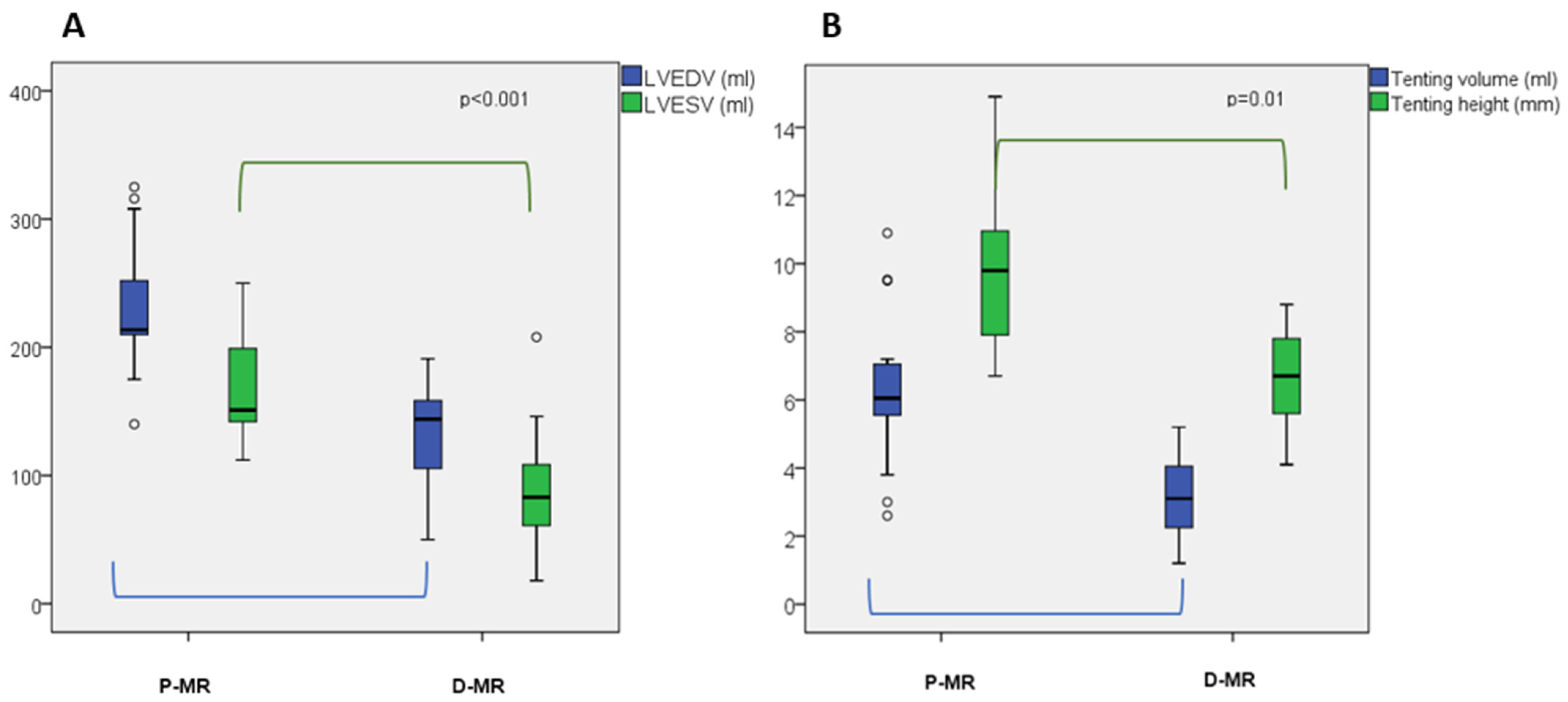
| Tenting volume, mL | 4.7 ± 2 | 3.2 ± 1.2 | 6.3 ± 2.3 | 0.01 |
| Tenting height, mm | 8 ± 1.9 | 6.5 ± 1.4 | 9.6 ± 2.4 | 0.01 |
| PL angle, ° | 47 ± 12 | 48 ± 12 | 46 ± 10 | 0.6 |
| AL angle, ° | 26 ± 9 | 24 ± 4 | 29 ± 8 | 0.08 |
| LVEF, % | 29 ± 8 | 32 ± 7 | 26 ± 5 | 0.003 |
| LVEDV, mL | 181 ± 49 | 135 ± 38 | 228 ± 48 | <0.001 |
| LVEDV/I, mL/m2 | 103 ± 28 | 80 ± 20 | 126 ± 27 | <0.001 |
| LVESV, mL | 127 ± 42 | 88 ± 40 | 167 ± 41 | <0.001 |
| LVESV/I, mL/m2 | 77 ± 24 | 60 ± 20 | 94 ± 23 | <0.001 |
| LV mass, gr | 275 ± 70 | 249 ± 63 | 301 ± 69 | 0.035 |
| LA Vol/I, mL/m2 | 60 ± 26 | 58 ± 27 | 62 ± 25 | 0.065 |
| RV ED area, cm2 | 18 ± 6 | 19 ± 5 | 18 ± 3 | 0.7 |
| RV ES area, cm2 | 11 ± 4 | 11 ± 3 | 10 ± 5 | 0.6 |
| RV FAC, % | 47 ± 9 | 50 ± 8 | 45 ± 9 | 0.07 |
| TAPSE, mm | 21 ± 5 | 20 ± 3 | 21 ± 4 | 0.8 |
| PASp, mmHg | 42 ± 14 | 40 ± 16 | 45 ± 12 | 0.07 |
| GLS, % | −8 (−11, −6) | −9 (−11, −7) | −7 (−8, −6) | 0.048 |
| Follow-up | ||||
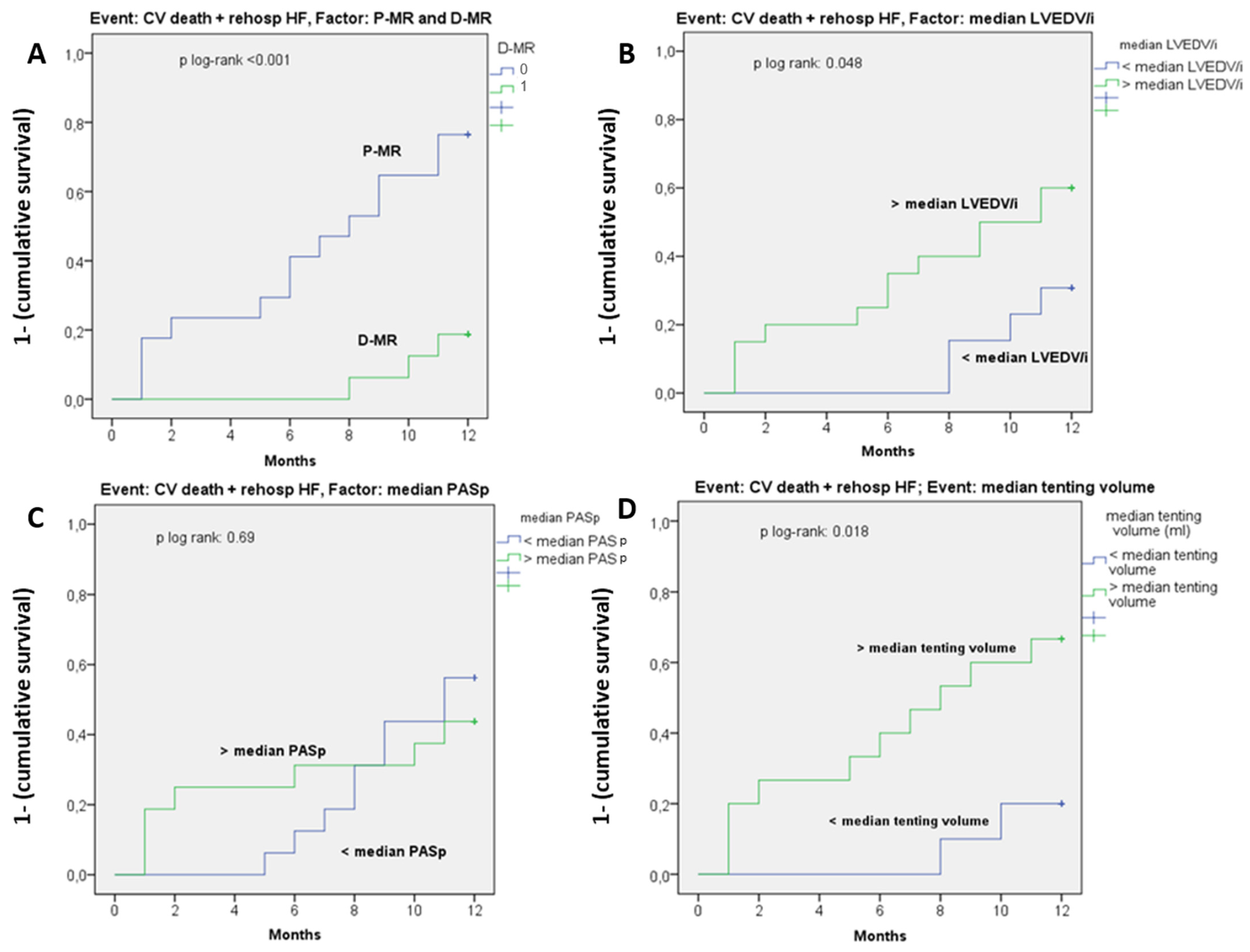
| CV death/ HF rehospitalization, n (%) | 15 (26) | 2 (7) | 13 (46) | <0.001 |
| 6-month echo, MR recurrence, n (%) | 20 (35) | 4 (14) | 16 (57) | <0.001 |
| Variables | HR (IC 95%) | p |
|---|---|---|
| Age | 0.96 (0.91–1) | 0.19 |
| Ischemic etiology | 0.98 (0.4–2.3) | 0.096 |
| CRF | 1.6 (0.66–1.18) | 0.29 |
| STS score | 1.001 (0.94–1.1) | 0.96 |
| LVEDV/i, mL/m2 | 1.007 (1.001–1.015) | 0.048 |
| LVESV/i, mL/m2 | 1.005 (0.997–1.013) | 0.23 |
| LV mass g/m2 | 1.012 (0.997–1.028) | 0.11 |
| LVEF, % | 1.002 (0.96–1.04) | 0.94 |
| PASp, mmHg | 1.03 (1.01–1.055) | 0.064 |
| Annulus ellipticity, % | 0.99 (0.98–1.01) | 0.92 |
| Annulus AP diameter, mm | 0.96 (0.88–1.055) | 0.45 |
| Tenting volume (mL) | 1.3 (1.08–1.57) | 0.005 |
| Tenting height (mm) | 1.19 (0.995–1.4) | 0.5 |
| GLS, % | 0.94 (0.7–1.2) | 0.94 |
| EROA, cm2 | 1.73 (0.39–7.4) | 0.47 |
| P-MR | 3.4 (1.3–8.6) | 0.009 |
| Variable | Intra-Observer Agreement | Inter-Observer Agreement |
|---|---|---|
| LVEF, % | 0.981 (0.92–0.996), p < 0.001 | 0.938 (0.75–0.986), p < 0.001 |
| LVEDV/i, mL/m2 | 0.996 (0.985–0.999), p < 0.001 | 0.994 (0.736–0.999), p < 0.001 |
| LVESV/i, mL/m2 | 0.998 (0.992–0.999), p < 0.001 | 0.997 (0.986–0.999), p < 0.001 |
| MVQ analysis | 0.998 (0.993–0.999), p < 0.001 | 0.996 (0.988–0.999), p < 0.001 |
Publisher’s Note: MDPI stays neutral with regard to jurisdictional claims in published maps and institutional affiliations. |
© 2022 by the authors. Licensee MDPI, Basel, Switzerland. This article is an open access article distributed under the terms and conditions of the Creative Commons Attribution (CC BY) license (https://creativecommons.org/licenses/by/4.0/).
Share and Cite
Cimino, S.; Agati, L.; Filomena, D.; Maestrini, V.; Monosilio, S.; Birtolo, L.I.; Mocci, M.; Mancone, M.; Sardella, G.; Grayburn, P.; et al. 3D Echo Characterization of Proportionate and Disproportionate Functional Mitral Regurgitation before and after Percutaneous Mitral Valve Repair. J. Clin. Med. 2022, 11, 645. https://doi.org/10.3390/jcm11030645
Cimino S, Agati L, Filomena D, Maestrini V, Monosilio S, Birtolo LI, Mocci M, Mancone M, Sardella G, Grayburn P, et al. 3D Echo Characterization of Proportionate and Disproportionate Functional Mitral Regurgitation before and after Percutaneous Mitral Valve Repair. Journal of Clinical Medicine. 2022; 11(3):645. https://doi.org/10.3390/jcm11030645
Chicago/Turabian StyleCimino, Sara, Luciano Agati, Domenico Filomena, Viviana Maestrini, Sara Monosilio, Lucia Ilaria Birtolo, Michele Mocci, Massimo Mancone, Gennaro Sardella, Paul Grayburn, and et al. 2022. "3D Echo Characterization of Proportionate and Disproportionate Functional Mitral Regurgitation before and after Percutaneous Mitral Valve Repair" Journal of Clinical Medicine 11, no. 3: 645. https://doi.org/10.3390/jcm11030645