Review

Cardiac Hypertrophy and Related Dysfunctions in Cushing Syndrome Patients—Literature Review

1
Department of Internal Medicine, Hyogo College of Medicine, Sasayama Medical Center, Sasayama 669-2321, Hyogo, Japan
2
Department of Diabetes, Endocrinology and Clinical Immunology, Hyogo College of Medicine, Nishinomiya 663-8131, Hyogo, Japan
*
Author to whom correspondence should be addressed.
J. Clin. Med. 2022, 11(23), 7035; https://doi.org/10.3390/jcm11237035
Submission received: 1 November 2022 / Revised: 19 November 2022 / Accepted: 21 November 2022 / Published: 28 November 2022
(This article belongs to the Special Issue Current Management of Adrenal Tumors)

Abstract

:
The survival rate of adrenal Cushing syndrome patients has been greatly increased because of the availability of appropriate surgical and pharmacological treatments. Nevertheless, increased possibility of a heart attack induced by a cardiovascular event remains a major risk factor for the survival of affected patients. In experimental studies, hypercortisolemia has been found to cause cardiomyocyte hypertrophy via glucocorticoid receptor activation, including the possibility of cross talk among several hypertrophy signals related to cardiomyocytes and tissue-dependent regulation of 11β-hydroxysteroid dehydrogenase type 1. However, the factors are more complex in clinical cases, as both geometric and functional impairments leading to heart failure have been revealed, and their associations with a wide range of factors such as hypertension are crucial. In addition, knowledge regarding such alterations in autonomous cortisol secretion, which has a high risk of leading to heart attack as well as overt Cushing syndrome, is quite limited. When considering the effects of treatment, partial improvement of structural alterations is expected, while functional disorders are controversial. Therefore, whether the normalization of excess cortisol attenuates the risk related to cardiac hypertrophy has yet to be fully elucidated.

1. Introduction

Since Harvey Cushing initially reported a 23-year-old woman with a pituitary adenoma in 1910, 50% of affected patients have died within five years of diagnosis, mainly because of a heart attack-inducing cardiovascular event, as a normal history of Cushing syndrome (CS) [1]. Numerous investigations regarding early diagnosis and appropriate treatment have been performed, with some small-population studies showing relative long-term mortality (within five to seven years) after treatment in patients with CS similar to that in the general population [2,3,4,5]. Nevertheless, diagnostic delay remains a serious issue [6] and a number of studies have found that the survival rate of CS patients has been compromised after successful treatment [3,7,8,9,10,11,12]. In particular, a large-scale population study revealed a higher frequency of life-threatening events such as heart attack, with a standardized mortality ratio of 3.0 (95% CI: 2.4–3.7) among 1127 patients with benign adrenal CS [13].
Since the 1990s, subclinical adrenal CS, which features autonomous secretion of excess cortisol without detectable related physical characteristics [14], has gained attention. In 2016, a European Society of Endocrinology Clinical Practice Guideline was presented that endorses this pathophysiology as “autonomous cortisol secretion (ACS)” and “possible (p)ACS” [15]. Those conditions are on the same spectrum and classified based on the results of a 1-mg dexamethasone test (DST), with ACS showing >138 nmol/L (>5.0 µg/dL) and pACS 51–138 nmol/L (1.9–5.0 µg/dL). These pathological conditions have also been shown to have a relationship with an increased risk of diabetes mellitus [16], hypertension [17], and heart attack-inducing cardiovascular events [18,19,20], as well as overt CS, whereas pathophysiological differences have also been considered. For example, approximately two-thirds of cases of ACS do not develop into overt CS [21] and inherent differences in genetic background have been revealed, such as the somatic mutation of PRKACA that encodes the catalytic subunit α of cyclic adenosine monophosphate (cAMP)-dependent protein kinase (PKA), which is common in overt adrenal CS (35~66%) cases, though uncommon in ACS (~11%) [22,23].
Cardiac hypertrophy is a well-known condition that indicates the possibility of hypercortisolemia-inducing ACS. Because of its relationship with induction of heart failure and cardiac death, timely detection of such an alteration can lead to early diagnosis and contribute to reducing mortality. However, there are few literature reviews regarding related experimental mechanisms or the clinical features of hypercortisolemia. The aim of the present study was to provide an updated review of findings obtained in both experimental and clinical medicine investigations showing the influence of excess cortisol on heart structure and function, as well as to summarize the effects of therapeutic interventions on cardiac impairment in affected patients.

2. Physiology of Cortisol

2.1. Glucocorticoid Receptor and Regulating Enzymes

The effects of bioactive cortisol, which is synthesized in the adrenal cortex, are exerted following connection to a glucocorticoid (GC) receptor (GR) or mineralocorticoid receptor (MR). GR belongs to a ligand-dependent transcription factor nuclear receptor superfamily that is comprised of three functional domains: an N-terminal transactivation domain (NTD), central DNA-binding domain (DBD), and C-terminal ligand-binding domain (LBD) [24]. Nearly all human tissues and organs express GR, which forms a multiprotein complex in cytoplasm [25]. Cortisol binding induces GR nuclear translocation with dissociation of the complex, including heat shock protein 90 [26], after which GC signaling enhances genomic and rapid non-genomic effects. MR is also a member of the ligand-dependent transcription factor nuclear receptor superfamily, showing a structural homology with GR, and induces genomic and rapid non-genomic effects in the same way as GR, though the outcome differs largely from that of GR activation and limited localization has been shown, such as cortical collecting ducts, epithelium of the large intestine and cerebrum, endothelium of blood vessels, and cardiac muscle tissue. Although both cortisol and aldosterone can function as a ligand to MR, it should be noted that cortisol has 100- to 1000-fold higher concentration in blood, and a 10- to 30-fold higher affinity to MR as compared with aldosterone. Furthermore, cortisol potentially shows both agonist and antagonist characteristics for MR [27]. However, MR transcription is seldom activated without stress or tissue injury [28,29,30], and GC was shown to function as an MR agonist in a rat heart failure model [31].
To produce an appropriate effect of cortisol and aldosterone, 11β-hydroxysteroid dehydrogenase type 1 and 2 (11β-HSD1, 2) play essential roles [32,33]. 11β-HSD1 converts inert 11-keto forms (cortisone, 11-dehydrocorticosterone) into cortisol by using the co-substrate NADPH provided by hexose-6-phophate dehydrogenase (H6PDH) [34], which results in an increase in local active cortisol concentration [35,36,37]. On the other hand, 11β-HSD2 inactivates cortisol [38,39], and has a high affinity but low capacity for NAD-dependent dehydration [40,41]. Therefore, the degree of the effect of cortisol on individual target cells is dependent on the balance of an intracellular GC-activating enzyme (11β-HSD1) and -inactivating enzyme (11β-HSD2) [42], with 11β-HSD1 inferior to 11β-HSD2 in regard to the strength of cortisol binding. For example, a suppressed effect of GC would be expected in the kidneys, where 11β-HSD2 is significantly expressed, unlike in the heart [28].

2.2. Physiological Role of Glucocorticoid in Rodent Cardiomyocytes

Endogenous GC contributes to maintain heart performance for regulating the life cycle of cardiomyocytes involved in growth, differentiation, metabolism, and apoptosis [43]. GC action via GR matures fetal cardiomyocytes and myofibrillar, and boosts its mitochondrial activation, by which contractile force is reinforced [44]. Narayanan et al. assessed the potential therapeutic benefits of dexamethasone treatment on myocardial function in senescent rats and demonstrated that it can reverse contractile performance by approximately two-fold caused by increased uptake of ATP-energized Ca2+ in the sarcoplasmic reticulum [45]. In addition, GC potentially inhibits cardiomyocyte apoptosis [46,47] by activating serum and glucocorticoid-responsive kinase (SGK-1), B-cell lymphoma-extra large (Bcl-xL), and growth arrest specific 2 (Gas2) [48,49]. Additionally, GR potentially protects cardiomyocytes from DNA damage and drug-induced cell death by regulating the expression of Kruppel-like factor 13 (KLF13), a major mediator of GR [50].
H6PDH activity has been identified in cardiomyocytes and fibroblasts in rats [51,52], while HSD11B1 mRNA has been observed in human hearts [53], though a physiologically normal condition restricts GC regeneration [54,55,56]. Nevertheless, 11β-HSD1 is essential for maintaining contractile force generation [54] and heart growth [57]. Interestingly, White et al. reported that cardiomyocytes lacking 11β-HSD1 in perinatal mice matured with a shortened length, though a phenotypically normal heart was successfully developed in those animals [58]. In relation to these findings, Rahman et al. presented the possibility of a reduction in left ventricular mass caused by a single nucleotide polymorphism in the HSD11B1 gene [59].

2.3. Harmful Effects to Cardiomyocytes by Experimental Excessive Glucocorticoid Exposure

2.3.1. Excessive Glucocorticoid and Cardiomyocyte Hypertrophy

Regarding the effects of excessive GC in therapeutic and pathologic conditions, abundant knowledge showing its influence with exposure to the heart has been accumulated. For example, antenatal corticosteroid therapy, known to reduce neonatal death [60], has been found to have a limited contribution to preterm birth [61], whereas it is associated with high risks of cardiovascular disease, hypertension, and type 2 diabetes in adults [62,63,64,65,66,67]. Ren et al. explored the direct effects of excessive GC on cardiomyocytes by exposing a rat embryonic cardiomyocyte cell line (H9C2) and primary cardiomyocytes to dexamethasone [50]. The results showed a significant increase in cell size along with elevated levels of expression of cardiomyocyte hypertrophic markers, such as atrial natriuretic factor (ANF), β-myosin heavy chain (β-MHC), and skeletal muscle α-actin (α-SKA) (Figure 1). Moreover, Ingenuity Pathway Analysis revealed that 58 genes were associated with cardiomyocyte hypertrophy signaling at 48 h after dexamethasone treatment. Additionally, dexamethasone was found to exhibit an anti-apoptotic effect on cardiomyocytes with exposure to tumor necrosis factor (TNF)-α with serum deprivation, while its deprivation abolished the effect of dexamethasone to elevate the expression levels of those hypertrophic maker genes, except for β-MHC. These results were diminished by addition of a GR antagonist or knock-down of GR expression, while suppression of MR activity did not have such effects, indicating the essential relationship of GR activity. Lister et al. presented findings that verified altered corticosteroid signaling in cardiomyocyte hypertrophy induced by phenylephrine (α-adrenergic receptor agonist) and phorbol ester (protein kinase C (PKC) activator), and as such signal activation is known to be involved in hypertension and diabetes [68]. Consequently, a hypertrophic response was found to accompany a significant increase in atrial natriuretic peptide mRNA (8- to 12-fold increase in both) and rDNA transcription (2-fold increase), which exhibit corticosteroid effects, and also GR and MR expression (2-fold increase) with enhanced receptor signaling. When priming with phenylephrine was performed, corticosteroids potentiated a hypertrophic response via GR, while phorbol ester-induced hypertrophy exhibited increased 11β-HSD1 expression and its reductase activity. These results indicate cross talk between corticosteroid receptor-activated pathways, and both α-adrenergic and PKC signals (Figure 1).
On the other hand, intracellular sodium kinetics in ischemia, heart failure, and hypertrophy has been considered [69,70,71,72,73]. According to a report by Katoh et al., cultured neonatal rat ventricular myocytes were treated for 24 h with corticosterone, aldosterone, and dexamethasone, and the results showed a nearly 1.5-fold increase in intracellular sodium concentration, though that occurred in a dose-dependent manner [74]. In addition, a GR antagonist reduced intracellular sodium concentration, and positive correlations between hypertrophic gene expressions and concentration were observed, findings not obtained with an MR antagonist. Additional experiments then revealed that dexamethasone upregulated the mRNA of sodium-calcium exchanger (NCX)1 and its protein, which is related to intracellular sodium homeostasis and influences calcium efflux in cardiomyocytes [72]. These findings suggest that exposure to excessive GC increases the concentration of intracellular sodium via GR, while dexamethasone treatment explains, at least in part, the direct effect of that increase, leading to cardiomyocyte hypertrophy (Figure 1).

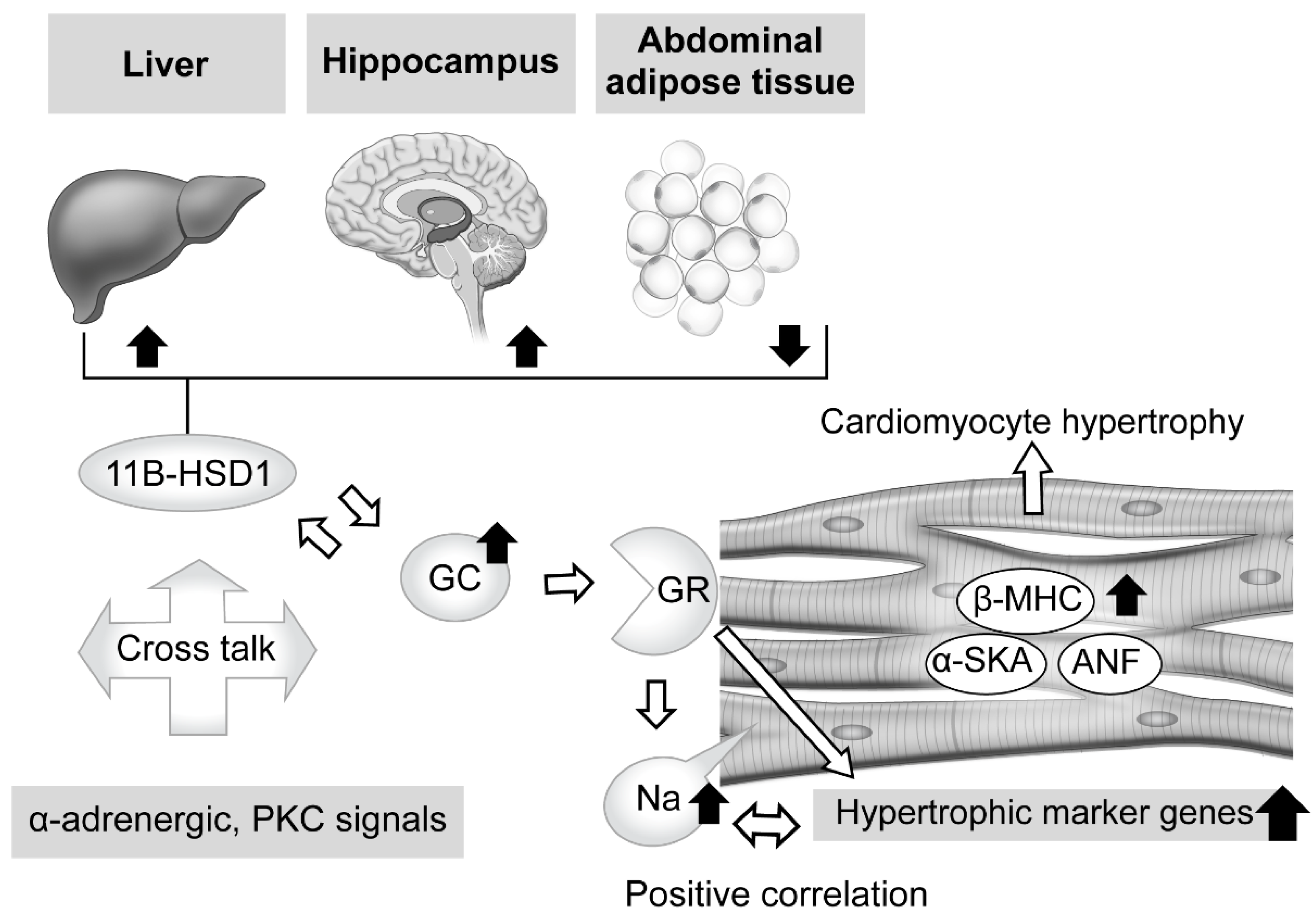
2.3.2. Tissue-Specific Role of 11β-Hydroxysteroid Dehydrogenase Type 1 in Glucocorticoid Excess

When considering local regulation of GC, its association with 11β-HSD1 is another related issue. Nishiyama et al. recently confirmed the influence of 11β-HSD1 in mouse tissues showing persistent GC excess [75]. Two weeks of administration of excessive corticosterone to male mice decreased expression levels of 11β-HSD1 mRNA in the hippocampus and liver, while those were increased in abdominal adipose tissue. Furthermore, similar results were obtained with male corticotropin releasing hormone (CRH)-overexpressing transgenic mice, an animal model of CD [76,77], while performance of an adrenalectomy reversed those changes, suggesting a tissue-dependent action (Figure 1). Huang et al. also noted the role of 11β-HSD1 in cardiomyocytes [78]. Briefly, treatment of primary neonatal rat ventricular cardiomyocytes (NRCMs), which show upregulated 11β-HSD1 expression, with palmitic acid induced a significant enlargement of cell size and increased the mRNA levels of cardiomyocyte hypertrophy-specific genes, including ANF, SKA, and β-MHC, whereas either a selective inhibitor of 11β-HSD1-treated or 11β-HSD1-deficient NRCMs caused a decrease in cell size. Moreover, they also confirmed marked attenuation of 11β-HSD1-induced hypertrophy of cardiomyocytes in not only the presence of a GR antagonist (RU486), but also with the MR antagonist spironolactone, which inhibits nuclear MR translocation [79].

3. Effects of Hypercortisolemia on Heart Structure and Function in Clinical Findings

In clinical cases, the pathophysiological relationship of hypercortisolemia with cardiac hypertrophy is complex and studies that assessed the structural alterations of cardiac muscle including hypertrophy in CS patients have been presented. Although patients with adrenocorticotrophic hormone (ACTH)-dependent and -independent CS participated in those investigations, few studies have focused on overt adrenal CS, while knowledge regarding cardiac hypertrophy in ACS cases is also quite limited (Table 1, Figure 2).

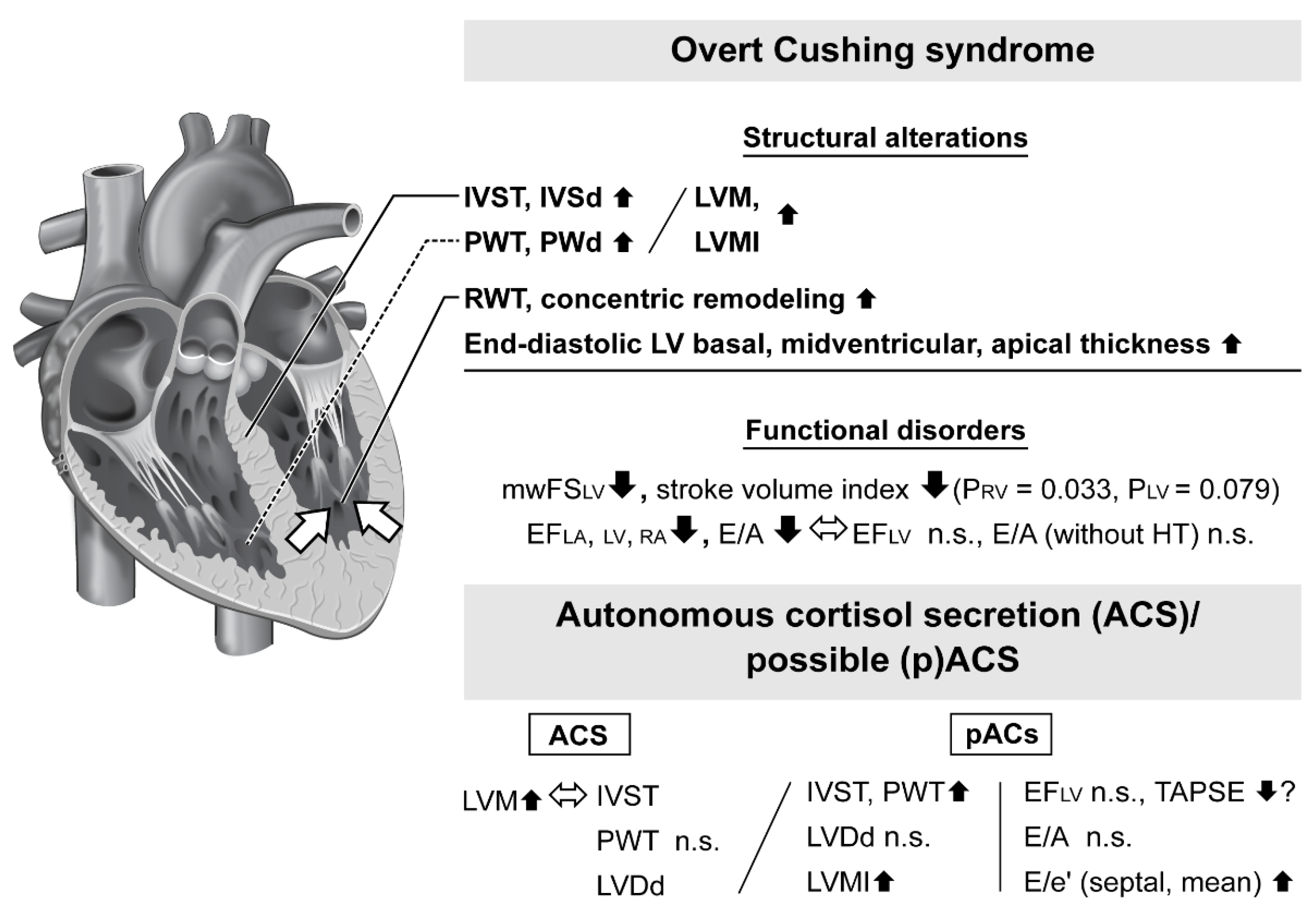
3.1. Heart Structural Alterations in Patients with Overt Cushing Syndrome

A common finding in echocardiographic studies of patients with overt CS is left ventricular hypertrophy (LVH), known as a risk factor for myocardial ischemia and development of heart failure [88]. Although the population was small, Sugihara et al. examined 12 patients (27–57 years old; eight females, four males) with overt CS, including nine with adrenal CS, and provided suggestive results [80]. Nine of those patients showed LVH with an interventricular septum thickness (IVST) diameter of >15 mm at end-diastole (mean ± standard deviation, 17.8 ± 6.2 mm), which was more severe than that in patients with essential hypertension (n = 53, 11.5 ± 2.9 mm, p < 0.01) or primary aldosteronism (n = 8, 11.3 ± 1.2 mm, p < 0.01). Interestingly, posterior wall thickness (PWT) diameter at end-diastole was relatively mild in all groups (11.3 ± 1.8 mm in CS group, 10.4 ± 1.6 mm in essential hypertension group, 10.8 ± 1.5 mm in primary aldosteronism group). Furthermore, a significant higher ratio of IVST to PWT (IVST/PWT) exceeding 1.3 was seen only in the CS group, suggesting existing asymmetric septal hypertrophy (ASH). Such disproportional hypertrophy is uncommon in both essential hypertension [89,90] and secondary hypertensive disease cases, including renovascular hypertension [91,92], primary aldosteronism [92], and pheochromocytoma [93]. A possible mechanism for this structural alteration has been speculated to be related to the increased rate of regional stress [94], while loss of diurnal pattern of cortisol causing nighttime high blood pressure (BP) [95] might also have an effect on this change. However, that was a small-scale study and at least two other studies have reported contrasting evidence. First, Muiesan et al. surveyed 42 patients with CS (36 with CD, 6 with adrenal adenoma, 1 each with adrenal carcinoma and ectopic ACTH production) and noted significantly larger LV interventricular septum diameter (IVSd) (10.1 ± 2.2 vs. 8.8 ± 1.3 mm, p = 0.05) and LV posterior wall diameter (PWd) (9.8 ± 1.7 vs. 7.8 ± 1.1 mm, p = 0.005) measurements as compared with control subjects matched for age, sex, body mass index (BMI), smoking habit, lipid level, BP level, and duration of hypertension [81]. In another study, Toja et al. noted that both IVST (10.2 ± 0.17 vs. 9.4 ± 0.17 mm, p < 0.05) and PWT (9.7 ± 0.16 vs. 8.5 ± 0.12 mm, p < 0.05) were significantly thicker in patients with CS than controls matched for age, sex, and BMI, while none of the CS patients presented ASH [82]. The results of those studies suggest that asymmetric change is not an essential phenotype in CS cases.
Although LVH is a well-known finding in CS patients, recent studies have revealed the frequency to be less than 50% [81,82,83], while relative wall thickness (RWT) and concentric remodeling, an LV geometric pattern category showing normal LV mass (LVM) but with concentric geometry [96], have recently received attention. A ten-year follow-up study that evaluated the relationships of LVM and concentric geometry with cardiovascular events and death in patients with essential hypertension found a 2.3-fold greater risk of cardiovascular events per 100 patient-years, even in cases with a concentric remodeling state, as compared with those with normal geometry [97]. Furthermore, 3% of the patients in that study with concentric remodeling died due to progression of hypertrophy. Therefore, RWT and concentric remodeling may also be of great concern for CS patients. Toja et al. divided CS patients and a control group into hypertension and normotension sub-groups [82]. Their results showed that RWT was indeed greater in both hypertensive (0.44 ± 0.01 vs. 0.36 ± 0.01) and normotensive (0.41 ± 0.01 vs. 0.36 ± 0.02) CS patients as compared with the controls (both p < 0.05). Additionally, among the CS patients, the hypertensive group had a significant greater score as compared with the normotensive group. However, LV mass index (LVMI, generally described as LVM/body surface area) in the CS with normotension group was similar to that seen in the control group (42.9 ± 2.34 vs. 39.3 ± 1.91 g/m2.7), while that was significantly larger in the CS with hypertension group (49.3 ± 2.12 mm) as compared with the normotensive CS (p < 0.05) and control with hypertension (42.1 ± 1.61 mm, p < 0.05) groups. In another study, Kamenický et al. demonstrated that LVMI is associated with systolic BP (sBP) [120 (113; 130) mmHg (median and interquartile range) in CS group vs. 112 (108; 117) mmHg in control group, p = 0.009; r = 0.72, p = 0.045] [84]. Indeed, hypertension can cause pressure overload leading to heart failure through LV remodeling rather than attenuation of myocardial function [98], while arterial hypercoagulability seen in CS [99] potentially exacerbates hypertension. On the other hand, Fallo et al. reported findings showing that RWT was not related to BP level in patients with CS [100]. Additionally, Avenetti et al. found that neither LVM nor geometry had an association with 24 h ambulatory BP (ABP) monitoring, with no difference in all-day BP values between CS and control groups (CS group: 138 ± 14/85 ± 10 mmHg, control group: 134 ± 15/84 ± 11 mmHg; p = 0.36 and 0.66, respectively), though a significant greater prevalence of pressure non-dipping profile in those CS patients was noted (56% vs. 16%, p < 0.05) [85]. Together, these findings indicate that the effects of a hypertensive condition remain to be fully revealed and might depend, at least in part, on duration or severity.

3.2. Cardiac Dysfunction in Patients with Overt Cushing Syndrome

In addition to structural alterations, cardiac dysfunction in CS patients is another factor for consideration. Toja et al. found no significant difference for left ventricular ejection fraction (EFLV), or ratio of early (E) and late (A) wave diastolic filling velocity between CS cases and a control group with normotension (65.2 ± 0.65% vs. 65.9 ± 0.42%, 1.37 ± 0.10 vs. 1.48 ± 0.06, respectively), while a significant attenuation of EFLV was only observed in the CS group with hypertension as compared with the controls with hypertension (63.7 ± 0.51% vs. 67.4 ± 0.74%, p < 0.05) [82]. In addition, midwall fractional shortening (mwFS), an index of absolute and stress-adjusted systolic dysfunction, was significantly attenuated in 20% of the CS patients as compared with the control group regardless of BP level (normotensive: 17.4 ± 0.41% vs. 18.5 ± 0.52%, p < 0.05; hypertensive: 16.4 ± 0.28% vs. 18.8 ± 0.47%, p < 0.05) [82]. Muiesan et al. also reported that mwFS in CS patients was significantly decreased as compared with BP level, duration of hypertension, and other similar factors in a matched control group (16.2 ± 3.0% vs. 20.5 ± 4.5%, p < 0.01), as was stress-adjusted endocardial FS [81]. Additionally, they found that mwFS was independently associated with CS (F = 22.6, p < 0.001) and hypertension (F = 5.78, p = 0.012), while no significant difference was observed in regard to endocardial FS. In a comparison of LV diastolic function between CS patients and controls, a reduced ratio of E/A velocity (0.94 ± 0.27 vs. 1.18 ± 0.32, p = 0.03), along with decreased E velocity (55.6 ± 12 cm/s vs. 78 ± 21 cm/s, p = 0.005) and prolonged E-wave deceleration time (205 ± 36 ms vs. 150 ± 30 ms, p < 0.001) were observed. Notably, analysis regarding the association of those diastolic parameters to CS and hypertension revealed that E-wave deceleration time was independently associated with CS (F = 12.9, p = 0.001) but not with hypertension (F = 1.93, p = 0.18), while E/A ratio was independently associated with both CS (F = 12.9, p = 0.001) and hypertension (F = 17.3, p < 0.001). Finally, the authors concluded that the cardiac structural alterations described above are associated with attenuation of both systolic and diastolic performance, such as mwFS, E/A ratio, and E-wave deceleration time, which may partially explain the high risk of heart attack occurring in patients with CS.

3.3. Cardiac Magnetic Resonance Imaging Test for Evaluation of Patients with Overt Cushing Syndrome

Cardiac magnetic resonance (CMR) imaging testing has been shown to provide results that are useful for highly accurate and comprehensive evaluations of cardiac geometry and function, as well as myocardial fibrosis [101,102,103], and those cardiac parameters have been assessed in patients with CS [84]. Comparisons of LV and right ventricular (RV) parameters in healthy controls and CS patients matched for age, sex, and BMI showed similar results for both parameters, as end-systolic volume was higher (LV; 31.8 (24.2; 37.4) mL/m2 vs. 22.4 (16.7; 29.4) mL/m2, p = 0.002; RV; 36.6 (32.6; 42.5) mL/m2 vs. 27.1 (21.6; 35.3) mL/m2, p = 0.012), though end-diastolic volume was not significantly different. Therefore, stroke volume index was significantly smaller in the RV (34.1 (26.9; 40.3) mL/m2 vs. 41.9 (37.1; 48.3) mL/m2, p = 0.033) and tended to be smaller in the LV (34.9 (31.4; 46.4) mL/m2 vs. 44.8 (37.5; 49.7) mL/m2, p = 0.079), and lower EF values were also observed (EFLV: 55.0 (51.0; 58.4)% vs. 64.3 (60.9; 69.6)%, p < 0.01; EFRV: 49.3 (42.5; 53.4)% vs. 60.0 (54.6; 64.3)%, p < 0.01). Additionally, left atrial (LA) parameters showed a significant increase in minimal volume (20.8 (15.5; 247)) mL/m2 vs. 15.6 (13.1; 20.7) mL/m2, p = 0.042) but not in maximal volume; consequently, EFLA was markedly lower in the CS patients as compared with the controls (39.2 (34.8; 48.4)% vs. 56.1 (48.6; 61.2)%, p < 0.01). Finally, end-diastolic LV segmental thickness shown in basal, midventricular, and apical slices was markedly increased (11.2 (8.96; 12.2) mm vs. 7.99 (6.78; 8.61) mm, p < 0.01; 10.3 (8.43; 11.3) mm vs. 7.26 (6.33; 8.21) mm, p < 0.01; 8.45 (7.86; 9.52) mm vs. 5.90 (5.16; 6.68) mm, p < 0.01, respectively). Furthermore, late gadolinium enhancement testing, which depicts dense myocardial fibrosis [101], revealed no delayed enhancement, suggesting the absence of dense replacement myocardial fibrosis in the CS cases. Nevertheless, the authors noted a limitation related to the method, in that the presence of diffuse interstitial fibrosis could not be precluded. Altogether, this CMR study reinforced evidence regarding LV structure and function as well as echocardiographic results, and further provided informative findings related to RA and LA systolic functions.

3.4. Contributions of Human Serum and Urinary Cortisol Levels to Cardiac Impairment

It is rather surprising that the associations of serum and urinary cortisol levels with cardiac hypertrophy in humans have yet to be clarified. A multivariate analysis of patients with adrenal CS (n = 50) conducted by Takagi et al. demonstrated that serum cortisol (24.3 ± 16.6 µg/dL) and potassium (3.8 ± 0.6 mEq/L) levels independently contributed to LVMI (r2 = 0.647; p < 0.05 and p < 0.01, respectively), while hemoglobin A 1c (HbA1c) (6.2 ± 1.6%) and serum potassium levels independently contributed to EFLV (r2 = 0.529; p < 0.05 and p < 0.05, respectively) [97]. They also explained that the diverse mechanisms of cortisol had indirect effects on such factors as hypertension [104], noradrenalin and angiotensin II [105], and the local RAA system [106], as well as direct effects, such as stimulation of cell cycle activity in cardiomyocytes leading to proliferation [107] and accelerating of cardiomyocyte hypertrophy by expression of angiotensinogen mRNA [108]. Moreover, Toja et al. presented multifactorial analysis of variance results, along with a theory that cortisol excess and hypertension interact to worsen cardiac parameters (e.g., F = 7.7, p < 0.05 for RWT) [82]. The authors considered this to be due to the 3.01-fold higher risk of increase in LVMI seen in CS patients with hypertension as compared with hypertensive controls, and because patients with cured CS with residual hypertension still showed significant cardiac impairments in both structural and functional parameters (LVMI, IVSd, PWd, RWT, EFLV, mwFS, E/A) as compared with the hypertensive controls. However, a separate series of studies revealed no correlation of cortisol level with impairment in patients with CS, based on the following findings: (1) the levels of urinary 17-hydroxycorticosteroids, serum cortisol, urinary adrenalin and noradrenalin, plasma aldosterone, and plasma renin activity were not correlated with IVST or PWT [80]; (2) there was no correlation of urinary cortisol with RWT, LVMI, or diastolic disfunction [81]; (3) 24 h urinary cortisol (1016.72 ± 549 nmol/24 h) was not associated with LVM (r = 0.1, p = 0.5), RWT (r = 0.02, p = 0.89), or all-day BP values [100]; and (4) CMR test results of LV and RV function also had no correlation with biological variables, including 24 h urinary cortisol and plasma cortisol levels [84]. Presently, the underlying mechanisms related to cortisol level alterations remain controversial and complex, and further study is needed, including an analysis matched for duration of CS as well as hypertension.

3.5. Geometric and Functional Impairment in Autonomous Cortisol Secretion Patients

There is only limited knowledge regarding alterations of heart structure and function associated with ACS, though its association with a high risk of heart attack-inducing cardiovascular events has been noted. Iacobellis et al. examined 46 consecutive patients (22 males, 24 females) with incidentaloma, including 40 with non-functional incidentaloma (mean age 61.6 ± 10.7 years) and six with mild adrenal CS (mean age 69.3 ± 12.5 years), defined when two or more of the following abnormalities were present: urinary-free cortisol level > 70 µg/day, >5 µg/dL of serum cortisol after 1 mg DST, and ACTH level < 10 pg/mL in the morning (08:00) [86]. The study protocol excluded patients with known malignancy or hormone-secreting tumors, such as pheochromocytoma and/or aldosterone-secreting adenoma, as well as severe or paroxysmal hypertension, hypokalemia, or clinical signs of hypercortisolism or hyperandrogenism. Echocardiographic examinations were performed to determine LVM and epicardial fat thickness, the latter of which reflects visceral fat deposition in the heart [109,110] and thought to be an indicator of cardiovascular risk [111,112,113], with the results compared with those of 30 healthy controls (mean age 59.6 ± 10 years, 20 males, 10 females). A significantly greater epicardial fat thickness and increase in LVM including height2.7-adjusted LVM, which is more sensitive for detecting LVH in patients with obesity [114], were observed (7.9 ± 0.8 mm vs. 7.4 ± 0.6 mm, p = 0.01; 49.7 ± 10 g/m2.7 vs. 47 ± 8 g/m2.7, p = 0.05; respectively). The results suggested that epicardial fat shown in echocardiogram findings was a non-invasive marker of earlier cardiac abnormalities in patients with adrenal incidentaloma. Interestingly, LVMh2.7 and LVM were obviously greater in mild CS patients than in patients with non-functional adenomas (62.6 ± 10 g/m2.7 vs. 47.7 ± 9 g/m2.7, p = 0.01; 223.7 ± 35 g vs. 200.5 ± 45 g, p = 0.01; respectively), whereas epicardial fat thickness was not significantly different (8.0 ± 0.7 mm vs. 7.9 ± 0.9 mm). On the other hand, Evran et al. found no echocardiographic differences in five patients (mean age 49.60 ± 8.64 years) with adrenal ‘subclinical CS (SCS)’, defined based on non-suppressive cortisol level (>50 nmol/L) in post-DST results, and ACTH < 10 pg/mL as compared to 76 patients (mean age 52.09 ± 9.78 years) with nonfunctional adrenal incidentaloma (NFAI) [115]. None of the 81 patients had a history of type 2 diabetes, hypertension, artery disease, dyslipidemia, drug abuse, steroid use, or functional adrenal mass (CS, primary hyperaldosteronism, pheochromocytoma). As compared with an age-, sex-, and BMI-matched healthy control group (n = 33), both the SCS and NFAI groups showed larger waist circumference (p = 0.00) and impaired glucose metabolism profiles (fasting plasma glucose and insulin levels, HOMA-IR; p = 0.01, 0.00, and 0.00, respectively), and higher triglyceride level (p = 0.01). The differences in hormonal parameters between the patients with SCS and NFAI reflected pathology, with no differences for basal serum level of cortisol, ACTH, or dehydroepiandrosterone sulfate (DHEAS), but a higher post-DST cortisol level in the SCS group (4.84 ± 0.47 vs. 1.32 ± 0.09, p = 0.00). When assessing echocardiographic measurements between the SCS and NFAI groups, there were no differences found for IVST (11.20 ± 2.49 mm vs. 10.38 ± 1.47 mm), PWT (10.80 ± 1.78 mm vs. 10.31 ± 1.45 mm), or end-diastolic diameter (49.00 ± 2.00 mm vs. 47.5 ± 3.49 mm).
Of note, a recent study by Sbaedella et al. disclosed a mild correlation between serum cortisol level especially in post-DST and LVH in echocardiography in patients with pACS [87]. Seventy-one consecutive patients with adrenal incidentaloma were enrolled in that study who were not affected by an overly active or malignant adrenal tumor, including overt CS, pheochromocytoma, Conn syndrome, adrenal carcinoma, late-onset congenital adrenal hyperplasia, myelolipoma, adrenal metastasis, and adrenal hemorrhage cases. Furthermore, none had a history of myocardial infarction or unstable angina, psychiatric disease or alcohol abuse, or were taking medications affecting glucocorticoid production. To diagnose ACS, 1 mg-DST based on the 2016 European Society of Endocrinology guidelines [15] and a two-day 2 mg Liddle test (2 mg-DST), which has greater specificity than 1 mg-DST (97–100% vs. 87.5%) [116], were performed. All 71 participants underwent 1 mg-DST, then those showing a cortisol level > 50 nmol/L (1.8 µg/dL) (n = 37) underwent 2 mg-DST. Only 3 patients showed suppressed cortisol (≤50 nmol/L) and 34 were within a range of 51–138 nmol/L, considered to be pACS; thus, the remaining 37 were categorized as having a non-functioning adenoma (NFA). Their clinical and biochemical characteristics at the baseline were similar, such as mean age 68 (57–73) years in the pACS group vs. 67 (60–72) years in the NFA group, male sex percentage 38.2% vs. 29.7%, and BMI 26.7 (22.6–29.3) vs. 25.9 (23.7–31.2), while other metabolic or electrocyte profiles and cardiovascular risk factors were also similar, except for adenoma diameter (27.4 (23.4–31.5) mm vs 20.2 (17.2–23.1) mm, p = 0.004). As for hormone parameters including baseline serum cortisol, ACTH, aldosterone/renin ratio, 17OH progesterone, DHEAS, urinary free cortisol, androstenedione, urine metanephrine, and lower levels of ACTH (18 (15–20) pg/mL vs. 25 (19–31) pg/mL, p = 0.028) and DHEAS (37.5 (18–54) µg/dL vs. 77.8 (45–189) µg/dL, p = 0.012) were observed in the pACS group. A covariance model with fixed age, BMI, sBP, LDL cholesterol, and HbA1c, revealed geometric and functional differences as follows. (1) IVST and PWT were significantly thicker in the pACS as compared with the NFA group (10.7 (95% CI: 10.2–11.2) mm vs. 9.9 (95% CI: 9.4–10.4) mm, p = 0.015; 9.1 (95% CI: 8.5–9.6) mm vs. 8.4 (95% CI: 7.9–9.0) mm, respectively; p = 0.047), while LV end-diastolic diameter (LVDd) and RWT were not. (2) There was a greater increase in LVMI for both LV massh2.7 and body surface area (BSA)-adjusted LVM (LVMBSA) confirmed in the pACS group (46.1 (95% CI: 42.5–49.6) g/m2 vs. 36.2 (95% CI: 32.8–39.6) g/m2, p = 0.01; 95.3 (95% CI: 88.0–102.5) g/m2 vs. 76.6 (95% CI: 69.6–83.6) g/m2, p = 0.001, respectively). (3) LVEF was similar between the two groups, while tricuspid annular plane systolic excursion (TAPSE), which partially represents RV function, was slightly reduced in the pACS patients, though the mean value was not highly suggestive of so-called RV dysfunction (<17 mm) (20.2 (95% CI: 18.9–21.6) mm). (4) Significantly higher values for septal and mean (lateral and septal) E/e’ were found in the pACS group (10.3 (95% CI: 9.0–11.6) vs. 8.3 (95% CI: 7.2–9.5), p = 0.028; 8.85 (95% CI: 7.8–9.8) vs. 7.4 (95% CI: 6.6–8.3), respectively; p = 0.044), though the difference for E/A was not significant. Furthermore, a stepwise method showed the main contributor to LVMI to be post-DST cortisol level, which accounted for 13.7%, followed by sBP (9.9%) and HbA1c (8.1%). When considering these findings, the higher prevalence of diastolic function abnormality, especially in septal E/e’, found in the pACS patients is notable. Elevation of E/e’ value generally follows a re-increase in E wave velocity after reduction in both E and e’ velocities. In this study, lateral, septal, and mean e’ were not significantly different between the pACS and NFA cases, though there was a tendency of relaxation impairment shown by septal and mean e’ values observed in the pACS group (7.4 (95% CI: 6.4–8.2) cm/s vs. 8.5 (95% CI: 7.7–9.3) cm/s, p = 0.059; 8.3 (95% CI: 7.4–9.2) cm/s vs. 9.4 (95% CI: 8.6–10.2) cm/s, respectively; p = 0.067), suggesting that the pACS patients might have been above a certain level of diastolic dysfunction.
On the other hand, A-wave velocity has received focus as a predictor of arterial stiffness [117] and also reported to be a useful index for evaluating LV diastolic disfunction [118], though related results for patients with adrenal CS are limited. We are currently conducting the Hyogo Sleep Cardio-Autonomic Atherosclerosis (HSCAA) study, a single-center cohort investigation performed at the hospital of Hyogo College of Medicine in Japan, to examine cardiovascular risk factors in patients with metabolic and endocrine diseases, including adrenal CS [119]. According to preliminary results of echocardiographic examinations, increased A-wave velocity has been observed in age- and metabolic profile-matched patients with adrenal ACS (mean age 58.8 ± 14.7 years, n = 25) as compared with those with a non-functioning adrenal adenoma (56.4 ± 13.9 years, n = 101) (75.3 ± 19.3 cm/s vs. 67.3 ± 15.5 cm/s, p = 0.04), while no significant differences for either e’ velocity or E/e’ between the groups have been confirmed (6.8 ± 2.4 cm/s vs. 7.1 ± 1.6 cm/s, p = 0.25; 10.2 ± 3.4 vs. 9.1 ± 2.8, p = 0.10, respectively). These findings might be useful in regard to early detection of ACS, though accumulation of additional cases is needed.

4. Treatment of Adrenal Cushing Syndrome and Effects on Cardiac Impairment

An adrenalectomy is recommended as standard care for overt adrenal CS, especially in unilateral CS cases, only a few studies with small populations have demonstrated low to very low levels of evidence regarding surgery for ACS [15]. Surgical treatment for ACS may contribute to reduce BP level and improve some metabolic profiles, such as glucose and lipid metabolism, as compared with conservative management [120,121,122,123], though differences related to long-term outcome have not been elucidated.
Nevertheless, structure alterations of the human heart in CS have been reported to be partially reversible after remission. Sugihara et al. followed nine patients after surgery for periods from three months to six years three months [80]. Consequently, seven showed decreases in IVST and PWT, and hypertension persisted in only two. Kamenický et al. presented results of an MRI study of 17 patients, 13 of whom reached surgical remission including all with adrenal adenomas and four who received steroidogenesis inhibitors, including their re-assessments at six months after remission [84]. The results showed improvements in both LV geometry, such as basal, midventricular, and apical wall thickness, and function including EFLV and stroke volume index. In addition, RV and LA functions were partially improved. On the other hand, Toja et al. performed follow-up examinations of 22 patients for a longer term after surgery (median 28.8 months). The results showed that functional parameters, including EF, mwFS, E/A ratio, E-wave deceleration time, and isovolumetric relaxation time, did not have significant recovery, though amelioration but not full recovery of IVST, PWT, LVMI, and RWT was demonstrated in association with improved BP level, as well as glucose and triglyceride profiles [82]. Together, these results suggest that functional improvement remains controversial, though it may follow structural amelioration.
Since the severity of LVH is correlated with cardiovascular risk, normalization of excess cortisol may contribute to reduce mortality. Nevertheless, additional studies focused on long-term outcomes of cured adrenal CS patients, including differences between surgery and conservative treatment, such as steroidogenesis inhibitor administration, are needed, while results of investigations of adrenal ACS cases are also crucial.

5. Conclusions and Perspective

In summary, excess cortisol has been experimentally shown to induce cardiomyocyte hypertrophy via activated GC signaling through GR. On the other hand, the pathophysiology in humans is more complex, indicating associations with several different factors including hypertension. Based on this literature review, it is concluded that a hypertensive state may exacerbate LVH in CS patients, though is possibly dependent, at least in part, on duration or severity. Furthermore, both systolic and diastolic function impairments were found to be independently associated with CS and hypertension. When considering treatment, partial improvement of structural alterations is expected after normalization of excess cortisol, whereas the possibility of improvements in functional disorders remains controversial. Although to what extent these findings are related to mortality of patients with overt CS or ACS remains vague, evidence presented in this review strongly demonstrates that hypercortisolemia itself has a relationship with geometric and/or functional alterations, as shown in clinical results as well as experimental findings. Nevertheless, the findings of this investigation require further elucidation regarding whether such alterations in pre-symptomatic state contribute to early diagnosis of hypercortisolemia, while evaluations of the effects of treatment on long-term mortality are also necessary.

Author Contributions

Writing: original draft preparation, A.K.; review and editing, M.K., S.K. and H.K. Figure and table preparation: A.K. All authors have read and agreed to the published version of the manuscript.

Funding

This research received no external funding.

Institutional Review Board Statement

Not applicable.

Informed Consent Statement

Not applicable.

Data Availability Statement

Not applicable.

Conflicts of Interest

The authors declare no conflict of interest.

References

  1. Plotz, C.M.; Knowlton, A.I.; Ragan, C. The natural history of Cushing’s syndrome. Am. J. Med. 1952, 13, 597–614. [Google Scholar] [CrossRef] [PubMed]
  2. Pikkarainen, L.; Sane, T.; Reunanen, A. The survival and well-being of patients treated for Cushing’s syndrome. J. Intern. Med. 1999, 245, 463–468. [Google Scholar] [CrossRef] [PubMed] [Green Version]
  3. Lindholm, J.; Juul, S.; Jørgensen, J.O.; Astrup, J.; Bjerre, P.; Feldt-Rasmussen, U.; Hagen, C.; Jørgensen, J.; Kosteljanetz, M.; Kristensen, L.; et al. Incidence and late prognosis of cushing’s syndrome: A population-based study. J. Clin. Endocrinol. Metab. 2001, 86, 117–123. [Google Scholar] [CrossRef]
  4. Yaneva, M.; Kalinov, K.; Zacharieva, S. Mortality in Cushing’s syndrome: Data from 386 patients from a single tertiary referral center. Eur. J. Endocrinol. 2013, 169, 621–627. [Google Scholar] [CrossRef] [PubMed] [Green Version]
  5. Ntali, G.; Asimakopoulou, A.; Siamatras, T.; Komninos, J.; Vassiliadi, D.; Tzanela, M.; Tsagarakis, S.; Grossman, A.B.; Wass, J.A.; Karavitaki, N. Mortality in Cushing’s syndrome: Systematic analysis of a large series with prolonged follow-up. Eur. J. Endocrinol. 2013, 169, 715–723. [Google Scholar] [CrossRef] [Green Version]
  6. Favero, V.; Cremaschi, A.; Parazzoli, C.; Falchetti, A.; Gaudio, A.; Gennari, L.; Scillitani, A.; Vescini, F.; Morelli, V.; Aresta, C.; et al. Pathophysiology of Mild Hypercortisolism: From the Bench to the Bedside. Int. J. Mol. Sci. 2022, 23, 673. [Google Scholar] [CrossRef]
  7. Newell-Price, J.; Bertagna, X.; Grossman, A.B.; Nieman, L.K. Cushing’s syndrome. Lancet 2006, 367, 1605–1617. [Google Scholar] [CrossRef]
  8. Javanmard, P.; Duan, D.; Geer, E.B. Mortality in Patients with Endogenous Cushing’s Syndrome. Endocrinol. Metab. Clin. North Am. 2018, 47, 313–333. [Google Scholar] [CrossRef]
  9. Barbot, M.; Zilio, M.; Scaroni, C. Cushing’s syndrome: Overview of clinical presentation, diagnostic tools and complications. Best Pract. Res. Clin. Endocrinol. Metab. 2020, 34, 101380. [Google Scholar] [CrossRef]
  10. Ntali, G.; Hakami, O.; Wattegama, M.; Ahmed, S.; Karavitaki, N. Mortality of Patients with Cushing’s Disease. Exp. Clin. Endocrinol. Diabetes. 2021, 129, 203–207. [Google Scholar] [CrossRef]
  11. Dekkers, O.M.; Horváth-Puhó, E.; Jørgensen, J.O.; Cannegieter, S.C.; Ehrenstein, V.; Vandenbroucke, J.P.; Pereira, A.M.; Sørensen, H.T. Multisystem morbidity and mortality in Cushing’s syndrome: A cohort study. J. Clin. Endocrinol. Metab. 2013, 98, 2277–2284. [Google Scholar] [CrossRef] [PubMed]
  12. Ntali, G.; Grossman, A.; Karavitaki, N. Clinical and biochemical manifestations of Cushing’s. Pituitary 2015, 18, 181–187. [Google Scholar] [CrossRef] [PubMed]
  13. Ahn, C.H.; Kim, J.H.; Park, M.Y.; Kim, S.W. Epidemiology and Comorbidity of Adrenal Cushing Syndrome: A Nationwide Cohort Study. J. Clin. Endocrinol. Metab. 2021, 106, e1362–e1372. [Google Scholar] [CrossRef] [PubMed]
  14. Reincke, M.; Nieke, J.; Krestin, G.P.; Saeger, W.; Allolio, B.; Winkelmann, W. Preclinical Cushing’s syndrome in adrenal “incidentalomas”: Comparison with adrenal Cushing’s syndrome. J. Clin. Endocrinol. Metab. 1992, 75, 826–832. [Google Scholar] [CrossRef] [PubMed]
  15. Fassnacht, M.; Arlt, W.; Bancos, I.; Dralle, H.; Newell-Price, J.; Sahdev, A.; Tabarin, A.; Terzolo, M.; Tsagarakis, S.; Dekkers, O.M. Management of adrenal incidentalomas: European Society of Endocrinology Clinical Practice Guideline in collaboration with the European Network for the Study of Adrenal Tumors. Eur. J. Endocrinol. 2016, 175, G1–G34. [Google Scholar] [CrossRef] [Green Version]
  16. Masserini, B.; Morelli, V.; Palmieri, S.; Eller-Vainicher, C.; Zhukouskaya, V.; Cairoli, E.; Orsi, E.; Beck-Peccoz, P.; Spada, A.; Chiodini, I. Lipid abnormalities in patients with adrenal incidentalomas: Role of subclinical hypercortisolism and impaired glucose metabolism. J. Endocrinol. Invest. 2015, 38, 623–628. [Google Scholar] [CrossRef]
  17. Aresta, C.; Favero, V.; Morelli, V.; Giovanelli, L.; Parazzoli, C.; Falchetti, A.; Pugliese, F.; Gennari, L.; Vescini, F.; Salcuni, A.; et al. Cardiovascular complications of mild autonomous cortisol secretion. Best Pract. Res. Clin. Endocrinol. Metab. 2021, 35, 101494. [Google Scholar] [CrossRef]
  18. Dalmazi, G.D.; Vicennati, V.; Garelli, S.; Casadio, E.; Rinaldi, E.; Giampalma, E.; Mosconi, C.; Golfieri, R.; Paccapelo, A.; Pagotto, U.; et al. Cardiovascular events and mortality in patients with adrenal incidentalomas that are either non-secreting or associated with intermediate phenotype or subclinical Cushing’s syndrome: A 15-year retrospective study. Lancet Diabetes Endocrinol. 2014, 2, 396–405. [Google Scholar] [CrossRef]
  19. Patrova, J.; Kjellman, M.; Wahrenberg, H.; Falhammar, H. Increased mortality in patients with adrenal incidentalomas and autonomous cortisol secretion: A 13-year retrospective study from one center. Endocrine 2017, 58, 267–275. [Google Scholar] [CrossRef]
  20. Debono, M.; Bradburn, M.; Bull, M.; Harrison, B.; Ross, R.J.; Newell-Price, J. Cortisol as a marker for increased mortality in patients with incidental adrenocortical adenomas. J. Clin. Endocrinol. Metab. 2014, 99, 4462–4470. [Google Scholar] [CrossRef]
  21. Barzon, L.; Sonino, N.; Fallo, F.; Palu, G.; Boscaro, M. Prevalence and natural history of adrenal incidentalomas. Eur. J. Endocrinol. 2003, 149, 273–285. [Google Scholar] [CrossRef] [PubMed] [Green Version]
  22. Sato, Y.; Maekawa, S.; Ishii, R.; Sanada, M.; Morikawa, T.; Shiraishi, Y.; Yoshida, K.; Nagata, Y.; Sato-Otsubo, A.; Yoshizato, T.; et al. Recurrent somatic mutations underlie corticotropin-independent Cushing’s syndrome. Science 2014, 344, 917–920. [Google Scholar] [CrossRef] [PubMed]
  23. Calebiro, D.; Dalmazi, G.D.; Bathon, K.; Ronchi, C.L.; Beuschlein, F. cAMP signaling in cortisol-producing adrenal adenoma. Eur. J. Endocrinol. 2015, 173, M99–M106. [Google Scholar] [CrossRef] [PubMed] [Green Version]
  24. Kumar, R.; Thompson, E.B. Gene regulation by the glucocorticoid receptor: Structure: Function relationship. J. Steroid Biochem. Mol. Biol. 2005, 94, 383–394. [Google Scholar] [CrossRef] [PubMed]
  25. Evans, R.M. The steroid and thyroid hormone receptor superfamily. Science 1988, 240, 889–895. [Google Scholar] [CrossRef] [PubMed]
  26. Grad, I.; Picard, D. The glucocorticoid responses are shaped by molecular chaperones. Mol. Cell Endocrinol. 2007, 275, 2–12. [Google Scholar] [CrossRef]
  27. Funder, J.W. Aldosterone and mineralocorticoid receptors: A personal reflection. Mol. Cell Endocrinol. 2012, 350, 146–150. [Google Scholar] [CrossRef]
  28. Funder, J.; Myles, K. Exclusion of corticosterone from epithelial mineralocorticoid receptors is insufficient for selectivity of aldosterone action: In Vivo binding studies. Endocrinology 1996, 137, 5264–5268. [Google Scholar] [CrossRef]
  29. Funder, J.W. Mineralocorticoid receptor antagonists: Emerging roles in cardiovascular medicine. Integr. Blood Press. Control 2013, 6, 129–138. [Google Scholar] [CrossRef] [Green Version]
  30. Funder, J.W. Mineralocorticoid receptor activation and oxidative stress. Hypertension 2007, 50, 840–841. [Google Scholar] [CrossRef]
  31. Mihailidou, A.S.; Le, T.Y.L.; Mardini, M.; Funder, J.W. Glucocorticoids activate cardiac mineralocorticoid receptors during experimental myocardial infarction. Hypertension 2009, 54, 1306–1312. [Google Scholar] [CrossRef] [PubMed] [Green Version]
  32. Seckl, J.R. 11beta-hydroxysteroid dehydrogenases: Changing glucocorticoid action. Curr. Opin. Pharmacol. 2004, 4, 597–602. [Google Scholar] [CrossRef] [PubMed]
  33. Hadoke, P.W.; Iqbal, J.; Walker, B.R. Therapeutic manipulation of glucocorticoid metabolism in cardiovascular disease. Br. J. Pharmacol. 2009, 156, 689–712. [Google Scholar] [CrossRef] [PubMed] [Green Version]
  34. Atanasov, A.G.; Nashev, L.G.; Gelman, L.; Legeza, B.; Sack, R.; Portmann, R.; Odermatt, A. Direct protein-protein interaction of 11beta-hydroxysteroid dehydrogenase type 1 and hexose-6-phosphate dehydrogenase in the endoplasmic reticulum lumen. Biochim. Biophys. Acta 2008, 1783, 1536–1543. [Google Scholar] [CrossRef] [Green Version]
  35. Morgan, S.A.; McCabe, E.L.; Gathercole, L.L.; Hassan-Smith, Z.K.; Larner, D.P.; Bujalska, I.J.; Stewart, P.M.; Tomlinson, J.W.; Lavery, G.G. 11β-HSD1 is the major regulator of the tissue-specific effects of circulating glucocorticoid excess. Proc. Natl. Acad. Sci. USA 2014, 111, E2482–E2491. [Google Scholar] [CrossRef] [Green Version]
  36. Tomlinson, J.W.; Walker, E.A.; Bujalska, I.J.; Draper, N.; Lavery, G.G.; Cooper, M.S.; Hewison, M.; Stewart, P.M. 11beta-hydroxysteroid dehydrogenase type 1, a tissue-specific regulator of glucocorticoid response. Endocr. Rev. 2004, 25, 831–866. [Google Scholar] [CrossRef]
  37. Atanasov, A.G.; Nashev, L.G.; Schweizer, R.A.; Frick, C.; Odermatt, A. Hexose-6-phosphate dehydrogenase determines the reaction direction of 11beta-hydroxysteroid dehydrogenase type 1 as an oxoreductase. FEBS Lett. 2004, 571, 129–133. [Google Scholar] [CrossRef] [Green Version]
  38. Walker, B.R. Glucocorticoids and cardiovascular disease. Eur. J. Endocrinol. 2007, 157, 545–559. [Google Scholar] [CrossRef] [Green Version]
  39. Yang, S.; Zhang, L. Glucocorticoids and vascular reactivity. Curr. Vasc. Pharmacol. 2004, 2, 1–12. [Google Scholar] [CrossRef]
  40. Albiston, A.L.; Obeyesekere, V.R.; Smith, R.E.; Krozowski, Z.S. Cloning and tissue distribution of the human 11 beta-hydroxysteroid dehydrogenase type 2 enzyme. Mol. Cell Endocrinol. 1994, 105, R11–R17. [Google Scholar] [CrossRef]
  41. Brown, R.W.; Chapman, K.E.; Murad, P.; Edwards, C.R.; Seckl, J.R. Purification of 11beta-hydroxysteroid dehydrogenase type 2 from human placenta utilizing a novel affinity labelling technique. Biochem. J. 1996, 313, 997–1005. [Google Scholar] [CrossRef] [PubMed]
  42. Walker, B.R. Extra-adrenal regeneration of glucocorticoids by 11 beta-hydroxysteroid dehydrogenase type 1, physiological regulator and pharmacological target for energy partitioning. Proc. Nutr. Soc. 2007, 66, 1–8. [Google Scholar] [CrossRef] [Green Version]
  43. Cain, D.W.; Cidlowski, J.A. Specificity and sensitivity of glucocorticoid signaling in health and disease. Best Pract. Res. Clin. Endocrinol. Metab. 2015, 29, 545–556. [Google Scholar] [CrossRef] [Green Version]
  44. Rog-Zielinska, E.A.; Thomson, A.; Kenyon, C.J.; Brownstein, D.G.; Moran, C.M.; Szumska, D.; Michailidou, Z.; Richardson, J.; Owen, E.; Watt, A.; et al. Glucocorticoid receptor is required for foetal heart maturation. Hum. Mol. Genet. 2013, 22, 3269–3282. [Google Scholar] [CrossRef] [PubMed] [Green Version]
  45. Narayanan, N.; Yang, C.; Xu, A. Dexamethasone treatment improves sarcoplasmic reticulum function and contractile performance in aged myocardium. Mol. Cell. Biochem. 2004, 266, 31–36. [Google Scholar] [CrossRef] [PubMed]
  46. Ren, R.; Cidlowski, J.A. Glucocorticoid signaling in cardiac disease. Horm. Mol. Biol. Clin. Investig. 2010, 4, 559–564. [Google Scholar] [CrossRef]
  47. Pearl, J.M.; Nelson, D.P.; Schwartz, S.M.; Wagner, C.J.; Bauer, S.M.; Setser, E.A.; Duffy, J.Y. Glucocorticoids reduce ischemia-reperfusion-induced myocardial apoptosis in immature hearts. Ann. Thorac. Surg. 2002, 74, 830–836; discussion 836–837. [Google Scholar] [CrossRef] [PubMed]
  48. Aoyama, T.; Matsui, T.; Novikov, M.; Park, J.; Hemmings, B.; Rosenzweig, A. Serum and glucocorticoid-responsive kinase-1 regulates cardiomyocyte survival and hypertrophic response. Circulation 2005, 111, 1652–1659. [Google Scholar] [CrossRef] [PubMed] [Green Version]
  49. Ren, R.; Oakley, R.H.; Cruz-Topete, D.; Cidlowski, J.A. Dual role for glucocorticoids in cardiomyocyte hypertrophy and apoptosis. Endocrinology 2012, 153, 5346–5360. [Google Scholar] [CrossRef] [Green Version]
  50. Cruz-Topete, D.; He, B.; Xu, X.; Cidlowski, J.A. Krüppel-like Factor 13 Is a Major Mediator of Glucocorticoid Receptor Signaling in Cardiomyocytes and Protects These Cells from DNA Damage and Death. J. Biol. Chem. 2016, 291, 19374–19386. [Google Scholar] [CrossRef]
  51. Brereton, P.S.; van Driel, R.R.; Suhaimi, F.B.; Koyama, K.; Dilley, R.; Krozowski, Z. Light and electron microscopy localization of the 11beta-hydroxysteroid dehydrogenase type I enzyme in the rat. Endocrinology 2001, 142, 1644–1651. [Google Scholar] [CrossRef] [PubMed]
  52. Gomez-Sanchez, E.P.; Romero, D.G.; de Rodriguez, A.F.; Warden, M.P.; Krozowski, Z.; Gomez-Sanchez, C.E. Hexose-6-phosphate dehydrogenase and 11beta-hydroxysteroid dehydrogenase-1 tissue distribution in the rat. Endocrinology 2008, 149, 525–533. [Google Scholar] [CrossRef] [PubMed] [Green Version]
  53. Chai, W.; Hofland, J.; Jansen, P.M.; Garrelds, I.M.; de Vries, R.; van den Bogaerdt, A.J.; Feelders, R.A.; de Jong, F.H.; Danser, A.H. Steroidogenesis vs. steroid uptake in the heart: Do corticosteroids mediate effects via cardiac mineralocorticoid receptors? J. Hypertens. 2010, 28, 1044–1053. [Google Scholar] [CrossRef]
  54. Lefer, A.M. Influence of corticosteroids on mechanical performance of isolated rat papillary muscles. Am. J. Physiol. 1968, 214, 518–524. [Google Scholar] [CrossRef] [PubMed]
  55. Walker, B.R.; Yau, J.L.; Brett, L.P.; Seckl, J.R.; Monder, C.; Williams, B.C.; Edwards, C.R. 11 beta-hydroxysteroid dehydrogenase in vascular smooth muscle and heart: Implications for cardiovascular responses to glucocorticoids. Endocrinology 1991, 129, 3305–3312. [Google Scholar] [CrossRef] [PubMed]
  56. Iqbal, J.; Andrew, R.; Cruden, N.L.; Kenyon, C.J.; Hughes, K.A.; Newby, D.E.; Hadoke, P.W.; Walker, B.R. Displacement of cortisol from human heart by acute administration of a mineralocorticoid receptor antagonist. J. Clin. Endocrinol. Metab. 2014, 99, 915–922. [Google Scholar] [CrossRef] [PubMed] [Green Version]
  57. McSweeney, S.J.; Hadoke, P.W.; Kozak, A.M.; Small, G.R.; Khaled, H.; Walker, B.R.; Gray, G.A. Improved heart function follows enhanced inflammatory cell recruitment and angiogenesis in 11betaHSD1-deficient mice post-MI. Cardiovasc. Res. 2010, 88, 159–167. [Google Scholar] [CrossRef] [Green Version]
  58. White, C.I.; Jansen, M.A.; McGregor, K.; Mylonas, K.J.; Richardson, R.V.; Thomson, A.; Moran, C.M.; Seckl, J.R.; Walker, B.R.; Chapman, K.E.; et al. Cardiomyocyte and Vascular Smooth Muscle-Independent 11β-Hydroxysteroid Dehydrogenase 1 Amplifies Infarct Expansion, Hypertrophy, and the Development of Heart Failure After Myocardial Infarction in Male Mice. Endocrinology 2016, 157, 346–357. [Google Scholar] [CrossRef] [Green Version]
  59. Rahman, T.J.; Mayosi, B.M.; Hall, D.; Avery, P.J.; Stewart, P.M.; Connell, J.M.; Watkins, H.; Keavney, B. Common variation at the 11-β hydroxysteroid dehydrogenase type 1 gene is associated with left ventricular mass. Circ. Cardiovasc. Genet. 2011, 4, 156–162. [Google Scholar] [CrossRef]
  60. McGoldrick, E.; Stewart, F.; Parker, R.; Dalziel, S.R. Antenatal corticosteroids for accelerating fetal lung maturation for women at risk of preterm birth. Cochrane Database Syst. Rev. 2020, 12, CD004454. [Google Scholar] [CrossRef]
  61. Agnew, E.J.; Ivy, J.R.; Stock, S.J.; Chapman, K.E. Glucocorticoids, antenatal corticosteroid therapy and fetal heart maturation. J. Mol. Endocrinol. 2018, 61, R61–R73. [Google Scholar] [CrossRef] [Green Version]
  62. Barker, D.J.; Martyn, C.N.; Osmond, C.; Hales, C.N.; Fall, C.H. Growth in utero and serum cholesterol concentrations in adult life. BMJ 1993, 307, 1524–1527. [Google Scholar] [CrossRef] [PubMed] [Green Version]
  63. Hales, C.N.; Barker, D.J.; Clark, P.M.; Cox, L.J.; Fall, C.; Osmond, C.; Winter, P.D. Fetal and infant growth and impaired glucose tolerance at age 64. BMJ 1991, 303, 1019–1022. [Google Scholar] [CrossRef] [PubMed] [Green Version]
  64. Law, C.M.; Shiell, A.W. Is blood pressure inversely related to birth weight? The strength of evidence from a systematic review of the literature. J. Hypertens. 1996, 14, 935–941. [Google Scholar] [CrossRef] [PubMed]
  65. Rich-Edwards, J.W.; Stampfer, M.J.; Manson, J.E.; Rosner, B.; Hankinson, S.E.; Colditz, G.A.; Willett, W.C.; Hennekens, C.H. Birth weight and risk of cardiovascular disease in a cohort of women followed up since 1976. BMJ 1997, 315, 396–400. [Google Scholar] [CrossRef] [PubMed] [Green Version]
  66. McMillen, I.C.; Robinson, J.S. Developmental origins of the metabolic syndrome: Prediction, plasticity, and programming. Physiol. Rev. 2005, 85, 571–633. [Google Scholar] [CrossRef] [PubMed]
  67. Matthews, S.G. Foetal experience: Lifelong consequences. J. Neuroendocrinol. 2007, 19, 73–74. [Google Scholar] [CrossRef] [PubMed]
  68. Lister, K.; Autelitano, D.J.; Jenkins, A.; Hannan, R.D.; Sheppard, K.E. Cross talk between corticosteroids and alpha-adrenergic signaling augments cardiomyocyte hypertrophy: A possible role for SGK1. Cardiovasc. Res. 2006, 70, 555–565. [Google Scholar] [CrossRef] [PubMed]
  69. Despa, S.; Islam, M.A.; Weber, C.R.; Pogwizd, S.M.; Bers, D.M. Intracellular Na(+) concentration is elevated in heart failure but Na/K pump function is unchanged. Circulation 2002, 105, 2543–2548. [Google Scholar] [CrossRef] [Green Version]
  70. Baartscheer, A.; Schumacher, C.A.; Belterman, C.N.; Coronel, R.; Fiolet, J.W. [Na+] i and the driving force of the Na+/Ca2+-exchanger in heart failure. Cardiovasc. Res. 2003, 57, 986–995. [Google Scholar] [CrossRef]
  71. Pogwizd, S.M.; Sipido, K.R.; Verdonck, F.; Bers, D.M. Intracellular Na in animal models of hypertrophy and heart failure: Contractile function and arrhythmogenesis. Cardiovasc. Res. 2003, 57, 887–896. [Google Scholar] [CrossRef] [Green Version]
  72. Pieske, B.; Houser, S.R. [Na+] i handling in the failing human heart. Cardiovasc. Res. 2003, 57, 874–886. [Google Scholar] [CrossRef]
  73. Murphy, E.; Eisner, D.A. Regulation of intracellular and mitochondrial sodium in health and disease. Circ. Res. 2009, 104, 292–303. [Google Scholar] [CrossRef] [PubMed] [Green Version]
  74. Katoh, D.; Hongo, K.; Ito, K.; Yoshino, T.; Kayama, Y.; Kawai, M.; Date, T.; Yoshimura, M. Corticosteroids increase intracellular free sodium ion concentration via glucocorticoid receptor pathway in cultured neonatal rat cardiomyocytes. Int. J. Cardiol. Heart Vessel. 2014, 3, 49–356. [Google Scholar] [CrossRef] [Green Version]
  75. Nishiyama, M.; Iwasaki, Y.; Nakayama, S.; Okazaki, M.; Taguchi, T.; Tsuda, M.; Makino, S.; Fujimoto, S.; Terada, Y. Tissue-specific regulation of 11β hydroxysteroid dehydrogenase type-1 mRNA expressions in Cushing’s syndrome mouse model. Steroids 2022, 183, 109021. [Google Scholar] [CrossRef] [PubMed]
  76. Stenzel-Poore, M.P.; Cameron, V.A.; Vaughan, J.; Sawchenko, P.E.; Vale, W. Development of Cushing’s syndrome in corticotropin-releasing factor transgenic mice. Endocrinology 1992, 130, 3378–3386. [Google Scholar] [CrossRef] [PubMed]
  77. Nakayama, S.; Nishiyama, M.; Iwasaki, Y.; Shinahara, M.; Okada, Y.; Tsuda, M.; Okazaki, M.; Tsugita, M.; Taguchi, T.; Makino, S.; et al. Corticotropin-releasing hormone (CRH) transgenic mice display hyperphagia with increased Agouti-related protein mRNA in the hypothalamic arcuate nucleus. Endocr. J. 2011, 58, 279–286. [Google Scholar] [CrossRef] [Green Version]
  78. Huang, M.; Liu, J.; Sheng, Y.; Lv, Y.; Yu, J.; Qi, H.; Di, W.; Lv, S.; Zhou, S.; Ding, G. 11β-hydroxysteroid dehydrogenase type 1 inhibitor attenuates high-fat diet induced cardiomyopathy. J. Mol. Cell. Cardiol. 2018, 125, 106–116. [Google Scholar] [CrossRef] [PubMed]
  79. Lavall, D.; Schuster, P.; Jacobs, N.; Kazakov, A.; Böhm, M.; Laufs, U. Rac1 GTPase regulates 11β hydroxysteroid dehydrogenase type 2 and fibrotic remodeling. J. Biol. Chem. 2017, 292, 7542–7553. [Google Scholar] [CrossRef] [Green Version]
  80. Sugihara, N.; Shimizu, M.; Kita, Y.; Shimizu, K.; Ino, H.; Miyamori, I.; Nakabayashi, H.; Takeda, R. Cardiac characteristics and postoperative courses in Cushing’s syndrome. Am. J. Cardiol. 1992, 69, 1475–1480. [Google Scholar] [CrossRef] [PubMed]
  81. Muiesan, M.L.; Lupia, M.; Salvetti, M.; Grigoletto, C.; Sonino, N.; Boscaro, M.; Rosei, E.A.; Mantero, F.; Fallo, F. Left ventricular structural and functional characteristics in Cushing’s syndrome. J. Am. Coll. Cardiol. 2003, 41, 2275–2279. [Google Scholar] [CrossRef] [PubMed] [Green Version]
  82. Toja, P.M.; Branzi, G.; Ciambellotti, F.; Radaelli, P.; Martin, M.D.; Lonati, L.M.; Scacchi, M.; Parati, G.; Cavagnini, F.; Giraldi, F.P. Clinical relevance of cardiac structure and function abnormalities in patients with Cushing’s syndrome before and after cure. Clin. Endocrinol. 2012, 76, 332–338. [Google Scholar] [CrossRef] [PubMed]
  83. Takagi, S.; Tanabe, A.; Tsuiki, M.; Naruse, M.; Takano, K. Hypokalemia, diabetes mellitus, and hypercortisolemia are the major contributing factors to cardiac dysfunction in adrenal Cushing’s syndrome. Endocr. J. 2009, 56, 1009–1018. [Google Scholar] [CrossRef] [PubMed] [Green Version]
  84. Kamenický, P.; Redheuil, A.; Roux, C.; Salenave, S.; Kachenoura, N.; Raissouni, Z.; Macron, L.; Guignat, L.; Jublanc, C.; Azarine, A.; et al. Cardiac structure and function in Cushing’s syndrome: A cardiac magnetic resonance imaging study. J. Clin. Endocrinol. Metab. 2014, 99, E2144–E2153. [Google Scholar] [CrossRef] [Green Version]
  85. Avenatti, E.; Rebellato, A.; Iannaccone, A.; Battocchio, M.; Dassie, F.; Veglio, F.; Milan, A.; Fallo, F. Left ventricular geometry and 24-h blood pressure profile in Cushing’s syndrome. Endocrine 2017, 55, 547–554. [Google Scholar] [CrossRef]
  86. Iacobellis, G.; Petramala, L.; Barbaro, G.; Kargi, A.Y.; Serra, V.; Zinnamosca, L.; Colangelo, L.; Marinelli, C.; Ciardi, A.; De Toma, G.; et al. Epicardial fat thickness and left ventricular mass in subjects with adrenal incidentaloma. Endocrine 2013, 44, 532–536. [Google Scholar] [CrossRef] [PubMed]
  87. Sbardella, E.; Minnetti, M.; D’Aluisio, D.; Rizza, L.; Di Giorgio, M.R.; Vinci, F.; Pofi, R.; Giannetta, E.; Venneri, M.A.; Vestri, A.; et al. Cardiovascular features of possible autonomous cortisol secretion in patients with adrenal incidentalomas. Eur. J. Endocrinol. 2018, 178, 501–511. [Google Scholar] [CrossRef] [PubMed] [Green Version]
  88. Verdecchia, P.; Angeli, F.; Achilli, P.; Castellani, C.; Broccatelli, A.; Gattobigio, R.; Cavallini, C. Echocardiographic left ventricular hypertrophy in hypertension: Marker for future events or mediator of events? Curr. Opin. Cardiol. 2007, 22, 329–334. [Google Scholar] [CrossRef]
  89. Wicker, P.; Roudaut, R.; Haissaguere, M.; Villega-Arino, P.; Clementy, J.; Dallocchio, M. Prevalence and significance of asymmetric septal hypertrophy in hypertension: An echocardiographic and clinical study. Eur. Heart J. 1983, 4, 1–5. [Google Scholar] [CrossRef]
  90. Yokota, Y.; Teng, S.S.; Emoto, R.; Miki, T.; Takarada, A.; Seo, T.; Sano, H.; Fukuzaki, H. Mechanism of development of asymmetric septal hypertrophy in patients with essential systemic hypertension. Jpn. Circ. J. 1989, 53, 1173–1184. [Google Scholar] [CrossRef]
  91. Vensel, L.A.; Devereux, R.B.; Pickering, T.G.; Herrold, E.M.; Borer, J.S.; Laragh, J.H. Cardiac structure and function in renovascular hypertension produced by unilateral and bilateral renal artery stenosis. Am. J. Cardiol. 1986, 58, 575–582. [Google Scholar] [CrossRef] [PubMed]
  92. Suzuki, T.; Abe, H.; Nagata, S.; Saitoh, F.; Iwata, S.; Ashizawa, A.; Kuramochi, M.; Omae, T. Left ventricular structural characteristics in unilateral renovascular hypertension and primary aldosteronism. Am. J. Cardiol. 1988, 62, 1224–1227. [Google Scholar] [CrossRef] [PubMed]
  93. Shub, C.; Cueto-Garcia, L.; Sheps, S.G.; Ilstrup, D.M.; Tajik, A.J. Echocardiographic findings in pheochromocytoma. Am. J. Cardiol. 1986, 57, 971–975. [Google Scholar] [CrossRef] [PubMed]
  94. Heng, M.K.; Janz, R.F.; Jobin, J. Estimation of regional stress in the left ventricular septum and free wall: An echocardiographic study suggesting a mechanism for asymmetric septal hypertrophy. Am. Heart J. 1985, 110, 84–90. [Google Scholar] [CrossRef]
  95. Barbot, M.; Ceccato, F.; Scaroni, C. The Pathophysiology and Treatment of Hypertension in Patients with Cushing’s Syndrome. Front. Endocrinol. 2019, 10, 321. [Google Scholar] [CrossRef]
  96. Ganau, A.; Devereux, R.B.; Roman, M.J.; de Simone, G.; Pickering, T.G.; Saba, P.S.; Vargiu, P.; Simongini, I.; Laragh, J.H. Patterns of left ventricular hypertrophy and geometric remodeling in essential hypertension. J. Am. Coll. Cardiol. 1992, 19, 1550–1558. [Google Scholar] [CrossRef] [Green Version]
  97. Koren, M.J.; Devereux, R.B.; Casale, P.N.; Savage, D.D.; Laragh, J.H. Relation of left ventricular mass and geometry to morbidity and mortality in uncomplicated essential hypertension. Ann. Intern. Med. 1991, 114, 345–352. [Google Scholar] [CrossRef]
  98. Norton, G.R.; Woodiwiss, A.J.; Gaasch, W.H.; Mela, T.; Chung, E.S.; Aurigemma, G.P.; Meyer, T.E. Heart failure in pressure overload hypertrophy: The relative roles of ventricular remodeling and myocardial dysfunction. J. Am. Coll. Cardiol. 2002, 39, 664–671. [Google Scholar] [CrossRef] [Green Version]
  99. Suarez, M.G.; Stack, M.; Hinojosa-Amaya, J.M.; Mitchell, M.D.; Varlamov, E.V.; Yedinak, C.G.; Cetas, J.S.; Sheppard, B.; Fleseriu, M. Hypercoagulability in Cushing Syndrome, Prevalence of Thrombotic Events: A Large, Single-Center, Retrospective Study. J. Endocr. Soc. 2019, 4, bvz033. [Google Scholar] [CrossRef] [Green Version]
  100. Fallo, F.; Budano, S.; Sonino, N.; Muiesan, M.L.; Agabiti-Rosei, E.; Boscaro, M. Left ventricular structural characteristics in Cushing’s syndrome. J. Hum. Hypertens. 1994, 8, 509–513. [Google Scholar] [PubMed]
  101. Grothues, F.; Smith, G.C.; Moon, J.C.; Bellenger, N.G.; Collins, P.; Klein, H.U.; Pennell, D.J. Comparison of interstudy reproducibility of cardiovascular magnetic resonance with two-dimensional echocardiography in normal subjects and in patients with heart failure or left ventricular hypertrophy. Am. J. Cardiol. 2002, 90, 29–34. [Google Scholar] [CrossRef]
  102. Codella, N.C.; Lee, H.Y.; Fieno, D.S.; Chen, D.W.; Hurtado-Rua, S.; Kochar, M.; Finn, J.P.; Judd, R.; Goyal, P.; Schenendorf, J.; et al. Improved left ventricular mass quantification with partial voxel interpolation: In vivo and necropsy validation of a novel cardiac MRI segmentation algorithm. Circ. Cardiovasc. Imaging 2012, 5, 137–146. [Google Scholar] [CrossRef] [PubMed] [Green Version]
  103. Moon, J.C.; McKenna, W.J.; McCrohon, J.A.; Elliott, P.M.; Smith, G.C.; Pennell, D.J. Toward clinical risk assessment in hypertrophic cardiomyopathy with gadolinium cardiovascular magnetic resonance. J. Am. Coll. Cardiol. 2003, 41, 1561–1567. [Google Scholar] [CrossRef] [PubMed] [Green Version]
  104. Pirpiris, M.; Sudhir, K.; Yeung, S.; Jennings, G.; Whitworth, J.A. Pressor responsiveness in corticosteroid-induced hypertension in humans. Hypertension 1992, 19, 567–574. [Google Scholar] [CrossRef] [PubMed] [Green Version]
  105. Sudhir, K.; Jennings, G.L.; Esler, M.D.; Korner, P.I.; Blombery, P.A.; Lambert, G.W.; Scoggins, B.; Whitworth, J.A. Hydrocortisone-induced hypertension in humans: Pressor responsiveness and sympathetic function. Hypertension 1989, 13, 416–421. [Google Scholar] [CrossRef] [Green Version]
  106. Saruta, T.; Suzuki, H.; Handa, M.; Igarashi, Y.; Kondo, K.; Senba, S. Multiple factors contribute to the pathogenesis of hypertension in Cushing’s syndrome. J. Clin. Endocrinol. Metab. 1986, 62, 275–279. [Google Scholar] [CrossRef]
  107. Giraud, G.D.; Louey, S.; Jonker, S.; Schultz, J.; Thornburg, K.L. Cortisol stimulates cell cycle activity in the cardiomyocyte of the sheep fetus. Endocrinology 2006, 147, 3643–3649. [Google Scholar] [CrossRef] [Green Version]
  108. Lumbers, E.R.; Boyce, A.C.; Joulianos, G.; Kumarasamy, V.; Barner, E.; Segar, J.L.; Burrell, J.H. Effects of cortisol on cardiac myocytes and on expression of cardiac genes in fetal sheep. Am. J. Physiol. Regul. Integr. Comp. Physiol. 2005, 288, R567–R574. [Google Scholar] [CrossRef] [Green Version]
  109. Iacobellis, G.; Corradi, D.; Sharma, A.M. Epicardial adipose tissue: Anatomic, biomolecular and clinical relationships with the heart. Nat. Clin. Pract. Cardiovasc. Med. 2005, 2, 536–543. [Google Scholar] [CrossRef]
  110. Iacobellis, G.; Bianco, A.C. Epicardial adipose tissue: Emerging physiological, pathophysiological and clinical features. Trends Endocrinol. Metab. 2011, 22, 450–457. [Google Scholar] [CrossRef]
  111. Iacobellis, G.; Ribaudo, M.C.; Zappaterreno, A.; Iannucci, C.V.; Leonetti, F. Relation between epicardial adipose tissue and left ventricular mass. Am. J. Cardiol. 2004, 94, 1084–1087. [Google Scholar] [CrossRef] [PubMed]
  112. Iacobellis, G.; Ribaudo, M.C.; Assael, F.; Vecci, E.; Tiberti, C.; Zappaterreno, A.; Mario, U.D.; Leonetti, F. Echocardiographic epicardial adipose tissue is related to anthropometric and clinical parameters of metabolic syndrome: A new indicator of cardiovascular risk. J. Clin. Endocrinol. Metab. 2003, 88, 5163–5168. [Google Scholar] [CrossRef] [PubMed] [Green Version]
  113. Iacobellis, G.; Willens, H.J. Echocardiographic epicardial fat: A review of research and clinical applications. J. Am. Soc. Echocardiogr. 2009, 22, 1311–1319. [Google Scholar] [CrossRef] [PubMed]
  114. Cuspidi, C.; Meani, S.; Negri, F.; Giudici, V.; Valerio, C.; Sala, C.; Zanchetti, A.; Mancia, G. Indexation of left ventricular mass to body surface area and height to allometric power of 2.7, is the difference limited to obese hypertensives? J. Hum. Hypertens. 2009, 23, 728–734. [Google Scholar] [CrossRef] [PubMed]
  115. Evran, M.; Akkuş, G.; Bozdoğan, İ.B.; Gök, M.; Deniz, A.; Sert, M.; Tetiker, T. Carotid Intima-Media Thickness as the Cardiometabolic Risk Indicator in Patients with Nonfunctional Adrenal Mass and Metabolic Syndrome Screening. Med. Sci. Monit. 2016, 22, 991–997. [Google Scholar] [CrossRef] [Green Version]
  116. Newell-Price, J.; Trainer, P.; Besser, M.; Grossman, A. The diagnosis and differential diagnosis of Cushing’s syndrome and pseudo-Cushing’s states. Endocr. Rev. 1998, 19, 647–672. [Google Scholar] [CrossRef] [Green Version]
  117. Korkmaz, L.; Akyüz, A.R.; Gurbak, I.; Erkan, H.; Dursun, I.; Celik, S. Presystolic A wave may predict increased arterial stiffness in asymptomatic individuals. Blood Press. Monit. 2016, 21, 144–148. [Google Scholar] [CrossRef]
  118. El-Aziz, T.A.A. A-wave acceleration: A new Doppler echocardiographic index for evaluation of left ventricular diastolic dysfunction in elderly patients. Angiology 2008, 59, 435–441. [Google Scholar] [CrossRef] [PubMed]
  119. Morimoto, A.; Kadoya, M.; Kakutani-Hatayama, M.; Kosaka-Hamamoto, K.; Miyoshi, A.; Shoji, T.; Goda, A.; Asakura, M.; Koyama, H. Subclinical decrease in cardiac autonomic and diastolic function in patients with metabolic disorders: HSCAA study. Metabol. Open 2020, 5, 100025. [Google Scholar] [CrossRef]
  120. Toniato, A.; Merante-Boschin, I.; Opocher, G.; Pelizzo, M.R.; Schiavi, F.; Ballotta, E. Surgical versus conservative management for subclinical Cushing syndrome in adrenal incidentalomas: A prospective randomized study. Ann. Surg. 2009, 249, 388–391. [Google Scholar] [CrossRef]
  121. Tsuiki, M.; Tanabe, A.; Takagi, S.; Naruse, M.; Takano, K. Cardiovascular risks and their long-term clinical outcome in patients with subclinical Cushing’s syndrome. Endocr. J. 2008, 55, 737–745. [Google Scholar] [CrossRef] [PubMed] [Green Version]
  122. Chiodini, I.; Morelli, V.; Salcuni, A.S.; Eller-Vainicher, C.; Torlontano, M.; Coletti, F.; Iorio, L.; Cuttitta, A.; Ambrosio, A.; Vicentini, L.; et al. Beneficial metabolic effects of prompt surgical treatment in patients with an adrenal incidentaloma causing biochemical hypercortisolism. J. Clin. Endocrinol. Metab. 2010, 95, 2736–2745. [Google Scholar] [CrossRef] [PubMed]
  123. Iacobone, M.; Citton, M.; Viel, G.; Boetto, R.; Bonadio, I.; Mondi, I.; Tropea, S.; Nitti, D.; Favia, G. Adrenalectomy may improve cardiovascular and metabolic impairment and ameliorate quality of life in patients with adrenal incidentalomas and subclinical Cushing’s syndrome. Surgery 2012, 152, 991–997. [Google Scholar] [CrossRef] [PubMed]
Figure 1. Effects of excessive glucocorticoid in cardiomyocytes and modulators of its signaling, including tissue-dependent regulation of 11β-hydroxysteroid dehydrogenase type 1. Excessive glucocorticoid (GC) enlarged the cell size of rat cardiomyocytes, with elevation of cardiomyocyte hypertrophic markers and related genes, such as atrial natriuretic factor (ANF), β-myosin heavy chain (β-MHC), and skeletal muscle α-actin (α-SKA). The glucocorticoid receptor (GR) antagonist and knockdown of GR expression attenuated that reaction, whereas suppression of mineralocorticoid receptor (MR) activity did not, indicating an essential role for GR in such a response. Phenylephrine (α-adrenergic receptor agonist) and phorbol ester (protein kinase C (PKC) activator), known to induce cardiomyocyte hypertrophy and related to hypertension and diabetes, potentiate the effects of corticosteroid and increase activated GR and MR expression. In addition, phorbol ester-induced hypertrophy exhibited increased 11β-hydroxysteroid dehydrogenase type 1 (11β-HSD1) expression and reductase activity. These results indicate possible cross talk between corticosteroid receptor-activated pathways, and both α-adrenergic and PKC signals. Corticosteroid also induces an increase in intracellular sodium concentration, which is related to ischemia, heart failure, and hypertrophy. That increase was revealed to have a positive correlation with hypertrophic gene expression. Furthermore, excessive corticosterone increased the expression level of 11β-HSD1 mRNA in abdominal adipose tissue, while that was decreased in the hippocampus and liver, suggesting regulation in a tissue-dependent manner.
Jcm 11 07035 g001
Figure 2. Structural and functional impairments related to overt Cushing syndrome (CS) and autonomous cortisol secretion (ACS), including possible (p)ACS. Overt CS patients show significantly greater interventricular septum thickness (IVST) and posterior wall thickness (PWT), as well as larger LV interventricular septum diameter (IVSd) and posterior wall diameter (PWd), with increased end-diastolic LV segmental thickness found in basal, midventricular, and apical slices. Furthermore, significantly thicker relative wall thickness (RWT) has been observed, and a hypertensive condition may promote increased left ventricular mass (LVM) and LVM index (LVMI) in overt CS patients. These structural alterations are associated with attenuation of heart performance, at least in regard to midwall fractional shortening (mwFS) and stroke volume index. As for ACA and pACS patients, only limited knowledge regarding structural and functional alterations is available. LVM or LVMI is significantly increased, while IVST, PWT, and LV diastolic diameter remain controversial. In patients with pACS, a slight reduction in tricuspid annular plane systolic excursion (TAPSE) with no impairment of LV ejection fraction (EF) may occur, as well as significantly higher values for septal and mean E/e’ without a decrease in E/A, suggesting that diastolic dysfunction above a certain level of might exist. LVM and LVMI were calculated as follows: LVM = 0.8 × [1.04 × (LVDd + IVST + PWT)3 − LVDd3] + 0.6; LVMI = LV mass/body surface area.
Jcm 11 07035 g002
Table 1. Effects of hypercortisolemia on heart structure and function in Cushing syndrome patients.
Details and Numbers of SubjectsAge, YearsSex, F/MBMI, kg/m2Duration to Diagnosis of CS (Months)Complications (No.)AssessmentPrimary FindingsReference
Overt CS (12)27–578/4--Diabetes mellitus (7)Echocardiography
  • LVH:
    (1)
    More severe IVST than essential hypertension and primary aldosteronism group
    (2)
    Significantly higher frequency of ASH
  • Abnormalities in electrocardiogram: high-voltage QRS complexes and negative T wave in CS group
  • Effects of treatment: improvement in echocardiography and normalization in electrocardiogram findings
[80]
CD (2) Impaired glucoseElectrocardiogram
Adrenal adenoma (9) tolerance (4)
Adrenal carcinoma (1)
CS (42)39 ± 1230/1228 ± 4.810.8 ± 14Hypertension (32)Echocardiography
  • LVH: larger IVSd and PWd, greater RWT and LVMI than those of the control
  • Higher frequency of concentric remodeling in CS group
  • Endocardial and mwFS: reduced in CS group
  • LV diastolic function: reduction and prolongation in transmitral E and A flow velocities and E deceleration time in CS group
[81]
CD (36) Diabetes mellitus (5)
Adrenal adenoma (6)
Adrenal carcinoma (1)
Ectopic ACTH production (1)
CS (71)40.1 ± 1.4661/1029.4 ± 1.139.4 ± 5.49HypertensiveEchocardiography
  • LVH and remodeling: noted in up to 70% of patients
  • LV systolic and diastolic functions: not impaired, including EF and E/A ratio
  • LVH after remission: considerably ameliorated, but still more frequent
  • Contributions to cardiac mass alteration: both excess cortisol and hypertensive condition
[82]
CD (65) condition (30)
Adrenal adenoma (2)
Ectopic ACTH production (2) Diabetes mellitus (8)
ACTH-independent adrenal hyperplasia (2)
Overt CS (50)46.6 ± 14.344/670.8 ± 69.6HypertensiveEchocardiography
  • LVH: significant increase in LVMI and RWT, but not in IVST or PWT
  • Geometric pattern: normal (35.1%), concentric hypertrophy (32.4%), concentric remodeling (24.4%), eccentric hypertrophy (8.1%)
  • EFLV and E/A ratio: significant decrease in 24% and 62%, respectively, of patients with CS
[83]
Adrenal CS (50) condition (41)
Diabetes mellitus (25)
Dyslipidemia (38)
CS (18)35.0 16/227.5 Hypertension (9)Cardiac magnetic
  • Baseline cardiac parameters: lower EF in both sides of ventricle and LA, and increased end-diastolic LV segmental thickness as compared with control
  • Effect of treatments: improvements in ventricular and atrial systolic performance, and decreased LVM
  • Late gadolinium enhancement of myocardium in CS patients: absent in all
[84]
CD (15)[26.2; 47.2] [22.2; 33.0] Diabetes mellitus (5)resonance imaging
Adrenal adenoma (2) Impaired glucose tolerance (4)
Ectopic ACTH production (1) Dyslipidemia (6)
CS (25)44.6 ± 11.321/427.1 HypertensiveEchocardiography
  • LVM and RWT: increased in patients with CS as compared with control
  • Prevalence of pressure non-dipping profile: greater in CS patients than control, with no significant association with LVM or geometry
  • Association between cortisol levels and LVM and RWT: no association between 24 h urinary cortisol levels and both LVM and RWT
[85]
CD (21)(25.4; 32.4) condition (16)
Adrenal adenoma (4) Diabetes mellitus (6)
Incidentaloma (46)69.3 ± 12.524/2230.3 ± 7.5Mean blood pressure (mmHg); 134 ± 9/82 ± 4Echocardiography
  • Epicardial fat thickness: higher in both NFA and mild CS patients as compared with control
  • LVM: highest for the CS group, and higher for NFA group than control
  • Best correlation parameter with LVM in multiple regression analysis: epicardial fat thickness
[86]
Mild CS (6)
NFA (40) Mean fasting glucose (mg/dL): 128 ± 25.8
Incidentaloma (71)67 24/4726.3 Hypertension (22)Echocardiography,
  • LVMI: increased in patients with pACS as compared to NFA and mildly correlated with post-DST cortisol level
  • LV functional impairments in pACS patients: higher prevalence of diastolic dysfunction than that of NFA
  • Arterial stiffness in pACS patients: worse than that of NFA
[87]
pACS (34)[59; 72] [23.5; 29.4] Diabetes mellitus (7)brachial oscillometric
NFA (37) Dyslipidemia (24)blood pressure waves
Shown is a summary of findings of previous studies that evaluated heart structure and function in patients with Cushing syndrome (CS). A common structural alteration found was left ventricular hypertrophy (LVH), while relative wall thickness (RWT) and concentric remodeling were also concerning factors noted. Although systolic and diastolic function disorders remain controversial, midwall fractional shortening (mwFS), and E or A wave parameters may be useful. Interestingly, correlations of serum or urinary levels of cortisol with heart impairments were not routinely noted. Nevertheless, the diverse effects of cortisol should be considered. Effective treatment may improve these impairments, though long-term outcomes have not been fully elucidated. CS: Cushing syndrome; CD: Cushing disease; pACS: possible autonomous cortisol secretion; NFA: non-functioning adenoma; LV: left ventricular; LVH: left ventricular hypertrophy; LVM: left ventricular mass; LVMI: left ventricular mass index; IVST: interventricular septum thickness; IVSd: interventricular septum diameter; PWT: posterior wall thickness; PWd: posterior wall diameter; ASH: asymmetric septal hypertrophy; RWT: relative wall thickness; LA: left atrium; EF: ejection fraction; mwFS: midwall fractional shortening; DST: dexamethasone test. Data are expressed as range from minimum to maximum, mean ± SD, or median and interquartile range.
Publisher’s Note: MDPI stays neutral with regard to jurisdictional claims in published maps and institutional affiliations.

