
Cross-Sectional Imaging Instead of Colonoscopy in Inflammatory Bowel Diseases: Lights and Shadows

 , ,
, , 
Abstract
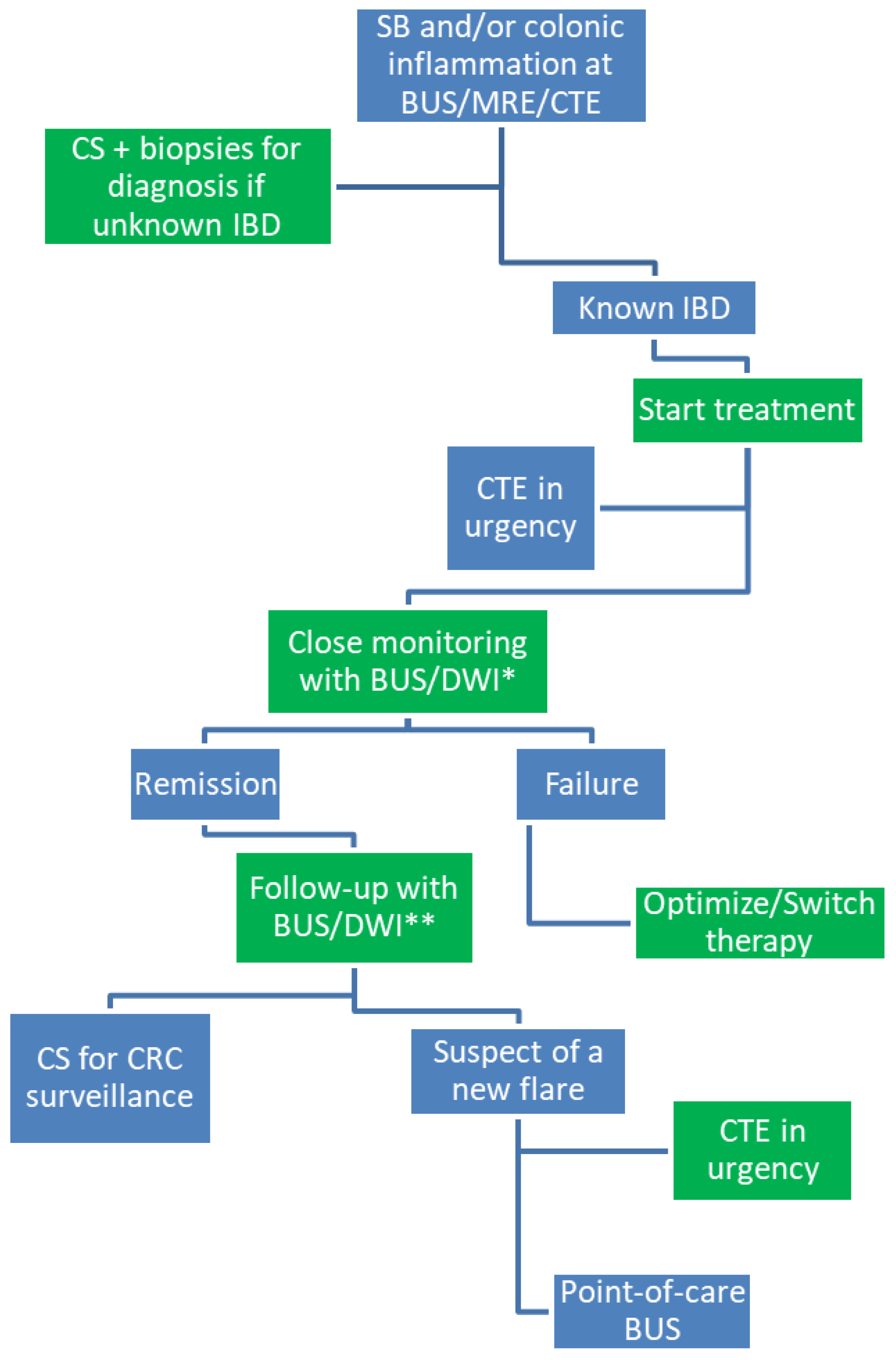
:1. Introduction
2. Crohn’s Disease
2.1. Cross-Sectional Imaging Techniques in Crohn’s Disease
2.2. Computed Tomography Enterography for the Assessment of Disease Activity and Complications
2.3. Magnetic Resonance Enterography
2.3.1. Assessment of Disease Activity and Complications
2.3.2. Monitoring and Prediction of Outcomes
2.4. Diffusion Weighted Imaging
2.4.1. Assessing Disease Activity
2.4.2. Monitoring Patients
2.5. Other New Magnetic Resonance Imaging-Based Techniques
2.6. Bowel Ultrasound
2.6.1. Assessment of Disease Activity and Complications
2.6.2. Predicting Outcomes and Monitoring
2.7. Artificial Intelligence
3. Ulcerative Colitis—Cross-Sectional Imaging Techniques in Ulcerative Colitis
3.1. Computed Tomography Enterography and Magnetic Resonance Enterography for Assessing Disease Activity
3.2. Diffusion Weighted Imaging
3.2.1. Assessing Disease Activity
3.2.2. Monitoring of Disease
3.3. Bowel Ultrasound
3.3.1. Assessing Disease Activity
3.3.2. Predicting Outcomes and Monitoring
4. Discussion
Author Contributions
Funding
Conflicts of Interest
References
- Torres, J.; Mehandru, S.; Colombel, J.-F.; Peyrin-Biroulet, L. Crohn’s disease. Lancet 2017, 389, 1741–1755. [Google Scholar] [CrossRef]
- Ungaro, R.; Mehandru, S.; Allen, P.B.; Peyrin-Biroulet, L.; Colombel, J.F. Ulcerative colitis. Lancet 2017, 389, 1756–1770. [Google Scholar] [CrossRef]
- Turner, D.; Ricciuto, A.; Lewis, A.; D’Amico, F.; Dhaliwal, J.; Griffiths, A.M.; Bettenworth, D.; Sandborn, W.J.; Sands, B.E.; Reinisch, W.; et al. STRIDE-II: An Update on the Selecting Therapeutic Targets in Inflammatory Bowel Disease (STRIDE) Initiative of the International Organization for the Study of IBD (IOIBD): Determining Therapeutic Goals for Treat-to-Target strategies in IBD. Gastroenterology 2021, 160, 1570–1583. [Google Scholar] [CrossRef] [PubMed]
- Colombel, J.-F.; D’Haens, G.; Lee, W.-J.; Petersson, J.; Panaccione, R. Outcomes and Strategies to Support a Treat-to-target Approach in Inflammatory Bowel Disease: A Systematic Review. J. Crohn’s Colitis 2019, 14, 254–266. [Google Scholar] [CrossRef] [PubMed] [Green Version]
- Magro, F.; Gionchetti, P.; Eliakim, R.; Ardizzone, S.; Armuzzi, A.; Barreiro-de Acosta, M.; Burisch, J.; Gecse, K.B.; Hart, A.L.; Hindryckx, P.; et al. Third European evidence-based consensus on diagnosis and management of ulcerative colitis. Part 1: Definitions, Diagnosis, Extra-intestinal Manifestations, Pregnancy, Cancer Surveillance, Surgery, and Ileo-anal Pouch Disorders. J. Crohn’s Colitis 2017, 11, 649–670. [Google Scholar] [CrossRef]
- Gomollón, F.; Dignass, A.; Annese, V.; Tilg, H.; Van Assche, G.; Lindsay, J.O.; Peyrin-Biroulet, L.; Cullen, G.J.; Daperno, M.; Kucharzik, T.; et al. 3rd European Evidence-based Consensus on the Diagnosis and Management of Crohn’s Disease 2016: Part 1: Diagnosis and Medical Management. J. Crohn’s Colitis 2017, 11, 3–25. [Google Scholar] [CrossRef] [PubMed] [Green Version]
- Louis, E.; Collard, A.; Oger, A.F.; Degroote, E.; El Yafi, F.A.N.; Belaiche, J. Behaviour of Crohn’s disease according to the Vienna classification: Changing pattern over the course of the disease. Gut 2001, 49, 777–782. [Google Scholar] [CrossRef] [Green Version]
- Lohsiriwat, V. Colonoscopic perforation: Incidence, risk factors, management and outcome. World J. Gastroenterol. 2010, 16, 425–430. [Google Scholar] [CrossRef]
- Noiseux, I.; Veilleux, S.; Bitton, A.; Kohen, R.; Vachon, L.; Guay, B.W.; Rioux, J.D. Inflammatory bowel disease patient perceptions of diagnostic and monitoring tests and procedures. BMC Gastroenterol. 2019, 19, 1–11. [Google Scholar] [CrossRef]
- Panes, J.; Bouzas, R.; Chaparro, M.; García-Sánchez, V.; Gisbert, J.P.; Martínez de Guereñu, B.; Mendoza, J.L.; Paredes, J.M.; Quiroga, S.; Ripollés, T.; et al. Systematic review: The use of ultrasonography, computed tomography and magnetic resonance imaging for the diagnosis, assessment of activity and abdominal complications of Crohn’s disease. Aliment. Pharmacol. Ther. 2011, 34, 125–145. [Google Scholar] [CrossRef]
- Desmond, A.N.; O’Regan, K.; Curran, C.; McWilliams, S.; Fitzgerald, T.; Maher, M.M.; Shanahan, F. Crohn’s disease: Factors associated with exposure to high levels of diagnostic radiation. Gut 2008, 57, 1524–1529. [Google Scholar] [CrossRef] [PubMed]
- Rimola, J.; Rodriguez, S.; Garcia-Bosch, O.; Ordas, I.; Ayala, E.; Aceituno, M.; Pellise, M.; Ayuso, C.; Ricart, E.; Donoso, L.; et al. Magnetic resonance for assessment of disease activity and severity in ileocolonic Crohn’s disease. Gut 2009, 58, 1113–1120. [Google Scholar] [CrossRef] [Green Version]
- Ordás, I.; Rimola, J.; García-Bosch, O.; Rodríguez, S.; Gallego, M.; Etchevers, M.J.; Pellisé, M.; Feu, F.; González-Suárez, B.; Ayuso, C.; et al. Diagnostic accuracy of magnetic resonance colonography for the evaluation of disease activity and severity in ulcerative colitis: A prospective study. Gut 2013, 62, 1566–1572. [Google Scholar] [CrossRef] [PubMed]
- Miles, A.; Bhatnagar, G.; Hallian, S.; Gupta, A.; Tolan, D.; Zealley, I.; Taylor, S.A.; METRIC Investigators. Magnetic resonance enterography, small bowel ultrasound and colonoscopy to diagnose and stage Crohn’s disease: Patient acceptability and perceived burden. Eur. Radiol. 2019, 29, 1083–1093. [Google Scholar] [CrossRef] [PubMed] [Green Version]
- Pouillon, L.; Laurent, V.; Pouillon, M.; Bossuyt, P.; Bonifacio, C.; Danese, S.; Deepak, P.; Loftus, E.; Bruining, D.H.; Peyrin-Biroulet, L. Diffusion-weighted MRI in inflammatory bowel disease. Lancet Gastroenterol. Hepatol. 2018, 3, 433–443. [Google Scholar] [CrossRef]
- Bryant, R.V.; Friedman, A.B.; Wright, E.K.; Taylor, K.M.; Begun, J.; Maconi, G.; Maaser, C.; Novak, K.L.; Kucharzik, T.; Atkinson, N.S.S.; et al. Gastrointestinal ultrasound in inflammatory bowel disease: An underused resource with potential paradigm-changing application. Gut 2018, 67, 973–985. [Google Scholar] [CrossRef]
- Maaser, C.; Sturm, A.; Vavricka, S.R.; Kucharzik, T.; Fiorino, G.; Annese, V.; Calabrese, E.; Baumgart, D.C.; Bettenworth, D.; Borralho Nunes, P.; et al. ECCO-ESGAR guideline for diagnostic assessment in IBD Part 1: Initial diagnosis, monitoring of known IBD, detection of complications. J. Crohn’s Colitis 2019, 13, 144–164. [Google Scholar] [CrossRef] [Green Version]
- Horsthuis, K.; Bipat, S.; Bennink, R.J.; Stoker, J. Inflammatory Bowel Disease Diagnosed with US, MR, Scintigraphy, and CT: Meta-analysis of Prospective Studies. Radiology 2008, 247, 64–79. [Google Scholar] [CrossRef]
- Horsthuis, K.; Bipat, S.; Stokkers, P.C.F.; Stoker, J. Magnetic resonance imaging for evaluation of disease activity in Crohn’s disease: A systematic review. Eur. Radiol. 2009, 19, 1450–1460. [Google Scholar] [CrossRef] [Green Version]
- Hammer, M.R.; Podberesky, D.J.; Dillman, J.R. Multidetector computed tomographic and magnetic resonance enterography in children: State of the art. Radiol. Clin. N. Am. 2013, 51, 615–636. [Google Scholar] [CrossRef] [PubMed]
- Nguyen, G.C.; Low, D.; Chong, R.Y.; Diong, C.; Chawla, T. Utilization of Diagnostic Imaging and Ionization Radiation Exposure Among an Inflammatory Bowel Disease Inception Cohort. Inflamm. Bowel Dis. 2019, 26, 898–906. [Google Scholar] [CrossRef]
- Ilangovan, R.; Burling, D.; George, A.; Gupta, A.; Marshall, M.; Taylor, S.A. CT enterography: Review of technique and practical tips. Br. J. Radiol. 2012, 85, 876–886. [Google Scholar] [CrossRef] [PubMed] [Green Version]
- Rimola, J.; Ordas, I.; Rodriguez, S.; García-Bosch, O.; Aceituno, M.; Llach, J.; Ayuso, C.; Ricart, E.; Panés, J. Magnetic resonance imaging for evaluation of Crohn’s disease: Validation of parameters of severity and quantitative index of activity. Inflamm. Bowel Dis. 2011, 17, 1759–1768. [Google Scholar] [CrossRef] [PubMed]
- Rimola, J.; Alvarez-Cofiño, A.; Perez, T.; Ayuso, C.; Alfaro, I.; Rodríguez, S.; Ricart, E.; Ordás, I.; Panes, J. Comparison of three magnetic resonance enterography indices for grading activity in Crohn’s disease. J. Gastroenterol. 2016, 52, 585–593. [Google Scholar] [CrossRef] [Green Version]
- Chavoshi, M.; Mirshahvalad, S.A.; Kasaeian, A.; Djalalinia, S.; Kolahdoozan, S.; Radmard, A.R. Diagnostic accuracy of Magnetic Resonance Enterography in the Evaluation of Colonic Abnormalities in Crohn’s Disease: A Systematic Review and Meta-Analysis. Acad. Radiol. 2021, 28, S192–S202. [Google Scholar] [CrossRef]
- Ordas, I.; Rimola, J.; Rodríguez, S.; Paredes, J.M.; Martínez-Pérez, M.J.; Blanc, E.; Arévalo, J.A.; Aduna, M.; Andreu, M.; Radosevic, A.; et al. Accuracy of Magnetic Resonance Enterography in Assessing Response to Therapy and Mucosal Healing in Patients with Crohn’s Disease. Gastroenterology 2014, 146, 374–382.e1. [Google Scholar] [CrossRef] [PubMed]
- Garcia-Bosch, O.; Ordas, I.; Aceituno, M.; Rodríguez, S.; Ramírez, A.M.; Gallego, M.; Ricart, E.; Rimola, J.; Panes, J. Comparison of diagnostic accuracy and impact of magnetic resonance imaging and colonoscopy for the management of Crohn’s disease. J. Crohn’s Colitis 2016, 10, 663–669. [Google Scholar] [CrossRef] [Green Version]
- Buisson, A.; Hordonneau, C.; Goutorbe, F.; Allimant, C.; Goutte, M.; Reymond, M.; Pereira, B.; Bommelaer, G. Bowel wall healing assessed using magnetic resonance imaging predicts sustained clinical remission and decreased risk of surgery in Crohn’s disease. J. Gastroenterol. 2019, 54, 312–320. [Google Scholar] [CrossRef] [PubMed]
- Fernandes, S.R.; Rodrigues, R.V.; Bernardo, S.; Cortez-Pinto, J.; Rosa, I.; da Silva, J.P.; Gonçalves, A.R.; Valente, A.; Baldaia, C.; Santos, P.M.; et al. Transmural healing is associated with improved long-term outcomes of patients with Crohn’s disease. Inflamm. Bowel Dis. 2017, 23, 1403–1409. [Google Scholar] [CrossRef]
- Choi, S.H.; Kim, K.W.; Lee, J.Y.; Kim, K.J.; Park, S.H. Diffusion-weighted magnetic resonance enterography for evaluating bowel inflammation in Crohn’s disease: A systematic review and meta-analysis. Inflamm. Bowel Dis. 2016, 22, 669–679. [Google Scholar] [CrossRef]
- Oussalah, A.; Laurent, V.; Bruot, O.; Bressenot, A.; Bigard, M.A.; Régent, D.; Peyrin-Biroulet, L. Diffusion-weighted magnetic resonance without bowel preparation for detecting colonic inflammation in inflammatory bowel disease. Gut 2010, 59, 1056–1065. [Google Scholar] [CrossRef] [PubMed]
- Buisson, A.; Joubert, A.; Montoriol, P.-F.; Ines, D.D.; Hordonneau, C.; Pereira, B.; Garcier, J.-M.; Bommelaer, G.; Petitcolin, V. Diffusion-weighted magnetic resonance imaging for detecting and assessing ileal inflammation in Crohn’s disease. Aliment. Pharmacol. Ther. 2013, 37, 537–545. [Google Scholar] [CrossRef]
- Hordonneau, C.; Buisson, A.; Scanzi, J.; Goutorbe, F.; Pereira, B.; Borderon, C.; Da Ines, D.; Montoriol, P.F.; Garcier, J.M.; Boyer, L.; et al. Diffusion-Weighted Magnetic Resonance Imaging in Ileocolonic Crohn’s Disease: Validation of Quantitative Index of Activity. Am. J. Gastroenterol. 2014, 109, 89–98. [Google Scholar] [CrossRef]
- Buisson, A.; Hordonneau, C.; Goutte, M.; Boyer, L.; Pereira, B.; Bommelaer, G. Diffusion-weighted magnetic resonance imaging is effective to detect ileocolonic ulcerations in Crohn’s disease. Aliment. Pharmacol. Ther. 2015, 42, 452–460. [Google Scholar] [CrossRef] [PubMed] [Green Version]
- Huh, J.; Kim, K.J.; Park, S.H.; Park, S.H.; Yang, S.-K.; Ye, B.D.; Park, S.H.; Han, K.; Kim, A.Y. Diffusion-Weighted MR Enterography to Monitor Bowel Inflammation after Medical Therapy in Crohn’s Disease: A Prospective Longitudinal Study. Korean J. Radiol. 2017, 18, 162–172. [Google Scholar] [CrossRef] [Green Version]
- Bilgili, M.Y. Reproducibility of apparent diffusion coefficients measurements in diffusion-weighted MRI of the abdomen with different b values. Eur. J. Radiol. 2012, 81, 2066–2068. [Google Scholar] [CrossRef] [PubMed]
- Thierry, M.-L.; Rousseau, H.; Pouillon, L.; Girard-Gavanier, M.; Baumann, C.; Lopez, A.; Danese, S.; Laurent, V.; Peyrin-Biroulet, L. Accuracy of Diffusion-weighted Magnetic Resonance Imaging in Detecting Mucosal Healing and Treatment Response, and in Predicting Surgery, in Crohn’s Disease. J. Crohn’s Colitis 2018, 12, 1180–1190. [Google Scholar] [CrossRef] [PubMed]
- Seo, N.; Park, S.H.; Kim, K.-J.; Kang, B.-K.; Lee, Y.; Yang, S.-K.; Ye, B.D.; Park, S.H.; Kim, S.Y.; Baek, S.; et al. MR Enterography for the Evaluation of Small-Bowel Inflammation in Crohn Disease by Using Diffusion-weighted Imaging without Intravenous Contrast Material: A Prospective Noninferiority Study. Radiology 2016, 278, 762–772. [Google Scholar] [CrossRef]
- Deepak, P.; Axelrad, J.E.; Ananthakrishnan, A.N. The Role of the Radiologist in Determining Disease Severity in Inflammatory Bowel Diseases. Gastrointest. Endosc. Clin. N. Am. 2019, 29, 447–470. [Google Scholar] [CrossRef]
- Pazahr, S.; Blume, I.; Frei, P.; Chuck, N.; Nanz, D.; Rogler, G.; Patak, M.; Boss, A. Magnetization transfer for the assessment of bowel fibrosis in patients with Crohn’s disease: Initial experience. Magma Magn. Reson. Mater. Phys. Biol. Med. 2012, 26, 291–301. [Google Scholar] [CrossRef] [PubMed] [Green Version]
- Rieder, F.; Latella, G.; Magro, F.; Yuksel, E.S.; Higgins, P.D.; Di Sabatino, A.; de Bruyn, J.R.; Rimola, J.; Brito, J.; Bettenworth, D.; et al. European Crohn’s and Colitis Organization topical review on prediction, diagnosis and management of fibrostenosing Crohn’s disease. J. Crohn’s Colitis 2016, 10, 873–885. [Google Scholar] [CrossRef]
- Li, X.H.; Mao, R.; Huang, S.Y.; Sun, C.H.; Cao, Q.H.; Fang, Z.N.; Zhang, Z.W.; Huang, L.; Lin, J.J.; Chen, Y.J.; et al. Characterization of degree of intestinal fibrosis in patients with Crohn disease by using magnetization transfer MR imaging. Radiology 2018, 287, 494–503. [Google Scholar] [CrossRef] [Green Version]
- Fang, Z.N.; Li, X.H.; Lin, J.J.; Huang, S.Y.; Cao, Q.H.; Chen, Z.H.; Sun, C.H.; Zhang, Z.W.; Rieder, F.; Rimola, J.; et al. Magnetization transfer imaging adds information to conventional MRIs to differentiate inflammatory from fibrotic components of small intestinal strictures in Crohn’s disease. Eur. Radiol. 2020, 30, 1938–1947. [Google Scholar] [CrossRef]
- Bickelhaupt, S.; Pazahr, S.; Chuck, N.; Blume, I.; Froehlich, J.M.; Cattin, R.; Raible, S.; Bouquet, H.; Bill, U.; Rogler, G.; et al. Crohn’s disease: Small bowel motility impairment correlates with inflammatory-related markers C-reactive protein and calprotectin. Neurogastroenterol. Motil. 2013, 25, 467–473. [Google Scholar] [CrossRef] [PubMed]
- Menys, A.; Puylaert, C.; Nolthenius, C.E.T.; Plumb, A.A.; Makanyanga, J.; Tielbeek, J.; Pendse, D.; Brosens, L.A.; Rodriguez-Justo, M.; Atkinson, D.; et al. Quantified Terminal Ileal Motility during MR Enterography as a Biomarker of Crohn Disease Activity: Prospective Multi-Institution Study. Radiology 2018, 289, 428–435. [Google Scholar] [CrossRef] [PubMed]
- Plumb, A.A.; Menys, A.; Russo, E.; Prezzi, D.; Bhatnagar, G.; Vega, R.; Halligan, S.; Orchard, T.R.; Taylor, S.A. Magnetic resonance imaging-quantified small bowel motility is a sensitive marker of response to medical therapy in Crohn’s disease. Aliment. Pharmacol. Ther. 2015, 42, 343–355. [Google Scholar] [CrossRef] [PubMed] [Green Version]
- Puylaert, C.A.; Tielbeek, J.A.; Bipat, S.; Stoker, J. Grading of Crohn’s disease activity using CT, MRI, US and scintigraphy: A meta-analysis. Eur. Radiol. 2015, 25, 3295–3313. [Google Scholar] [CrossRef] [Green Version]
- Calabrese, E.; Maaser, C.; Zorzi, F.; Kannengiesser, K.; Hanauer, S.B.; Bruining, D.H.; Iacucci, M.; Maconi, G.; Novak, K.L.; Panaccione, R.; et al. Bowel ultrasonography in the management of Crohn’s disease. A review with recommendations of an international panel of experts. Inflamm. Bowel Dis. 2016, 22, 1168–1183. [Google Scholar] [CrossRef]
- Imperatore, N.; Rispo, A.; Testa, A.; Mainenti, P.; De Palma, G.; Rea, M.; Nardone, O.; Luglio, G.; Caporaso, N.; Castiglione, F. OC.08.4: Bowel Damage in Crohn’s Disease: Direct Comparison of Ultrasonography- and Magnetic Resonance-Based Lemann Index. Dig. Liver Dis. 2017, 49, e97–e98. [Google Scholar] [CrossRef]
- Taylor, S.A.; Mallett, S.; Bhatnagar, G.; Baldwin-Cleland, R.; Bloom, S.; Gupta, A.; Hamlin, P.J.; Hart, A.L.; Higginson, A.; Jacobs, I.; et al. Diagnostic accuracy of magnetic resonance enterography and small bowel ultrasound for the extent and activity of newly diagnosed and relapsed Crohn’s disease (METRIC): A multicentre trial. Lancet Gastroenterol. Hepatol. 2018, 3, 548–558. [Google Scholar] [CrossRef]
- Allocca, M.; Fiorino, G.; Bonifacio, C.; Furfaro, F.; Gilardi, D.; Argollo, M.; Peyrin-Biroulet, L.; Danese, S. Comparative Accuracy of Bowel Ultrasound Versus Magnetic Resonance Enterography in Combination With Colonoscopy in Assessing Crohn’s Disease and Guiding Clinical Decision-making. J. Crohn’s Coliti 2018, 12, 1280–1287. [Google Scholar] [CrossRef]
- Ripollés, T.; Martínez-Pérez, M.J.; Paredes, J.M.; Vizuete, J.; Martin, G. The role of intravenous contrast agent in the sonographic assessment of Crohn’s disease activity: Is contrast agent injection necessary? J. Crohn’s Colitis 2019, 13, 585–592. [Google Scholar] [CrossRef]
- Ripollés, T.; Rausell, N.; Paredes, J.M.; Grau, E.; Martínez, M.J.; Vizuete, J. Effectiveness of contrast-enhanced ultrasound for characterisation of intestinal inflammation in Crohn’s disease: A comparison with surgical histopathology analysis. J. Crohn’s Colitis 2013, 7, 120–128. [Google Scholar] [CrossRef]
- Pescatori, L.C.; Mauri, G.; Savarino, E.; Pastorelli, L.; Vecchi, M.; Sconfienza, L.M. Bowel Sonoelastography in Patients with Crohn’s Disease: A Systematic Review. Ultrasound Med. Biol. 2018, 44, 297–302. [Google Scholar] [CrossRef] [Green Version]
- Lu, C.; Merrill, C.; Medellin, A.; Novak, K.; Wilson, S.R. Bowel Ultrasound State of the Art: Grayscale and Doppler Ultrasound, Contrast Enhancement, and Elastography in Crohn Disease. J. Ultrasound Med. 2019, 38, 271–288. [Google Scholar] [CrossRef] [PubMed] [Green Version]
- Bots, S.; Nylund, K.; Löwenberg, M.; Gecse, K.; Gilja, O.H.; D’Haens, G. Ultrasound for assessing disease activity in IBD patients: A systematic review of activity scores. J. Crohn’s Colitis 2018, 12, 920–929. [Google Scholar] [CrossRef] [PubMed] [Green Version]
- Sævik, F.; Eriksen, R.; Eide, G.E.; Gilja, O.H.; Nylund, K. Development and Validation of a Simple Ultrasound Activity Score for Crohn’s Disease. J. Crohn’s Colitis 2020, 15, 115–124. [Google Scholar] [CrossRef] [PubMed]
- Novak, K.L.; Nylund, K.; Maaser, C.; Petersen, F.; Kucharzik, T.; Lu, C.; Allocca, M.; Maconi, G.; de Voogd, F.; Christensen, B.; et al. Expert Consensus on Optimal Acquisition and Development of the International Bowel Ultrasound Segmental Activity Score [IBUS-SAS]: A Reliability and Inter-rater Variability Study on Intestinal Ultrasonography in Crohn’s Disease. J. Crohn’s Colitis 2021, 15, 609–616. [Google Scholar] [CrossRef]
- Sagami, S.; Kobayashi, T.; Miyatani, Y. Accuracy of Ultrasound for Evaluation of Colorectal Segments in Patients with Inflam-matory Bowel Diseases: A Systematic Review and Meta-analysis. Clin. Gastroenterol. Hepatol. 2021, 19, 908–921.e6. [Google Scholar] [CrossRef]
- Panes, J.; Rimola, J. Perianal fistulizing Crohn’s disease: Pathogenesis, diagnosis and therapy. Nat. Rev. Gastroenterol. Hepatol. 2017, 14, 652–664. [Google Scholar] [CrossRef]
- Maconi, G.; Greco, M.T.; Asthana, A.K. Transperineal Ultrasound for Perianal Fistulas and Abscesses—A Systematic Review and Meta-Analysis. Ultraschall der Med.-Eur. J. Ultrasound 2017, 38, 265–272. [Google Scholar] [CrossRef]
- Rispo, A.; Imperatore, N.; Testa, A.; Nardone, O.M.; Luglio, G.; Caporaso, N.; Castiglione, F. Diagnostic Accuracy of Ultrasonography in the Detection of Postsurgical Recurrence in Crohn’s Disease: A Systematic Review with Meta-analysis. Inflamm. Bowel Dis. 2018, 24, 977–988. [Google Scholar] [CrossRef] [PubMed]
- Allocca, M.; Craviotto, V.; Bonovas, S.; Furfaro, F.; Zilli, A.; Peyrin-Biroulet, L.; Fiorino, G.; Danese, S. Predictive Value of Bowel Ultrasound in Crohn’s Disease: A 12-Month Prospective Study. Clin. Gastroenterol. Hepatol. 2021. [Google Scholar] [CrossRef] [PubMed]
- Allocca, M.; Craviotto, V.; Dell’Avalle, C.; Furfaro, F.; Zilli, A.; D’Amico, F.; Bonovas, S.; Peyrin-Biroulet, L.; Fiorino, G.; Danese, S. Bowel ultrasound score is accurate in assessing response to therapy in patients with Crohn’s disease. Aliment. Pharmacol. Ther. 2021. [Google Scholar] [CrossRef] [PubMed]
- Novak, K.; Tanyingoh, D.; Petersen, F.; Kucharzik, T.; Panaccione, R.; Ghosh, S.; Kaplan, G.G.; Wilson, A.; Kannengiesser, K.; Maaser, C. Clinic-based point of care transabdominal ultrasound for monitoring Crohn’s disease: Impact on clinical decision making. J. Crohn’s Colitis 2015, 9, 795–801. [Google Scholar] [CrossRef] [Green Version]
- Moreno, N.; Ripollés, T.; Paredes, J.M.; Ortiz, I.; Martínez, M.J.; López, A.; Delgado, F.; Moreno-Osset, E. Usefulness of abdominal ultrasonography in the analysis of endoscopic activity in patients with Crohn’s disease: Changes following treatment with immunomodulators and/or anti-TNF antibodies. J. Crohn’s Colitis 2014, 8, 1079–1087. [Google Scholar] [CrossRef]
- Ripollés, T.; Paredes, J.M.; Martínez-Pérez, M.J.; Rimola, J.; Jauregui-Amezaga, A.; Bouzas, R.; Martin, G.; Moreno-Osset, E. Ultrasonographic changes at 12 weeks of anti-TNF drugs predict 1-year sonographic response and clinical outcome in Crohn’s disease: A multicenter study. Inflamm. Bowel Dis. 2016, 22, 2465–2473. [Google Scholar] [CrossRef] [PubMed]
- Zorzi, F.; Ghosh, S.; Chiaramonte, C.; Lolli, E.; Ventura, M.; Onali, S.; De Cristofaro, E.; Fantini, M.C.; Biancone, L.; Monteleone, G.; et al. Response Assessed by Ultrasonography as Target of Biological Treatment for Crohn’s Disease. Clin. Gastroenterol. Hepatol. 2020, 18, 2030–2037. [Google Scholar] [CrossRef]
- Serban, E.D. Treat-to-target in Crohn’s disease: Will transmural healing become a therapeutic endpoint? World J. Clin. Cases 2018, 6, 501–513. [Google Scholar] [CrossRef]
- Castiglione, F.; Imperatore, N.; Testa, A.; De Palma, G.D.; Nardone, O.M.; Pellegrini, L.; Caporaso, N.; Rispo, A. One-year clinical outcomes with biologics in Crohn’s disease: Transmural healing compared with mucosal or no healing. Aliment. Pharmacol. Ther. 2019, 49, 1026–1039. [Google Scholar] [CrossRef]
- Kucharzik, T.; Wittig, B.M.; Helwig, U.; Börner, N.; Rössler, A.; Rath, S.; Maaser, C.; Naumann, A.; Pelster, G.; Spengler, J.; et al. Use of Intestinal Ultrasound to Monitor Crohn’s Disease Activity. Clin. Gastroenterol. Hepatol. 2016, 15, 535–542.e2. [Google Scholar] [CrossRef]
- Calabrese, E.; Rispo, A.; Zorzi, F.; De Cristofaro, E.; Testa, A.; Costantino, G.; Viola, A.; Bezzio, C.; Ricci, C.; Prencipe, S.; et al. Ultrasonography Tight Control and Monitoring in Crohn’s Disease during Different Biological Therapies: A Multicenter Study. Clin. Gastroenterol. Hepatol. 2021; in press. [Google Scholar] [CrossRef] [PubMed]
- Kucharzik, T.; Wilkens, R.; Maconi, G.; D’Agostino, M.A.; Le Bars, M.; Nazar, M.; Sloan, S.; Lahaye, M.; Ni, L.; Ercole, E.; et al. DOP10 Intestinal ultrasound response and transmural healing after ustekinumab induction in Crohn’s disease: Week 16 interim analysis of the STARDUST trial substudy. J. Crohn’s Colitis 2020, 14, S046–S048. [Google Scholar] [CrossRef]
- Le Berre, C.; Sandborn, W.J.; Aridhi, S.; Devignes, M.D.; Fournier, L.; Smaïl-Tabbone, M.; Danese, S.; Peyrin-Biroulet, L. Application of Artificial Intelligence to Gastroenterology and Hepatology. Gastroenterology 2020, 158, 76–94.e2. [Google Scholar] [CrossRef] [Green Version]
- Stidham, R.W.; Enchakalody, B.; Waljee, A.K.; Higgins, P.D.R.; Wang, S.C.; Su, G.; Wasnik, A.P.; Al-Hawary, M. Assessing Small Bowel Stricturing and Morphology in Crohn’s Disease Using Semi-automated Image Analysis. Inflamm. Bowel Dis. 2019, 26, 734–742. [Google Scholar] [CrossRef] [PubMed]
- Lamash, Y.; Kurugol, S.; Freiman, M.; Perez-Rossello, J.M.; Callahan, M.J.; Bousvaros, A.; Warfield, S.K. Curved planar reformatting and convolutional neural network-based segmentation of the small bowel for visualization and quantitative assessment of pediatric Crohn’s disease from MRI. J. Magn. Reson. Imaging 2019, 49, 1565–1576. [Google Scholar] [CrossRef]
- Yang, S.; Lemke, C.; Cox, B.F.; Newton, I.P.; Näthke, I.; Cochran, S. A learning-based microultrasound system for the detection of inflammation of the gastrointestinal tract. IEEE Trans. Med. Imaging 2021, 40, 38–47. [Google Scholar] [CrossRef]
- Andersen, K.; Vogt, C.; Blondin, D.; Beck, A.; Heinen, W.; Aurich, V.; Häussinger, D.; Mödder, U.; Cohnen, M. Multi-detector CT-colonography in inflammatory bowel disease: Prospective analysis of CT-findings to high-resolution video colonoscopy. Eur. J. Radiol. 2006, 58, 140–146. [Google Scholar] [CrossRef] [PubMed]
- Johnson, K.T.; Hara, A.K.; Johnson, C.D. Evaluation of colitis: Usefulness of CT enterography technique. Emerg. Radiol. 2009, 16, 277–282. [Google Scholar] [CrossRef]
- Yu, L.-L.; Yang, H.-S.; Zhang, B.-T.; Lv, Z.-W.; Wang, F.-R.; Zhang, C.-Y.; Chen, W.-B.; Zhang, H.-M. Diffusion-weighted magnetic resonance imaging without bowel preparation for detection of ulcerative colitis. World J. Gastroenterol. 2015, 21, 9785–9792. [Google Scholar] [CrossRef]
- Laurent, V.; Naudé, S.; Vuitton, L.; Zallot, C.; Baumann, C.; Girard-Gavanier, M.; Peyrin-Biroulet, L. Accuracy of Diffusion-weighted Magnetic Resonance Colonography in Assessing Mucosal Healing and the Treatment Response in Patients with Ulcerative Colitis. J. Crohn’s Colitis 2016, 11, 716–723. [Google Scholar] [CrossRef] [Green Version]
- Antonelli, E.; Giuliano, V.; Casella, G.; Villanacci, V.; Baldini, V.; Baldoni, M.; Morelli, O.; Bassotti, G. Ultrasonographic assessment of colonic wall in moderate–severe ulcerative colitis: Comparison with endoscopic findings. Dig. Liver Dis. 2011, 43, 703–706. [Google Scholar] [CrossRef]
- Allocca, M.; Fiorino, G.; Bonovas, S.; Furfaro, F.; Gilardi, D.; Argollo, M.; Magnoni, P.; Peyrin-Biroulet, L.; Danese, S. Accuracy of Humanitas Ultrasound Criteria in Assessing Disease Activity and Severity in Ulcerative Colitis: A Prospective Study. J. Crohn’s Coliti 2018, 12, 1385–1391. [Google Scholar] [CrossRef] [Green Version]
- Allocca, M.; Filippi, E.; Costantino, A.; Bonovas, S.; Fiorino, G.; Furfaro, F.; Peyrin-Biroulet, L.; Fraquelli, M.; Caprioli, F.; Danese, S. Milan ultrasound criteria are accurate in assessing disease activity in ulcerative colitis: External validation. United Eur. Gastroenterol. J. 2021, 9, 438–442. [Google Scholar] [CrossRef]
- Sagami, S.; Kobayashi, T.; Aihara, K.; Umeda, M.; Morikubo, H.; Matsubayashi, M.; Kiyohara, H.; Nakano, M.; Ohbu, M.; Hibi, T. Transperineal ultrasound predicts endoscopic and histological healing in ulcerative colitis. Aliment. Pharmacol. Ther. 2020, 51, 1373–1383. [Google Scholar] [CrossRef]
- Parente, F.; Molteni, M.; Marino, B.; Colli, A.; Ardizzone, S.; Greco, S.; Sampietro, G.M.; Gallus, S. Bowel Ultrasound and Mucosal Healing in Ulcerative Colitis. Dig. Dis. 2009, 27, 285–290. [Google Scholar] [CrossRef]
- Allocca, M.; Dell’Avalle, C.; Furfaro, F.; Craviotto, V.; Zilli, A.; D’Amico, F.; Peyrin-Biroulet, L.; Fiorino, G.; Danese, S. OP21 Predictive value of Milan Ultrasound Criteria in Ulcerative Colitis: A prospective observational cohort study. J. Crohn’s Colitis 2021, 15, S020–S021. [Google Scholar] [CrossRef]
- Maconi, G.; Ardizzone, S.; Parente, F.; Bianchi Porro, G. Ultrasonography in the evaluation of extension, activity, and follow-up of ulcerative colitis. Scand. J. Gastroenterol. 1999, 34, 1103–1107. [Google Scholar] [CrossRef] [PubMed]
- Maaser, C.; Petersen, F.; Helwig, U.; Fischer, I.; Roessler, A.; Rath, S.; Lang, D.; Kucharzik, T. Intestinal ultrasound for monitoring therapeutic response in patients with ulcerative colitis: Results from the TRUST&UC study. Gut 2019, 69, 1629–1636. [Google Scholar] [CrossRef] [PubMed] [Green Version]
- Shah, S.C.; Colombel, J.F.; Sands, B.E.; Narula, N. Systematic review with meta-analysis: Mucosal healing is associated with im-proved long-term outcomes in Crohn’s disease. Aliment. Pharmacol. Ther. 2016, 43, 317–333. [Google Scholar] [CrossRef]
- American Society for Gastrointestinal Endoscopy Standards of Practice Committee; Shergill, A.K.; Lightdale, J.R.; Bruining, D.H.; Acosta, R.D.; Chandrasekhara, V.; Chathadi, K.V.; Decker, G.A.; Early, D.S.; Evans, J.A.; et al. The role of endoscopy in inflammatory bowel disease. Gastrointest. Endosc. 2015, 81, 1101–1121.e13. [Google Scholar] [CrossRef] [PubMed]
- Calabrese, E.; Kucharzik, T.; Maaser, C.; Maconi, G.; Strobel, D.; Wilson, S.R.; Zorzi, F.; Novak, K.L.; Bruining, D.H.; Iacucci, M.; et al. Real-time interobserver agreement in bowel ultrasonography for diagnostic assessment in patients with Crohn’s disease: An international multicenter study. Inflamm. Bowel Dis. 2018, 24, 2001–2006. [Google Scholar] [CrossRef] [PubMed] [Green Version]


| Technique | Sensitivity | Specificity | Strengths | Limitations |
|---|---|---|---|---|
| CTE | Low costs, high availability, short examination time | Radiation exposure, intravenous contrast agent, bowel preparation | ||
| MRE | Radiation-free, detailed high-quality imaging | Intravenous contrast agent, bowel preparation, high costs, poor availability, long-lasting procedure | ||
| DWI |
|
| Intravenous contrast agent not required, easier and quicker than MRE, fasting and bowel preparation needed only for SB assessment | Scanners and examinations are heterogeneous Less precise anatomic view than MRE |
| BUS | Low costs, radiation free, high availability and acceptability, easy, performed at the point-of-care | Conventionally regarded as operator-dependent |
| Technique | Sensitivity | Specificity | Strengths | Limitations |
|---|---|---|---|---|
| CTE | 74% [81] | >85% [81] | High affordability, short-lasting examination, cheap | Radiation exposure, intravenous contrast agent, bowel cleansing |
| MRE | 87% [13] | 88% [13] | No radiation exposure | Intravenous contrast agent, bowel preparation, time-consuming procedure, costly |
| DWI | 89.4% [31] | 86.7% [31] | Fast, no radiation exposure, no intravenous contrast agent, no fasting, no bowel preparation | Not standardized DWI scanners and procedures |
| BUS | 90% [18] | 96% [18] | No radiation exposure, available, tolerable, cheap, performed at the point-of-care | Traditionally considered operator-dependent |
Publisher’s Note: MDPI stays neutral with regard to jurisdictional claims in published maps and institutional affiliations. |
© 2022 by the authors. Licensee MDPI, Basel, Switzerland. This article is an open access article distributed under the terms and conditions of the Creative Commons Attribution (CC BY) license (https://creativecommons.org/licenses/by/4.0/).
Share and Cite
Alfarone, L.; Dal Buono, A.; Craviotto, V.; Zilli, A.; Fiorino, G.; Furfaro, F.; D’Amico, F.; Danese, S.; Allocca, M. Cross-Sectional Imaging Instead of Colonoscopy in Inflammatory Bowel Diseases: Lights and Shadows. J. Clin. Med. 2022, 11, 353. https://doi.org/10.3390/jcm11020353
Alfarone L, Dal Buono A, Craviotto V, Zilli A, Fiorino G, Furfaro F, D’Amico F, Danese S, Allocca M. Cross-Sectional Imaging Instead of Colonoscopy in Inflammatory Bowel Diseases: Lights and Shadows. Journal of Clinical Medicine. 2022; 11(2):353. https://doi.org/10.3390/jcm11020353
Chicago/Turabian StyleAlfarone, Ludovico, Arianna Dal Buono, Vincenzo Craviotto, Alessandra Zilli, Gionata Fiorino, Federica Furfaro, Ferdinando D’Amico, Silvio Danese, and Mariangela Allocca. 2022. "Cross-Sectional Imaging Instead of Colonoscopy in Inflammatory Bowel Diseases: Lights and Shadows" Journal of Clinical Medicine 11, no. 2: 353. https://doi.org/10.3390/jcm11020353