Article

Patient-Derived Training Simulator for Image-Guided Adaptive Brachytherapy of Locally Advanced Cervical Cancers: Development and Initial Use

1
Department of Radiation Oncology, Gunma University Graduate School of Medicine, 3-39-22 Showa-machi, Maebashi 371-8511, Gunma, Japan
2
Gunma University Heavy Ion Medical Center, 3-39-22 Showa-machi, Maebashi 371-8511, Gunma, Japan
*
Author to whom correspondence should be addressed.
J. Clin. Med. 2022, 11(11), 3103; https://doi.org/10.3390/jcm11113103
Submission received: 21 March 2022 / Revised: 20 May 2022 / Accepted: 27 May 2022 / Published: 30 May 2022
(This article belongs to the Special Issue Clinical Advances in Gynaecological Cancer Management)

Abstract

:
Image-guided adaptive brachytherapy (IGABT) using intracavitary and interstitial (IC/IS) techniques plays a pivotal role in definitive radiotherapy for locally advanced cervical cancers. However, the training opportunities for interstitial needle application are limited, preventing this technique from becoming widespread. This study aimed to develop a training simulator for IC/IS brachytherapy. The simulator consists of a soft silicone tumor phantom and acrylic tube mimicking the vagina; it has high visibility because of translucent materials and is compatible with computed tomography (CT) and magnetic resonance imaging (MRI). A patient harboring a typical bulky and irregular-shaped cervical tumor was selected from 495 in-house IGABT-treated candidates, and a tumor phantom (68 × 49 × 45 mm) modeled on this patient was produced from three-dimensional real-scale measurements of the MRI-based high-risk clinical target volume at first brachytherapy. In trial use by two physicians with different levels of IGABT skills, a Fletcher-Suit Asian Pacific applicator, and a Venezia applicator with interstitial needles were nicely applied to the simulator, facilitating successful creation of CT-based treatment plans consistent with clinical practice. Thus, the training simulator can be useful for the training of IC/IS brachytherapy, and warrants further research employing a greater number of phantoms and practitioners to verify its educational value.

1. Introduction

Cervical cancer causes more than 300,000 deaths worldwide annually, and its mortality ranks fourth among all cancers [1]. Radiotherapy plays a pivotal role in the definitive treatment of locally advanced cervical cancers [2], with definitive radiotherapy approaches including external beam radiotherapy (EBRT) and brachytherapy [3]. For the latter, image-guided adaptive brachytherapy (IGABT) using computed tomography (CT) or magnetic resonance imaging (MRI) has become widespread in recent years [4,5]. The introduction of IGABT technologies into definitive radiotherapy practice has dramatically improved the oncologic outcomes of cervical cancers [6,7].
Despite the technological advancements, IGABT using a tandem and ovoid applicator alone results in suboptimal dose coverage for bulky or irregular-shaped tumors, leading to insufficient tumor control [8,9]. To deliver a sufficient dose to such tumors, the tandem and ovoid applicator can be supplemented with a few interstitial needles. In 2011, our group introduced a combination of a few interstitial needles with a Fletcher-Suit Asian Pacific applicator (Elekta, Stockholm, Sweden) under in-room CT guidance [10]. Multiple studies have shown that this combined intracavitary and interstitial (IC/IS) brachytherapy has excellent outcomes for locally advanced disease [7,11]. In addition, brachytherapy applicators equipped with a template for needle insertion have been developed by several groups. In 2006, Dimopoulos et al. introduced the Vienna applicator consisting of a tandem and a ring that enables needle insertion from predefined directions guided through the holes in the ring [12]. In 2009, Jürgenliemk-Schulz et al. introduced the Utrecht applicator consisting of a tandem and two oval ovoids that enable needle insertion from more-varied directions than the Vienna applicator, with the needles guided through the holes in the ovoids [13]. In 2018, Walter et al. introduced the Venezia applicator consisting of a tandem and two lunar-shaped ovoids (that form a ring in combination) functioning as a needle template [14].
The key to the success of IC/IS brachytherapy is to insert interstitial needles in the locations appropriate to achieving a highly conformal dose to the targeted tumor volume. However, the training opportunities for needle application are limited, thereby preventing this technique from becoming widely used by radiation oncologists. To address this issue, this study aimed to develop a training simulator for IC/IS brachytherapy for locally advanced cervical cancer.

2. Materials and Methods

2.1. Foundation of the Simulator

The foundation of the simulator was developed using transparent acryl to provide visibility. No metal was used to ensure that the simulator was CT and MRI compatible. To enable a physician to approach through the vagina, an acrylic tube mimicking the vagina (50 mm in diameter and 90 mm in length) was installed on an acrylic plate (200 × 100 mm).

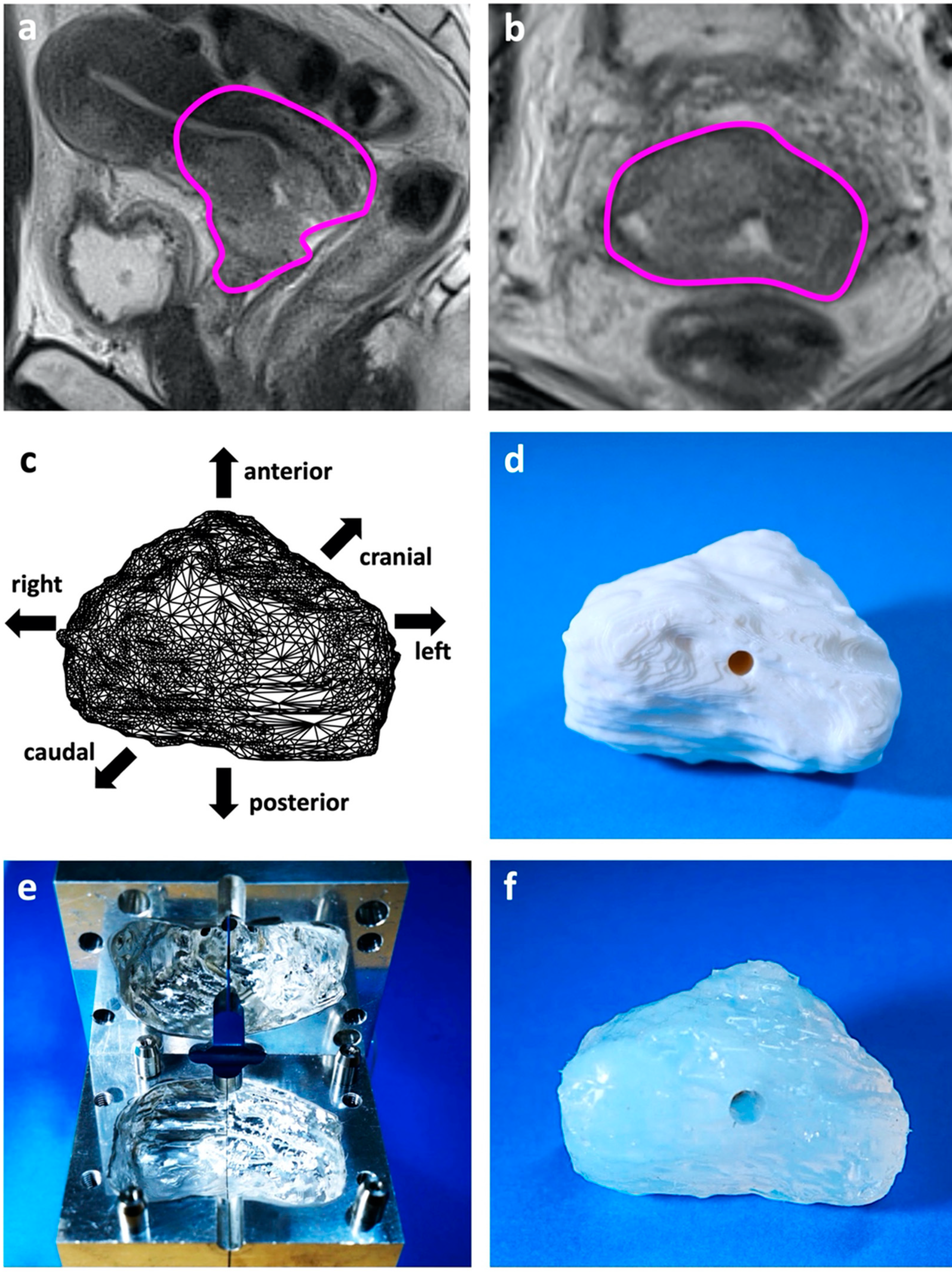
2.2. Patient-Derived Tumor Phantom

A tumor phantom representing a typical bulky and irregular-shaped tumor was produced. The case on which the tumor phantom was modeled was selected by a review of in-house IGABT records. The inclusion criteria for the review were as follows: (i) patients with newly-diagnosed and pathologically-confirmed cervical cancer treated with definitive radiotherapy using IGABT [7] at Gunma University Hospital (Maebashi, Gunma, Japan) between 2012 and 2020; and (ii) staged as IIIB based on the International Federation of Gynecology and Obstetrics 2009 staging system. For the eligible patients, MRI obtained at the first brachytherapy was inspected by three radiation oncologists (KT, KA, and DI), and ten patients who harbored a bulky tumor with extensive bilateral parametrial involvement were selected. For these ten patients, the high-risk clinical target volume (HR-CTV) was delineated on the T2-weighted MRI according to the recommendations of The Groupe Européen de Curiethérapie and the European Society for Radiotherapy and Oncology [15,16] using MIM Maestro (version 6.8.7, MIM Software Inc., Cleveland, OH, USA). The HR-CTV contour data were then transferred to OsiriX 64-bit (version 12.5.2, Pixmeo, Bernex, Switzerland) and subjected to three-dimensional (3D)-volume rendering. The three radiation oncologists inspected the 3D-reconstructed HR-CTVs from various angles and identified the best case for use as the phantom.
The HR-CTV data of the model patient were then transferred to a computer-aided design software (version 3.0 Creo Parametric, PTC, Boston, MA, USA) and a real-scale 3D volume was exported in acrylonitrile butadiene styrene (ABS) plastic using a 3D printer (Value 3D Magix MF-1100, Mutoh Industries Ltd., Tokyo, Japan). An aluminum mold of the ABS plastic tumor phantom was created using a computerized numerical control milling center (ACCUMILL 4000, DMG Mori Co., Ltd., Nagoya, Aichi, Japan). Finally, a translucent soft silicon tumor phantom was created using the mold.

2.3. Trial Use of the IC/IS Training Simulator

Trial use of the developed IC/IS training simulator (consisting of a combination of foundation parts and the tumor phantom) was performed by following the clinical flow of IGABT. An expert or resident radiation oncologist performed the application using either a Fletcher-Suit Asian Pacific applicator or a Venezia applicator, with or without interstitial needles. The expert was a Japanese Radiological Society (JRS) board-certified radiation oncologist with 14 years of clinical experience and more than 800 sessions of IC/IS brachytherapy. The resident was a first-grade JRS resident who had experience in approximately 10 sessions of IC/IS brachytherapy. For the sake of simplicity, the number of needles used was fixed at four. No constraints were set on the insertion locations of the needles. With the Fletcher-Suit Asian Pacific applicator, the needles were inserted in a freehand manner, as described previously [10].
After each application, the expert conducted CT-based treatment planning. CT images were obtained using an in-room scanner and the HR-CTV (i.e., the tumor phantom) was delineated using MIM Maestro. Then, the CT images of the model patient used for the actual first brachytherapy were superimposed on those of each trial application, and the contours of the rectum and bladder in the former were reproduced in each trial application. The contour data were transferred to Oncentra (version 4.5.3, Elekta, Stockholm, Sweden) and treatment plans were created based on a high-dose-rate 192Ir remote-after-loading system (microSelectron, Elekta, Stockholm, Sweden) [7]. The highest possible dose was prescribed to the HR-CTV while keeping the dose constraints for the rectum and bladder set at D2cc (i.e., the maximum dose at which any 2 cc of the volume is irradiated) below 6 Gy and 7.6 Gy, respectively; these dose constraints are based on the standard protocol for definitive radiotherapy in Japan that employs a central shielding technique for the later 10–20 Gy of EBRT to the whole pelvis [17,18], where the total equivalent dose in 2 Gy per fraction with an α/β ratio of 3 for D2cc falls broadly within the hard constraints defined in the EMBRACE-II study protocol [19]. D90 (i.e., the minimum dose at which any 90% of the volume is irradiated) was utilized as the endpoint of dose coverage to the HR-CTV [18].

3. Results

In-house IGABT records of 495 eligible patients were reviewed and 70 stage IIIB patients were identified. Detailed inspection of 3D-volume-rendered data led to the identification of the model patient who harbored the HR-CTV at the first brachytherapy of 68 (left–right) × 49 (anterior–posterior) × 45 (cranial–caudal) mm in size (Figure 1a,b, Supplementary Materials Data S1). Using the HR-CTV data, the life-size tumor phantom was created (Figure 1c–f). Finally, the IC/IS training simulator was developed by combining the tumor phantom with the acrylic foundation parts (Figure 2a,b).
In accord with the clinical flow, the trial use of the developed IC/IS training simulator was performed by the two physicians with different levels of IGABT skills (an expert and a resident). Two applicators (a Fletcher-Suit Asian Pacific applicator and a Venezia applicator) were used with or without interstitial needles. The needles were nicely inserted into the tumor phantom via a vagina-mimicking tube with high visibility (Figure 2a,b), followed by the successful creation of CT-based treatment plans. The Fletcher-Suit Asian Pacific applicator inserted by the expert resulted in an unacceptably low HR-CTV D90 of 4.23 Gy (Table 1; Figure 3a). The addition of four needles improved the HR-CTV D90 up to 5.69 Gy (by the resident) or 6.70 Gy (by the expert) (Table 1; Figure 3b,c). These trends in dose-volume parameters were broadly consistent when the Venezia applicator was used (Table 1; Figure 3d–f).

4. Discussion

Despite its efficacy, multiple reports suggest that the use of brachytherapy in cervical cancer treatment has been diminishing in recent decades. In 2013, Han et al. reported a decrease in the brachytherapy utilization rate from 83% in 1988 to 58% in 2009, using data from the National Cancer Institute’s Surveillance, Epidemiology, and End Results program covering 7359 patients with stage IB2 to IVA cervical cancer who were treated with EBRT [20]. In their study, the use of brachytherapy was independently associated with better cause-specific and overall survival. More recently, Robin et al. reported that less than 50% of cervical cancer patients diagnosed between 2004 and 2012 received a standard of care including EBRT and brachytherapy according to the National Cancer Database [21]. More importantly, the number of brachytherapies performed by residents decreased over the period 2006–2007 to 2010–2011 according to the Accreditation Council of Graduate Medical Education final resident case logs, where the number of interstitial procedures decreased by 25% over the period [22]. These data highlight an increasing need for the education and training of IC/IS brachytherapy, especially for early-career physicians.
Accumulating evidence suggests the efficacy of IC/IS brachytherapy for locally advanced cervical cancers. In a large-scale retrospective study (retroEMBRACE), Fokdal et al. reported that IC/IS brachytherapy resulted in significantly greater HR-CTV D90 compared with intracavitary brachytherapy alone, with no difference in doses to organs at risk [23]. In their study, the 3-year local control rate for patients with an HR-CTV > 30 cm3 was significantly greater when they were treated with IC/IS brachytherapy. More recently, in a multicenter prospective observational study (EMBRACE-I), IC/IS brachytherapy was used in 43.0% of 1416 cervical cancers, and the overall 5-year local control was 92% [6]. In addition, a sub-group analysis of this cohort reported that IC/IS brachytherapy was used for 69.8% of the cases with bladder wall infiltration [24]. Consistent with these studies, we found in the present study that treatment plans with a tandem and ovoids without interstitial needles resulted in an insufficient dose to a bulky HR-CTV model (approximately 70 cc), whereas the addition of interstitial needles contributed to an HR-CTV D90 greater than 6 Gy regardless of applicator type. It is worthy to note that an ongoing multicenter prospective interventional study (EMBRACE-II) employs stricter dose constraints than those for the previous studies [25]. These data suggest high demand for the IC/IS technique in the near future. From this standpoint, the training simulator developed in the present study can contribute to the radiation oncology community by supporting education and dissemination of the IC/IS technique.
Optimization of the intratumoral needle location is important for broadening the therapeutic window of IC/IS brachytherapy [26]. Freehand needle application has the advantage of a high degree of freedom in needle position and direction; however, this, in turn, has the disadvantage of poor reproducibility and difficulty in educating beginners. By contrast, the use of a template for needle application (e.g., the Vienna, Utrecht, and Venezia applicator) provides high reproducibility at the cost of reduced degrees of freedom in needle direction [12,13,14]. To date, no clear indication has been established as to the case types most suited to either the freehand method or templates. In this study, the HR-CTV D90 of the plan with freehand needles exceeded that with the Venezia template when the application was performed by the expert. By contrast, the HR-CTV D90 of the Venezia-based plan outperformed the freehand-based plan when the application was performed by the resident. These findings may indicate the presence of applicator- or practitioner-specific caveats when educating practitioners in needle insertion.
The training simulator developed in the present study can be easily replicated from the one-off aluminum mold, which will enable inter-practitioner, -modality, and -institute comparisons of IGABT simulation under standardized settings in the future. We can also use this simulator to examine improvements in the performance of the same practitioner or institute. The 3D-tumor printing pipeline reported in this study is applicable to the production of tumor phantoms tailored to individual patients, which could be used in pre-treatment or post-treatment evaluation of IGABT plans in routine clinical practice. Furthermore, 3D-printing technologies can be used in combination with emerging artificial intelligence to improve brachytherapy outcomes [27].
The following limitations to the present study should be noted. First, we developed only one tumor phantom and have reported the results of trial use by only two practitioners, an expert and a resident. The feasibility of the training simulator for educating practitioners in IC/IS brachytherapy should be investigated further by employing multiple phantoms modeled on various irregular-shaped tumors and procedures performed by a greater number of practitioners. Second, we did not assess the dose to the bowel, another organ at risk in IGABT for cervical cancer, and this should be pursued in future studies.

5. Conclusions

To support IC/IS brachytherapy training for bulky and irregular-shaped cervical cancers, we developed a training simulator consisting of a patient-derived soft silicon tumor phantom and an acrylic tube mimicking the vagina. In trial use by two physicians with different levels of IGABT skills, a Fletcher-Suit Asian Pacific applicator, and a Venezia applicator with interstitial needles were nicely applied to the simulator, facilitating successful creation of CT-based treatment plans consistent with clinical practice. Further evaluation employing a greater number of phantoms and practitioners is warranted to confirm its educational value.

Supplementary Materials

The following supporting information can be downloaded at: https://www.mdpi.com/article/10.3390/jcm11113103/s1. STL-format 3D data of the phantom tumor. Drag-and-drop of the tumor allows inspection from any angle.

Author Contributions

Conceptualization, T.O. (Takahiro Oike) and D.I.; methodology, K.T., M.S., H.S. and T.O. (Takahiro Oike); software, M.S. and H.S.; validation, K.T., T.O. (Takahiro Oike) and K.A.; formal analysis, K.T.; investigation, K.T., K.A. and D.I.; resources, T.O. (Takahiro Oike); data curation, K.T.; writing—original draft preparation, K.T.; writing—review and editing, T.O. (Takahiro Oike); visualization, K.T. and K.A.; supervision, T.O. (Tatsuya Ohno); project administration, T.O. (Takahiro Oike) and T.O. (Tatsuya Ohno); funding acquisition, T.O. (Takahiro Oike). All authors have read and agreed to the published version of the manuscript.

Funding

This research was funded by grants-in-aid from (i) MicroSelectron HDR Kenkyukai; (ii) the Ministry of Education, Culture, Sports, Science, and Technology of Japan for programs for Leading Graduate Schools, Cultivating Global Leaders in Heavy Ion Therapeutics and Engineering; and (iii) Gunma University Heavy Ion Medical Center. The APC was funded by (ii).

Institutional Review Board Statement

The study was conducted in accordance with the Declaration of Helsinki, and approved by the institutional ethical review committee of Gunma University Hospital (approval number: H2019-226).

Informed Consent Statement

Patient consent was waived due to the retrospective observational nature of the study.

Data Availability Statement

Data is contained within the article.

Acknowledgments

We thank Nobuyuki Kamei of DIP Co., Ltd. (Ota, Japan) for technical assistance in developing the training simulators. We also thank Keishiro Mochida and Yoji Hamano of Gunma University for technical assistance with the trial sessions.

Conflicts of Interest

Kento Tomizawa, Ken Ando, Daisuke Irie, Makoto Sakai, Hirofumi Shimada, and Tatsuya Ohno declare no conflict of interest. Takahiro Oike is supported by MicroSelectron HDR Kenkyukai. The funder had no role in the design of the study; in the collection, analyses, or interpretation of data; in the writing of the manuscript, or in the decision to publish the results.

References

  1. Sung, H.; Ferlay, J.; Siegel, R.L.; Laversanne, M.; Soerjomataram, I.; Jemal, A.; Bray, F. Global cancer statistics 2020: GLOBOCAN estimates of incidence and mortality worldwide for 36 cancers in 185 countries. CA Cancer J. Clin. 2021, 71, 209–249. [Google Scholar] [CrossRef] [PubMed]
  2. Marth, C.; Landoni, F.; Mahner, S.; McCormack, M.; Gonzalez-Martin, A.; Colombo, N.; Committee, E.G. Cervical cancer: ESMO Clinical Practice Guidelines for diagnosis, treatment and follow-up. Ann. Oncol. 2017, 28, iv72–iv83. [Google Scholar] [CrossRef]
  3. Monk, B.J.; Tewari, K.S.; Koh, W.J. Multimodality therapy for locally advanced cervical carcinoma: State of the art and future directions. J. Clin. Oncol. 2007, 25, 2952–2965. [Google Scholar] [CrossRef]
  4. Grover, S.; Longo, J.; Einck, J.; Puri, P.; Brown, D.; Chino, J.; Mahantshetty, U.; Yashar, C.; Erickson, B. The Unique Issues With Brachytherapy in Low- and Middle-Income Countries. Semin. Radiat. Oncol. 2017, 27, 136–142. [Google Scholar] [CrossRef] [PubMed]
  5. Viswanathan, A.N.; Thomadsen, B.; American Brachytherapy Society Cervical Cancer Recommendations Committee; American Brachytherapy Society. American Brachytherapy Society consensus guidelines for locally advanced carcinoma of the cervix. Part I: General principles. Brachytherapy 2012, 11, 33–46. [Google Scholar] [CrossRef]
  6. Pötter, R.; Tanderup, K.; Schmid, M.P.; Jürgenliemk-Schulz, I.; Haie-Meder, C.; Fokdal, L.U.; Sturdza, A.E.; Hoskin, P.; Mahantshetty, U.; Segedin, B.; et al. MRI-guided adaptive brachytherapy in locally advanced cervical cancer (EMBRACE-I): A multicentre prospective cohort study. Lancet Oncol. 2021, 22, 538–547. [Google Scholar] [CrossRef]
  7. Ohno, T.; Noda, S.E.; Okonogi, N.; Murata, K.; Shibuya, K.; Kiyohara, H.; Tamaki, T.; Ando, K.; Oike, T.; Ohkubo, Y.; et al. In-room computed tomography-based brachytherapy for uterine cervical cancer: Results of a 5-year retrospective study. J. Radiat. Res. 2017, 58, 543–551. [Google Scholar] [CrossRef] [PubMed] [Green Version]
  8. Pötter, R.; Dimopoulos, J.; Georg, P.; Lang, S.; Waldhäusl, C.; Wachter-Gerstner, N.; Weitmann, H.; Reinthaller, A.; Knocke, T.H.; Wachter, S.; et al. Clinical impact of MRI assisted dose volume adaptation and dose escalation in brachytherapy of locally advanced cervix cancer. Radiother. Oncol. 2007, 83, 148–155. [Google Scholar] [CrossRef]
  9. Toita, T.; Kitagawa, R.; Hamano, T.; Umayahara, K.; Hirashima, Y.; Aoki, Y.; Oguchi, M.; Mikami, M.; Takizawa, K.; Cervical Cancer (Vulva Cancer); et al. Phase II study of concurrent chemoradiotherapy with high-dose-rate intracavitary brachytherapy in patients with locally advanced uterine cervical cancer: Efficacy and toxicity of a low cumulative radiation dose schedule. Gynecol. Oncol. 2012, 126, 211–216. [Google Scholar] [CrossRef]
  10. Wakatsuki, M.; Ohno, T.; Yoshida, D.; Noda, S.E.; Saitoh, J.; Shibuya, K.; Katoh, H.; Suzuki, Y.; Takahashi, T.; Nakano, T. Intracavitary combined with CT-guided interstitial brachytherapy for locally advanced uterine cervical cancer: Introduction of the technique and a case presentation. J. Radiat. Res. 2011, 52, 54–58. [Google Scholar] [CrossRef] [Green Version]
  11. Okazaki, S.; Murata, K.; Noda, S.E.; Kumazaki, Y.; Hirai, R.; Igari, M.; Abe, T.; Komatsu, S.; Nakano, T.; Kato, S. Dose-volume parameters and local tumor control in cervical cancer treated with central-shielding external-beam radiotherapy and CT-based image-guided brachytherapy. J. Radiat. Res. 2019, 60, 490–500. [Google Scholar] [CrossRef] [PubMed] [Green Version]
  12. Dimopoulos, J.C.; Kirisits, C.; Petric, P.; Georg, P.; Lang, S.; Berger, D.; Pötter, R. The Vienna applicator for combined intracavitary and interstitial brachytherapy of cervical cancer: Clinical feasibility and preliminary results. Int. J. Radiat. Oncol. Biol. Phys. 2006, 66, 83–90. [Google Scholar] [CrossRef]
  13. Jürgenliemk-Schulz, I.M.; Tersteeg, R.J.; Roesink, J.M.; Bijmolt, S.; Nomden, C.N.; Moerland, M.A.; de Leeuw, A.A. MRI-guided treatment-planning optimisation in intracavitary or combined intracavitary/interstitial PDR brachytherapy using tandem ovoid applicators in locally advanced cervical cancer. Radiother. Oncol. 2009, 93, 322–330. [Google Scholar] [CrossRef]
  14. Walter, F.; Maihöfer, C.; Schüttrumpf, L.; Well, J.; Burges, A.; Ertl-Wagner, B.; Mahner, S.; Belka, C.; Gallwas, J.; Corradini, S. Combined intracavitary and interstitial brachytherapy of cervical cancer using the novel hybrid applicator Venezia: Clinical feasibility and initial results. Brachytherapy 2018, 17, 775–781. [Google Scholar] [CrossRef]
  15. Haie-Meder, C.; Pötter, R.; Van Limbergen, E.; Briot, E.; De Brabandere, M.; Dimopoulos, J.; Dumas, I.; Hellebust, T.P.; Kirisits, C.; Lang, S.; et al. Recommendations from Gynaecological (GYN) GEC-ESTRO Working Group (I): Concepts and terms in 3D image based 3D treatment planning in cervix cancer brachytherapy with emphasis on MRI assessment of GTV and CTV. Radiother. Oncol. 2005, 74, 235–245. [Google Scholar] [CrossRef] [PubMed]
  16. Pötter, R.; Haie-Meder, C.; Van Limbergen, E.; Barillot, I.; De Brabandere, M.; Dimopoulos, J.; Dumas, I.; Erickson, B.; Lang, S.; Nulens, A.; et al. Recommendations from gynaecological (GYN) GEC ESTRO working group (II): Concepts and terms in 3D image-based treatment planning in cervix cancer brachytherapy-3D dose volume parameters and aspects of 3D image-based anatomy, radiation physics, radiobiology. Radiother. Oncol. 2006, 78, 67–77. [Google Scholar] [CrossRef]
  17. Ebina, Y.; Mikami, M.; Nagase, S.; Tabata, T.; Kaneuchi, M.; Tashiro, H.; Mandai, M.; Enomoto, T.; Kobayashi, Y.; Katabuchi, H.; et al. Japan Society of Gynecologic Oncology guidelines 2017 for the treatment of uterine cervical cancer. Int J. Clin. Oncol. 2019, 24, 1–19. [Google Scholar] [CrossRef] [PubMed]
  18. Otani, Y.; Ohno, T.; Ando, K.; Murata, K.; Kato, S.; Noda, S.E.; Murofushi, K.; Ushijima, H.; Yoshida, D.; Okonogi, N.; et al. Dosimetric feasibility of computed tomography-based image-guided brachytherapy in locally advanced cervical cancer: A Japanese prospective multi-institutional study. J. Radiat. Res. 2021, 62, 502–510. [Google Scholar] [CrossRef]
  19. EMBRACE II Study Protocol v.1.0. Image Guided Intensity Modulated External Beam Radiochemotherapy and MRI Based Adaptive Brachytherapy in Locally Advanced Cervical Cancer. Available online: https://www.embracestudy.dk/UserUpload/PublicDocuments/EMBRACE%20II%20Protocol.pdf (accessed on 20 March 2022).
  20. Han, K.; Milosevic, M.; Fyles, A.; Pintilie, M.; Viswanathan, A.N. Trends in the utilization of brachytherapy in cervical cancer in the United States. Int. J. Radiat. Oncol. Biol. Phys. 2013, 87, 111–119. [Google Scholar] [CrossRef] [PubMed]
  21. Robin, T.P.; Amini, A.; Schefter, T.E.; Behbakht, K.; Fisher, C.M. Disparities in standard of care treatment and associated survival decrement in patients with locally advanced cervical cancer. Gynecol. Oncol. 2016, 143, 319–325. [Google Scholar] [CrossRef]
  22. Compton, J.J.; Gaspar, L.E.; Shrieve, D.C.; Wilson, L.D.; Griem, K.L.; Amdur, R.J.; Lee, W.R. Resident-reported brachytherapy experience in ACGME-accredited radiation oncology training programs. Brachytherapy 2013, 12, 622–627. [Google Scholar] [CrossRef] [PubMed]
  23. Fokdal, L.; Sturdza, A.; Mazeron, R.; Haie-Meder, C.; Tan, L.T.; Gillham, C.; Šegedin, B.; Jürgenliemk-Schultz, I.; Kirisits, C.; Hoskin, P.; et al. Image guided adaptive brachytherapy with combined intracavitary and interstitial technique improves the therapeutic ratio in locally advanced cervical cancer: Analysis from the retroEMBRACE study. Radiother. Oncol. 2016, 120, 434–440. [Google Scholar] [CrossRef]
  24. Spampinato, S.; Fokdal, L.U.; Pötter, R.; Haie-Meder, C.; Lindegaard, J.C.; Schmid, M.P.; Sturdza, A.; Jürgenliemk-Schulz, I.M.; Mahantshetty, U.; Segedin, B.; et al. Risk factors and dose-effects for bladder fistula, bleeding and cystitis after radiotherapy with imaged-guided adaptive brachytherapy for cervical cancer: An EMBRACE analysis. Radiother. Oncol. 2021, 158, 312–320. [Google Scholar] [CrossRef] [PubMed]
  25. Pötter, R.; Tanderup, K.; Kirisits, C.; de Leeuw, A.; Kirchheiner, K.; Nout, R.; Tan, L.T.; Haie-Meder, C.; Mahantshetty, U.; Segedin, B.; et al. The EMBRACE II study: The outcome and prospect of two decades of evolution within the GEC-ESTRO GYN working group and the EMBRACE studies. Clin. Transl. Radiat. Oncol. 2018, 9, 48–60. [Google Scholar] [CrossRef] [PubMed] [Green Version]
  26. Oike, T.; Ohno, T.; Noda, S.E.; Kiyohara, H.; Ando, K.; Shibuya, K.; Tamaki, T.; Takakusagi, Y.; Sato, H.; Nakano, T. Can combined intracavitary/interstitial approach be an alternative to interstitial brachytherapy with the Martinez Universal Perineal Interstitial Template (MUPIT) in computed tomography-guided adaptive brachytherapy for bulky and/or irregularly shaped gynecological tumors? Radiat. Oncol. 2014, 9, 222. [Google Scholar]
  27. Fionda, B.; Boldrini, L.; D’Aviero, A.; Lancellotta, V.; Gambacorta, M.A.; Kovács, G.; Patarnello, S.; Valentini, V.; Tagliaferri, L. Artificial intelligence (AI) and interventional radiotherapy (brachytherapy): State of art and future perspectives. J. Contemp. Brachyther. 2020, 12, 497–500. [Google Scholar] [CrossRef]
Figure 1. Production of the patient-derived tumor phantom using 3D printing: (a) T2-weighted MRI of the model patient obtained at the first brachytherapy session. HR-CTV is delineated in magenta in the sagittal plane and (b) axial plane. (c) HR-CTV data in computer-aided design software. (d) HR-CTV model printed in ABS plastic using a 3D printer. (e) Aluminum mold created against the ABS plastic HR-CTV model. (f) Translucent soft silicon HR-CTV model created from the aluminum mold.
Jcm 11 03103 g001
Figure 2. Presentation of the IC/IS training simulator: (a) With Fletcher-Suit Asian Pacific applicator and interstitial needles. (b) With Venezia applicator and interstitial needles.
Jcm 11 03103 g002
Figure 3. Representative dose-distributions for the treatment plans in the trial use of the IC/IS training simulator with: (a) Fletcher-Suit Asian Pacific applicator inserted by an expert; (b) Fletcher-Suit Asian Pacific applicator and interstitial needles inserted by the resident; (c) Fletcher-Suit Asian Pacific applicator and interstitial needles inserted by an expert; (d) Venezia applicator inserted by an expert; (e) Venezia applicator and interstitial needles inserted by the resident; or (f) Venezia applicator and interstitial needles inserted by expert.
Jcm 11 03103 g003
Table 1. Dose-volume parameters for the treatment plans in the trial use of the IC/IS training simulator.
ApplicatorNeedlesPractitionerHR-CTV D90Rectum D2ccBladder D2cc
Fletcher-Suit No Expert4.23 Gy5.99 Gy3.73 Gy
Fletcher-Suit YesResident5.69 Gy5.99 Gy4.11 Gy
Fletcher-Suit YesExpert6.70 Gy5.98 Gy4.05 Gy
VeneziaNo Expert4.16 Gy5.99 Gy3.55 Gy
VeneziaYesResident6.20 Gy5.99 Gy3.61 Gy
VeneziaYesExpert6.45 Gy5.99 Gy3.83 Gy
The highest possible dose was prescribed to the HR-CTV while keeping the dose constraints for the rectum and bladder set at D2cc below 6 Gy and 7.6 Gy. Fletcher-Suit, Fletcher-Suit Asian Pacific applicator.
Publisher’s Note: MDPI stays neutral with regard to jurisdictional claims in published maps and institutional affiliations.

