
Journey through Crohn’s Disease Complication: From Fistula Formation to Future Therapies

 , ,
, ,  , , ,
, , ,  and
and 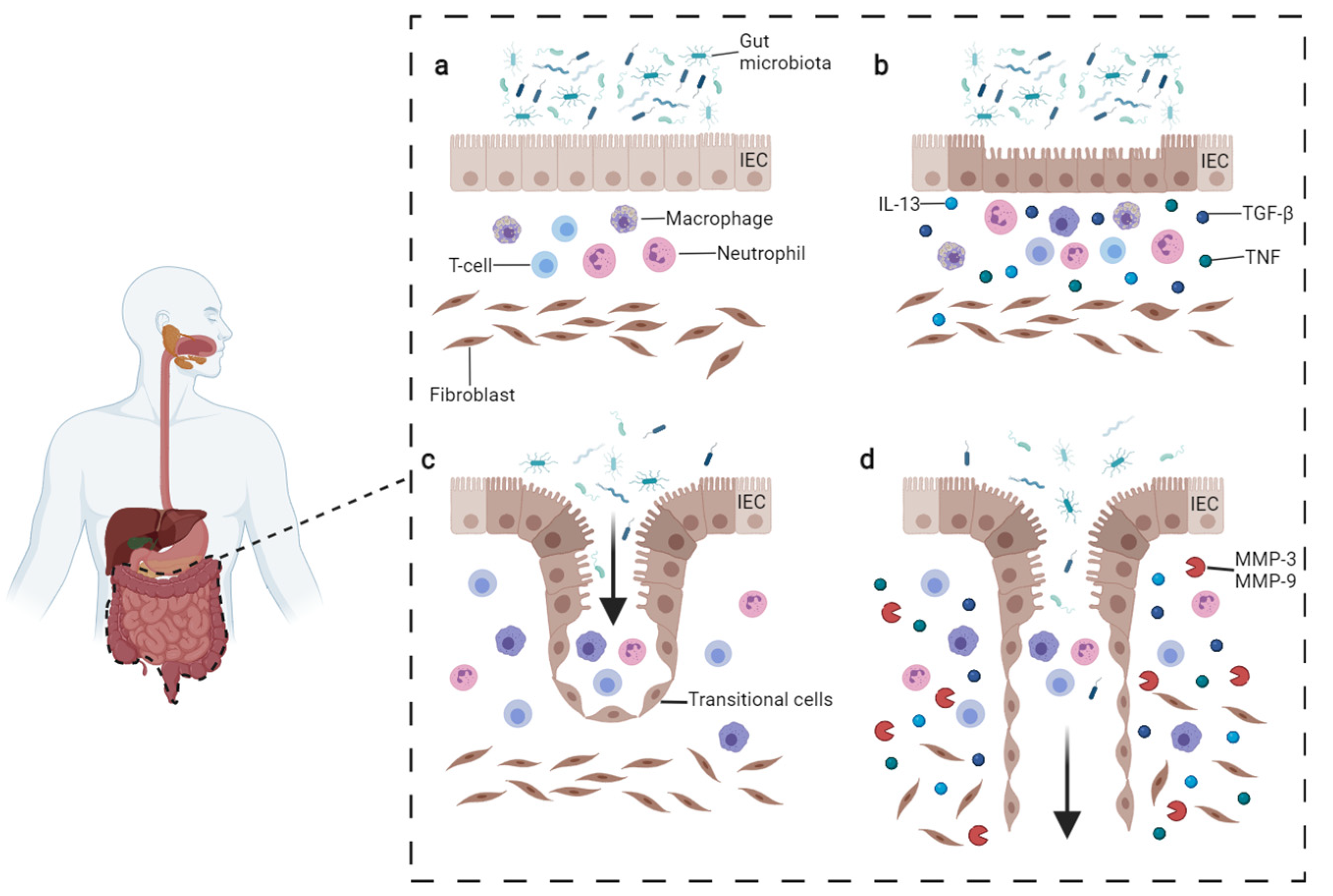
Abstract
:1. Common Features of Fistulae in Crohn’s Disease
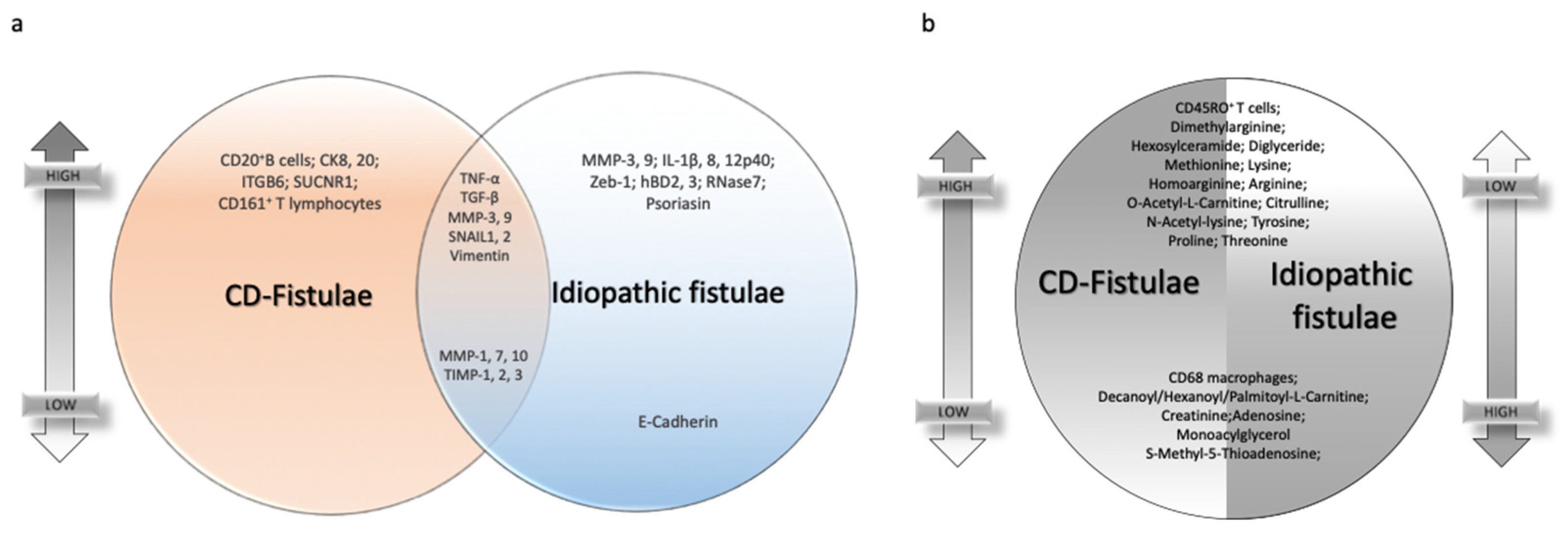
2. Signature Differences Distinguish CD-Related Fistulae from Idiopathic Ones
3. EMT Process in Fistula Formation
4. Mucinous Adenocarcinoma as a Complication in CD Patients with Fistula
5. Therapeutic Approaches to CD-Related Fistulae
6. Conclusions
Author Contributions
Funding
Institutional Review Board Statement
Informed Consent Statement
Data Availability Statement
Conflicts of Interest
References
- Piovani, D.; Danese, S.; Peyrin-Biroulet, L.; Nikolopoulos, G.K.; Lytras, T.; Bonovas, S. Environmental Risk Factors for Inflammatory Bowel Diseases: An Umbrella Review of Meta-Analyses. Gastroenterology 2019, 157, 647–659.e4. [Google Scholar] [CrossRef] [Green Version]
- Baumgart, D.C.; Sandborn, W.J. Crohn’s disease. Lancet 2012, 380, 1590–1605. [Google Scholar] [CrossRef] [Green Version]
- Torres, J.; Mehandru, S.; Colombel, J.F.; Peyrin-Biroulet, L. Crohn’s disease. Lancet 2017, 389, 1741–1755. [Google Scholar] [CrossRef]
- Peyrin-Biroulet, L.; Harmsen, W.S.; Tremaine, W.J.; Zinsmeister, A.R.; Sandborn, W.J.; Loftus, E.V. Cumulative Length of Bowel Resection in a Population-Based Cohort of Patients With Crohn’s Disease. Clin. Gastroenterol. Hepatol. 2016, 14, 1439–1444. [Google Scholar] [CrossRef] [Green Version]
- Thia, K.T.; Sandborn, W.J.; Harmsen, W.S.; Zinsmeister, A.R.; Loftus, E.V. Risk Factors Associated with Progression to Intestinal Complications of Crohn’s Disease in a Population-Based Cohort. Gastroenterology 2010, 139, 1147–1155. [Google Scholar] [CrossRef] [PubMed] [Green Version]
- Adegbola, S.O.; Dibley, L.; Sahnan, K.; Wade, T.; Verjee, A.; Sawyer, R.; Mannick, S.; McCluskey, D.; Yassin, N.; Phillips, R.K.S.; et al. Burden of disease and adaptation to life in patients with Crohn’s perianal fistula: A qualitative exploration. Health Qual. Life Outcomes 2020, 18, 370. [Google Scholar] [CrossRef] [PubMed]
- Bataille, F.; Klebl, F.; Rümmele, P.; Schroeder, J.; Farkas, S.; Wild, P.-J.; Fürst, A.; Hofstädter, F.; Schölmerich, J.; Herfarth, H.; et al. Morphological characterisation of Crohn’s disease fistulae. Gut 2004, 53, 1314–1321. [Google Scholar] [CrossRef] [Green Version]
- Meier, J.K.H.; Scharl, M.; Miller, S.N.; Brenmoehl, J.; Hausmann, M.; Kellermeier, S.; Schölmerich, J.; Rogler, G. Specific differences in migratory function of myofibroblasts isolated from Crohn’s disease fistulae and strictures. Inflamm. Bowel Dis. 2011, 17, 202–212. [Google Scholar] [CrossRef] [PubMed] [Green Version]
- Botaille, F.; Rohrmeier, C.; Bates, R.; Weber, A.; Reider, F.; Brenmoehl, J.; Strauch, U.; Farkas, S.; Fǔrst, A.; Hofstäáter, F.; et al. Evidence for a role of epithelial mesenchymal transition during pathogenesis of fistulae in Crohn’s disease. Inflamm. Bowel Dis. 2008, 14, 1514–1527. [Google Scholar] [CrossRef] [PubMed] [Green Version]
- Feagan, B.G.; Schwartz, D.; Danese, S.; Rubin, D.T.; Lissoos, T.W.; Xu, J.; Lasch, K. Efficacy of Vedolizumab in Fistulising Crohn’s Disease: Exploratory Analyses of Data from GEMINI 2. J. Crohn’s Colitis 2018, 12, 621–626. [Google Scholar] [CrossRef] [Green Version]
- Sokol, H.; Landman, C.; Seksik, P.; Berard, L.; Montil, M.; Nion-Larmurier, I.; Bourrier, A.; Le Gall, G.; Lalande, V.; De Rougemont, A.; et al. Fecal microbiota transplantation to maintain remission in Crohn’s disease: A pilot randomized controlled study. Microbiome 2020, 8, 12. [Google Scholar] [CrossRef] [PubMed]
- Haac, B.E.; Palmateer, N.C.; Seaton, M.E.; Van Yperen, R.; Fraser, C.M.; Bafford, A.C. A Distinct Gut Microbiota Exists within Crohn’s Disease–Related Perianal Fistulae. J. Surg. Res. 2019, 242, 118–128. [Google Scholar] [CrossRef] [PubMed]
- Bullman, S.; Pedamallu, C.S.; Sicinska, E.; Clancy, T.E.; Zhang, X.; Cai, D.; Neuberg, D.; Huang, K.; Guevara, F.; Nelson, T.; et al. Analysis of Fusobacterium persistence and antibiotic response in colorectal cancer. Science 2017, 358, 1443–1448. [Google Scholar] [CrossRef] [Green Version]
- Yu, T.; Guo, F.; Yu, Y.; Sun, T.; Ma, D.; Han, J.; Qian, Y.; Kryczek, I.; Sun, D.; Nagarsheth, N.; et al. Fusobacterium nucleatum Promotes Chemoresistance to Colorectal Cancer by Modulating Autophagy. Cell 2017, 170, 548–563.e16. [Google Scholar] [CrossRef] [Green Version]
- Panés, J.; Rimola, J. Perianal fistulizing Crohn’s disease: Pathogenesis, diagnosis and therapy. Nat. Rev. Gastroenterol. Hepatol. 2017, 14, 652–664. [Google Scholar] [CrossRef]
- Tozer, P.J.; Whelan, K.; Phillips, R.K.S.; Hart, A.L. Etiology of perianal Crohn’s disease: Role of genetic, microbiological, and immunological factors. Inflamm. Bowel Dis. 2009, 15, 1591–1598. [Google Scholar] [CrossRef] [PubMed]
- Parks, A.G. Pathogenesis and Treatment of Fistula-in-Ano. Br. Med. J. 1961, 1, 463–469. [Google Scholar] [CrossRef] [Green Version]
- Yassin, N.A.; Bernardo, D.; Mallappa, S. The presence of endotoxins in Crohn’s and idiopathic perianal fistula tracks. United Eur. Gastroenterol. J. 2013, 1, A506. [Google Scholar]
- van Onkelen, R.S.; Mitalas, L.E.; Gosselink, M.P.; van Belkum, A.; Laman, J.D.; Schouten, W.R. Assessment of microbiota and peptidoglycan in perianal fistulas. Diagn. Microbiol. Infect. Dis. 2013, 75, 50–54. [Google Scholar] [CrossRef]
- Kirkegaard, T.; Hansen, A.; Bruun, E.; Brynskov, J. Expression and localisation of matrix metalloproteinases and their natural inhibitors in fistulae of patients with Crohn’s disease. Gut 2004, 53, 701–709. [Google Scholar] [CrossRef] [Green Version]
- Van Onkelen, R.S.; Gosselink, M.P.; Van Meurs, M.; Melief, M.J.; Schouten, W.R.; Laman, J.D. Pro-inflammatory cytokines in cryptoglandular anal fistulas. Tech. Coloproctol. 2016, 20, 619–625. [Google Scholar] [CrossRef] [PubMed] [Green Version]
- Plevy, S.E.; Landers, C.J.; Prehn, J.; Carramanzana, N.M.; Deem, R.L.; Shealy, D.; Targan, S.R. A role for TNF-alpha and mucosal T helper-1 cytokines in the pathogenesis of Crohn’s disease. J. Immunol. 1997, 159, 6276–6282. [Google Scholar]
- Ortiz-Masiá, D.; Gisbert-Ferrándiz, L.; Bauset, C.; Coll, S.; Mamie, C.; Scharl, M.; Esplugues, J.V.; Alós, R.; Navarro, F.; Cosín-Roger, J.; et al. Succinate Activates EMT in Intestinal Epithelial Cells through SUCNR1: A Novel Protagonist in Fistula Development. Cells 2020, 9, 1104. [Google Scholar] [CrossRef]
- Kiehne, K.; Fincke, A.; Brunke, G.; Lange, T.; Fölsch, U.R.; Herzig, K.-H. Antimicrobial peptides in chronic anal fistula epithelium. Scand. J. Gastroenterol. 2007, 42, 1063–1069. [Google Scholar] [CrossRef] [PubMed]
- Ratto, C.; Litta, F.; Lucchetti, D.; Parello, A.; Boninsegna, A.; Arena, V.; Donisi, L.; Calapà, F.; Sgambato, A. Immunopathological characterization of cryptoglandular anal fistula: A pilot study investigating its pathogenesis. Color. Dis. 2016, 18, O436–O444. [Google Scholar] [CrossRef]
- Maggi, L.; Capone, M.; Giudici, F.; Santarlasci, V.; Querci, V.; Liotta, F.; Ficari, F.; Maggi, E.; Tonelli, F.; Annunziato, F.; et al. CD4+CD161+ T lymphocytes infiltrate crohn’s disease-associated perianal fistulas and are reduced by anti-TNF-α local therapy. Int. Arch. Allergy Immunol. 2013, 161, 81–86. [Google Scholar] [CrossRef]
- Haddow, J.B.; Musbahi, O.; Macdonald, T.T.; Knowles, C.H. Comparison of cytokine and phosphoprotein profiles in idiopathic and Crohn’s disease-related perianal fistula. World J. Gastrointest. Pathophysiol. 2019, 10, 42–53. [Google Scholar] [CrossRef]
- Adegbola, S.O.; Sarafian, M.; Sahnan, K.; Ding, N.S.; Faiz, O.D.; Warusavitarne, J.; Phillips, R.K.S.; Tozer, P.J.; Holmes, E.; Hart, A.L. Differences in amino acid and lipid metabolism distinguish Crohn’s from idiopathic/cryptoglandular perianal fistulas by tissue metabonomic profiling and may offer clues to underlying pathogenesis. Eur. J. Gastroenterol. Hepatol. 2021, 33, 1469–1479. [Google Scholar] [CrossRef] [PubMed]
- Jia, D.; Park, J.H.; Kaur, H.; Jung, K.H.; Yang, S.; Tripathi, S.; Galbraith, M.; Deng, Y.; Jolly, M.K.; Kaipparettu, B.A.; et al. Towards decoding the coupled decision-making of metabolism and epithelial-to-mesenchymal transition in cancer. Br. J. Cancer 2021, 124, 1902–1911. [Google Scholar] [CrossRef]
- Kalluri, R.; Weinberg, R.A. The basics of epithelial-mesenchymal transition. J. Clin. Investig. 2009, 119, 1420–1428. [Google Scholar] [CrossRef] [Green Version]
- Scharl, M.; Weber, A.; Fürst, A.; Farkas, S.; Jehle, E.; Pesch, T.; Kellermeier, S.; Fried, M.; Rogler, G. Potential role for snail family transcription factors in the etiology of Crohn’s disease-associated fistulae. Inflamm. Bowel Dis. 2011, 17, 1907–1916. [Google Scholar] [CrossRef]
- Scharl, M.; Frei, S.; Pesch, T.; Kellermeier, S.; Arikkat, J.; Frei, P.; Fried, M.; Weber, A.; Jehle, E.; Rühl, A.; et al. Interleukin-13 and transforming growth factor β synergise in the pathogenesis of human intestinal fistulae. Gut 2013, 62, 63–72. [Google Scholar] [CrossRef]
- Wynn, T.A. IL-13 Effector Functions. Annu. Rev. Immunol. 2003, 21, 425–456. [Google Scholar] [CrossRef]
- Chiaramonte, M.G.; Donaldson, D.D.; Cheever, A.W.; Wynn, T.A. An IL-13 inhibitor blocks the development of hepatic fibrosis during a T-helper type 2–dominated inflammatory response. J. Clin. Investig. 1999, 104, 777–785. [Google Scholar] [CrossRef] [PubMed] [Green Version]
- Zhu, Z.; Homer, R.J.; Wang, Z.; Chen, Q.; Geba, G.P.; Wang, J.; Zhang, Y.; Elias, J.A. Pulmonary expression of interleukin-13 causes inflammation, mucus hypersecretion, subepithelial fibrosis, physiologic abnormalities, and eotaxin production. J. Clin. Investig. 1999, 103, 779–788. [Google Scholar] [CrossRef] [PubMed] [Green Version]
- Hasegawa, M.; Fujimoto, M.; Kikuchi, K.; Takehara, K. Elevated serum levels of interleukin 4 (IL-4), IL-10, and IL-13 in patients with systemic sclerosis. J. Rheumatol. 1997, 24, 328–332. [Google Scholar] [PubMed]
- Aguilera, O.; Peña, C.; García, J.M.; Larriba, M.J.; Ordóñez-Morán, P.; Navarro, D.; Barbáchano, A.; de Silanes, I.L.; Ballestar, E.; Fraga, M.F.; et al. The Wnt antagonist DICKKOPF-1 gene is induced by 1α,25-dihydroxyvitamin D3 associated to the differentiation of human colon cancer cells. Carcinogenesis 2007, 28, 1877–1884. [Google Scholar] [CrossRef] [PubMed]
- González-Sancho, J.M.; Aguilera, O.; García, J.M.; Pendás-Franco, N.; Peña, C.; Cal, S.; De Herreros, A.G.; Bonilla, F.; Muñoz, A. The Wnt antagonist DICKKOPF-1 gene is a downstream target of β-catenin/TCF and is downregulated in human colon cancer. Oncogene 2005, 24, 1098–1103. [Google Scholar] [CrossRef] [Green Version]
- Frei, S.M.; Hemsley, C.; Pesch, T.; Lang, S.; Weber, A.; Jehle, E.; Rühl, A.; Fried, M.; Rogler, G.; Scharl, M. The Role for Dickkopf-Homolog-1 in the Pathogenesis of Crohn’s Disease-Associated Fistulae. PLoS ONE 2013, 8, e78882. [Google Scholar] [CrossRef] [PubMed]
- Ortiz-Masià, D.; Salvador, P.; Macias-Ceja, D.C.; Gisbert-Ferrándiz, L.; Esplugues, J.V.; Manyé, J.; Alós, R.; Navarro-Vicente, F.; Mamie, C.; Scharl, M.; et al. WNT2b Activates Epithelial-mesenchymal Transition Through FZD4: Relevance in Penetrating Crohńs Disease. J. Crohn’s Colitis 2020, 14, 230–239. [Google Scholar] [CrossRef]
- Goffin, L.; Fagagnini, S.; Vicari, A.; Mamie, C.; Melhem, H.; Weder, B.; Lutz, C.; Lang, S.; Scharl, M.; Rogler, G.; et al. Anti-MMP-9 Antibody: A promising therapeutic strategy for treatment of inflammatory bowel disease complications with fibrosis. Inflamm. Bowel Dis. 2016, 22, 2041–2057. [Google Scholar] [CrossRef] [Green Version]
- Bruckner, R.S.; Spalinger, M.R.; Barnhoorn, M.C.; Feakins, R.; Fuerst, A.; Jehle, E.C.; Rickenbacher, A.; Turina, M.; Niechcial, A.; Lang, S.; et al. Contribution of CD3+CD8− and CD3+CD8+ T Cells to TNF-α Overexpression in Crohn Disease–Associated Perianal Fistulas and Induction of Epithelial-Mesenchymal Transition in HT-29 Cells. Inflamm. Bowel Dis. 2021, 27, 538–549. [Google Scholar] [CrossRef]
- Bautista, J.M.; Marín-Jiménez, I.; Moreno, L.H. A Perianal Mass in a Crohn’s Disease Patient. Gastroenterology 2012, 142, 12–187. [Google Scholar] [CrossRef]
- Yang, B.-L.; Shao, W.-J.; Sun, G.-D.; Chen, Y.-Q.; Huang, J.-C. Perianal mucinous adenocarcinoma arising from chronic anorectal fistulae: A review from single institution. Int. J. Colorectal Dis. 2009, 24, 1001–1006. [Google Scholar] [CrossRef]
- Ky, A.; Sohn, N.; Weinstein, M.A.; Korelitz, B.I. Carcinoma arising in anorectal fistulas of Crohn’s disease. Dis. Colon Rectum 1998, 41, 992–996. [Google Scholar] [CrossRef] [PubMed]
- Iesalnieks, I.; Gaertner, W.B.; Glaß, H.; Strauch, U.; Hipp, M.; Agha, A.; Schlitt, H.J. Fistula-associated anal adenocarcinoma in Crohn’s disease. Inflamm. Bowel Dis. 2010, 16, 1643–1648. [Google Scholar] [CrossRef]
- Smith, R.; Hicks, D.; Tomljanovich, P.I.; Lele, S.B.; Rajput, A.; Bullard Dunn, K. Adenocarcinoma arising from chronic perianal Crohn’s disease: Case report and review of the literaturetle. Am. Surg. 2008, 74, 59–61. [Google Scholar] [CrossRef] [PubMed]
- Santos, M.D.; Nogueira, C.; Lopes, C. Mucinous Adenocarcinoma Arising in Chronic Perianal Fistula: Good Results with Neoadjuvant Chemoradiotherapy Followed by Surgery. Case Rep. Surg. 2014, 2014, 386150. [Google Scholar] [CrossRef] [PubMed]
- Hugen, N.; van Beek, J.J.P.; De Wilt, J.H.W.; Nagtegaal, I.D. Insight into Mucinous Colorectal Carcinoma: Clues from Etiology. Ann. Surg. Oncol. 2014, 21, 2963–2970. [Google Scholar] [CrossRef] [PubMed]
- Prasad, S.N.; Razik, A.; Siddiqui, F.; Lal, H. Mucinous adenocarcinoma arising from chronic perianal fistula mimicking horseshoe abscess. BMJ Case Rep. 2018, 2018, bcr-2017. [Google Scholar] [CrossRef] [PubMed]
- Díaz-Vico, T.; Fernández-Martínez, D.; García-Gutiérrez, C.; Suárez-Sánchez, A.; Cifrián-Canales, I.; Mendoza-Pacas, G.E.; Sánchez-Farpón, H.; Truán-Alonso, N. Mucinous adenocarcinoma arising from chronic perianal fistula-a multidisciplinary approach. J. Gastrointest. Oncol. 2019, 10, 589–596. [Google Scholar] [CrossRef] [PubMed]
- Papaconstantinou, I.; Mantzos, D.S.; Kondi-Pafiti, A.; Koutroubakis, I.E. Anal adenocarcinoma complicating chronic Crohn’s disease. Int. J. Surg. Case Rep. 2015, 10, 201–203. [Google Scholar] [CrossRef] [PubMed] [Green Version]
- Schwartz, D.A.; Ghazi, L.J.; Regueiro, M.; Fichera, A.; Zoccali, M.; Ong, E.M.W.; Mortelé, K.J. Guidelines for the multidisciplinary management of Crohn’s perianal fistulas: Summary statement. Inflamm. Bowel Dis. 2015, 21, 723–730. [Google Scholar] [CrossRef] [PubMed]
- Kotze, P.G.; Shen, B.; Lightner, A.; Yamamoto, T.; Spinelli, A.; Ghosh, S.; Panaccione, R. Modern management of perianal fistulas in Crohn’s disease: Future directions. Gut 2018, 67, 1181–1194. [Google Scholar] [CrossRef]
- van Koperen, P.J.; Safiruddin, F.; Bemelman, W.A.; Slors, J.F.M. Outcome of surgical treatment for fistula in ano in Crohn’s disease. Br. J. Surg. 2009, 96, 675–679. [Google Scholar] [CrossRef] [PubMed]
- Torres, J.; Bonovas, S.; Doherty, G.; Kucharzik, T.; Gisbert, J.P.; Raine, T.; Adamina, M.; Armuzzi, A.; Bachmann, O.; Bager, P.; et al. ECCO Guidelines on Therapeutics in Crohn’s Disease: Medical Treatment. J. Crohn’s Colitis 2020, 14, 4–22. [Google Scholar] [CrossRef]
- Adamina, M.; Bonovas, S.; Raine, T.; Spinelli, A.; Warusavitarne, J.; Armuzzi, A.; Bachmann, O.; Bager, P.; Biancone, L.; Bokemeyer, B.; et al. ECCO Guidelines on Therapeutics in Crohn’s Disease: Surgical Treatment. J. Crohn’s Colitis 2020, 14, 155–168. [Google Scholar] [CrossRef] [PubMed] [Green Version]
- Sands, B.E.; Anderson, F.H.; Bernstein, C.N.; Chey, W.Y.; Feagan, B.G.; Fedorak, R.N.; Kamm, M.A.; Korzenik, J.R.; Lashner, B.A.; Onken, J.E.; et al. Infliximab Maintenance Therapy for Fistulizing Crohn’s Disease. N. Engl. J. Med. 2004, 350, 876–885. [Google Scholar] [CrossRef] [PubMed]
- Regueiro, M.; Mardini, H. Treatment of Perianal Fistulizing Crohn’s Disease with Infliximab Alone or as an Adjunct to Exam under Anesthesia with Seton Placement. Inflamm. Bowel Dis. 2003, 9, 98–103. [Google Scholar] [CrossRef]
- Poggioli, G.; Laureti, S.; Pierangeli, F.; Rizzello, F.; Ugolini, F.; Gionchetti, P.; Campieri, M. Local Injection of Infliximab for the Treatment of Perianal Crohn’s Disease. Dis. Colon Rectum 2005, 48, 768–774. [Google Scholar] [CrossRef] [PubMed]
- Tonelli, F.; Giudici, F.; Asteria, C.R. Effectiveness and safety of local adalimumab injection in patients with fistulizing perianal crohn’s disease: A pilot study. Dis. Colon Rectum 2012, 55, 870–875. [Google Scholar] [CrossRef] [PubMed]
- Asteria, C.R.; Ficari, F.; Bagnoli, S.; Milla, M.; Tonelli, F. Treatment of perianal fistulas in Crohn’s disease by local injection of antibody to TNF-α accounts for a favourable clinical response in selected cases: A pilot study. Scand. J. Gastroenterol. 2006, 41, 1064–1072. [Google Scholar] [CrossRef]
- Sica, G.S.; Di Carlo, S.; Tema, G.; Montagnese, F.; Blanco, G.D.V.; Fiaschetti, V.; Maggi, G.; Biancone, L. Treatment of peri-anal fistula in Crohn’s disease. World J. Gastroenterol. 2014, 20, 13205–13210. [Google Scholar] [CrossRef]
- De Parades, V.; Far, H.S.; Etienney, I.; Zeitoun, J.-D.; Atienza, P.; Bauer, P. Seton drainage and fibrin glue injection for complex anal fistulas. Color. Dis. 2010, 12, 459–463. [Google Scholar] [CrossRef] [PubMed]
- Senéjoux, A.; Siproudhis, L.; Abramowitz, L.; Munoz-Bongrand, N.; Desseaux, K.; Bouguen, G.; Bourreille, A.; Dewit, O.; Stefanescu, C.; Vernier, G.; et al. Fistula Plug in Fistulising Ano-Perineal Crohn’s Disease: A Randomised Controlled Trial. J. Crohn’s Colitis 2016, 10, 141–148. [Google Scholar] [CrossRef] [PubMed]
- Mennigen, R.; Laukötter, M.; Senninger, N.; Rijcken, E. The OTSC® proctology clip system for the closure of refractory anal fistulas. Tech. Coloproctol. 2015, 19, 241–246. [Google Scholar] [CrossRef] [PubMed]
- Tozer, P.J.; Burling, D.; Gupta, A.; Phillips, R.K.S.; Hart, A.L. Review article: Medical, surgical and radiological management of perianal Crohn’s fistulas. Aliment. Pharmacol. Ther. 2011, 33, 5–22. [Google Scholar] [CrossRef] [Green Version]
- Stellingwerf, M.E.; van Praag, E.M.; Tozer, P.J.; Bemelman, W.A.; Buskens, C.J. Systematic review and meta-analysis of endorectal advancement flap and ligation of the intersphincteric fistula tract for cryptoglandular and Crohn’s high perianal fistulas. BJS Open 2019, 3, 231–241. [Google Scholar] [CrossRef]
- Meinero, P.; Mori, L. Video-assisted anal fistula treatment (VAAFT): A novel sphincter-saving procedure for treating complex anal fistulas. Tech. Coloproctol. 2011, 15, 417–422. [Google Scholar] [CrossRef] [Green Version]
- Wilhelm, A.; Fiebig, A.; Krawczak, M. Five years of experience with the FiLaCTM laser for fistula-in-ano management: Long-term follow-up from a single institution. Tech. Coloproctol. 2017, 21, 269–276. [Google Scholar] [CrossRef] [Green Version]
- Guadalajara, H.; García-Arranz, M.; Herreros, M.D.; Borycka-Kiciak, K.; Lightner, A.L.; García-Olmo, D. Mesenchymal stem cells in perianal Crohn’s disease. Tech. Coloproctol. 2020, 24, 883–889. [Google Scholar] [CrossRef]
- Jiang, X.-X.; Zhang, Y.; Liu, B.; Zhang, S.-X.; Wu, Y.; Yu, X.-D.; Mao, N. Human mesenchymal stem cells inhibit differentiation and function of monocyte-derived dendritic cells. Blood 2005, 105, 4120–4126. [Google Scholar] [CrossRef] [Green Version]
- Panés, J.; García-Olmo, D.; Van Assche, G.; Colombel, J.F.; Reinisch, W.; Baumgart, D.C.; Dignass, A.; Nachury, M.; Ferrante, M.; Kazemi-Shirazi, L.; et al. Expanded allogeneic adipose-derived mesenchymal stem cells (Cx601) for complex perianal fistulas in Crohn’s disease: A phase 3 randomised, double-blind controlled trial. Lancet 2016, 388, 1281–1290. [Google Scholar] [CrossRef]
- Aguirre, J.E.; Beswick, E.J.; Grim, C.; Uribe, G.; Tafoya, M.; Palma, G.C.; Samedi, V.; McKee, R.; Villeger, R.; Fofanov, Y.; et al. Matrix metalloproteinases cleave membrane-bound PD-L1 on CD90+ (myo-)fibroblasts in Crohn’s disease and regulate Th1/Th17 cell responses. Int. Immunol. 2020, 32, 57–68. [Google Scholar] [CrossRef] [PubMed]
- Fontani, F.; Marcucci, T.; Picariello, L.; Tonelli, F.; Vincenzini, M.T.; Iantomasi, T. Redox regulation of MMP-3/TIMP-1 ratio in intestinal myofibroblasts: Effect of N-acetylcysteine and curcumin. Exp. Cell Res. 2014, 323, 77–86. [Google Scholar] [CrossRef] [PubMed]
- Dulai, P.S.; Gleeson, M.W.; Taylor, D.; Holubar, S.D.; Buckey, J.C.; Siegel, C.A. Systematic review: The safety and efficacy of hyperbaric oxygen therapy for inflammatory bowel disease. Aliment. Pharmacol. Ther. 2014, 39, 1266–1275. [Google Scholar] [CrossRef] [PubMed]
- Lansdorp, C.A.; Gecse, K.B.; Buskens, C.J.; Löwenberg, M.; Stoker, J.; Bemelman, W.A.; D’Haens, G.R.A.M.; van Hulst, R.A. Hyperbaric oxygen therapy for the treatment of perianal fistulas in 20 patients with Crohn’s disease. Aliment. Pharmacol. Ther. 2021, 53, 587–597. [Google Scholar] [CrossRef]


Publisher’s Note: MDPI stays neutral with regard to jurisdictional claims in published maps and institutional affiliations. |
© 2021 by the authors. Licensee MDPI, Basel, Switzerland. This article is an open access article distributed under the terms and conditions of the Creative Commons Attribution (CC BY) license (https://creativecommons.org/licenses/by/4.0/).
Share and Cite
Rubbino, F.; Greco, L.; di Cristofaro, A.; Gaiani, F.; Vetrano, S.; Laghi, L.; Bonovas, S.; Piovani, D. Journey through Crohn’s Disease Complication: From Fistula Formation to Future Therapies. J. Clin. Med. 2021, 10, 5548. https://doi.org/10.3390/jcm10235548
Rubbino F, Greco L, di Cristofaro A, Gaiani F, Vetrano S, Laghi L, Bonovas S, Piovani D. Journey through Crohn’s Disease Complication: From Fistula Formation to Future Therapies. Journal of Clinical Medicine. 2021; 10(23):5548. https://doi.org/10.3390/jcm10235548
Chicago/Turabian StyleRubbino, Federica, Luana Greco, Alessio di Cristofaro, Federica Gaiani, Stefania Vetrano, Luigi Laghi, Stefanos Bonovas, and Daniele Piovani. 2021. "Journey through Crohn’s Disease Complication: From Fistula Formation to Future Therapies" Journal of Clinical Medicine 10, no. 23: 5548. https://doi.org/10.3390/jcm10235548