Article

Uric Acid as a Predictor for Early Allograft Dysfunction after Living Donor Liver Transplantation: A Prospective Observational Study

1
Department of Anesthesiology, Chang Gung Memorial Hospital, Linkou Branch, Taoyuan 333, Taiwan
2
College of Medicine, Chang Gung University, Taoyuan 333, Taiwan
3
Department of General Surgery, Chang Gung Memorial Hospital, Linkou Branch, Taoyuan 333, Taiwan
4
Center for Big Data Analytics and Statistics, Chang Gung Memorial Hospital, Linkou Branch, Taoyuan 333, Taiwan
5
Department of Liver and Transplant Surgery, Chang Gung Memorial Hospital, Linkou Branch, Taoyuan 333, Taiwan
*
Author to whom correspondence should be addressed.
H.-I.T. and L.-M.H. contributed equally to this work as first authors.
J. Clin. Med. 2021, 10(12), 2729; https://doi.org/10.3390/jcm10122729
Submission received: 7 April 2021 / Revised: 20 May 2021 / Accepted: 1 June 2021 / Published: 21 June 2021
(This article belongs to the Section Gastroenterology & Hepatopancreatobiliary Medicine)

Abstract

:
Early allograft dysfunction (EAD) is a postoperative complication that may cause graft failure and mortality after liver transplantation. The objective of this study was to examine whether the preoperative serum uric acid (SUA) level may predict EAD. We performed a prospective observational study, including 61 donor/recipient pairs who underwent living donor liver transplantation (LDLT). In the univariate and multivariate analysis, SUA ≤4.4 mg/dL was related to a five-fold (odds ratio (OR): 5.16, 95% confidence interval (CI): 1.41–18.83; OR: 5.39, 95% CI: 1.29–22.49, respectively) increased risk for EAD. A lower preoperative SUA was related to a higher incidence of and risk for EAD. Our study provides a new predictor for evaluating EAD and may exert a protective effect against EAD development.

1. Introduction

One of the curative treatments for end-stage liver disease is liver transplantation. As a result of the urgent need for liver transplantation and a critical lack of liver grafts from deceased donors, living donor liver transplantation (LDLT) became a solution to this plight in 1994 [1]. However, early allograft dysfunction (EAD) is a common complication leading to postoperative morbidity and mortality after liver transplantation [2,3]. EAD is the result of ischemia/reperfusion injury from reactive oxygen species following graft injury [4]. EAD patients are at greater risk for postoperative graft dysfunction or even graft failure that may require re-transplantation. That said, only a few of these patients undergo re-transplantation due to the scarce supply of matched liver donors. Donor- and recipient-related risk factors have been established in association with the development of EAD, such as donor body mass index (BMI), donor age, recipient age, cold ischemia time, and recipient liver mass [5,6], all of which should be taken into consideration before and during liver transplantation.
Recently, uric acid (UA) has been widely studied for its antioxidant capacity. UA is a terminal product of purine metabolism and a potent water-soluble molecule that accounts for more than half of the antioxidant capacity in human plasma [7,8]. UA can stabilize vitamin C in serum and eliminate peroxynitrite, which may result in nitric oxide donor formation in vitro [9]. Traditionally, UA is notorious for its relationship with gout, cardiovascular disease, and metabolic syndrome [7]. However, interestingly, a low serum concentration of UA was reported to be associated with a higher prevalence of and deterioration in some neurological diseases, including Alzheimer’s disease and Parkinson’s disease, as well as a worse outcome after acute ischemic stroke [10,11,12]. Increasing evidence has shown that UA plays a crucial role as an antioxidant and contributes to hepatic antioxidant capacity [13]. We hypothesized that higher preoperative serum uric acid (SUA) level may exert a protective effect against EAD.

2. Materials and Methods

2.1. Objectives

This prospective, observational, single-center, hospital-based study was executed after receiving approvals from the Institutional Review Board of Chang Gung Memorial Hospital, Linkou Branch, Taiwan (Registration Numbers: 201800847A3, CMRPG3K1881, CMRPG3H1191, CMRPG3H11912 CMRPG3H1193). Septic patients, patients with shock, patients with preoperative pulmonary hypertension wedge pressure higher than 35 mmHg, and patients who declined to participate were excluded from the study. A total of 61 pairs of donors and recipients who performed LDLT at Taiwan Chang Gung Memorial Hospital (Taoyuan, Taiwan) from October 2018 to March 2020 were recruited into the study, as shown in Figure 1.

2.2. Data Collection and Variable Definition

In all cases, a biological model of the end-stage liver disease (MELD) score was measured on the day of the operation. All the patients were fasted at least 8 hours before liver transplantation. Before the induction of general anesthesia, blood samples were obtained from the arterial catheter and urine samples from the indwelling urinary catheter. Alanine aminotransferase (ALT), blood urea nitrogen (BUN), creatinine (Cre), SUA and urine uric acid (UUA) were measured in the hospital’s clinical laboratory. The estimated glomerular filtration rate (eGFR) was measured using the formula: eGFR (mL/min/1.73 m2) = 186 × Cre−1.154 × age−0.203 × (0.742 if women). Chronic kidney disease (CKD) was defined as a GFR less than 60 mL/min/1.73 m2. Cold ischemia time (CIT) was determined as the interval from the cold storage solution preservation to liver graft implantation. Warm ischemia time (WIT) was determined as the interval from hepatic vein reconstruction to portal vein reperfusion. The postoperative EAD was diagnosed according to the Otholff criteria as one or more of the following conditions: international normalized ratio ≥ 1.6 on Day 7, total bilirubin ≥ 10 mg/dL on Day 7, and alanine or aspartate aminotransferases ≥ 2000 IU/L within 7 days after liver transplantation [14].

2.3. Statistical Analysis
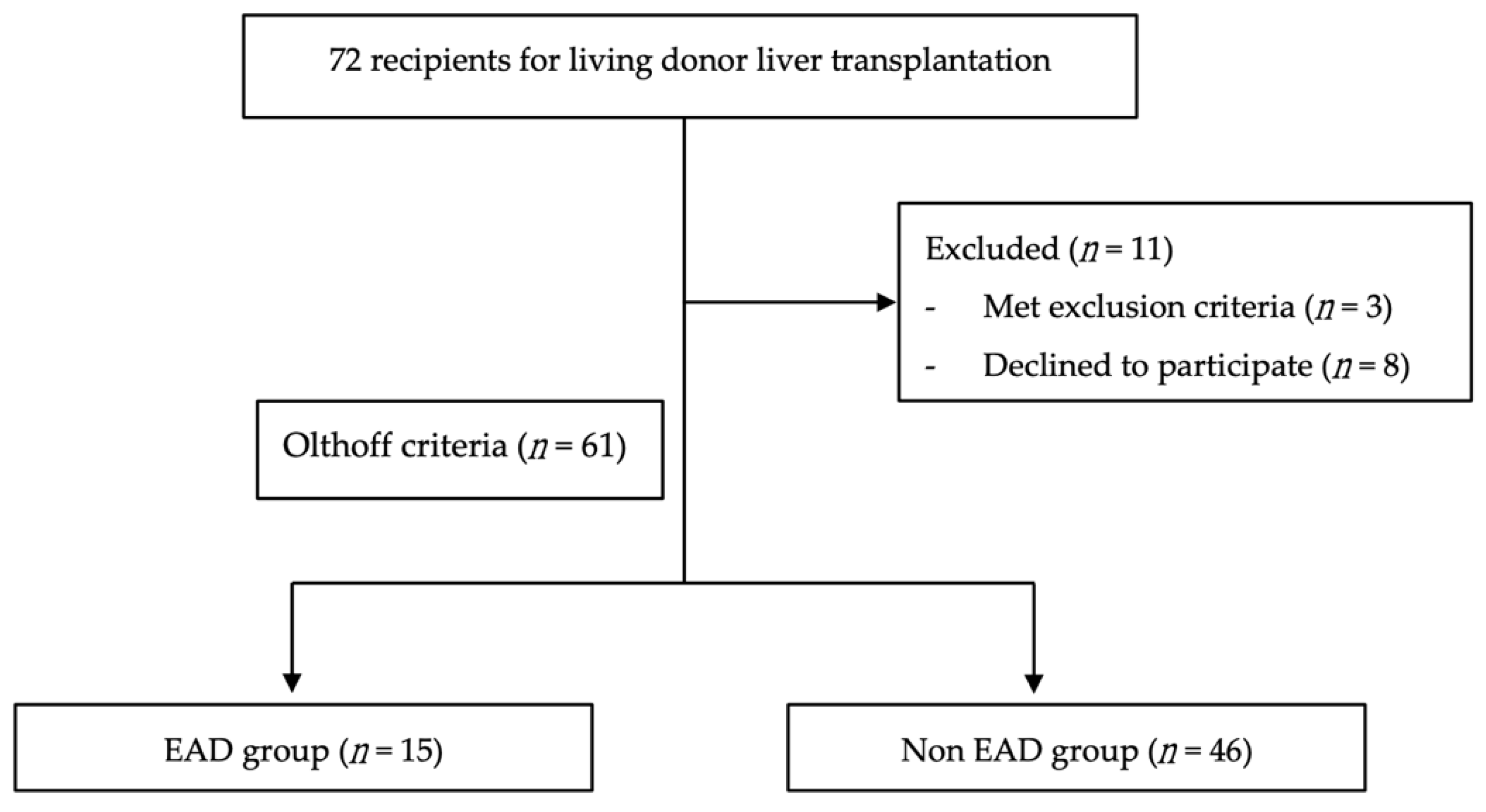
Established risk factors for EAD in this cohort were identified from previous reports [15,16]. Identified recipient-related risk factors were age, BMI, ALT, and MELD score. Donor-related risk factors were age, BMI, liver mass, and CIT. Numerical variables including MELD score, age, BMI, ALT, UUA, BUN, Cre, eGFR, graft size, graft recipient weight ratio (GRWR), intraoperative blood loss, CIT, and WIT were expressed as the mean and standard deviation. Categorical variables including SUA, blood type, sex, type of virology, and hepatocellular carcinoma (HCC) are shown as numbers and percentages. The association of the threshold SUA, comorbidities, demographic variables, and laboratory variables with the incidence of EAD was identified by univariate logistic regression analysis. With no selection criteria, the aforementioned potential risk factors of EAD following LDLT were then analyzed in the multiple logistic regression model to establish the independent influence of SUA on EAD. The strength of the association of each variable with EAD was summarized by calculating the odds ratio (OR) and corresponding 95% confidence interval (CI) from the coefficients estimated in the logistic regression models. Kaplan–Meier plots and Cox regression models were generated for survival analysis and prognostic factor analysis. All of the aforementioned data were analyzed using SAS 9.4 (SAS Institute Inc., Cary, NC, USA) statistical software. A p-value <0.05 was considered statistically significant.

3. Results

3.1. Baseline Characteristics

A total of 72 recipients were initially recruited to the study. After excluding three who met the exclusion criteria and eight who declined to participate, 61 recipients remained in this study. In this cohort, the mean age of the donors and recipients was 32.34 ± 9.21 and 55.13 ± 10.43 years, respectively. Among the recipients, 44 (72.13%) were males and 17 (27.86%) were females. Furthermore, 32 had hepatitis B virus (HBV)-related cirrhosis, 11 had hepatitis C virus (HCV)-related cirrhosis, 20 had alcoholic cirrhosis, and the remaining 27 had HCC. Intraoperatively, the graft size was, on average, 640 ± 131.43 g, with a GRWR of 0.98% ± 0.21%. Cold and warm ischemia times were on average 39.77 ± 25.97 and 16.41 ± 4.19 min, respectively. Additionally, 15 (24.59%) recipients developed EAD postoperatively and 11 died within 1 year of the operation. After evaluating the continuous relationship between the development of SUA and EAD, the SUA threshold was determined to be 4.4 mg/dL. Subjects were then divided into low SUA group (defined as ≤4.4 mg/dL; n = 27) and high SUA group (defined as >4.4 mg/dL; n = 34). The clinical characteristics of the patients are presented in Table 1 and Table 2.

3.2. SUA Level in Different Subgroups

We analyzed SUA levels in subgroups as shown in Table 3. In our study group, no patients had a history of gout in EAD group and thus no further analysis was conducted in regards to the effect of uric acid lowering drug for SUA level. Male recipients appeared to have lower UUA than female recipients. In patients who had alcoholism, chronic kidney disease (CKD), diabetes mellitus (DM) and hypertension (HTN), their UUA levels were lower. However, no statistical significance was observed in all aforementioned subgroups.

3.3. Risk Factors for Early Allograft Dysfunction

The clinical definition of hyperuricemia corresponds to a urate concentration exceeding 7 mg/dL for men and 6.5 mg/dL for women. We aimed to investigate the optimal cutoff value of SUA with maximal odds ratio for the prediction of EAD. The optimal cutoff value using the receiver operating characteristic (ROC) result in our study with SUA ≤ 4.4 mg/dL was related to a maximal odds ratio, as observed in Figure 2. Furthermore, we use SUA as a categorical variable instead of continuous variable for further evaluation. In the univariate analysis, SUA ≤ 4.4 mg/dL was related to a five-fold (OR: 5.16, 95% CI: 1.41–18.83) increased risk for EAD. Similarly, in the multivariate analysis, SUA ≤ 4.4 mg/dL also was associated with a five-fold (OR: 5.39, 95% CI: 1.29–22.49) increased risk for EAD. Potential risk factors for EAD, including blood type O, SUA, and MELD score, were established on the basis of the results of the univariate analysis. All significant factors in the univariate analysis were included to create a multivariate logistic regression model. Only SUA remained as an independent prognostic factor in the multivariate logistic regression model (Table 4).

3.4. SUA and the Association with Early Allograft Dysfunction

To further demonstrate that lower preoperative SUA was associated with a higher risk for EAD, the SUA data were divided into quartiles. The incidences of EAD in association with SUA are shown in Figure 3. We demonstrate that a higher SUA was related to a lower incidence of EAD.

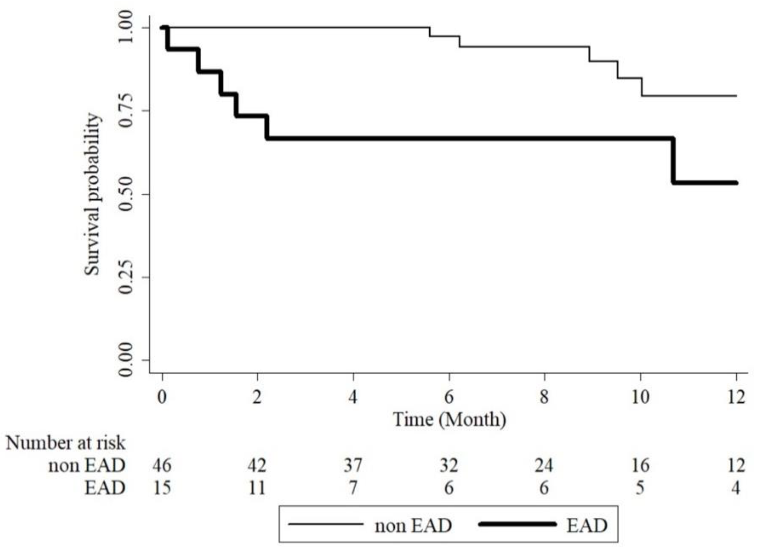
3.5. Early Allograft Dysfunction and One Year Mortality

Kaplan–Meier survival curves showed that the EAD group had a lower survival proportion than the EAD group (log-rank test, p = 0.0006), as shown in Figure 4. When adjusting for several variables using a Cox proportional hazards model, the EAD group had a significantly higher hazard ratio (HR) than the non-EAD group (HR: 6.69, 95% CI: 1.66–26.92, p = 0.008), as shown in Table 5. We also noted the trend between increasing SUA related to decreasing mortality but not statistically significant.

4. Discussion

As a result of the urgent need for liver transplantation and a critical lack of grafts from donation after circulatory death or donation after brain death, living donor liver donation has become a solution to the organ shortage dilemma. However, EAD, with an incidence of 15–27%, is a common postoperative complication that may lead to morbidity and mortality after liver transplantation [3,5,17]. Some risk factors for the development of EAD, whether donor-, recipient-, or surgery-related, have been identified that may help transplant surgeons take precautions before or during liver transplantation. EAD is the result of ischemia/reperfusion injury from reactive oxygen species following graft injury. Oxidative stress is related to the gradual increase in reactive oxygen species and adversely affects the pathogenesis of ischemia/reperfusion injury. Thus, safe and effective therapeutic agents with antioxidant properties to mitigate oxidative damage may provide another different treatment strategy.
Serum UA has been recently studied for its antioxidant activity in various diseases. UA is a final breakdown product of purine nucleotides, and its metabolism involves factors that regulate both hepatic production and renal excretion. Initially, two purine nucleotides, adenine and guanine, are converted into inosine and guanosine, before being further converted into the purine bases hypoxanthine and guanine, respectively. Hypoxanthine and guanine are then oxidized and deaminated by hypoxanthine oxidase (XO) and guanine deaminase to form xanthine, which is again oxidized by XO to form UA [9]. Disturbances of production and excretion can lead to abnormal SUA levels whether hypouricemia or hyperuricemia. The liver is the major site of UA production. In severe hepatocellular injury, XO activity is reduced for the production of UA [18]. The kidneys are the major site for UA excretion. About 65–75% of UA produced daily is excreted via kidneys [19]. SUA levels are proved positively correlated to urinary excretion of uric acid [20]. Ischemia/reperfusion injury after liver transplantation is complex and involves various pathways such as the activation of Toll-like receptors (TLRs), changes in messenger RNA (mRNA) expression, the generation of reactive oxygen species (ROS), the regulation of autophagy, and the activation of hypoxia-inducible factors [21]. UA has shown a protective role in reducing TLR4/nuclear factor kappa B (NF-κB) activation, reducing ROS production, and regulating apoptosis [22,23,24]. Interestingly, in healthy humans, the acute elevation of UA seems to prevent the increase in oxidative stress and arterial stiffness caused by hyperoxia [25]. UA treatment was also reported to prevent the worsening of early ischemic injury related to reperfusion after acute stroke in patients receiving thrombolysis [26,27]. Thus, a low UA level may imply the severity of initial liver injury and susceptibility to later reperfusion injury following liver transplantation.
In our study, we demonstrated that recipients with a lower preoperative SUA (≤4.4 mg/dL) are at a five-fold increased risk for the development of EAD postoperatively. Furthermore, those who developed EAD were at a six-fold increased risk of mortality, one year postoperatively. Such findings are exciting; however, limitations still apply to this study. Even though dietary habits may affect SUA level, all patients had SUA levels within normal range and maintained fasted for at least 8 hours preoperatively [28]. The small population size limited to Asian ethnicity and living donor transplantation patients can be a potential limitation, and validation in a large cohort including different ethnic groups is warranted. Even though SUA is an antioxidant, it is an approved risk factor for metabolic syndrome and cardiovascular complications [7,29]. The balance between risks and benefits can be challenging. Additional research is required to examine the altered enzymatic activities in the UA pathway in liver-diseased individuals and to validate the hypothesis that UA therapy may help in lowering the risk for EAD.

5. Conclusions

UA plays an important role in the human body due to its antioxidant capacity. We identified that preoperative SUA may be a potential biomarker to predict the development of EAD following LDLT. Higher preoperative SUA may exert a protective effect against EAD development.

Author Contributions

Conceptualization, H.-I.T. and H.-P.Y.; methodology, L.-M.H.; formal analysis, H.-M.C.; investigation, H.-I.T. and L.-M.H.; data curation, C.-W.L. and W.-C.L.; writing—original draft preparation, L.-M.H.; writing—review and editing, H.-I.T. and H.-P.Y.; supervision, H.-P.Y.; funding acquisition, H.-I.T. and H.-P.Y. All authors have read and agreed to the published version of the manuscript.

Funding

This work was funded by the Chang Gung Memorial Hospital (CMRPG3K1881, CMRPG3H1191, CMRPG3H11912, CMRPG3H1193).

Institutional Review Board Statement

The study was approved by the Institutional Review Board of Chang Gung Memorial Hospital, Linkou Branch, Taiwan (Registration Number 201800847A3, CMRPG3K1881).

Informed Consent Statement

Informed consent was obtained from all subjects involved in the study.

Data Availability Statement

The data presented in this study are available on request from the corresponding author. The data are not publicly available.

Acknowledgments

The authors wish to thank Ting-Yang Huang for his support and encouragement. The authors also thank the Maintenance Project of the Center for Big Data Analytics and Statistics (Grant CLRPG3D0046) at Chang Gung Memorial Hospital for statistical assistance and support in terms of study design and monitoring, data analysis, and interpretation.

Conflicts of Interest

The authors declare no conflict of interest.

References

  1. Lo, C.-M.; Fan, S.-T.; Liu, C.-L.; Lo, R.J.W.; Lai, C.-L.; Chan, J.K.F.; Ng, L.; Fung, I.A.; Wong, J. Adult-to-Adult Living Donor Liver Transplantation Using Extended Right Lobe Grafts. Ann. Surg. 1997, 226, 261–270. [Google Scholar] [CrossRef]
  2. Brea-Gómez, E.; Villar-Quintana, R.; Plata-Illescas, C.; Zambudio-Carroll, N.; Lopez-Garrido, M.A.; Nogueras-Lopez, F.; Muffak-Granero, K.; Becerra-Massare, A.; Villegas-Herrera, M.T.; Segura Jiménez, I.; et al. Analysis of the Predictive Ability for Graft Loss and Mortality of Two Criteria for Early Allograft Dysfunction After Liver Transplantation. Transplant. Proc. 2018, 50, 605–609. [Google Scholar] [CrossRef]
  3. Wadei, H.M.; Lee, D.D.; Croome, K.P.; Mai, M.L.; Golan, E.; Brotman, R.; Keaveny, A.P.; Taner, C.B. Early Allograft Dysfunction after Liver Transplantation Is Associated with Short- and Long-Term Kidney Function Impairment. Am. J. Transplant. 2016, 16, 850–859. [Google Scholar] [CrossRef] [PubMed] [Green Version]
  4. Deschenes, M. Early Allograft Dysfunction: Causes, Recognition, and Management. Liver Transplant. 2013, 19, S6–S8. [Google Scholar] [CrossRef] [PubMed]
  5. Lee, D.D.; Croome, K.P.; Shalev, J.A.; Musto, K.R.; Sharma, M.; Keaveny, A.P.; Taner, C.B. Early Allograft Dysfunction after Liver Transplantation: An Intermediate Outcome Measure for Targeted Improvements. Ann. Hepatol. 2016, 15, 53–60. [Google Scholar] [CrossRef] [PubMed]
  6. Pomposelli, J.J.; Goodrich, N.P.; Emond, J.C.; Humar, A.; Baker, T.B.; Grant, D.R.; Fisher, R.A.; Roberts, J.P.; Olthoff, K.M.; Gillespie, B.W.; et al. Patterns of Early Allograft Dysfunction in Adult Live Donor Liver Transplantation: The A2ALL Experience. Transplantation 2016, 100, 1490–1499. [Google Scholar] [CrossRef]
  7. Álvarez-Lario, B.; MacArrón-Vicente, J. Is There Anything Good in Uric Acid? QJM Int. J. Med. 2011, 104, 1015–1024. [Google Scholar] [CrossRef] [PubMed]
  8. Ndrepepa, G. Uric Acid and Cardiovascular Disease. Clin. Chim. Acta 2018, 484, 150–163. [Google Scholar] [CrossRef]
  9. Maiuolo, J.; Oppedisano, F.; Gratteri, S.; Muscoli, C.; Mollace, V. Regulation of Uric Acid Metabolism and Excretion. Int. J. Cardiol. 2016, 213, 8–14. [Google Scholar] [CrossRef] [Green Version]
  10. Wen, M.; Zhou, B.; Chen, Y.-H.; Ma, Z.-L.; Gou, Y.; Zhang, C.-L.; Yu, W.-F.; Jiao, L. Serum Uric Acid Levels in Patients with Parkinson’s Disease: A Meta-Analysis. PLoS ONE 2017, 12, e0173731. [Google Scholar] [CrossRef] [Green Version]
  11. Fernández-Gajardo, R.; Matamala, J.M.; Gutiérrez, R.; Lozano, P.; Cortés-Fuentes, I.A.; Sotomayor, C.G.; Bustamante, G.; Pasten, J.A.; Vargas, G.; Guerrero, R.; et al. Relationship between Infarct Size and Serum Uric Acid Levels during the Acute Phase of Stroke. PLoS ONE 2019, 14, e0219402. [Google Scholar] [CrossRef] [PubMed] [Green Version]
  12. Boccardi, V.; Carino, S.; Marinelli, E.; Lapenna, M.; Caironi, G.; Bianco, A.R.; Cecchetti, R.; Ruggiero, C.; Mecocci, P.; Defanti, C.A.; et al. Uric Acid and Late-Onset Alzheimer’s Disease: Results from the ReGAl 2.0 Project. Aging Clin. Exp. Res. 2020, 33, 361–366. [Google Scholar] [CrossRef]
  13. Mikami, T.; Sorimachi, M. Uric Acid Contributes Greatly to Hepatic Antioxidant Capacity besides Protein. Physiol. Res. 2017, 66, 1001–1007. [Google Scholar] [CrossRef]
  14. Olthoff, K.M.; Kulik, L.; Samstein, B.; Kaminski, M.; Abecassis, M.; Emond, J.; Shaked, A.; Christie, J.D. Validation of a Current Definition of Early Allograft Dysfunction in Liver Transplant Recipients and Analysis of Risk Factors. Liver Transplant. 2010, 16, 943–949. [Google Scholar] [CrossRef] [PubMed]
  15. Yang, L.; Xin, E.Y.; Liao, B.; Lai, L.J.; Han, M.; Wang, X.P.; Ju, W.Q.; Wang, D.P.; Guo, Z.Y.; He, X.S. Development and Validation of a Nomogram for Predicting Incidence of Early Allograft Dysfunction Following Liver Transplantation. Transplant. Proc. 2017, 49, 1357–1363. [Google Scholar] [CrossRef] [PubMed]
  16. Ko, Y.C.; Tsai, H.I.; Lee, C.W.; Lin, J.R.; Lee, W.C.; Yu, H.P. A Nomogram for Prediction of Early Allograft Dysfunction in Living Donor Liver Transplantation. Medicine 2020, 99, e22749. [Google Scholar] [CrossRef] [PubMed]
  17. Li, L.; Wang, H.; Yang, J.; Jiang, L.; Yang, J.; Wang, W.; Yan, L.; Wen, T.; Li, B.; Xu, M. Immediate Postoperative Low Platelet Counts after Living Donor Liver Transplantation Predict Early Allograft Dysfunction. Medicine 2015, 94, e1373. [Google Scholar] [CrossRef]
  18. Michelis, M.F.; Warms, P.C.; Fusco, R.D.; Davis, B.B. Hypouricemia and Hyperuricosuria in Laennec cirrhosis. Arch. Intern. Med. 1974, 134, 681–683. [Google Scholar] [CrossRef]
  19. de Oliveira, E.P.; Burini, R.C. High Plasma Uric Acid Concentration: Causes and Consequences. Diabetol. Metab. Syndr. 2012, 4, 12. [Google Scholar] [CrossRef] [Green Version]
  20. Mrug, S.; Mrug, M. Uric Acid Excretion Predicts Increased Aggression in Urban Adolescents. Physiol. Behav. 2016, 163, 144–148. [Google Scholar] [CrossRef]
  21. Dar, W.A.; Sullivan, E.; Bynon, J.S.; Eltzschig, H.; Ju, C. Ischaemia Reperfusion Injury in Liver Transplantation: Cellular and Molecular Mechanisms. Liver Int. 2019, 39, 788–801. [Google Scholar] [CrossRef] [Green Version]
  22. Zhang, B.; Yang, N.; Lin, S.; Zhang, F. Suitable Concentrations of Uric Acid Can Reduce Cell Death in Models of OGD and Cerebral Ischemia–Reperfusion Injury. Cell. Mol. Neurobiol. 2017, 37, 931–939. [Google Scholar] [CrossRef] [PubMed]
  23. Cheng, G.; Wang, R.; Zhang, B.; Deng, X. The Protective Effect of Uric Acid in Reducing TLR4/NF-ΚB Activation through the Inhibition of HMGB1 Acetylation in a Model of Ischemia–Reperfusion Injury in Vitro. Mol. Biol. Rep. 2020, 47, 3233–3240. [Google Scholar] [CrossRef]
  24. Ya, B.; Liu, Q.; Li, H.; Cheng, H.; Yu, T.; Chen, L.; Wang, Y.; Yuan, L.; Li, W.; Liu, W.; et al. Uric Acid Protects against Focal Cerebral Ischemia/Reperfusion-Induced Oxidative Stress via Activating Nrf2 and Regulating Neurotrophic Factor Expression. Oxidative Med. Cell. Longev. 2018, 2018, 6069150. [Google Scholar] [CrossRef] [PubMed]
  25. Vukovic, J.; Modun, D.; Budimir, D.; Sutlovic, D.; Salamunic, I.; Zaja, I.; Boban, M. Acute, Food-Induced Moderate Elevation of Plasma Uric Acid Protects against Hyperoxia-Induced Oxidative Stress and Increase in Arterial Stiffness in Healthy Humans. Atherosclerosis 2009, 207, 255–260. [Google Scholar] [CrossRef]
  26. Kikuchi, K.; Setoyama, K.; Tanaka, E.; Otsuka, S.; Terashi, T.; Nakanishi, K.; Takada, S.; Sakakima, H.; Ampawong, S.; Kawahara, K.; et al. Uric Acid Enhances Alteplase-Mediated Thrombolysis as an Antioxidant. Sci. Rep. 2018, 8, 15844. [Google Scholar] [CrossRef] [Green Version]
  27. Chamorro, Á.; Amaro, S.; Castellanos, M.; Segura, T.; Arenillas, J.; Martí-Fábregas, J.; Gállego, J.; Krupinski, J.; Gomis, M.; Cánovas, D.; et al. Safety and Efficacy of Uric Acid in Patients with Acute Stroke (URICO-ICTUS): A Randomised, Double-Blind Phase 2b/3 Trial. Lancet Neurol. 2014, 13, 453–460. [Google Scholar] [CrossRef]
  28. Ekpenyong, C.E.; Daniel, N. Roles of Diets and Dietary Factors in the Pathogenesis, Management and Prevention of Abnormal Serum Uric Acid Levels. Pharma Nutr. 2015, 3, 29–45. [Google Scholar] [CrossRef]
  29. Kanbay, M.; Jensen, T.; Solak, Y.; Le, M.; Roncal-Jimenez, C.; Rivard, C.; Lanaspa, M.A.; Nakagawa, T.; Johnson, R.J. Uric Acid in Metabolic Syndrome: From an Innocent Bystander to a Central Player. Eur. J. Intern. Med. 2016, 29, 3–8. [Google Scholar] [CrossRef] [Green Version]
Figure 1. Flow diagram of patient selection and allocation. EAD, early allograft dysfunction.
Jcm 10 02729 g001
Figure 2. Receiver operating characteristic (ROC) analysis to determine the cutoff value of serum uric acid for the prediction of early allograft dysfunction.
Jcm 10 02729 g002
Figure 3. Incidence of EAD in association with the levels of preoperative SUA. SUA, serum uric acid; EAD, early allograft dysfunction.
Jcm 10 02729 g003
Figure 4. The one year survival curves of EAD versus non-EAD patients. EAD, early allograft dysfunction.
Jcm 10 02729 g004
Table 1. Baseline characteristics of liver transplantation patients.
CharacteristicsRecipient (N = 61)Donor (N = 61)
Age (years) 55.13 ± 10.4332.34 ± 9.21
MELD 16.67 ± 8.09
BMI (kg/m2)25.28 ± 4.2723.00 ± 2.64
Preoperative ALT (U/L)69.31 ± 149.85
Preoperative SUA (mg/dL)4.83 ± 2.58
Preoperative UUA (mg/dL)54.40 ± 59.65
Preoperative BUN (mg/dL)18.01 ± 11.78
Preoperative Cre (mg/dL)0.87 ± 0.49
Preoperative eGFR (mL/min/1.73 m2)113.69 ± 50.12
Gender
Male44 (72.13)33 (54.10)
Female17 (27.87)28 (45.90)
Blood type
A20 (32.79)17 (27.87)
B15 (24.59)11 (18.03)
O24 (39.34)33 (54.10)
AB2 (3.28)0 (0)
ABO incompatibility5 (8.20)
Comorbidity
HBV32 (52.46)
HCV11 (18.03)
Alcoholism20 (32.79)
HCC27 (44.26)
CKD8 (13.11)
DM15 (24.59)
HTN21 (34.43)
Gout3 (4.92)
Medication
(Diuretics/ACEi/ARB)
42 (68.85)
Intraoperative Parameters
Graft size (g)640.41 ± 131.43
Blood loss (mL)1977.38 ± 2181.03
GRWR (%)0.98 ± 0.21
Cold ischemia time (min)39.77 ± 25.97
Warm ischemia time (min)16.41 ± 4.19
Outcomes
EAD15 (24.59)
1 year mortality11 (18.03)
Data are presented as the mean ± SD or number and percentage in parenthesis. MELD, model of end-stage liver disease; BMI, body mass index; ALT, alanine aminotransferase; SUA, serum uric acid; BUN, blood urea nitrogen; UUA, urine uric acid; Cre, creatinine; eGFR, estimated glomerular filtration rate; HBV, hepatitis B virus; HCV, hepatitis C virus; HCC, hepatocellular carcinoma; CKD, chronic kidney disease; DM, diabetes mellitus; HTN, hypertension; ACEi, angiotensin converting enzyme inhibitor; ARB, angiotensin receptor blocker; GRWR, graft recipient weight ratio; EAD, early allograft dysfunction.
Table 2. A comparison of non-EAD and EAD patients.
CharacteristicsNon-EAD (N = 46)EAD (N = 15)p-Value
Recipient
Age (years)56.37 ± 10.4151.33 ± 9.880.105
MELD15.20 ± 7.6621.20 ± 7.920.011
BMI (kg/m2)25.13 ± 4.1025.71 ± 4.860.656
Sex
Male33 (71.74)11 (73.33)1.000
Female13 (28.26)4 (26.67)
Blood type
A18 (39.13)2 (13.33)0.162
B11 (23.91)4 (26.67)
O15 (32.61)9 (60.00)
AB2 (4.35)0 (0.00)
ABO incompatible3 (6.52)2 (13.33)0.589
Preoperative ALT (U/L)68.13 ± 169.172.93 ± 65.73)0.874
Preoperative SUA (mg/dL)5.26 ± 2.573.51 ± 2.190.021
Low UA (≤4.4 mg/dL)16 (34.78)11 (73.33)0.009
High UA (>4.4 mg/dL)30 (65.22)4 (26.67)
Preoperative UUA (mg/dL)59.45 ± 66.8038.93 ± 23.790.082
Preoperative BUN (mg/dL)17.89 ± 11.9218.37 ± 11.740.891
Preoperative Cre (mg/dL)0.83 ± 0.460.98 ± 0.580.309
Preoperative eGFR (mL/min/1.73 m2)114.61 ± 44.77110.88 ± 65.680.805
Comorbidity
HBV25 (54.35)7 (46.67)0.605
HCV7 (15.22)4 (26.67)0.439
Alcoholism16 (34.78)4 (26.67)0.754
HCC23 (50.00)4 (26.67)0.114
CKD5 (10.87)5 (33.33)0.101
DM10 (21.74)5 (33.33)0.491
HTN16 (34.78)5 (33.33)0.918
Gout3 (6.52)0 (0.00)0.569
Medication
(Diuretics/ACEi/ARB)
32 (69.57)10 (66.67)0.833
Donor
Age (years)31.61 ± 9.1334.60 ± 9.380.278
BMI (kg/m2)22.87 ± 2.8523.38 ± 1.870.521
Sex
Male25 (54.35)8 (53.33)0.945
Female21 (45.65)7 (46.67)
Blood type
A15 (32.61)2 (13.33)0.345
B8 (17.39)3 (20.00)
O23 (50.00)10 (66.67)
AB0 (0.00)0 (0.00)
Intraoperative Parameters
Graft size (g)635.50 ± 127.60655.30 ± 146.200.617
Blood loss (mL)1704.80 ± 2035.602813.30 ± 2464.800.087
GRWR (%)0.97 ± 0.201.00 ± 0.270.588
Cold ischemia (min)39.07 ± 25.441.93 ± 28.480.714
Warm ischemia (min)16.07 ± 4.4117.47 ± 3.310.264
Outcomes
1 year mortality5 (10.87)6 (40.00)0.019
Data are presented as the mean ± SD or number and percentage in parenthesis. MELD, model of end-stage liver disease; BMI, body mass index; ALT, alanine aminotransferase; SUA, serum uric acid; BUN, blood urea nitrogen; UUA, urine uric acid; Cre, creatinine; eGFR, estimated glomerular filtration rate; HBV, hepatitis B virus; HCV, hepatitis C virus; HCC, hepatocellular carcinoma; CKD, chronic kidney disease; DM, diabetes mellitus; HTN, hypertension; ACEi, angiotensin converting enzyme inhibitor; ARB, angiotensin receptor blocker; GRWR, graft recipient weight ratio; EAD, early allograft dysfunction.
Table 3. SUA levels in different patient groups.
SexFemale (N = 17)Male (N = 44)p-Value
SUA (mg/dL)4.81 ± 2.684.84 ± 2.570.960
UUA (mg/dL)60.36 ± 108.1152.10 ± 24.420.759
AlcoholismNon alcoholism (N = 41)Alcoholism (N = 20)p-Value
SUA (mg/dL)4.87 ± 2.734.77 ± 2.300.888
UUA (mg/dL)56.62 ± 71.2549.85 ± 22.740.582
CKDNon-CKD (N = 51)CKD (N = 10)p-Value
SUA (mg/dL)4.51 ± 2.136.48 ± 3.940.155
UUA (mg/dL)58.02 ± 63.8535.97 ± 24.940.073
DMNon-DM (N = 46)DM (N = 15)p-Value
SUA (mg/dL)4.56 ± 2.315.67 ± 3.210.151
UUA (mg/dL)58.05 ± 66.9143.323 ± 26.100.220
HTNNon-HTN (N = 40)HTN (N = 21)p-Value
SUA (mg/dL)5.01 ± 2.564.49 ± 2.650.458
UUA (mg/dL)60.11 ± 71.6543.54 ± 21.780.183
Data are presented as the mean ± SD in parenthesis. MELD, model of end-stage liver disease; BMI, body mass index; ALT, alanine aminotransferase; SUA, serum uric acid; BUN, blood urea nitrogen; UUA, urine uric acid; Cre, creatinine; eGFR, estimated glomerular filtration rate; HBV, hepatitis B virus; HCV, hepatitis C virus; HCC, hepatocellular carcinoma; CKD, chronic kidney disease; DM, diabetes mellitus; HTN, hypertension; ACEi, angiotensin converting enzyme inhibitor; ARB, angiotensin receptor blocker; GRWR, graft recipient weight ratio; EAD, early allograft dysfunction.
Table 4. Univariate analysis and logistic regression analysis.
CharacteristicsUnivariate AnalysisMultivariate Analysis
(Stepwise Selection)
OR (95% CI)p-ValueOR (95% CI)p-Value
Recipient
Age0.96 (0.91–1.01)0.117
MELD1.09 (1.02–1.18)0.018
BMI1.03 (0.90–1.18)0.650
Sex
Male1.08 (0.29–4.02)0.905
FemaleRef
Blood type
ARef
B3.27 (0.51–20.93)0.215
O5.40 (1.01–28.93)0.049
AB
ABO incompatible2.21 (0.33–14.65)0.413
Preoperative SUA
Low UA (≤4.4 mg/dL)5.16 (1.41–18.83)0.0135.39 (1.29–22.49)0.021
High UA (>4.4 mg/dL)Ref
ALT1.00 (1.00–1.00)0.914
Comorbidity
HBV0.74 (0.23–2.37)0.606
HCV2.03 (0.50–8.21)0.323
Alcoholism0.68 (0.19–2.49)0.562
HCC0.36 (0.10–1.31)0.122
CKD4.10 (0.99–16.95)0.051
DM1.80 (0.50–6.49)0.369
HTN0.94 (0.27–3.22)0.918
Gout
Medication
(Diuretics/ACEi/ARB)
0.88 (0.25–3.04)0.833
Donor
Age1.04 (0.97–1.10)0.276
BMI1.08 (0.86–1.35)0.514
Sex
Male0.96 (0.30–3.09)0.945
FemaleRef
Blood type
ARef
B2.81 (0.39–20.46)0.307
O3.26 (0.63–17.01)0.161
AB
Intraoperative Parameters
Graft1.00 (1.00–1.01)0.610
Blood loss1.00 (1.00–1.00)0.096
GRWR2.12 (0.15–30.71)0.582
Cold ischemia time1.00 (0.98–1.03)0.709
Warm ischemia time1.08 (0.94–1.24)0.264
OR, odds ratio; CI, confidence interval; Ref, reference; MELD, model of end-stage liver disease; BMI, body mass index; ALT, alanine aminotransferase; SUA, serum uric acid; BUN, blood urea nitrogen; UUA, urine uric acid; Cre, creatinine; eGFR, estimated glomerular filtration rate; HBV, hepatitis B virus; HCV, hepatitis C virus; HCC, hepatocellular carcinoma; CKD, chronic kidney disease; DM, diabetes mellitus; HTN, hypertension; ACEi, angiotensin converting enzyme inhibitor; ARB, angiotensin receptor blocker; GRWR, graft recipient weight ratio; EAD, early allograft dysfunction.
Table 5. Cox regression.
CharacteristicsUnivariate AnalysisMultivariate Analysis
(Stepwise Selection)
HR (95% CI)p-ValueHR (95% CI)p-Value
Recipient
Age1.05 (0.97–1.14)0.217
MELD0.99 (0.92–1.08)0.879
BMI0.88 (0.74–1.04)0.129
Sex
Male1.91 (0.41–8.86)0.409
FemaleRef
Blood type
ARef
B1.14 (0.16–8.14)0.893
O3.78 (0.78–18.31)0.098
AB0.995
ABO incompatible1.18 (0.15–9.23)0.876
Preoperative SUA
Low UA (≤4.4 mg/dL)1.18 (0.36–2.87)0.785
High UA (>4.4 mg/dL)Ref
ALT0.99 (0.98–1.01)0.451
Comorbidity
HBV1.45 (0.42–4.96)0.555
HCV0.62 (0.08–4.88)0.617
Alcoholism0.49 (0.11–2.29)0.367
HCC1.58 (0.48–5.51)0.450
CKD1.56 (0.34–7.26)0.567
DM1.33 (0.34–7.40)0.563
HTN1.75 (0.53–5.74)0.357
Gout2.91 (0.62–13.69)0.177
Medication
(Diuretics/ACEi/ARB)
0.94 (0.28–3.22)0.923
EAD4.4 (1.33–14.53)0.0156.69 (1.66–26.92)0.008
Donor
Age1.00 (0.94–1.06)0.962
BMI0.95 (0.76–1.20)0.662
Sex
Male1.84 (0.54–6.33)0.332
FemaleRef
Blood type
ARef
B1.07 (0.18–6.43)0.939
O1.25 (0.31–5.01)0.754
AB
Intraoperative Parameters
Graft1.00 (0.99–1.00)0.253
Blood loss1.00 (1.00–1.00)0.451
GRWR0.78 (0.06–10.98)0.856
Cold ischemia1.00 (0.98–1.02)0.961
Warm ischemia 0.93 (0.80–1.07)0.292
HR, hazard ratio; CI, confidence interval; Ref, reference; MELD, model of end-stage liver disease; BMI, body mass index; ALT, alanine aminotransferase; SUA, serum uric acid; HBV, hepatitis B virus; HCV, hepatitis C virus; HCC, hepatocellular carcinoma; CKD, chronic kidney disease; DM, diabetes mellitus; HTN, hypertension; ACEi, angiotensin converting enzyme inhibitor; ARB, angiotensin receptor blocker; GRWR, graft recipient weight ratio; EAD, early allograft dysfunction.
Publisher’s Note: MDPI stays neutral with regard to jurisdictional claims in published maps and institutional affiliations.

