Article

Long-Term Hypermethylation of FcγR2B in Leukocytes of Patients with Kawasaki Disease

1
Department of Pediatrics and Kawasaki Disease Center, Kaohsiung Chang Gung Memorial Hospital, Chang Gung University College of Medicine, Kaohsiung 83301, Taiwan
2
Institute for Translational Research in Biomedicine, Kaohsiung Chang Gung Memorial Hospital, Kaohsiung 83301, Taiwan
3
Congenital Structural Heart Disease Center, Department of Pediatrics, Kaohsiung Veterans General Hospital, Kaohsiung 813, Taiwan
4
School of Medicine, National Yang Ming Chiao Tung University, Taipei 711, Taiwan
5
Department of Physical Therapy, Shu-Zen College of Medicine and Management, Kaohsiung 821, Taiwan
*
Authors to whom correspondence should be addressed.
J. Clin. Med. 2021, 10(11), 2347; https://doi.org/10.3390/jcm10112347
Submission received: 19 March 2021 / Revised: 8 May 2021 / Accepted: 23 May 2021 / Published: 27 May 2021
(This article belongs to the Section Hematology)

Abstract

:
The Fc gamma receptor family contains several activating receptors and the only inhibitory receptor, FcγR2B. In this study, we investigated the dynamic methylation change of FcγR2B in different stages of Kawasaki disease (KD). We enrolled a total of 116 participants, which included patients with febrile diseases as controls and KD patients. Whole blood cells of KD patients were collected prior to intravenous immunoglobulin (IVIG) treatment (KD1), three to seven days after IVIG (KD2), three weeks after IVIG treatment (KD3), six months after IVIG (KD4), and one year after IVIG treatment (KD5). In total, 76 KD patients provided samples in every stage. Leukocytes of controls were also recruited. We performed DNA extraction and pyrosequencing. FcγR2B methylation levels were higher in KD3 compared to both the controls and KD1. A significantly higher methylation of FcγR2B was found in KD5 when compared with KD1. FcγR2B methylation levels in the IVIG-resistant group were lower than those in the IVIG-responsive group at KD1-3 (p = 0.004, 0.004, 0.005 respectively). This study is the first to report the dynamic change of FcγR2B methylation and to demonstrate long-term hypermethylation one year after disease onset. Hypomethylation of FcγR2B is associated with IVIG resistance.

1. Introduction

Kawasaki disease (KD) is a form of vasculitis characterized by such symptoms as conjunctivitis, strawberry tongue or erythema and cracking lips, skin rash, erythema and edema of the limbs, and cervical lymphadenopathy, in addition to a fever lasting at least five days, for the primary diagnostic criteria [1,2]. An association between Fc receptors (FcR), inflammation, and KD has already been well-recognized [3,4]. FcγRIIB encoded by FcγR2B is the only inhibitory FcR and is expressed on neutrophils, eosinophils, mast cells, memory B cells, plasma cells, monocytes, and macrophages [5,6]. Meanwhile, Fc gamma receptors (FCGRs) play a vital role in mediating the inflammatory suppression by intravenous immunoglobulin (IVIG) through a Th2 pathway [7,8]. Xia et al. previously identified lower FcγRIIB expression on monocytes in KD patients with coronary artery aneurysms (CAAs) and the expression increased following IVIG treatment [7]. They observed that interleukin (IL)-6 and TNF-α mRNA in monocytes had a negative correlation with FcγRIIB expression on monocytes. Genetic studies have shown that functional FcγR2B gene variants FcγR2B (-120T/A) influence IVIG response in patients with KD in Caucasians [9]. Shrestha et al. demonstrated a higher likelihood of being responders with a greater number of A allele, which generated higher receptor expression as a result of promoter activity confirmed by in vitro luciferase reporter assays before [10,11]. However, the A allele at FcγR2B-120T/A was completely absent in Asian groups.
In our previous study, we reported no significant difference in the mRNA expression of FcγR2B in leukocytes between KD patients and controls [12]. Furthermore, the expression levels of FcγR2B mRNA did not differ significantly in leukocytes of KD patients with regard to CAA formation or IVIG treatment response in samples collected from pre-IVIG to three weeks after recovery from KD. Previous studies showed that IVIG increased FcγRIIB expression on monocytes/macrophages by type 2 inflammatory cytokines IL-4 in mice while others showed that this was not the case in KD patients [8,13].
DNA methylation, which is among the primary epigenetic mechanisms, has been implicated in the pathogenesis of KD. Our previous study demonstrated that the methylation of FcγR2A, a low-affinity activating FCGR, was lower in patients with KD and in those KD patients with IVIG resistance [14]. These findings supported higher FcγR2A transcriptional levels in KD patients. By using methylation array, our previous study revealed the upregulation of FcγR2B methylation following IVIG infusion in KD [12]. However, there is no literature tracking FcγR2B methylation in KD patients with a long-term follow-up. Whether these epigenetic modifications have long-term persistence is unclear, but this question is important since KD patients often develop type 2 inflammation-related allergic diseases later in life [15,16]. IL-4 levels were higher in KD compared to controls. The essential role of DNA methylation is long-term transcriptional regulation according to other models. For example, sublingual immunotherapy decreased DNA methylation significantly in memory regulatory T cells after one year of treatment [17,18]. The current study aimed to investigate the long-term epigenetic changes of the FcγR2B gene in KD.

2. Methods

2.1. Subjects

For this prospective study, we recruited 116 subjects from Kaohsiung Chang Gung Memorial Hospital, a major medical center in Taiwan. Of those, 40 subjects belonged to the control group, which consisted of patients who had fever but were not diagnosed with KD. We recruited patients with febrile diseases, including bacterial or viral infections as controls, but we excluded any patients with underlying diseases or congenital diseases, including neurological diseases, hematologic diseases, congenital heart diseases, or malignant tumors. The other 76 subjects were KD patients diagnosed and hospitalized in the ward according to the KD diagnostic criteria set forth by the American Heart Association (AHA) [2]. The diagnostic criteria of KD included any child with a fever lasting more than five days and four of the following five criteria: diffuse mucosal inflammation of the tongue and fissure lips, bilateral non-purulent conjunctivitis, angioedema over limbs, multiple rashes, and unilateral cervical lymphadenopathy [19]. According to the guidelines of the AHA, all patients with KD were treated with high-dose IVIG (2 g/kg) [20]. Inclusion criteria for the study were FcγR2B methylation measured at all five stages (Figure 1). Transient dilatations and incomplete KD patients were also included in the study. The exclusion criteria were FcγR2B methylation tests < 5 times or treatment at other hospitals. The 76 KD patients who were admitted to the hospital and received all five FcγR2B methylation tests were subjected to analysis in this study for kinetic changes of FcγR2B methylation after discharge. This study’s IVIG resistance subjects included six IVIG-resistant individuals. This study was approved by the institutional review board of Chang Gung Memorial Hospital and carried out in accordance with the Helsinki Declaration (IRB No. 104-9786A3). After providing a detailed explanation of the study, we obtained written informed consent from the parents or guardians of all participants. IVIG responsiveness (IVIG sensitivity) was defined as fever subsided within 48 h and then no fever (>38 °C) for seven days or more following initial IVIG treatment. We used two-dimensional cardiac ultrasound to determine the presence of CAA, which we defined as a coronary artery with a diameter greater than 3 mm or a diameter greater than 4 mm in patients over 5 years old or an internal diameter of a segment at least 1.5 times the size of an adjacent segment, or a z-score ≥2.5. We performed such laboratory tests as complete blood count (CBC), differential counts, and C-reactive protein (CRP) tests on the 76 children with KD.

2.2. Measurement of DNA Methylation Levels of FcγR2B Promoter Regions by Bisulfite Pyrosequencing

White blood cells were obtained from a total of 40 controls and 76 KD patients who participated in the study at KD1 to KD5. To extract DNA, we first treated the collected blood cells with a 0.5% SDS lysis buffer and then with protease K (1 mg/mL) for 4 h at 60 °C to digest the nuclear protein. DNA was isolated from blood samples using Non-Organic Solvent Reagents Purification in accordance with the manufacturer’s instructions. The DNA methylation of FcγR2B was measured using pyrosequencing. We further evaluated the relationship between FcγR2B methylation and patients’ clinical outcomes, including response to IVIG and CAA. As shown in Figure 2, one CpG methylation locus cg22436411 (Chr1, 159898477) was predicted by Pyromark Q24 software (Qiagen Inc., Valencia, CA, USA). One methylation site was identified using pyrosequencing for target-specific sequencing at cg22436411 in the FcγR2B promoter region. In short, 0.5 μg of genomic DNA were bisulfite modified using an EZ DNA methylation kit (Zymo Research, Irvine, CA, USA) and eluted in 20 μL of Tris buffer (10 mM) as previously described in another study [8]. Pursuant to the instructions, we used a polymerized chain reaction in 25 μL of bisulfite-converted DNA, 1×PyroMark PCR Master Mix (Qiagen Inc., Valencia, CA, USA), and 0.2 μM 25 μL of reaction mixture. The biotinylated primer of FcγR2B was 5’-ATGTTGAGGGTGAATAAATGG (forward) 5’-ATATTCCCCAAAAAATAAATTACCCCTAAC (reverse). After amplification, we purified and treated the product with a sequencing primer 5’-GGTAATGAGGATGATGATA, which was designed to bind adjacent to the CpG site of interest. We then applied the PyroMarkQ24 instrument and calculated the percentage of methylation using the software Qiagen 1.0.10 (Qiagen Inc., Valencia, CA, USA).

2.3. Cell Culture

THP-1 cells purchased from the American Type Culture Collection (TIB-202) were maintained in a humidified atmosphere of 5% CO2 at 37 °C [21]. The cells were seeded at a density of 1 × 10⁶ cells in six-well plates and cultured in a fetal bovine serum/antibiotics-containing RPMI 1640 medium. The THP-1 cells were pretreated with/without 0.1 μg/mL lipopolysaccharide (from Escherichia coli, 055:B5, Sigma-Aldrich, Inc., St. Louis, Missouri, USA) and then either maintained as the control or had 5 mg/mL IVIG further added. THP-1 cells were cultured for 2, 4, 6, and 8 h [22]. The cells were collected for DNA extraction and pyrosequencing subject to FcγR2B methylation analysis after culture at the indicated times.

2.4. Statistical Analysis

The data in this study is expressed as mean ± standard error (Mean ± SE). We analyzed the discontinuous variables of demographic data with Chi-square and analyzed the continuous variables with Student’s t-test, which we further employed to calculate the p-values of FcγR2B methylation between the patients and controls. Continuous data were analyzed using the corresponding non-parametric method, the Mann-Whitney U test, for FcγR2B methylation between the IVIG response and resistance groups. Statistical significance was calculated for change of FcγR2B before and after IVIG using the paired t-test. We utilized Pearson correlation to analyze the data between FcγR2B methylation and laboratory tests, including CBC, differential counts, and CRP. A p-value less than 0.05 was considered statistically significant. All analyses were performed using SPSS software (SPSS 14.0; SPSS, Inc., Chicago, IL, USA). We repeated the in vitro experiments at least three times. The statistical analysis was completed using analysis of variance (ANOVA) followed by Fisher’s Least Significant Difference test for multiple comparisons.

3. Results

3.1. Study Subjects

This study’s participants included 40 unrelated febrile individuals (21 male; 52.50%) and 76 KD subjects (42 male; 55.26%) that met the criteria for KD diagnosis. The flow diagram for the identification of KD patients is provided in Figure 1. IVIG at 2 g/kg in a single infusion were administered to 76 (100%) patients, including 6 IVIG-resistant KD patients who had persistent or recurring fever at least 36 h after the finishing of IVIG afflux and 70 IVIG-responsive KD patients [2]. Of the 76 KD patients enrolled in this study, 18 had CAA. As shown in Table 1, the gender distribution between the two groups demonstrated no significant difference, and the age of the KD patients (1.79 ± 0.14) had no significant difference when compared with the control subjects (2.29 ± 0.26, p = 0.10).

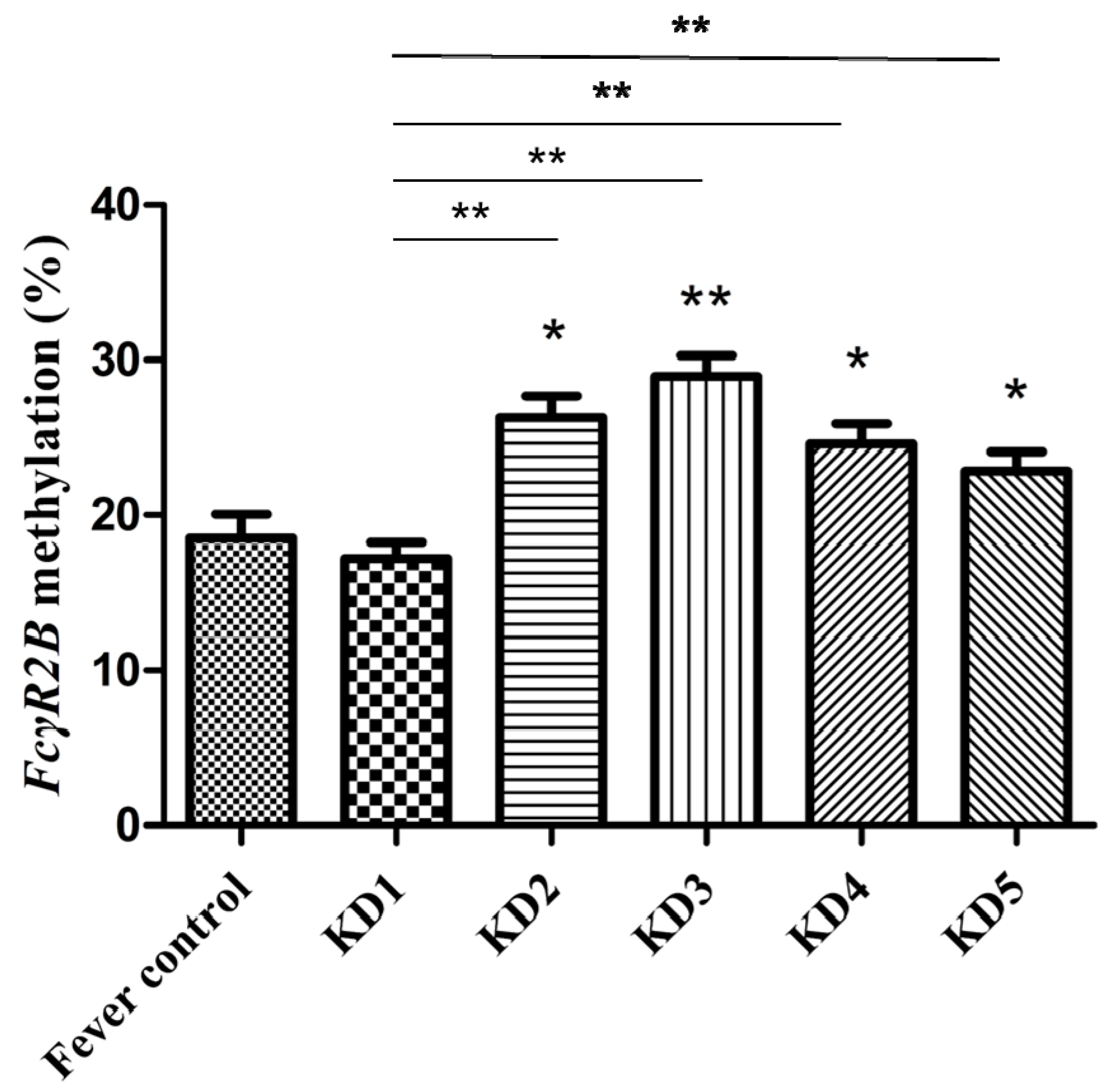
3.2. Methylation Levels of FcγR2B Promoter Regions on Leukocytes in Different Stages of Kawasaki Disease

We studied the FcγR2B methylation of the total white blood cells in paired blood samples from 76 KD patients (see Figure 3). We obtained peripheral blood samples from all 76 KD subjects at five different times: within 24 h prior to IVIG treatment (KD1), 3–7 days after IVIG (KD2), three weeks after IVIG (KD3), six months after IVIG (KD4), and one year after IVIG (KD5) [23]. We found no significant difference in FcγR2B methylation between KD patients and controls. FcγR2B methylation was significantly higher in samples taken three days after IVIG than in the first samples taken before IVIG (KD2: 26.29  ±  1.40 and KD1: 17.22  ±  1.02, paired t test, p < 0.001). The highest FcγR2B methylation was observed in KD3 samples (KD3: 28.96  ±  1.31, p = 0.015 when compared with KD2; p < 0.001 when compared with KD1). In KD5 patients, the FcγR2B methylation was still higher than in the children at KD1 (KD5: 22.86  ±  1.24, paired t test, p < 0.001). Many factors seemed to influence FcγR2B methylation, including IVIG, age, and lymphocyte counts. To rule out the possible effect of age on methylation levels, we checked the Pearson correlation between age and methylation levels, which demonstrated a negative correlation (p = 0.008). A generalized estimating equation was evaluated for correlations between multiple measures of the same KD patients using Bonferroni correction [24]. The methylation of FcγR2B increased significantly, after adjusting for lymphocyte counts and age (KD2, 3, 4, 5 vs. KD1: β = 8.43, 10.65, 8.58, 11.40; all p < 0.001).
Patients had similar FcγR2B methylation between incomplete (n = 15) and complete (n = 61) patients at KD1 (p  = 0.198). Of the 15 patients with steroids, steroids were administered in 10 patients after KD2. The KD3 FcγR2B methylation was significantly associated with KD1 FcγR2B methylation but not steroid usage by linear regression with KD1 FcγR2B methylation and steroid usage as covariates. No difference in methylation was found when clustered by years or different IVIG brands: Gamunex-C (Grifols Therapeutics, California, USA) and “TBSF” Human Immunoglobulin (Taiwan Blood Services Foundation, Taiwan) (p = 0.055 by the Kruskal-Wallis test; p = 0.336 by the Mann-Whitney U test) at KD2.
We discovered other CpG sites, cg22942846 and cg00831970, of FcγR2B from the M450 methylation array reported in our previous study [25]. There were no significant changes in methylation of the CpG sites cg22942846 and cg00831970 between non-febrile and febrile controls or non-febrile controls and KD.

3.3. FcγR2B Hypomethylation with IVIG Resistance in Kawasaki Disease

Patients with IVIG resistance (n = 6) had significantly lower FcγR2B methylation (7.33 ± 2.12 vs. 18.07 ± 1.04, p = 0.004) than the IVIG-responsive group (n = 70) (Figure 4). In IVIG non-responders, the significantly reduced FcγR2B methylation persisted to KD3. FcγR2B methylation levels in the IVIG-resistant group were lower than those in the IVIG-responsive group both 3–7 days and three weeks after IVIG (p = 0.004, 0.005). No significant correlation was found between FcγR2B methylation levels and coronary artery abnormalities in KD1, KD2, KD3, KD4, or KD5 (p = 0.509, 0.169, 0.219, 0.666, and 0.754, respectively). Analysis revealed that the FcγR2B methylation levels in the control subjects did not differ from those in KD patients with CAA (p  = 0.366).

3.4. Association between FcγR2B Methylation and Blood Parameters in Kawasaki Disease

We observed correlations between the lymphocyte proportion and acute or convalescent FcγR2B methylation in KD1, KD2, KD3, KD4, and KD5 (all p values < 0.001). We found that the proportion of segmented neutrophils was inversely correlated with FcγR2B methylation in KD1, KD2, KD3, KD4, and KD5 (all p values < 0.001), as well as total white blood cells in KD1, KD2, KD3, and KD5 (p = 0.023, 0.002, 0.046, 0.026). Furthermore, a reduced neutrophil-to-lymphocyte ratio was associated with FcγR2B methylation in KD1, KD2, KD3, KD4, and KD5 (p < 0.001 in KD15, respectively). Meanwhile, in patients with KD, FcγR2B methylation positively correlated with platelet counts (p = 0.047). Interestingly, this study identified a significant (p = 0.003) inverse association between CRP levels and FcγR2B methylation.

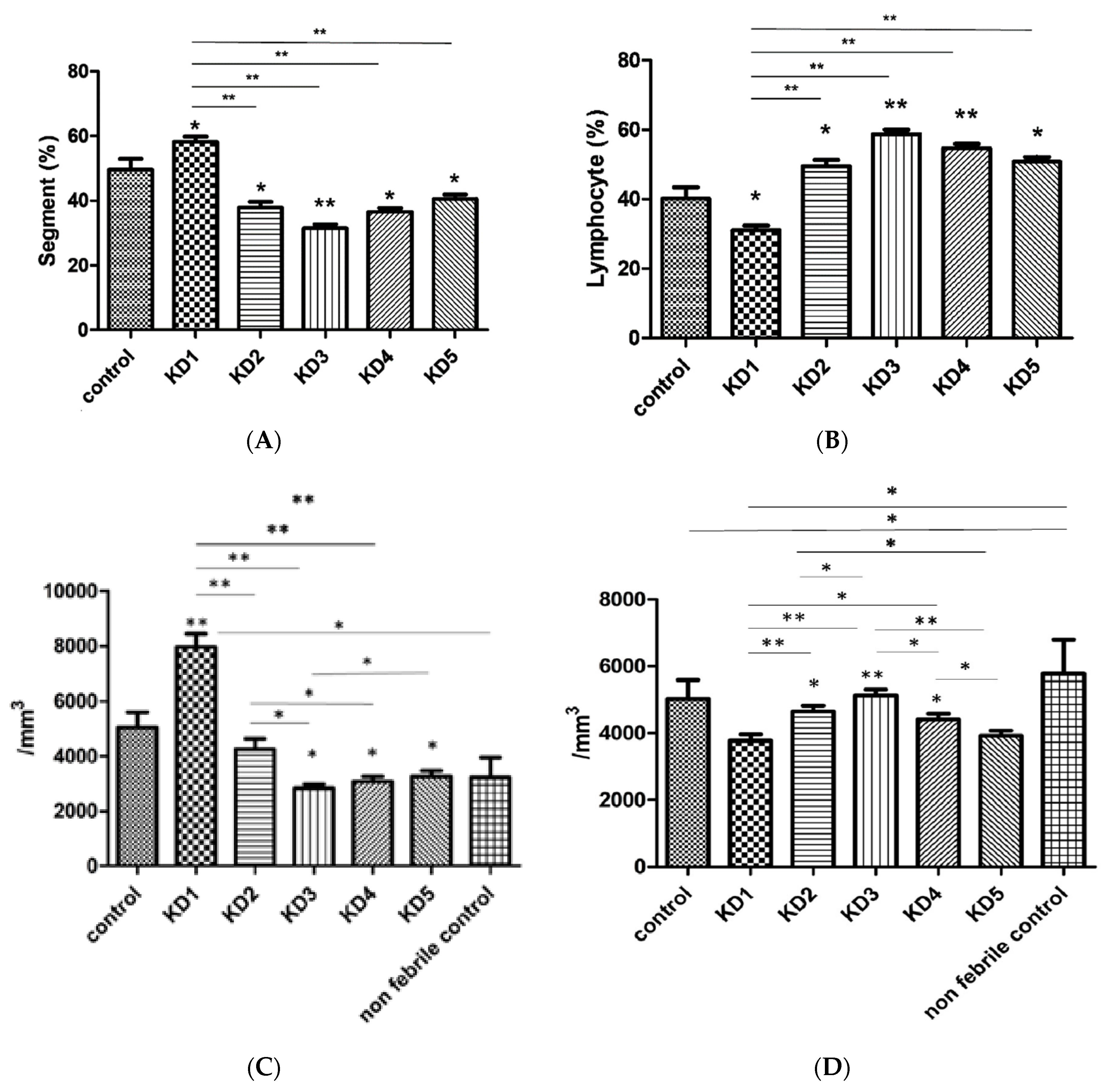
3.5. Hematological Changes in Kawasaki Disease

A considerable increase of segmented neutrophils was observed in KD patients compared with controls (p = 0.026) (Figure 5A). In the post-IVIG stage, significant reductions were observed in segmented neutrophils (58.18 ± 1.63% in KD1, 37.81  ±  1.89% in KD2, 31.40 ± 1.29% in KD3, 36.50 ± 1.26% in KD4, 40.50 ± 1.41% in KD5) while lymphocytes were significantly lower in patients with KD compared to controls (p = 0.012) (Figure 5B). We observed a significant increase of lymphocytes following IVIG treatment, and lymphocytes had the most significant increase at week three (KD2 vs. KD3 p = 0.015). This pattern of lymphocyte changes was almost identical in FcγR2B methylation, both exhibiting similar increases and their highest values in KD3. After IVIG treatment, the absolute counts of neutrophils and lymphocytes in KD patients returned to the levels of the six age- and gender-matched non-febrile controls (Figure 5C,D).

3.6. IVIG Regulates FcγR2B Methylation in THP-1 Cells

Due to the stronger inhibitory effect of IVIG on the activation of monocytes than that of T cells and patients’ different expressions of FcγR2B on monocytes, we investigated whether IVIG could regulate FcγR2B methylation in THP-1 cells (a human monocytic leukemia cell line) [7,22]. Pretreating cells with IVIG and lipopolysaccharide for six hours significantly increased FcγR2B methylation in THP-1 cells (p = 0.019) (Figure 6).

4. Discussion

Interestingly, we found that hypermethylation at the FcγR2B promoter persisted for more than one year in KD. FcγRIIB is generally considered to be the only inhibitory Fc receptor that can suppress an inflammatory response. Hypomethylation of FcγR2B is associated with IVIG resistance, indicating that greater FcγR2B expression is required for an inflammatory condition in patients with IVIG resistance to overcome excessive inflammation. FcγR2B may play a role in downregulating inflammation. Huang et al. found that DNA methyltransferase (DNMT1) was downregulated in patients with KD, and after three days of IVIG in patients with CAA, DNMT1 was lower than patients without CAA [26]. Our previous report identified IVIG resistance was involved in a scoring system for predicting CAA [27]. Therefore, we considered that the decrease in methylation of FcγR2A and B in patients with IVIG resistance may be due to the influence of DNMT1 [14].
Although the anti-inflammatory process is usually confined to the acute phase of the IVIG response, long-term immune changes may persist following IVIG treatment. An in vitro study was designed to explore the potential role of IVIG for FcγR2B methylation. We found significantly higher FcγR2B methylation after administering IVIG and confirmed the effect of IVIG on methylation changes suspected by the analysis of Illumina HumanMethylation27 BeadChip [12,28]. Evidence supporting the existence of long-term chronic inflammation and anti-inflammation is limited, so the mechanisms responsible for the chronic immune response in KD patients and their potential consequences on patients’ health remain unknown. Our results suggest that an epigenome at specific genes that control anti-inflammation stably changes throughout the course of KD. Persistent inflammation with higher high-sensitivity CRP levels was observed in patients with KD history [29]. Part of the proposed mechanism for durable epigenetic modification may come from the therapeutic IVIG. The mechanism of sustained epigenetic modification in KD needs further research to draw a clear conclusion.
A recent study found that a neutrophil-to-lymphocyte ratio ≥ 3.2 could predict IVIG resistance [30]. An earlier study found that IVIG-resistant patients had significantly lower platelet counts than IVIG-responsive patients [31]. These results were consistent with another finding of this study that the neutrophil-to-lymphocyte ratio, lymphocytes, segmented neutrophils, and platelets were all associated with FcγR2B methylation. The present study suggested that FcγR2B methylation was lower in the leukocytes of patients with IVIG-resistant KD than in IVIG-responsive subjects. In summary, these observations support the perspective that all of these features are unique to KD patients with IVIG resistance.
Furukawa et al. observed no significant difference in lymphocytes between the matched controls and KD (4.68 ± 0.25 vs. 4.06 ± 0.20 × 10⁹/L), and lymphocyte values did not differ between KD patients in the acute stage and in the convalescent stage (4.06 ± 0.20 vs. 4.89 ± 0.19 × 10⁹/L) [32,33]. Nevertheless, the long-term anti-inflammatory effect of IVIG is still being investigated. One study found that total lymphocyte counts between both groups with IVIG-and-aspirin versus aspirin (3.52 ± 1.05 vs. 3.32 ± 2.13 × 10⁹/L) were significantly lower than normal controls (4.33 ± 1.52 × 10⁹/L) [34]. Therefore, evidence is lacking to support the efficacy of IVIG for total lymphocytes in patients with KD. Although we revealed lower segmented neutrophils after IVIG treatment in our research, it has been reported that IVIG may induce polymorphonuclear cells in the peripheral blood [35].
This study has certain limitations. First, since this is a study in methylation, whether the findings remain constant over mRNA levels requires further study. The transcription of the FcγR2B gene is not only regulated by promoter methylation but also by regulatory elements with silencer activity [36,37]. Second, FcγR2B methylation was not measured for patients without IVIG infusion because IVIG is the standard treatment for KD. Perhaps due to the presence of CAA, patients were more willing to participate in regular follow-up visits, and we could also collect blood samples, which may have caused a selection bias. We enrolled the cases with all KD1–5 samples younger than the expected average age of KD, reflecting selection bias in this cohort study. Nationwide population-based cohort studies have surveyed KD children with mean age of 1.57 years and 16.72 months in Taiwan [38,39]. The validation of FcγR2B methylation in one CpG site was also too limited to explain IVIG resistance or its long-term hypermethylation.
In summary, this study is the first to show that FcγR2B may serve as an indicator of IVIG resistance in KD1-3, as well as the first to report long-term epigenetic alterations in the FcγR2B methylation of KD.

Author Contributions

Conceptualization, L.-S.C., K.-P.W. and H.-C.K.; Methodology, L.-S.C., H.-R.Y. and H.-C.K.; Validation, M.M.-H.G., K.-D.C. and Y.-H.H.; Formal Analysis, L.-S.C., K.-D.C.; Writing—Original Draft Preparation, L.-S.C.; Writing—Review and Editing, M.M.-H.G. and C.-L.C.; Visualization, H.-C.K.; Supervision, H.-C.K.; Funding Acquisition, L.-S.C. and H.-C.K. All authors have read and agreed to the published version of the manuscript.

Funding

This study was supported in part by grants from the Ministry of Science and Technology, Taiwan (MOST 108-2314-B-182 -037 -MY3) and Chang Gung Memorial Hospital (CPRPG8F0791, CMRPG8K0641-2). However, these institutions had no role in the study design, data collection and analysis, decision to publish, or preparation of the manuscript.

Institutional Review Board Statement

This study was approved by the institutional review board of Chang Gung Memorial Hospital and carried out in accordance with the Helsinki Declaration (IRB No. 104-9786A3).

Informed Consent Statement

After providing a detailed explanation of the study, we obtained written informed consent from the parents or guardians of all participants.

Data Availability Statement

The data are available from the author upon request.

Acknowledgments

We would like to thank Hsin-Yi Chien, Chih-Yun Lin, and Nien-Tzu Hsu, as well as the Biostatistics Center, Kaohsiung Chang Gung Memorial Hospital, for statistics work.

Conflicts of Interest

The authors declare that the research was conducted in the absence of any commercial or financial relationships that could be construed as a potential conflict of interest.

References

  1. Kawasaki, T.; Kosaki, F.; Okawa, S.; Shigematsu, I.; Yanagawa, H. A new infantile acute febrile mucocutaneous lymph node syndrome (MLNS) prevailing in Japan. Pediatrics 1974, 54, 271–276. [Google Scholar] [PubMed]
  2. McCrindle, B.W.; Rowley, A.H.; Newburger, J.W.; Burns, J.C.; Bolger, A.F.; Gewitz, M.; Baker, A.L.; Jackson, M.A.; Takahashi, M.; Shah, P.B.; et al. Diagnosis, Treatment, and Long-Term Management of Kawasaki Disease: A Scientific Statement for Health Professionals from the American Heart Association. Circulation 2017, 135, e927–e999. [Google Scholar] [CrossRef] [PubMed]
  3. Nakatani, K.; Takeshita, S.; Tsujimoto, H.; Kawamura, Y.; Kawase, H.; Sekine, I. Regulation of the expression of Fc gamma receptor on circulating neutrophils and monocytes in Kawasaki disease. Clin. Exp. Immunol. 1999, 117, 418–422. [Google Scholar] [CrossRef] [PubMed]
  4. Chang, L.-S.; Guo, M.M.-H.; Lo, M.-H.; Kuo, H.-C. Identification of increased expression of activating Fc receptors and novel findings regarding distinct IgE and IgM receptors in Kawasaki disease. Pediatr. Res. 2021, 89, 191–197. [Google Scholar] [CrossRef]
  5. Smith, K.G.; Clatworthy, M.R. FcgammaRIIB in autoimmunity and infection: Evolutionary and therapeutic implications. Nat. Rev. Immunol. 2010, 10, 328–343. [Google Scholar] [CrossRef] [Green Version]
  6. Jhou, J.P.; Chen, S.J.; Huang, H.Y.; Lin, W.W.; Huang, D.Y.; Tzeng, S.J. Upregulation of FcgammaRIIB by resveratrol via NF-kappaB activation reduces B-cell numbers and ameliorates lupus. Exp. Mol. Med. 2017, 49, 144. [Google Scholar] [CrossRef] [Green Version]
  7. Xia, Y.; Tian, X.; Li, Q.; Wang, G.; Li, C.; Yang, J. Expression of FcRs on monocytes among Kawasaki disease patients with coronary artery lesions. Int. Immunopharmacol. 2017, 45, 1–5. [Google Scholar] [CrossRef]
  8. Anthony, R.M.; Kobayashi, T.; Wermeling, F.; Ravetch, J.V. Intravenous gammaglobulin suppresses inflammation through a novel T(H)2 pathway. Nature 2011, 475, 110–113. [Google Scholar] [CrossRef]
  9. Shrestha, S.; Wiener, H.W.; Olson, A.K.; Edberg, J.C.; Bowles, N.E.; Patel, H.; Portman, M.A. Functional FCGR2B gene variants influence intravenous immunoglobulin response in patients with Kawasaki disease. J. Allergy Clin. Immunol. 2011, 128, 677–680.e1. [Google Scholar] [CrossRef] [Green Version]
  10. Li, X.; Ptacek, T.S.; Brown, E.E.; Edberg, J.C. Fcγ receptors: Structure, function and role as genetic risk factors in SLE. Genes Immun. 2009, 10, 380–389. [Google Scholar] [CrossRef] [Green Version]
  11. Su, K.; Wu, J.; Edberg, J.C.; Li, X.; Ferguson, P.; Cooper, G.S.; Langefeld, C.D.; Kimberly, R.P. A promoter haplotype of the immunoreceptor tyrosine-based inhibitory motif-bearing FcgammaRIIb alters receptor expression and associates with autoimmunity. I. Regulatory FCGR2B polymorphisms and their association with systemic lupus erythematosus. J. Immunol. 2004, 172, 7186–7191. [Google Scholar] [CrossRef] [PubMed] [Green Version]
  12. Chang, L.S.; Lo, M.H.; Li, S.C.; Yang, M.Y.; Hsieh, K.S.; Kuo, H.C. The effect of FcgammaRIIA and FcgammaRIIB on coronary artery lesion formation and intravenous immunoglobulin treatment responses in children with Kawasaki disease. Oncotarget 2017, 8, 2044–2052. [Google Scholar] [CrossRef] [Green Version]
  13. Ichiyama, T.; Ueno, Y.; Hasegawa, M.; Ishikawa, Y.; Matsubara, T.; Furukawa, S. Intravenous immunoglobulin does not increase FcgammaRIIB expression on monocytes/macrophages during acute Kawasaki disease. Rheumatology 2005, 44, 314–317. [Google Scholar] [CrossRef] [Green Version]
  14. Kuo, H.-C.; Chang, J.-C.; Kuo, H.-C.; Yu, H.-R.; Wang, C.-L.; Lee, C.-P.; Huang, L.-T.; Yang, K.D. Identification of an Association Between Genomic Hypomethylation ofFCGR2Aand Susceptibility to Kawasaki Disease and Intravenous Immunoglobulin Resistance by DNA Methylation Array. Arthritis Rheumatol. 2014, 67, 828–836. [Google Scholar] [CrossRef]
  15. Kuo, H.-C.; Chang, W.-C.; Yang, K.D.; Yu, H.-R.; Wang, C.-L.; Ho, S.-C.; Yang, C.-Y. Kawasaki disease and subsequent risk of allergic diseases: A population-based matched cohort study. BMC Pediatr. 2013, 13, 38. [Google Scholar] [CrossRef] [PubMed] [Green Version]
  16. Huang, P.-Y.; Huang, Y.-H.; Guo, M.M.-H.; Chang, L.-S.; Kuo, H.-C. Kawasaki Disease and Allergic Diseases. Front. Pediatr. 2021, 8, 614386. [Google Scholar] [CrossRef]
  17. Swamy, R.S.; Reshamwala, N.; Hunter, T.; Vissamsetti, S.; Santos, C.B.; Baroody, F.M.; Hwang, P.H.; Hoyte, E.G.; Garcia, M.A.; Nadeau, K.C. Epigenetic modifications and improved regulatory T-cell function in subjects undergoing dual sublingual immunotherapy. J. Allergy Clin. Immunol. 2012, 130, 215–224.e7. [Google Scholar] [CrossRef] [Green Version]
  18. Figge, D.A.; Jaunarajs, K.L.E.; Standaert, D.G. Dynamic DNA Methylation Regulates Levodopa-Induced Dyskinesia. J. Neurosci. 2016, 36, 6514–6524. [Google Scholar] [CrossRef] [Green Version]
  19. Kuo, H.-C.; Hsu, Y.-W.; Wu, M.-S.; Chien, S.-C.; Liu, S.-F.; Chang, W.-C. Intravenous immunoglobulin, pharmacogenomics, and Kawasaki disease. J. Microbiol. Immunol. Infect. 2016, 49, 1–7. [Google Scholar] [CrossRef] [Green Version]
  20. Newburger, J.W.; Takahashi, M.; Gerber, M.A.; Gewitz, M.H.; Tani, L.Y.; Burns, J.C.; Shulman, S.T.; Bolger, A.F.; Ferrieri, P.; Baltimore, R.S.; et al. Diagnosis, Treatment, and Long-Term Management of Kawasaki Disease: A Statement for Health Professionals from the Committee on Rheumatic Fever, Endocarditis, and Kawasaki Disease, Council on Cardiovascular Disease in the Young, American Heart Association. Circulation 2004, 110, 2747–2771. [Google Scholar] [CrossRef] [Green Version]
  21. Huang, E.-Y.; Chang, J.-C.; Chen, H.-H.; Hsu, C.-Y.; Hsu, H.-C.; Wu, K.-L. Carcinoembryonic antigen as a marker of radioresistance in colorectal cancer: A potential role of macrophages. BMC Cancer 2018, 18, 321. [Google Scholar] [CrossRef] [Green Version]
  22. Makata, H.; Ichiyama, T.; Uchi, R.; Takekawa, T.; Matsubara, T.; Furukawa, S. Anti-inflammatory effect of intravenous immunoglobulin in comparison with dexamethasone in vitro: Implication for treatment of Kawasaki disease. Naunyn-Schmiedeberg’s Arch. Pharmacol. 2006, 373, 325–332. [Google Scholar] [CrossRef]
  23. Chang, L.; Guo, M.M.; Yan, J.; Huang, Y.; Lo, M.; Kuo, H. Low FCMR mRNA expression in leukocytes of patients with Kawasaki disease six months after disease onset. Pediatr. Allergy Immunol. 2020, 31, 554–559. [Google Scholar] [CrossRef]
  24. Basu, S.K.; Pradhan, S.; Jacobs, M.B.; Said, M.; Kapse, K.; Murnick, J.; Whitehead, M.T.; Chang, T.; du Plessis, A.J.; Limperopoulos, C. Age and Sex Influences Gamma-aminobutyric Acid Concentrations in the Developing Brain of Very Premature Infants. Sci. Rep. 2020, 10, 1–10. [Google Scholar] [CrossRef]
  25. Huang, L.-H.; Kuo, H.-C.; Pan, C.-T.; Lin, Y.-S.; Huang, Y.-H.; Li, S.-C. Multiomics analyses identified epigenetic modulation of the S100A gene family in Kawasaki disease and their significant involvement in neutrophil transendothelial migration. Clin. Epigenetics 2018, 10, 1–13. [Google Scholar] [CrossRef] [Green Version]
  26. Huang, Y.-H.; Chen, K.-D.; Lo, M.-H.; Cai, X.-Y.; Chang, L.-S.; Kuo, Y.-H.; Huang, W.-D.; Kuo, H.-C. Decreased DNA methyltransferases expression is associated with coronary artery lesion formation in Kawasaki disease. Int. J. Med. Sci. 2019, 16, 576–582. [Google Scholar] [CrossRef] [Green Version]
  27. Chang, L.-S.; Lin, Y.-J.; Yan, J.-H.; Guo, M.M.-H.; Lo, M.-H.; Kuo, H.-C. Neutrophil-to-lymphocyte ratio and scoring system for predicting coronary artery lesions of Kawasaki disease. BMC Pediatr. 2020, 20, 1–7. [Google Scholar] [CrossRef]
  28. Li, S.-C.; Chan, W.-C.; Huang, Y.-H.; Guo, M.M.-H.; Yu, H.-R.; Huang, F.-C.; Kuo, H.-C.; Kuo, H.-C. Major methylation alterations on the CpG markers of inflammatory immune associated genes after IVIG treatment in Kawasaki disease. BMC Med. Genom. 2016, 9 (Suppl. 1), 37. [Google Scholar] [CrossRef] [Green Version]
  29. Chen, Y.-C.; Shen, C.-T.; Wang, N.-K.; Huang, Y.-L.; Chiu, H.-H.; Chen, C.-A.; Chiu, S.-N.; Lin, M.-T.; Wang, J.-K.; Wu, M.-H. High Sensitivity C Reactive Protein (hs-CRP) in Adolescent and Young Adult Patients with History of Kawasaki Disease. Acta Cardiol. Sin. 2015, 31, 473–477. [Google Scholar]
  30. Chantasiriwan, N.; Silvilairat, S.; Makonkawkeyoon, K.; Pongprot, Y.; Sittiwangkul, R. Predictors of intravenous immunoglobulin resistance and coronary artery aneurysm in patients with Kawasaki disease. Paediatr. Int. Child Health 2018, 38, 1–4. [Google Scholar] [CrossRef]
  31. Li, X.; Chen, Y.; Tang, Y.; Ding, Y.; Xu, Q.; Sun, L.; Qian, W.; Qian, G.; Qin, L.; Lv, H. Predictors of intravenous immunoglobulin-resistant Kawasaki disease in children: A meta-analysis of 4442 cases. Eur. J. Nucl. Med. Mol. Imaging 2018, 177, 1279–1292. [Google Scholar] [CrossRef] [Green Version]
  32. Matsubara, T.; Ichiyama, T.; Furukawa, S. Immunological profile of peripheral blood lymphocytes and monocytes/macrophages in Kawasaki disease. Clin. Exp. Immunol. 2005, 141, 381–387. [Google Scholar] [CrossRef]
  33. Furukawa, S.; Matsubara, T.; Yabuta, K. Mononuclear cell subsets and coronary artery lesions in Kawasaki disease. Arch. Dis. Child. 1992, 67, 706–708. [Google Scholar] [CrossRef]
  34. Lee, H.K.; Kim, D.S.; Noh, G.W.; Lee, K.Y. Effects of intravenous immune globulin on the peripheral lymphocyte phenotypes in Kawasaki disease. Yonsei Med. J. 1996, 37, 357–363. [Google Scholar] [CrossRef] [Green Version]
  35. Hall, P.D. Immunomodulation with intravenous immunoglobulin. Pharmacother. J. Hum. Pharmacol. Drug Ther. 1993, 13, 564–573. [Google Scholar]
  36. Bonnerot, C.; Daëron, M.; Varin, N.; Amigorena, S.; Hogarth, P.M.; Even, J.; Fridman, W.H. Methylation in the 5’ region of the murine beta Fc gamma R gene regulates the expression of Fc gamma receptor II. J. Immunol. 1988, 141, 1026–1033. [Google Scholar]
  37. Bonnerot, C.; Choukroun, V.; Marloie, M.A.; Fridman, W.H. Two distinct regions of the mouse beta Fc gamma R gene control its transcription. Immunobiology 1992, 185, 222–234. [Google Scholar] [CrossRef]
  38. Lei, W.-T.; Lin, C.-Y.; Kao, Y.-H.; Lee, C.-H.; Lin, C.-H.; Shyur, S.-D.; Yang, K.-D.; Chen, J.-H. The risk of hospitalization for respiratory tract infection (RTI) in children who are treated with high-dose IVIG in Kawasaki Disease: A nationwide population-based matched cohort study. PeerJ 2018, 6, e4539. [Google Scholar] [CrossRef]
  39. Wang, L.-J.; Kuo, H.-C. Cognitive Development After Kawasaki Disease—Clinical Study and Validation Using a Nationwide Population-Based Cohort. Circ. J. 2018, 82, 517–523. [Google Scholar] [CrossRef] [Green Version]
Figure 1. Flowchart of patients with Kawasaki disease. KD, Kawasaki disease; IVIG, intravenous immunoglobulin.
Jcm 10 02347 g001
Figure 2. Only one methylation site was identified using pyrosequencing for target-specific sequencing at cg22436411 in the FCGR2B promoter region.
Jcm 10 02347 g002
Figure 3. Prior to therapy, FcγR2B methylation did not differ significantly between Kawasaki disease (KD) and controls. After receiving intravenous immunoglobulin (IVIG) treatment, FcγR2B methylation increased and could last up to one year later. * denotes significance (* p < 0.05, ** p < 0.001). KD1, within 24 h prior to IVIG treatment; KD2, 3–7 days after IVIG; KD3, three weeks after IVIG, KD4, six months after IVIG; KD5, one year after IVIG.
Jcm 10 02347 g003
Figure 4. FcγR2B methylation was lower in the intravenous immunoglobulin (IVIG)-resistant group than in the IVIG-responsive group. KD, Kawasaki disease; KD1, within 24 h prior to IVIG treatment; KD2, 3–7 days after IVIG; KD3, three weeks after IVIG, KD4, six months after IVIG; KD5, one year after IVIG. * p < 0.05.
Jcm 10 02347 g004
Figure 5. Patients treated with intravenous immunoglobulin (IVIG) developed the lowest significant decrease in percentage of segmented neutrophils and an increase in the percentage of lymphocytes by the third week. (A) Segmented neutrophils were statistically significantly higher in the KD group compared with controls and decreased after receiving immunoglobulin for more than one year. (B) The percentage of lymphocytes was significantly lower in the KD group than in the control group, increasing after receiving immunoglobulin, and the increase lasted for more than one year. Asterisks denote significance compared with controls. (C) Absolute neutrophil counts. (D) Absolute lymphocyte counts. KD1, within 24 h prior to IVIG treatment; KD2, 3–7 days after IVIG; KD3, three weeks after IVIG, KD4, six months after IVIG; KD5, one year after IVIG. * p < 0.05, ** p < 0.001.
Jcm 10 02347 g005
Figure 6. (A) At 6 h, upregulated FcγR2B methylation was detected, compared with THP-1 cells without IVIG supplement. IVIG significantly increased the FcγR2B methylation in the LPS-treated THP-1 cells. (B) No change was detectable in THP-1 cells pretreated only with lipopolysaccharide at different time points. H, hours; I, intravenous immunoglobulin; L/LPS, lipopolysaccharide (* p < 0.05).
Jcm 10 02347 g006
Table 1. (A) Basal characteristics of patients with Kawasaki disease and controls. (B) Characteristics of controls.
A
Kawasaki Disease
n = 76
Controls
n = 40
p
Age1.79 ± 0.142.29 ± 0.260.10
Gender
Female/male
34/4219/210.78
Data are presented as mean with standard errors.
B
DiagnosisNumberDiagnosisNumber
Low respiratory infection/bronchitis, bronchiolitis6Acute gastritis1
Low respiratory infection/pneumonia, bronchopneumonia10Rotavirus infection1
Upper respiratory infection/tonsillitis, pharyngitis5Bacteremia1
Influenza A1Urinary tract infection1
Acute otitis media3Acute enteritis with salmonella infection1
Acute sinusitis4Hand-foot-mouth disease1
Croup1Infectious mononucleosis1
Acute mastoiditis1Herpetic gingivostomatitis1
Acute arthritis1
Publisher’s Note: MDPI stays neutral with regard to jurisdictional claims in published maps and institutional affiliations.

