Article

Enhanced Anti-Biofouling Properties of BWRO Membranes via the Deposition of Poly (Catechol/Polyamine) and Ag Nanoparticles

School of Chemical Engineering and Technology, Tianjin Key Laboratory of Membrane Science and Desalination Technology, State Key Laboratory of Chemical Engineering Tianjin University, Tianjin 300350, China
*
Authors to whom correspondence should be addressed.
Membranes 2023, 13(5), 530; https://doi.org/10.3390/membranes13050530
Submission received: 14 April 2023 / Revised: 28 April 2023 / Accepted: 15 May 2023 / Published: 19 May 2023

Abstract

:
The surface modification of reverse osmosis (RO) membranes to improve their anti-biofouling properties is gaining increased attention. Here, we modified the polyamide brackish water reverse osmosis (BWRO) membrane via the biomimetic co-deposition of catechol (CA)/tetraethylenepentamine (TEPA) and in situ growth of Ag nanoparticles. Ag ions were reduced into Ag nanoparticles (AgNPs) without extraneous reducing agents. The hydrophilic property of the membrane was improved, and the zeta potential was also increased after the deposition of poly (catechol/polyamine) and AgNPs. Compared with the original RO membrane, the optimized PCPA3-Ag10 membrane showed a slight reduction in water flux, and the salt rejection declined, but enhanced anti-adhesion and anti-bacterial activities were observed. The FDRt of the PCPA3-Ag10 membranes during the filtration of BSA, SA and DTAB solution were 5.63 ± 0.09%, 18.34 ± 0.33% and 34.12 ± 0.15%, respectively, much better than those of the original membrane. Moreover, the PCPA3-Ag10 membrane exhibited a 100% reduction in the number of viable bacteria (B. subtilis and E. coli) inoculated on the membrane. The stability of the AgNPs was also high enough, and these results verify the effectiveness of poly (catechol/polyamine) and the AgNP-based modification strategy for the control of fouling.

1. Introduction

Reverse osmosis (RO) membrane separation technology is considered a very promising technology for desalination and wastewater treatment. However, a significant obstacle to the high performance of RO technology is membrane biofouling, which impacts the water flux and the lifetime of membrane filters and modules [1,2,3,4,5]. Recently, several methods have been developed to monitor biofouling in RO systems, such as assimilable organic carbon (AOC), the membrane biofilm formation rate (mBFR) and bacterial growth potential (BGP) [6,7]. Many pretreatment processes have also been employed to decrease the fouling of RO membranes, such as the chlorination of feed water, ultrafiltration and microfiltration [8,9,10]. However, due to the self-replicating properties of bacteria, the above methods cannot completely reduce biofouling. Another strategy to address this problem is developing membranes with anti-bacterial properties on the membrane surface [11,12]. An anti-bacterial membrane can continuously inactivate bacteria and suppress biofilm formation, lowering the biofouling potential inside membrane modules during filtration.
Silver nanoparticles (AgNPs), due to the highly effective bactericides, have recently been widely used as anti-bacterial additives in the preparation of anti-biofouling RO membranes [13,14]. An early study on the preparation of AgNP-loaded RO membranes embedded AgNPs into the membrane matrix via the interfacial polymerization process [15,16,17,18]. However, this method usually involves complicated procedures and causes damage to separation layer structures. In addition, only a small percentage of AgNPs are exposed on the surface, and thus, the utilization of these AgNPs is low [17,19]. Therefore, the strategy of directly loading AgNPs on membrane surfaces has been developed to enhance the accessibility of these nanoparticles. In this strategy, there are usually appropriate sites on the membrane surfaces to bind and immobilize AgNPs. For example, Zhang et al. [20] immobilized AgNPs on membrane surfaces via polyacrylic acid sites. It was also reported that anti-bacterial RO membranes with cysteamine (NH2-(CH2)2-SH) sites can interact with AgNPs [21], AgNP-decorated silica particles [22] and AgNP -oxide nanosheets [23] through the covalent Ag-S bond. However, this strategy is restricted to limited grafting sites, which causes the low loading density of AgNPs.
Recently, polydopamine, a hydrophilic polymer with abundant hydroxyl and amine groups, has been widely used in the surface modification of RO membranes to reduce the adhesion properties of contaminants. In addition, polydopamine can also be employed as a reducing agent so that AgNPs can be easily incorporated onto polydopamine-coated membranes via the in situ reduction method [24]. However, the high cost of dopamine limits its extensive application in membrane surface modification [25]. Besides polydopamine, other polymer coatings, such as tannic acid-Fe3+-polyethyleneimine (TA-Fe-PEI), have also been used for the surface modification of RO membranes and the in situ generation of AgNPs to enhance the antibacterial properties [26,27]. These works are encouraging and help us to develop new substitutions for dopamine coatings to produce AgNPs on RO membranes. It is confirmed that catechol and polyamine play important roles in dopamine polymerization to form strong adhesion [28], and a low-cost poly(catechol-polyamine) (PCPA) coating has also been developed on the surface of polypropylene membranes without significant agglomeration. Just recently, PCPA modification with the co-deposition of AgNPs has been reported on the surface of various matrixes, including PVDF membranes [29], TiO2 [30] and aluminum beads [31]. However, to the best of our knowledge, there are few studies about the modification of RO membranes via the combination of PCPA coatings and AgNPs.
Therefore, based on the anti-adhesive property of PCPA and anti-bacterial property of AgNPs, we prepared a PCPA coating and studied AgNPs’ in situ growth on the surface of a commercial RO membrane. We demonstrated the successful loading of AgNPs on the surface and systematically tested the membrane properties. Then, we verified the anti-adhesion and anti-bacterial performance of the membrane. Finally, the stability of the AgNPs was tested using the static soaking method.

2. Experimental Section

2.1. Materials

Commercial brackish water reverse osmosis (BWRO) membrane (ESPA1) was purchased from Nitto Denko Hydranautics. Sodium bisulfite (NaHSO3) and nitric acid (HNO3) were obtained from Chemart Chemical Technology in Tianjin. Catechol (CA) (Aladdin, Shanghai, China), tris(hydroxymethyl)-aminomethane (Tris) (Aladdin, Shanghai, China) and Tetraethylenepentamine (TEPA) (Xianding biotechnology, Shanghai, China) were used for the crosslinking of the PCPA coating on the membrane surface. Sodium chloride (NaCl) (Chemart Chemical, Tianjin, China), bovine serum albumin (BSA) (Aladdin Bio-Chem Technology, Shanghai, China), sodium alginate (SA) (Meryer Chemical, Shanghai, China) and dodecyl trimethyl ammonium bromide (DTAB) (Meryer Chemical Technology, Shanghai, China) were used in the anti-adhesion test. Polyvinylpyrrolidone (PVP, K16-18) was purchased from Macklin Biochemical Co. Ltd. (Shanghai, China). Yeast extract, peptone, agar and lysozyme were purchased from Aoboxing Biotech Co. Ltd. (Beijing, China). Silver nitrate (AgNO3) (Guangfu Chemical Technology, Tianjin, China) was used for the formation of AgNPs. Bacillus subtilis (B. subtilis) (TransGen, Beijing, China) and Escherichia coli (E. coli) (TransGen, Beijing, China) were used in the anti-bacterial experiment. Deionized (DI) water (Distilled Water Department of Yongqingyuan, Tianjin, China) was used in all the experiments.

2.2. PCPA Coating and In Situ Formation of AgNPs

The preparation of the membranes consisted of two steps: the PCPA coating deposition and the in situ formation of AgNPs. The procedures used to prepare AgNP-loaded RO membranes are shown in Figure 1. The commercial membrane pieces were preserved in NaHSO3 solution (0.5 wt%). Before use, they were immersed in pure water for 1 day. The membrane pieces were fixed between the membrane frames (11 cm × 19 cm) with the active layer exposed to the modified solution. The modified solution was prepared by adding CA (2n mM, n = 2, 3, 4, 5, 6 and 7) and TEPA (n mM) into 40 mL of Tris-buffer (pH 8.5, 50 mM), and it was covered on the active layer of the RO membranes. The membrane frames was shaken on a rotating shaker for 15 min. The PCPA-coated membrane is denoted as PCPAn.
The PCPA3 membrane was washed with deionized water and then immersed in a mixture of solution containing of 15 mL of silver ammonia (m g·L−1, m = 5, 7.5, 10, 12.5 and 15) and 15 mL of PVP (1.5 g·L−1) for 15 min to obtain the membranes modified with AgNPs. The resulting membranes were referred to as PCPA3-Agm, where m is the concentration of silver ammonia solution. PVP was used as a dispersant and stabilizer to reduce the agglomeration of AgNPs. The above process was carried out on a shaker 85 times per minute. Finally, the PCPA3-Agm membranes were rinsed and kept in deionized water at 4 °C before use.

2.3. Characterization

X-ray photoelectron spectroscopy was used to characterize the chemical composition of the membrane surface (XPS, K-Alpha+, ESCALAB-250Xi, ThermoFisher Scientific, Loughborough, UK) with energy-dispersive X-ray spectroscopy (EDS, NanoSEM430, FEI, USA). The total number of AgNPs on the membranes was determined using Inductively Coupled Plasma Mass Spectrometry (ICP-MS, Agilent7700, Agilent, Santa Clara, CA, USA). Before the test, the membrane sample (3 cm × 3 cm) was digested using HNO3 solution. Then, the resulting solution was kept at 90 °C for 2 days to make all the AgNPs into Ag ions in the solution.
A field emission scanning electron microscope was used to characterize the surface morphology of the membranes (SEM S4800, Hitachi, Tokyo, Japan). Atomic force microscopy was employed to characterize the surface roughness (AFM, Dimension Icon, Bruker Co., Bremen, Germany). Before these tests, the samples were dried under vacuum at 60 °C for 1 day. The water contact angle was tested using an optical contact angle goniometer (OCA15EC, DataPhysics, Filderstadt, Germany). The water contact angle was tested at 10 different locations on one sample, and the average value was used. The Zeta potential of the surface was tested using a solid surface Zeta potential tester (SurPASS, Anton Paar GmbH, GmbH, Graz, Austria) using 1 mM KCl background solution at 25 °C.

2.4. Water Flux and Salt Rejection

A cross-flow device was employed to assess the water flux and salt rejection of all the membranes. The area (A) of the membranes was 31.16 cm2. The permselectivity of the membrane was tested at 25 °C and 1.55 ± 0.1 MPa. The feed was 2000 mg·L−1 NaCl solution. The cross-flow rate of the feed was 1.5 L·min−1 [32]. The water flux (J, L·m−2·h−1) of the RO membrane was calculated using Equation (1):
J = V A · T
in which V is the volume of permeated water (L), and T is the recorded measurement time (h). The salt rejection rates (R) of membranes were determined using Equation (2):
R = 1 C p C f × 100 % ,
in which Cp and Cf are the salt concentrations of the permeated and feed solutions, respectively. The salt concentrations were tested using a conductivity instrument (DDSJ-308A).

2.5. Anti-Adhesion Property Evaluation

Three model pollutants, BSA (150 mg·L−1), SA (100 mg·L−1) and DTAB (50 mg·L−1), were employed to determine the anti-adhesion performance of the PCPA3-Ag10 membrane.
First, the RO membranes were compacted in a solution of 2000 mg·L−1 NaCl for 60 min at 1.55 ± 0.1 MPa to obtain a stable flow. Then, the initial pure water flux (J0) was obtained at the cross-flow rate of 1.5 L·min−1 for 0.5 h. After the filtration of the NaCl solution for 30 min, the model contaminants were admitted to feed solution for the anti-adhesion tests. The anti-adhesion tests lasted 4.5 h, and the water flux (Jt) was recorded every 3 min. At the end of the filtration process, the virgin and PCPA3-Ag10 membranes were rinsed with DI water for 1 h with the cross-flow rate of 2 L·min−1 at 0.7 MPa. All membranes were tested in two filtration-rinse cycles [33].
The anti-adhesion performances of the membrane were described via the flux recovery rate after cleaning (FRRc, c = 1 or 2) and the flux decline rate (FDRt). The calculation Formulas (3) and (4) are offered as follows:
F R R c   =   J t , c J 0 × 100 %   c   =   1,2
F D R t   =   ( 1 J f J 0 ) × 100 % ,
in which Jt,c is the water flux of the RO membranes after first (c = 1) or second (c = 2) rinse, and Jf is the water flux after the second filtration test.

2.6. Anti-Bacterial and Stability Property of Membranes

The anti-bacterial performance was assessed via the vitality of bacteria after touching the membrane surface. The freeze-dried E. coli and B. subtilis cultures were refreshed in an LB via 12 h incubation in a 37 °C shaking incubator. Then, 100 μL of the refreshed culture was diluted 103 times with 10 mL of sterilized saline (0.9 wt%) to make a bacterial suspension. Membrane samples (2.5 cm × 7.6 cm) were sterilized under UV irradiation for 1 h before the test. In a Petri dish, membrane samples were placed on glass slides with the upward active layer. The 100 µL of E. coli suspension or B. subtilis suspension was dispersed on the surface of the membranes. The cell concentration of E. coli and B. subtilis subtilis suspension was approximately 1.0 × 10⁶ cfu·mL−1. Then, a cover glass slide was placed over the membrane surface to ensure full contact with the bacterial suspension. The Petri dish was kept in the incubator with the temperature of 37 °C for 1.5 h. The RO membrane samples and slides were fully rinsed with 20 mL of saline, and the wash solution was collected. Then, 100 µL of the wash solution was inoculated on the LB agar medium, and the number of bacterial colonies was marshalled after incubation in the incubator for 12 h. The mortality rate (E) was calculated using the following equation:
E   =   B A B × 100 % ,
in which A and B is the number of viable bacteria grown on LB agar plates using the microbial suspension contacted with and without the membrane, respectively.
The stability of immobilized AgNPs on membranes was investigated via static leaching experiments according to the previous report [27]. The membrane samples (3 cm × 3 cm) were immersed in 5% NaHCO3 solution (pH 8.2). The Ag ion concentration in the solutions was determined using ICP-MS.

3. Results and Discussion

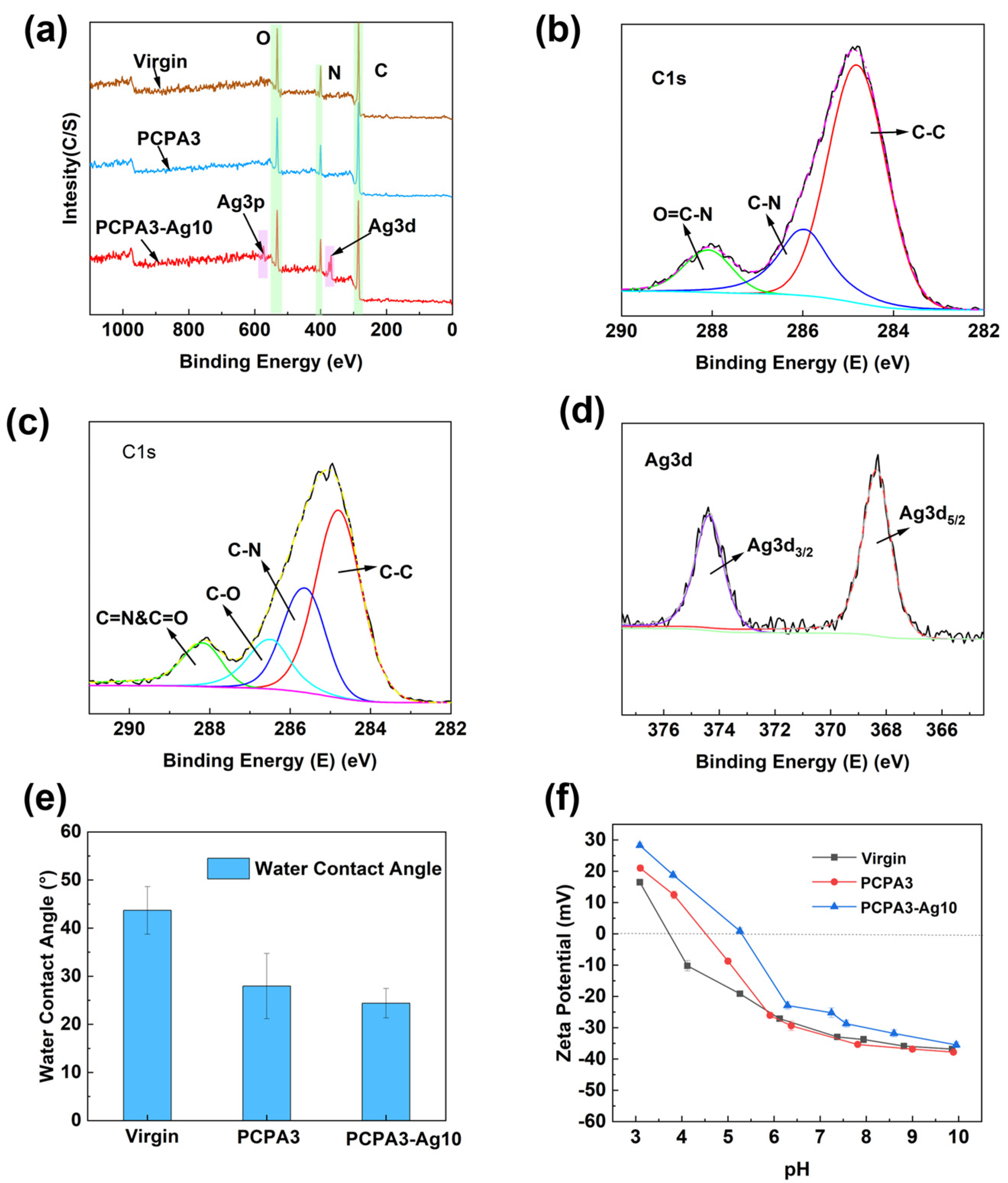
3.1. Membrane Characterizations

Figure 2a–c show the SEM micrographs of the virgin, PCPA3 and PCPA3-Ag10 membranes. All membranes had a distinct “ridge and valley” rough structure. The surface morphology of the PCPA3 membrane showed no significant change compared to the virgin one. However, after the immersion of PCPA3 in the silver ammonia solution, fine particles appeared on the membrane surface of PCPA3-Ag10.
We further investigated the loading amount of silver on the PCPA3-Ag10 membrane. The EDS elemental map images in Figure 2d show a clear signal of silver, nitrogen and oxygen elements, and the EDS elemental compositions show the atomic percent of Ag was 1.42% in Table 1.
According to the ICP-MS results, the total amount of AgNPs on the PCPA3-Ag10 membrane was 28.21 μg·cm−2. We also measured the total amount of AgNPs on the PCPA3-Agm membranes, and the results show that the total amount of AgNPs on the PCPA3-Agm membranes increased with the Ag concentration in silver ammonia solution (SI, Figure S1). The uniform PCPA interlayer could provide binding sites for the reduction of Ag ions, and the presence of catechol can reduce Ag ions into zero-valent AgNPs [24].
From the AFM images in Figure 2e–g, the average roughness (Ra) values of the virgin, PCPA 3 and PCPA3-Ag10 membranes were 80.3, 75.4 and 74.5 nm, respectively. Compared with the virgin membrane, the roughness of PCPA3 membranes was slightly reduced, and parts of the valley structures were filled. This is because the size of AgNPs is very small, and AgNPs have little contribution to the roughness of membranes. Hence, the PCPA3 and PCPA3-Ag10 membranes had similar roughness. Overall, the surface morphology of the RO membranes did not change much after the PCPA and AgNP modification.
The elemental compositions of membrane surfaces before and after the PCPA modification were captured using XPS. Figure 3a shows the full spectra of elements on the virgin, PCPA3 and PCPA3-Ag10 membrane surfaces. Table 2 shows the elemental composition and proportion of membranes via XPS, and the PCPA3 membrane had increased N/O content compared to the virgin membrane. This was attributed to the higher N content in the PCPA coating. The peak fits of C1s fine spectra from the virgin and PCPA3 membranes are shown in Figure 3b,c. Compared to the virgin membrane, the PCPA3 membrane showed a new peak at 286.6 eV due to the C-O bond, which was derived from the phenol structure in CA crosslinked on the membrane surface. In Figure 3d, the spectrum of the PCPA3-Ag10 membrane showed peaks for Ag 3d 3/2 and Ag 3d 5/2 with binding energies of 374.3 eV and 368.2 eV, which belonged to the peak of Ag0 [34].
The water contact angle was tested to evaluate the hydrophilicity of the membrane surface in Figure 3e. The water contact angle of the virgin membrane was 43.7 ± 4.9°. The PCPA3 membrane had smaller water contact angles than the virgin one due to the large numbers of hydrophilic amino and phenolic groups in the PCPA3 coating. The water contact angle of the PCPA3-Ag10 membrane was 24.7 ± 4.0°, which was attributed to the addition of AgNPs with high hydrophilicity. In addition, we also found that the water contact angle decreased continuously with the increase in Ag contents (SI, Figure S2).
The surface charge was demonstrated by measuring the streaming potential of the membrane surface in the range of pH 3–10. Figure 3f shows the amphoteric properties on the existing RO membrane surface. The Zeta potentials of the virgin membrane were 16.5 ± 0.9 mV at pH 3 and −36.8 ± 0.2 mV at pH 10. The zeta potentials of PCPA3 were 21.0 ± 0.9 mV at pH 3 and −37.8 ± 0.6 mV at pH 10. Additionally, the zeta potentials of PCPA3-Ag10 were 28.3 ± 0.8 mV at pH 3 and −35.5 ± 0.9 mV at pH 10. Compared to the virgin membrane, the Zeta potential of both modified membranes increased, with a rightward shift in the equipotential point. The PCPA coating contained a large amount of amine, which was protonated to increase the positive charge density under acidic conditions. Meanwhile, the partial dissolution of AgNP particles loaded on the membrane surface under acidic conditions may also have led to an enhanced positive charge on the surface of the membrane [14].

3.2. Membrane Permselectivity

To evaluate the permselectivity of the membranes, we investigated the water flux and salt rejection of the virgin, PCPA3 and PCPA3-Ag10 membranes. The water flux of the virgin membrane was 78.4 ± 2.7 L·m−2·h−1, as shown in Figure 4. The water flux of the PCPA3 and PCPA3-Ag10 membranes showed a certain decline in comparison to the virgin membrane. There were abundant polar groups (such as amino and hydroxyl groups) in the PCPA coating, which could have accelerated the penetration ability of water across the membrane. However, the PCPA coating could have also improved the mass transfer resistance of water across the membrane [29,35]. The water flux of the PCPA3-Ag10 membrane declined, because AgNPs could partly block water channels on the membrane surface [24]. The PCPA3 and PCPA3-Ag10 membranes showed a slight decrease in salt rejection in comparison to the virgin membrane. On the basis of the solution-diffusion model [36], whether the rejection of modification membrane was improved or decreased depended on the water/salt permeability ratio of modification membranes in comparison to the water/salt permeability ratio of the virgin one. The PCPA coating and the loading of AgNPs reduced the water permeability, leading to a low water/salt permeability ratio after modification [37,38].

3.3. Membrane Anti-Adhesion Properties

Three model pollutants, BSA (representing proteins in the water treatment process), SA (simulating polysaccharides) and DTAB (characterizing fouling surfactants in wastewater), were selected to study the anti-adhesion property of the PCPA3-Ag10 membrane. The relative flux Jt/J0 was used to evaluate the change in flux for the membrane fouling and recovery. In Figure 5, it is shown that the relative flux of the membranes decreased with time in each cycle.
After washing, the water flux of all membranes recovered to some extent. The FRR1 values were always higher than the FRR2 values in all cycles, which could be attributed to the irreversible adsorption with pollutants. Compared to the virgin one, the relative flux of PCPA3-Ag10 membrane showed a lower loss of water flux. The higher FRRc (c = 1 and 2) and lower FDRt of the PCPA3-Ag10 membrane is shown in Table 3, indicating that this modification method can efficiently reduce the adsorption of pollutants.
The decreased degrees of water flux were different in the three contaminated environments. The relative flux in the BSA-contaminated environment had the lowest decline, while the relative flux in the DTAB-contaminated environment had the highest decline. DTAB, as a quaternary ammonium salt, has a positive charge in aqueous solutions. The membranes with negative charges had a stronger adsorption effect on DTAB than BSA and SA. This is the reason for the highest loss of water flux in the DTAB-contaminated environment.

3.4. Membrane Anti-Bacterial Property

The anti-bacterial performance of the membranes was studied by growing two bacteria which were in contact with the membranes for 1.5 h. Figure 6 reveals the bacterial colonies grown on LB plates. The mortalities of the bacteria were obtained by counting the number of colonies, as shown in Table 4.
The mortalities of B. subtilis and E. coli which were in contact with the virgin membrane were 12.5 ± 1.7% and 15.5 ± 0.9%, respectively. Additionally, the mortality rates of B. subtilis and E. coli which were in contact with the PCPA3 membrane were 96.1 ± 0.4% and 83.7 ± 1.3%, respectively. There were abundant phenolic hydroxyl groups and amine groups in the PCPA coating. The hydroxyl (–OH) in phenol could interact with the cell membrane of the bacteria and disrupt their membrane structures. Amine groups can affect charge distribution in bacterial cells, which make bacterial cells deformed and finally dead [39,40]. As shown in Figure 6c,f, the mortality of the two bacteria in contact with the PCPA3-Ag10 membrane reached 100%, indicating the strong anti-bacterial property of AgNPs.

3.5. AgNPs Stability Test

Long-term stability was essential for the practical application of the PCPA3-Ag10 membrane. The stability of AgNPs on the membrane was assessed by measuring the amounts of Ag+ released in 7 days. As shown in Figure 7, a lot of silver (1.33 μg·cm−2·day−1) was released into the solution on the first day after the immersion in the NaHCO3 solution (pH 8.2). Then, the releasing rate of Ag slowed down to a stable rate (0.06 μg·cm−2·day−1). The sudden release of silver at the beginning of the immersion may have been due to the loss of AgNPs loosely attached on the membrane surface. It can be seen that there was still about 93.21% silver immobilized on the surface of the membrane after 7 days’ immersion.
We summarize the permselectivity and AgNP stability of the membranes prepared in this work with other similar membranes in Table 5. In general, the anti-bacterial property of all membranes with Ag incorporated in them can be improved. The PCPA3-Ag10 membrane reported here can maintain a long-term and efficient anti-bacterial property with a low release rate of Ag ions. These promising results demonstrate that the strategy of using a PCPA coating as the interlayer to load AgNPs on a membrane surface has a good application perspective.

4. Conclusions

In this work, the surface modification of RO membranes was proposed to immobilize AgNPs effectively via the deposition of the poly (catechol/polyamine) interlayer. After the loading of AgNPs, the water flux of PCPA3-Ag10 membrane was reduced to 72.5 ± 0.3 L·m−2·h−1, which was7.48% less than the virgin membrane. However, in the anti-adhesion experiments with three representative model pollutants, the FDRt values of the PCPA3-Ag10 membrane were much less than the virgin membrane. Additionally, in the anti-bacterial experiments, the bacterial mortality rates of the PCPA3-Ag10 membrane could reach 100%. The PCPA3-Ag10 membrane showed a strong anti-adhesion property and long-lasting anti-bacterial activity, with a small decrease in the permselectivity. Compared with previous methods of immobilizing AgNPs on RO membranes, the modification method proposed here can improve the anti-adhesion property of membranes and maintain a long-term and stable anti-bacterial ability with a low release rate of Ag ions.

Supplementary Materials

The following supporting information can be downloaded at: https://www.mdpi.com/article/10.3390/membranes13050530/s1, Figure S1: The elemental percentage of silver and the silver mass loading were obtained by EDS analysis and silver dissolution experiments; Figure S2: The water fluxes and salt rejection of Virgin, PCPA3 membrane and PCPA3-Agm membranes.

Author Contributions

Conceptualization, S.X., L.X. and W.Z.; Methodology, Y.L., L.X. and W.Z.; Validation, Y.L.; Writing—original draft, Y.L.; Writing—review & editing, L.X., S.X. and W.Z. All authors have read and agreed to the published version of the manuscript.

Funding

This research received no external funding.

Data Availability Statement

Not applicable.

Conflicts of Interest

The authors declare no conflict of interest.

References

  1. Fritzmann, C.; Lowenberg, J.; Wintgens, T.; Melin, T. State-of-the-art of reverse osmosis desalination. Desalination 2007, 216, 1–76. [Google Scholar] [CrossRef]
  2. Shannon, M.A.; Bohn, P.W.; Elimelech, M.; Georgiadis, J.G.; Marinas, B.J.; Mayes, A.M. Science and technology for water purification in the coming decades. Nature 2008, 452, 301–310. [Google Scholar] [CrossRef] [PubMed]
  3. Matin, A.; Khan, Z.; Zaidi, S.M.J.; Boyce, M.C. Biofouling in reverse osmosis membranes for seawater desalination: Phenomena and prevention. Desalination 2011, 281, 1–16. [Google Scholar] [CrossRef]
  4. Al-Juboori, R.A.; Yusaf, T. Biofouling in RO system: Mechanisms, monitoring and controlling. Desalination 2012, 302, 1–23. [Google Scholar] [CrossRef]
  5. Pontié, M.; Ben Rejeb, S.; Legrand, J. Anti-microbial approach onto cationic-exchange membranes. Sep. Purif. Technol. 2012, 101, 91–97. [Google Scholar] [CrossRef]
  6. Abushaban, A.; Salinas-Rodriguez, S.G.; Pastorelli, D.; Schippers, J.C.; Mondal, S.; Goueli, S.; Kennedy, M.D. Assessing Pretreatment Effectiveness for Particulate, Organic and Biological Fouling in a Full-Scale SWRO Desalination Plant. Membranes 2021, 11, 167. [Google Scholar] [CrossRef]
  7. Dhakal, N.; Salinas-Rodriguez, S.G.; Ampah, J.; Schippers, J.C.; Kennedy, M.D. Measuring Biofouling Potential in SWRO Plants with a Flow-Cytometry-Based Bacterial Growth Potential Method. Membranes 2021, 11, 76. [Google Scholar] [CrossRef] [PubMed]
  8. Henthome, L.; Boysen, B. State-of-the-art of reverse osmosis desalination pretreatment. Desalination 2015, 356, 129–139. [Google Scholar] [CrossRef]
  9. Madaeni, S.S.; Samieirad, S. Chemical cleaning of reverse osmosis membrane fouled by wastewater. Desalination 2010, 257, 80–86. [Google Scholar] [CrossRef]
  10. Ghayeni, S.B.S.; Beatson, P.J.; Schneider, R.P.; Fane, A.G. Water reclamation from municipal wastewater using combined microfiltration reverse osmosis (ME-RO): Preliminary performance data and microbiological aspects of system operation. Desalination 1998, 116, 65–80. [Google Scholar] [CrossRef]
  11. Rana, D.; Matsuura, T. Surface Modifications for Antifouling Membranes. Chem. Rev. 2010, 110, 2448–2471. [Google Scholar] [CrossRef] [PubMed]
  12. Kang, G.D.; Cao, Y.M. Development of antifouling reverse osmosis membranes for water treatment: A review. Water Res. 2012, 46, 584–600. [Google Scholar] [CrossRef]
  13. Morones, J.R.; Elechiguerra, J.L.; Camacho, A.; Holt, K.; Kouri, J.B.; Ramirez, J.T.; Yacaman, M.J. The bactericidal effect of silver nanoparticles. Nanotechnology 2005, 16, 2346–2353. [Google Scholar] [CrossRef] [PubMed]
  14. Zhang, C.Q.; Hu, Z.Q.; Deng, B.L. Silver nanoparticles in aquatic environments: Physiochemical behavior and antimicrobial mechanisms. Water Res. 2016, 88, 403–427. [Google Scholar] [CrossRef] [PubMed]
  15. Bian, S.J.; Wang, Y.Y.; Xiao, F.K.; Tong, Y.B.; Gao, C.J.; Zhu, G.R. Fabrication of polyamide thin-film nanocomposite reverse osmosis membrane with improved permeability and antibacterial performances using silver immobilized hollow polymer nanospheres. Desalination 2022, 539, 115953. [Google Scholar] [CrossRef]
  16. Firouzjaei, M.D.; Pejman, M.; Gh, M.S.; Aktij, S.A.; Zolghadr, E.; Rahimpour, A.; Sadrzadeh, M.; Shamsabadi, A.A.; Tiraferri, A.; Elliott, M. Functionalized polyamide membranes yield suppression of biofilm and planktonic bacteria while retaining flux and selectivity. Sep. Purif. Technol. 2022, 282, 119981. [Google Scholar] [CrossRef]
  17. Ben-Sasson, M.; Lu, X.L.; Bar-Zeev, E.; Zodrow, K.R.; Nejati, S.; Qi, G.G.; Giannelis, E.P.; Elimelech, M. In situ formation of silver nanoparticles on thin-film composite reverse osmosis membranes for biofouling mitigation. Water Res. 2014, 62, 260–270. [Google Scholar] [CrossRef]
  18. Seyedpour, S.F.; Firouzjaei, M.D.; Rahimpour, A.; Zolghadr, E.; Shamsabadi, A.A.; Das, P.; Afkhami, F.A.; Sadrzadeh, M.; Tiraferri, A.; Elliott, M. Toward Sustainable Tackling of Biofouling Implications and Improved Performance of TFC FO Membranes Modified by Ag-MOF Nanorods. ACS Appl. Mater. Interfaces 2020, 12, 38285–38298. [Google Scholar] [CrossRef]
  19. Yu, Y.Y.; Zhou, Z.B.; Huang, G.C.; Cheng, H.; Han, L.; Zhao, S.S.; Chen, Y.C.; Meng, F.A. Purifying water with silver nanoparticles (AgNPs)-incorporated membranes: Recent advancements and critical challenges. Water Res. 2022, 222, 118901. [Google Scholar] [CrossRef]
  20. Zhang, J.; Wang, G.; Zhang, J.H.; Xu, Z.G.; Zhao, Y.; Wang, Y.C.; She, F.H.; Gray, S.; Kong, L.X. Substrate-Independent, Regenerable Anti-Biofouling Coating for Polymeric Membranes. Membranes 2021, 11, 205. [Google Scholar] [CrossRef]
  21. Yin, J.; Yang, Y.; Hu, Z.Q.; Deng, B.L. Attachment of silver nanoparticles (AgNPs) onto thin-film composite (TFC) membranes through covalent bonding to reduce membrane biofouling. J. Membr. Sci. 2013, 441, 73–82. [Google Scholar] [CrossRef]
  22. Park, S.H.; Ko, Y.S.; Park, S.J.; Lee, J.S.; Cho, J.; Baek, K.Y.; Kim, I.T.; Woo, K.; Lee, J.H. Immobilization of silver nanoparticle-decorated silica particles on polyamide thin film composite membranes for antibacterial properties. J. Membr. Sci. 2016, 499, 80–91. [Google Scholar] [CrossRef]
  23. Soroush, A.; Ma, W.; Silvino, Y.; Rahaman, M.S. Surface modification of thin film composite forward osmosis membrane by silver-decorated graphene-oxide nanosheets. Environ. Sci.—Nano 2015, 2, 395–405. [Google Scholar] [CrossRef]
  24. Yang, Z.; Wu, Y.C.; Wang, J.Q.; Cap, B.; Tang, C.Y.Y. In Situ Reduction of Silver by Polydopamine: A Novel Antimicrobial Modification of a Thin-Film Composite Polyamide Membrane. Environ. Sci. Technol. 2016, 50, 9543–9550. [Google Scholar] [CrossRef] [PubMed]
  25. Wu, J.J.; Cai, C.; Zhou, Z.; Qian, H.; Zha, F.L.; Guo, J.; Feng, B.; He, T.X.; Zhao, N.; Xu, J. Low-cost mussel inspired poly(catechol/polyamine) coating with superior anti-corrosion capability on copper. J. Colloid Interface Sci. 2016, 463, 214–221. [Google Scholar] [CrossRef] [PubMed]
  26. Wu, J.H.; Wang, Z.; Yan, W.T.; Wang, Y.; Wang, J.X.; Wang, S.C. Improving the hydrophilicity and fouling resistance of RO membranes by surface immobilization of PVP based on a metal-polyphenol precursor layer. J. Membr. Sci. 2015, 496, 58–69. [Google Scholar] [CrossRef]
  27. Dong, C.X.; Wang, Z.; Wu, J.H.; Wang, Y.; Wang, J.X.; Wang, S.C. A green strategy to immobilize silver nanoparticles onto reverse osmosis membrane for enhanced anti-biofouling property. Desalination 2017, 401, 32–41. [Google Scholar] [CrossRef]
  28. Lee, H.; Dellatore, S.M.; Miller, W.M.; Messersmith, P.B. Mussel-inspired surface chemistry for multifunctional coatings. Science 2007, 318, 426–430. [Google Scholar] [CrossRef]
  29. Wei, Z.Z.; Jin, Y.; Li, J.; Jia, L.Y.; Ma, Y.J.; Chen, M. Preparation of superhydrophobic PVDF composite membrane via catechol/polyamine co-deposition and Ag nanoparticles in-situ growth for membrane distillation. Desalination 2022, 529, 115649. [Google Scholar] [CrossRef]
  30. Yang, D.; Ni, Y.F.; Kong, X.X.; Xue, H.; Guo, W.L.; Zhang, L.Q. Enhanced electromechanical properties of natural rubber using highly efficient and cost-effective mussel-inspired modification of TiO2 nanoparticles. Appl. Surf. Sci. 2019, 495, 143638. [Google Scholar] [CrossRef]
  31. Hao, M.Z.; Li, L.; Shao, X.M.; Tian, M.; Zou, H.; Zhang, L.Q.; Wang, W.C. Fabrication of Highly Conductive Silver-Coated Aluminum Microspheres Based on Poly(catechol/polyamine) Surface Modification. Polymers 2022, 14, 2727. [Google Scholar] [CrossRef] [PubMed]
  32. Xie, L.X.; He, X.; Liu, Y.Q.; Cao, C.P.; Zhang, W. Treatment of reverse osmosis membrane by sodium hypochlorite and alcohols for enhanced performance using the swelling-fastening effect. Chemosphere 2022, 292, 133444. [Google Scholar] [CrossRef] [PubMed]
  33. Xie, L.X.; Liu, Y.; Zhang, W.; Xu, S.C. A Dopamine/Tannic-Acid-Based Co-Deposition Combined with Phytic Acid Modification to Enhance the Anti-Fouling Property of RO Membrane. Membranes 2021, 11, 342. [Google Scholar] [CrossRef] [PubMed]
  34. Yang, D.; Wei, Q.G.; Li, B.Y.; Yu, L.Y.; Ni, Y.F.; Zhang, L.Q. High thermal conductive silicone rubber composites constructed by strawberry-structured Al2O3-PCPA-Ag hybrids. Compos. Part A—Appl. Sci. Manuf. 2021, 142, 106260. [Google Scholar] [CrossRef]
  35. Kasemset, S.; Lee, A.; Miller, D.J.; Freeman, B.D.; Sharma, M.M. Effect of polydopamine deposition conditions on fouling resistance, physical properties, and permeation properties of reverse osmosis membranes in oil/water separation. J. Membr. Sci. 2013, 425, 208–216. [Google Scholar] [CrossRef]
  36. Geise, G.M.; Park, H.B.; Sagle, A.C.; Freeman, B.D.; McGrath, J.E. Water permeability and water/salt selectivity tradeoff in polymers for desalination. J. Membr. Sci. 2011, 369, 130–138. [Google Scholar] [CrossRef]
  37. Yan, W.T.; Wang, Z.; Wu, J.H.; Zhao, S.; Wang, J.X.; Wang, S.C. Enhancing the flux of brackish water TFC RO membrane by improving support surface porosity via a secondary pore-forming method. J. Membr. Sci. 2016, 498, 227–241. [Google Scholar] [CrossRef]
  38. Tang, L.; Livi, K.J.T.; Chen, K.L. Polysulfone Membranes Modified with Bioinspired Polydopamine and Silver Nanoparticles Formed in Situ To Mitigate Biofouling. Environ. Sci. Technol. Lett. 2015, 2, 59–65. [Google Scholar] [CrossRef]
  39. Helander, I.M.; Nurmiaho-Lassila, E.L.; Ahvenainen, R.; Rhoades, J.; Roller, S. Chitosan disrupts the barrier properties of the outer membrane of Gram-negative bacteria. Int. J. Food Microbiol. 2001, 71, 235–244. [Google Scholar] [CrossRef]
  40. Zhai, X.F.; Ye, J.L.; He, Y.T.; Ahmatjan, Z.; Zhang, Y.F.; Lin, S.; Wang, C.H.; Hu, X.Y.; Meng, J.Q. Antibacterial Thin Film Composite Polyamide Membranes Prepared by Sequential Interfacial Polymerization. Macromol. Mater. Eng. 2020, 305, 2000114. [Google Scholar] [CrossRef]
  41. Gao, H.H.; Xue, Y.J.; Zhang, Y.F.; Zhang, Y.J.; Meng, J.Q. Engineering of Ag-nanoparticle-encapsulated intermediate layer by tannic acid-inspired chemistry towards thin film nanocomposite membranes of superior antibiofouling property. J. Membr. Sci. 2022, 641, 119922. [Google Scholar] [CrossRef]
Figure 1. Schematic of the in situ formation of AgNPs on RO surface.
Membranes 13 00530 g001
Figure 2. Surface SEM images of (a) virgin, (b) PCPA3 and (c) PCPA3-Ag10 membrane; (d) EDS mapping characterization of PCPA3-Ag10 membrane; AFM images of (e) virgin, (f) PCPA3 and (g) PCPA3-Ag10 membrane.
Membranes 13 00530 g002
Figure 3. (a) XPS survey scans of membranes; C1s fine XPS spectra of the (b) virgin and (c) PCPA3 membrane; (d) Ag 3d spectra of the PCPA3-Ag10 membrane; (e) water contact angles of RO membranes and (f) zeta potentials of virgin, PCPA3 and PCPA3-Ag10 membranes.
Membranes 13 00530 g003
Figure 4. The water flux and salt rejection of the virgin, PCPA3 and PCPA3−Ag10 membranes.
Membranes 13 00530 g004
Figure 5. Relative flux of water across membranes tested with (a) 150 mg·L−1 BSA, (b) 50 mg·L−1 DTAB and (c) 100 mg·L−1 SA in 2000 mg·L−1 NaCl solution.
Membranes 13 00530 g005
Figure 6. Photographs of LB agar of E. coli and B. subtilis colonies in contact with (a,d) virgin, (b,e) PCPA3 and (c,f) PCPA3-Ag10 membrane for 1.5 h. (inoculum level ca. 6.5 × 10⁷ cfu·m−2).
Membranes 13 00530 g006
Figure 7. Releasing rates of Ag+ from the PCPA3-Ag10 membrane and the weight percentage of AgNPs remaining on the membrane.
Membranes 13 00530 g007
Table 1. The EDS elemental compositions.
SampleAtomic Percent (%)
CNOAg
PCPA3-Ag1070.0317.6710.881.42
Table 2. The relative surface atomic concentration from XPS.
SamplesAtomic Percent (%)Atomic Ratio
CNOAgN/O
Virgin71.1011.6417.26-0.67
PCPA372.3712.7114.92-0.74
PCPA3-Ag1069.9617.0611.021.461.35
Table 3. The values of FRRc (c = 1 and 2) and FDRt of the virgin and PCPA3-Ag10 membranes during the filtration of BSA, SA and DTAB solutions (%).
BSASADTAB
VirginPCPA3-Ag10VirginPCPA3-Ag10VirginPCPA3-Ag10
FRR192.69 ± 0.1198.05 ± 0.1354.43 ± 0.2098.81 ± 0.4252.78 ± 2.4683.23 ± 0.71
FRR288.68 ± 0.0695.58 ± 0.0278.42 ± 0.3294.53 ± 0.2049.50 ± 0.7579.10 ± 0.28
FDRt12.34 ± 0.175.63 ± 0.0936.34 ± 0.2818.34 ± 0.3359.81 ± 0.3534.12 ± 0.15
Table 4. The mortalities of bacteria on the membrane samples (%).
SampleB. subtilisE. coli
Virgin12.5 ± 1.715.5 ± 0.9
PCPA396.1 ± 0.483.7 ± 1.3
PCPA3-Ag10100100
Table 5. Comparison of performances between Ag-incorporated membranes.
Base MembranesModified LayersLoaded Ag Amounts
(μg/cm2)
Release Rate of Ag+ (μg·cm−2·day−1)Water Flux (L·m−2·h−1)Rejection (%)The Mortalities of Bacteria (%)Refs.
Commercial PSF UF membraneTA/Fe–Ag TFN108.300.127 aN: ---
M:34.3
N: ---
M:98.1
---[41]
Commercial RO (RE4021-TE, Woongjin
Chemical Co.)
TA-Fe-PEI-Ag52.280.080 aN:45.7
M:52.9
N:98.9
M:99.2
100% for B. subtilis and E. coli[27]
Self-made PSU support layerTFC-S–AgNPs15.500.100 bN:49.8
M:69.4
N:95.9
M;93.6
---[21]
Commercial PSF UF membraneTFC-AgNP@SiO20.150.001 bN:30.0
M:29.0
N:99.0
M:98.8
92.7, 99.5 and 73.3% for E. coli, P. aeruginosa and S. aureus[22]
Commercial RO (SW30XL, Dow)In situ AgNPs3.700.028 bN:66.5
M: 58.5
N:98.8
M:98.6
78.0%, 91.0%, and 96.0% for E. coli, P. aeruginosa, and S. aureus bacteria colonies[17]
Commercial RO (ESPA1, Nitto Denko Hydranautics)PCPA3-Ag1028.210.060 aN:78.4
M:72.6
N:97.9
M:97.0
100% for B. subtilis and E. coliThis work
Remark: N, pristine membranes without modification; M, surface-modified membranes; a the RO membrane samples were immersed in NaHCO3 solution; b the RO membrane samples were immersed in DI water.
Disclaimer/Publisher’s Note: The statements, opinions and data contained in all publications are solely those of the individual author(s) and contributor(s) and not of MDPI and/or the editor(s). MDPI and/or the editor(s) disclaim responsibility for any injury to people or property resulting from any ideas, methods, instructions or products referred to in the content.

