Article

The MA Helix Is Important for Receptor Assembly and Function in the α4β2 nACh Receptor

by
Dorottya I. Fricska
,
Susanne M. Mesoy
and
Sarah C. R. Lummis
*
Department of Biochemistry, University of Cambridge, Tennis Court Road, Cambridge CB2 1QW, UK
*
Author to whom correspondence should be addressed.
Membranes 2023, 13(12), 891; https://doi.org/10.3390/membranes13120891
Submission received: 21 October 2023 / Revised: 22 November 2023 / Accepted: 24 November 2023 / Published: 29 November 2023

Abstract

:
Pentameric ligand-gated ion channels (pLGICs) are expressed throughout the central and peripheral nervous systems of vertebrates and modulate many aspects of human health and disease. Recent structural and computational data indicate that cation-selective pLGICs contain a long helical extension (MA) of one of the transmembrane helices. The MA helix has been shown to affect both the membrane expression of, and ion conductance levels through, these pLGICs. Here we probe the functional effects of 68 mutations in the MA region of the α4β2 nicotinic acetylcholine receptor (nAChR), using a voltage-sensitive membrane dye and radioligand binding to measure receptor function and expression/assembly. We found seven alanine mutations in a stretch of the MA helix that prevent correct receptor folding and/or assembly, as evidenced by the lack of both function and ligand binding. A further two alanine mutations resulted in receptors that were capable of binding ligand but showed no functional response, and we propose that, in these mutants, ligand binding is insufficient to trigger channel opening. The data clarify the effect of the MA helix, and as the effects of some of our mutations in the α4β2 nAChR differ from the effects of equivalent mutations in other cation-selective pLGICs, we suggest that residues in the MA helix may play subtly different roles in different receptors.

Graphical Abstract

1. Introduction

Pentameric ligand-gated ion channels (pLGICs) are neurotransmitter-gated ion channels that mediate fast synaptic transmission in the central nervous system, underpinning muscle action, gut activity, and a range of neurological functions. The archetypal pLGIC is the nicotinic acetylcholine receptor (nAChR), whose structure and function has been extensively studied over the last 50 years (e.g., [1,2,3,4,5]). The nAChR, like all pLGICs, consists of five subunits surrounding the ion channel pore. Neurotransmitter binding occurs in the extracellular domain (ECD), which is primarily β-sheet, while the α-helical transmembrane domain (TMD) is responsible for ion transduction [6,7]. In addition, vertebrate pLGICs contain a long (around 100–250 amino acids) intracellular domain (ICD) formed by the loop between transmembrane helices 3 and 4. This domain is often omitted from structural studies, but in some structures of cation-selective pLGICs, some parts have been elucidated; this includes two α-helices: the MX helix at the N-terminal end and the MA helix at the C-terminal end (Figure 1) [8].
Early structures revealed the presence of the MA helix in the ICD and its contribution to apertures close to the plasma membrane, named ‘lateral portals’ [9]. Since then, the MA helix has been shown to contribute both to membrane expression and single-channel conductance levels in cation-selective pLGICs, with recent structures showing significant helix unwinding (Figure 1) as the receptor moves from the closed to the open state [10,11,12,13].
Interestingly, sequence alignments indicate that anion-selective pLGICs may not have MA helices, as evidenced by the lack of sequence conservation after the M3 helix (Figure 2). This is in contrast to cation-selective pLGICs, which show some conservation of the MA region, and parallels the case of the MX helix at the other end of the ICD, which also shows sequence conservation in cation-selective, but not anion-selective, pLGICs [14]. Partial structures and AlphaFold structural predictions show the same pattern, consistently finding MA helices in most cation-selective pLGICs but not in anion-selective pLGICs [8,15,16,17].
In this work, we investigate the role of residues that make up the MA helix in the α4β2 nAChR and show that some residues are important for expression while others may play a role in allowing ion flux to occur in response to ligand binding. For ease of comparing MA helices between different receptors and subunits, we have instituted the numbering system shown in Figure 2, which starts at a largely conserved proline near the start of predicted MA helix, i.e., P562A (α4) and P417A (β2) in the nAChR are at position 0 and are referred to as PMA0A in the text and tables. The next residue after residue MA39 is an aspartic acid that we count as the first residue of the M4 helix (D4.0, as described in [18]) due to its high level of conservation (likely due to a structurally important salt bridge to a lysine on M2) to provide a consistent measure between pLGICs.

2. Materials and Methods

2.1. Cell Culture

HEK293 (human embryonic kidney) cells were grown at 37 °C in 7% CO2 in Dulbecco’s Modified Eagle’s Medium/Nutrient Mix F12 with GlutaMAX containing 10% fetal calf serum. Rat α4 and β2 nAChR genes with a L9’A mutation (Tapper et al. 2004) in pcDNA3.1 were modified by QuikChange site-directed mutagenesis and verified by nucleotide sequencing. For transfection, 5 μg of DNA in a α4:β2 1:2 ratio was incubated with 30 μg 25 kDa linear polyethyleneimine in DMEM/F12 for 10 min before being added to the HEK293 cells. Where relevant, 500 ng each of human NACHO (novel acetylcholine receptor chaperone) and human RIC-3 (resistance to inhibitors of cholinesterase-3) in pcDNA3.1 were also added [19,20].

2.2. FlexStation Analysis

As previously described [21], Flex buffer (10 mM HEPES, 115 mM NaCl, 1 mM KCl 1 mM CaCl2, 1 mM MgCl2, and 10 mM glucose, pH 7.4) containing blue fluorescent membrane potential dye (Molecular Devices) was added to cells 2 days post-transfection. After 45 min of incubation at 37 °C, the fluorescence responses to buffer or nicotine (added after 20 s) were measured every 2 s for 150 s on a FlexStation (Molecular Devices). Concentration–response curves were calculated using the equation F = F m i n + F m a x F m i n 1 + 10 n H ( l o g E C 50 A ) in GraphPad Prism v6.0, where Fmax and Fmin are the greatest and smallest recorded fluorescence values, [A] is the concentration of the agonist, and nH is the Hill coefficient.

2.3. Radioligand Binding

As previously described [22], a crude membrane preparation (which works well and uses less smaterial than a purified plasma membrane preparation) was prepared using cells harvested 2 days post-transfection, and this was incubated for 4 h at 4 °C in 50 mM Tris-HCl, pH 7.4 with [3H]epibatidine (62.2 Ci/mmol, PerkinElmer, Beaconsfield, UK). 300 μM nicotine was used to define nonspecific binding.

2.4. Protein Structure Prediction

AlphaFold2 [16,17] was used to predict the monomeric structures of full-length α4 and β2 sequences. These monomers were then aligned with their respective subunits in an experimentally determined truncated (α4)2 (β2)3 structure (PDB code 6CNJ) using PyMOL (The PyMOL Molecular Graphics System, Version 2.4.1, Schrödinger, LLC, New York, NY, USA), which was predicted to be the closed state. The open state was predicted using I-TASSER with default settings [23], using the α7 nAChR structure 7KOX as a structural template for both the monomers and then for the complete pentamer using PyMOL.

3. Results

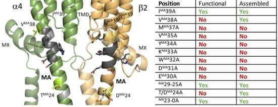
3.1. Nine Double-Alanine Mutations in the MA Helix Abolish Function

Wild-type rat α4β2 nAChRs containing an L9’A mutation in the M2 helix of the α4 subunits (to enhance receptor responses to ligand [24,25], referred to as WT in the following text) showed concentration-dependent fluorescent responses to nicotine addition, revealing a pEC50 of 7.6 ± 0.12 (EC50 = 25.3 nM), similar to previous work [18], and a Hill slope (nH) of 0.8 ± 0.2 (Figure 3). Mutant receptors with similar EC50 values exhibited similar concentration–response curves (Figure 3D).
To explore the role of the MA helix in α4β2 nAChR function, we mutated each pair of MA residues (equivalent residues in the α4 and β2 MA helices; see Figure 2) to alanine, as well as selected prolines near the potential start of the MA helix. In the initial screening of 40 mutants, 30 showed WT-like function, and 10 had no response (Table 1). Simultaneous co-expression with the two chaperones RIC-3 and NACHO ([26,27], indicated by a ‘+’ appended to the mutant name) rescued the WT-like receptor function of one double mutant (IMA39A, indicating that the lack of response of this mutant in the initial assay was due to poor folding and/or export) but had no effect on the remaining nine.

3.2. Alanine Mutations Are Less Disruptive in the α4 Than in the β2 MA Helix

To determine the contributions of each subunit type at the ten mutation-sensitive MA positions, we characterized the corresponding receptors with MA mutations in only one of the subunit types (i.e., alanine mutant α4 subunits with WT β2 subunits and vice versa). 14 of these 20 single mutants showed WT-like function, and only 2 α4 and 4 β2 single mutants remained nonresponsive to the ligand (Table 2). This apparent dependence on β2 subunit residues over α4 subunit residues could be due to the different roles of the two subunits but is more likely due to the (α4)2(β2)3 stoichiometry of receptors used in this study, where any β2 mutation occurs three times in each pentamer but an α4 mutation only twice. The IMA39A double mutant required co-expression with the chaperones to show detectable function (Table 1), but both single mutants here showed WT-like function without requiring chaperones. While a mutation could change the assembly preference and final stoichiometry of the receptors, the wild-type (α4)3(β2)2 receptor has an EC50 about 30-fold smaller than the wild-type (α4)2(β2)3 receptor [28], and we observed no such shifts, indicating that the stoichiometry was likely unchanged by the mutations.

3.3. Two of the Nine Nonfunctional Mutant Receptors Are Expressed

To probe the expression of the nine MA double-mutant receptors that showed no response in the functional assay, we measured the [3H]epibatidine binding (Figure 4). While seven of the receptors showed no measurable binding, indicating that they are deficient in subunit folding and/or assembly, two (VMA38A+ and T/DMA24A+) showed significant levels of binding. This indicates that the lack of response in the functional assay for these two mutants is either due to the mutation preventing channel opening in response to ligand binding or to the receptors (which are assembled and capable of binding ligand) not having reached the plasma membrane.

3.4. Non-Alanine Mutations Reveal Required Characteristics of Key MA Helix Residues

To explore the residue requirements at the nine positions identified as crucial to receptor assembly, export, and/or function, we assessed the effects of a range of amino acid substitutions at each position (Table 3). Three positions (VMA38, VMA35, and WMA32) showed highly specific residue requirements, where even conservative substitutions abolished receptor function, and four tolerated only one of the assayed substitutions (EMA30, DMA31, KMA33, and YMA34).

4. Discussion

The aim of this work was to explore the importance of the MA helix residues in receptor function by substituting them with alanine either in one subunit at a time (i.e., in two or three subunits of each pentamer) or in two subunits simultaneously (i.e., in all subunits of each pentamer). Alanine substitutions at 9 of the 40 positions tested abolished receptor responses, even on co-expression with chaperones RIC-3 and NACHO. Two of these non-responsive receptors showed ligand binding (VMA38A and T/DMA24A), indicating that those two residue pairs are involved either in receptor export to the plasma membrane or in allowing channel opening as a consequence of ligand binding. The remaining seven non-responsive mutant receptors showed no radioligand binding, indicating a disruption of receptor folding and/or assembly. Alanine substitutions at the remaining 30 positions tested had no measurable effect on receptor function.

4.1. Two Substitutions Abolished Detectable Ion Channel Function but Not Ligand Binding

The T/DMA24A and VMA38A mutants showed no detectable function in the fluorescence assay (Table 1) but retained their ligand binding ability (Figure 4). VMA38 sits in a pocket defined by hydrophobic MX residues (α: F, V, P, and L; β: F, L, P, and L), and Thr substitution does not rescue function. This is reminiscent of the neuromuscular nAChR αV46 pin-into-socket gating hypothesis, which proposes a critical link between the ECD and the TMD is the side chain of αV46 tucked into a pocket formed by M2 [9,29,30]. Studies of this Val show its replacement by the isosteric Thr is deleterious, indicating the side-chain polarity is critical, and mutagenesis with unnatural amino acids shows the αV46 side chain methyl groups differentially affect gating, indicating they are in different environments [31]. We suggest that a VMA38 pin-into-socket link could play a role in gating by forming an essential link between the MA and MX helices, allowing a conformational change that opens the portals. In support of this, the α7 nAChR structure shows that the MX helix moves into the membrane on receptor opening (Figure 1D), and VMA38 moves with MX, thereby remaining in this pocket (Figure 5B) even as the MA helix unwinds and the C-terminal end of the M4 helix moves outwards on receptor activation (Figure 1D). In support of this hypothesis in the α7 nAChR, VMA38A reduces ACh-induced currents [11].
An alternative explanation is that VMA38A could affect plasma membrane expression, as receptors located on internal membranes but unable to reach the plasma membrane would also be detected in our assay. In support of this, in the α7 nAChR, VMA38A reduces receptor expression levels at the plasma membrane, in addition to its effect on currents [11]. However, we consider this unlikely here, as the subunit specific mutant receptors αVMA38 + βVMA38A and αVMA38A + βVMA38 both showed WT-like function, indicating that the receptors containing these mutant subunits were capable of reaching the plasma membrane. Thus, we suggest that the receptors are correctly assembled and targeted but are unable to undergo the conformational changes necessary for ion channel opening without the Val link.
Individual alanine mutations at position MA24 indicate that αTMA24, but not βDMA24, is crucial to receptor function (Table 2). This work used the high-sensitivity (α4)2(β2)3 receptor, so the βDMA24A mutation occurs in 3/5 subunits but has no detectable effect, while the αTMA24A mutation occurs in only 2/5 subunits but abolishes receptor responses in the functional assay, ruling out a simple dosage effect as the explanation for the relative importance of this residue between the two subunits. The receptors were assembled, as shown by the binding assay, and while these might have been retained in internal membranes, we consider this unlikely, as discussed above. We suggest a better explanation is that there is a critical functional interaction between αTMA24 and another residue in the ICD; in support of this hypothesis, a study of the α7 nAChR ICD found that EMA24 forms a salt bridge with an arginine in a small helix in the ICD named h3 [10], although its importance is unknown. Our structures of the nAChR α4 (P09483) and β2 (P12390) subunits do show a similar helix to h3 running parallel to the MA helix (perhaps unsurprising, as α7 was the template here), but no interactions are visible, perhaps indicating that our model is not sufficiently accurate in this region. It is also possible that there is an interaction with one of the chaperone proteins or with the lipid headgroups. Nevertheless, if there is an important interaction here, it is not widely conserved, as the (five-fold) EMA24A mutation in the 5-HT3A receptor has only minor effects on receptor function (a slight decrease in single-channel conductance) [32].

4.2. Other Alanine Substitutions in the MA Helix

Alanine substitutions at seven positions in a row (MA30-MA37) abolished both receptor function and ligand binding (Table 1 and Figure 4 and Figure 5), indicating that these mutant receptors were not properly folded and/or assembled within the cell. The first residue in this stretch, EMA30, has been shown to affect single-channel conductance levels, as mutating it to Arg effects a >two-fold change in single-channel conductance, and changes here also affect single-channel conductance in the 5-HT3A receptor (R4′ or R440) [12,13]. This stretch is also equivalent to the stretch in the α7 nAChR that unwinds and moves out on receptor opening (Figure 1D), so alterations here would be expected to alter function. However, the effects of mutations in this stretch are less deleterious in the α7 nAChR; alanine mutations in MA32, MA33, MA34, and MA38 (MA35 and MA36 are already Ala) reduce but do not abolish expression and/or function [11].
Alanine substitutions at positions MA16-MA32 in a mutant 5-HT3A receptor (where three arginines in the MA helix have already been replaced by a Gln, Asp, and Ala triad) all retained detectable channel function (though the WMA32A mutant showed only low levels of function) [32]. Thus, the data indicate that the exact composition of the MA helix is less crucial to function in the 5-HT3AR and α7nAChR than in the α4β2 nAChR. [33] proposed that MA movement is crucial for ion conductance in the 5-HT3A receptor at the level of residues MA8 and MA9 but not at the level of MA15 and MA16, though what the effects are around MA24 and further C-terminal of that, remain to be determined.

4.3. Other Non-Alanine Substitutions in the MA Helix

Lysine substitutions at positions MA30 and MA31 in the α4β2 nAChR both abolished detectable receptor response to ligand. While not precisely comparable, we note that MA30 is an arginine in the 5-HT3A receptor, and the DMA31R mutation does not reduce receptor function [32], demonstrating that positively charged residues in the MA have different effects at the same positions in these two receptors. In fact, the negative charge is indicated to be specifically important at both these positions by the fact that DMA31E and EMA30D are both tolerated substitutions but DMA31N or EMA30Q (Table 3) are not.

5. Conclusions

In this work, we have shown that a region of the α4β2 nAChR MA helix (MA30-MA37) plays a role in receptor assembly while two residues (MA38 and MA24) are involved in receptor function. The latter contrasts with data from the α7 nAChR and 5-HT3AR, where equivalent mutations do not ablate function, suggesting that the specific roles of the MA helix residues can vary between different cation-selective pLGICs.

Author Contributions

Conceptualization, S.M.M. and S.C.R.L.; methodology, D.I.F., S.M.M. and S.C.R.L.; formal analysis, D.I.F., S.M.M. and S.C.R.L.; investigation, D.I.F., S.M.M. and S.C.R.L.; resources, S.M.M. and S.C.R.L.; data curation, D.I.F., S.M.M. and S.C.R.L.; writing—original draft preparation, D.I.F., S.M.M. and S.C.R.L.; writing—review and editing, D.I.F., S.M.M. and S.C.R.L.; supervision, S.M.M. and S.C.R.L.; project administration, S.C.R.L.; funding acquisition, S.M.M. and S.C.R.L. All authors have read and agreed to the published version of the manuscript.

Funding

This research was funded by AstraZeneca (studentship to SMM) and the MRC (grant No. MR/L021676 to S.C.R.L.).

Institutional Review Board Statement

Not applicable.

Data Availability Statement

Data is contained within the article.

Conflicts of Interest

The authors declare no conflict of interest.

References

  1. Bondarenko, V.; Wells, M.M.; Chen, Q.; Tillman, T.S.; Singewald, K.; Lawless, M.J.; Caporoso, J.; Brandon, N.; Coleman, J.A.; Saxena, S.; et al. Structures of highly flexible intracellular domain of human α7 nicotinic acetylcholine receptor. Nat. Commun. 2022, 13, 793. [Google Scholar] [CrossRef] [PubMed]
  2. Carland, J.E.; Cooper, M.A.; Livesey, M.R.; Hales, T.G.; Peters, J.A.; Lambert, J.J. Mutagenic Analysis of the Intracellular Portals of the Human 5-HT3A Receptor. J. Biol. Chem. 2013, 288, 31592–31601. [Google Scholar] [CrossRef] [PubMed]
  3. Cecchini, M.; Changeux, J.P. The nicotinic acetylcholine receptor and its prokaryotic homologues: Structure, conformational transitions & allosteric modulation. Neuropharmacology 2015, 96, e137–e149. [Google Scholar]
  4. Changeux, J.-P.; Galzi, J.-L.; Devillers-Thiéry, A.; Bertrand, D. The functional architecture of the acetylcholine nicotinic receptor explored by affinity labelling and site-directed mutagenesis. Q. Rev. Biophys. 1992, 25, 395–432. [Google Scholar] [CrossRef] [PubMed]
  5. Criado, M.; Mulet, J.; Gerber, S.; Sala, S.; Sala, F. A small cytoplasmic region adjacent to the fourth transmembrane segment of the α7 nicotinic receptor is essential for its biogenesis. FEBS Lett. 2011, 585, 2477–2480. [Google Scholar] [CrossRef] [PubMed]
  6. Dougherty, D.A. Cys-loop neuroreceptors: Structure to the rescue? Chem. Rev. 2008, 108, 1642–1653. [Google Scholar] [CrossRef] [PubMed]
  7. Fonck, C.; Cohen, B.N.; Nashmi, R.; Whiteaker, P.; Wagenaar, D.A.; Rodrigues-Pinguet, N.; Deshpande, P.; McKinney, S.; Kwoh, S.; Munoz, J.; et al. Novel Seizure Phenotype and Sleep Disruptions in Knock-In Mice with Hypersensitive α4* Nicotinic Receptors. J. Neurosci. 2005, 25, 11396–11411. [Google Scholar] [CrossRef] [PubMed]
  8. Gu, S.; Matta, J.A.; Lord, B.; Harrington, A.W.; Sutton, S.W.; Davini, W.B.; Bredt, D.S. Brain α7 Nicotinic Acetylcholine Receptor Assembly Requires NACHO. Neuron 2016, 89, 948–955. [Google Scholar] [CrossRef]
  9. Hales, T.G.; Dunlop, J.I.; Deeb, T.Z.; Carland, J.E.; Kelley, S.P.; Lambert, J.J.; Peters, J.A. Common Determinants of Single Channel Conductance within the Large Cytoplasmic Loop of 5-Hydroxytryptamine Type 3 and α4β2 Nicotinic Acetylcholine Receptors. J. Biol. Chem. 2006, 281, 8062–8071. [Google Scholar] [CrossRef]
  10. Hanek, A.P.; Lester, H.A.; Dougherty, D.A. A Stereochemical Test of a Proposed Structural Feature of the Nicotinic Acetylcholine Receptor. J. Am. Chem. Soc. 2008, 130, 13216–13218. [Google Scholar] [CrossRef]
  11. Hassaine, G.; Deluz, C.; Grasso, L.; Wyss, R.; Tol, M.B.; Hovius, R.; Graff, A.; Stahlberg, H.; Tomizaki, T.; Desmyter, A.; et al. X-ray structure of the mouse serotonin 5-HT3 receptor. Nature 2014, 512, 276–281. [Google Scholar] [CrossRef]
  12. Jumper, J.; Evans, R.; Pritzel, A.; Green, T.; Figurnov, M.; Ronneberger, O.; Tunyasuvunakool, K.; Bates, R.; Žídek, A.; Potapenko, A.; et al. Highly accurate protein structure prediction with AlphaFold. Nature 2021, 596, 583–589. [Google Scholar] [CrossRef]
  13. Kelley, S.P.; Dunlop, J.I.; Kirkness, E.F.; Lambert, J.J.; Peters, J.A. A cytoplasmic region determines single-channel conductance in 5-HT3 receptors. Nature 2003, 424, 321–324. [Google Scholar] [CrossRef]
  14. Lansdell, S.J.; Gee, V.J.; Harkness, P.C.; Doward, A.I.; Baker, E.R.; Gibb, A.J.; Millar, N.S. RIC-3 Enhances Functional Expression of Multiple Nicotinic Acetylcholine Receptor Subtypes in Mammalian Cells. Mol. Pharmacol. 2005, 68, 1431–1438. [Google Scholar] [CrossRef]
  15. Mazzaferro, S.; Whiteman, S.T.; Alcaino, C.; Beyder, A.; Sine, S.M. NACHO and 14-3-3 promote expression of distinct subunit stoichiometries of the α4β2 acetylcholine receptor. Cell Mol. Life Sci. 2020, 78, 1565–1575. [Google Scholar] [CrossRef]
  16. Mesoy, S.M.; Lummis, S.C.R. M4, the Outermost Helix, is Extensively Involved in Opening of the α4β2 nACh Receptor. ACS Chem. Neurosci. 2020, 12, 133–139. [Google Scholar] [CrossRef]
  17. Mocatta, J.; Mesoy, S.M.; Dougherty, D.A.; Lummis, S.C.R. 5-HT3 Receptor MX Helix Contributes to Receptor Function. ACS Chem. Neurosci. 2022, 13, 2338–2345. [Google Scholar] [CrossRef]
  18. Morales-Perez, C.L.; Noviello, C.M.; Hibbs, R.E. X-ray structure of the human α4β2 nicotinic receptor. Nature 2016, 538, 411–415. [Google Scholar] [CrossRef]
  19. Nelson, M.E.; Kuryatov, A.; Choi, C.H.; Zhou, Y.; Lindstrom, J.; Kohout, T.A.; Lefkowitz, R.J. Alternate Stoichiometries of α4β2 Nicotinic Acetylcholine Receptors. Mol. Pharmacol. 2003, 63, 332–341. [Google Scholar] [CrossRef]
  20. Nemecz, Á.; Prevost, M.S.; Menny, A.; Corringer, P.J. Emerging Molecular Mechanisms of Signal Transduction in Pentameric Ligand-Gated Ion Channels. Neuron 2016, 90, 452–470. [Google Scholar] [CrossRef]
  21. Nguyen, M.; Alfonso, A.; Johnson, C.D.; Rand, J.B. Caenorhabditis elegans mutants resistant to inhibitors of acetylcholinesterase. Genetics 1995, 140, 527–535. [Google Scholar] [CrossRef]
  22. Noviello, C.M.; Gharpure, A.; Mukhtasimova, N.; Cabuco, R.; Baxter, L.; Borek, D.; Sine, S.M.; Hibbs, R.E. Structure and gating mechanism of the α7 nicotinic acetylcholine receptor. Cell 2021, 184, 2121–2134.e13. [Google Scholar] [CrossRef]
  23. Plested, A.J.R. Structural mechanisms of activation and desensitization in neurotransmitter-gated ion channels. Nat. Struct. Mol. Biol. 2016, 23, 494–502. [Google Scholar] [CrossRef]
  24. Price, K.L.; Lummis, S.C. FlexStation examination of 5-HT3 receptor function using Ca2+- and membrane potential-sensitive dyes: Advantages and potential problems. J. Neurosci. Methods 2005, 149, 172–177. [Google Scholar] [CrossRef]
  25. Stuebler, A.G.; Jansen, M. Mobility of Lower MA-Helices for Ion Conduction through Lateral Portals in 5-HT3A Receptors. Biophys. J. 2020, 119, 2593–2603. [Google Scholar] [CrossRef]
  26. Tapper, A.R.; McKinney, S.L.; Nashmi, R.; Schwarz, J.; Deshpande, P.; Labarca, C.; Whiteaker, P.; Marks, M.J.; Collins, A.C.; Lester, H.A. Nicotine Activation of α4* Receptors: Sufficient for Reward, Tolerance, and Sensitization. Science 2004, 306, 1029–1032. [Google Scholar] [CrossRef]
  27. Thompson, A.; Lummis, S. Discriminating between 5-HT3A and 5-HT3AB receptors. Br. J. Pharmacol. 2013, 169, 736–747. [Google Scholar] [CrossRef]
  28. Thompson, A.J.; Lester, H.A.; Lummis, S.C.R. The structural basis of function in Cys-loop receptors. Q. Rev. Biophys. 2010, 43, 449–499. [Google Scholar] [CrossRef]
  29. Unwin, N.; Miyazawa, A.; Li, J.; Fujiyoshi, Y. Activation of the Nicotinic Acetylcholine Receptor Involves a Switch in Conformation of the α Subunits. J. Mol. Biol. 2002, 319, 1165–1176. [Google Scholar] [CrossRef]
  30. Unwin, N. Structure and action of the nicotinic acetylcholine receptor explored by electron microscopy. FEBS Lett. 2003, 555, 91–95. [Google Scholar] [CrossRef]
  31. Unwin, N. Refined Structure of the Nicotinic Acetylcholine Receptor at 4Å Resolution. J. Mol. Biol. 2005, 346, 967–989. [Google Scholar] [CrossRef] [PubMed]
  32. Varadi, M.; Anyango, S.; Deshpande, M.; Nair, S.; Natassia, C.; Yordanova, G.; Yuan, D.; Stroe, O.; Wood, G.; Laydon, A.; et al. AlphaFold Protein Structure Database: Massively expanding the structural coverage of protein-sequence space with high-accuracy models. Nucleic Acids Res. 2022, 50, D439–D444. [Google Scholar] [CrossRef]
  33. Yang, J.; Zhang, Y. I-TASSER server: New development for protein structure and function predictions. Nucleic Acids Res. 2015, 43, W174–W181. [Google Scholar] [CrossRef]
Figure 1. Structures of the α7 nAChR in the closed (dark blue, PDB 7KOO) and activated (light blue, 7KOX) states. (A) Single subunit showing major structural domains. (B) Receptor overview. (C) TMD and ICD of the open and closed states of two subunits. (D) M4, MX, and MA, showing unwinding of MA on receptor opening. The black line indicates the approximate location of the plasma membrane. Residue MA39 is the most C-terminal residue investigated in this study, and residue MA10 shows the relative positioning of the two helices below the membrane.
Membranes 13 00891 g001
Figure 2. Comparison of pLGIC MA sequences. (A) A sequence alignment for 3 different nAChR subunits showing residue identity (~70% between α4 and β2 subunits) and the MA sequence numbering system used here. (B) A multiple-sequence alignment of MA helices from all nAChR subunits and representative subunits of other pLGICs reveals the most conserved residues. The three arginine residues that contribute to single-channel conductance in the 5-HT3AR subunit are indicated with red circles. (C) A phylogenetic analysis of the same sequences with nAChR subunits shown in red, other cationic subunits in yellow, and anionic subunits in blue.
Membranes 13 00891 g002
Figure 3. Characterization of α4β2 nAChRs in HEK293 cells. (AC) Typical fluorescent responses (F, arbitrary units) to the addition of nicotine at 20 s to the mock transfected cells (A) or cells transfected with WT α4β2 nAChR (B) or WT α4β2 nAChR and chaperones RIC-3 and NACHO (C). (D) concentration–response curves from (B,C) and similar data (mean ± SEM, n ≥ 3).
Membranes 13 00891 g003
Figure 4. Specific binding of [3H]epibatidine relative to the WT in membranes from cells transfected with double-mutant nAChR subunit DNA, as indicated, and with RIC-3 and NACHO (MT indicates mock transfected cells). Black indicates significantly different to MT (p < 0.05). Data = mean ± SEM, n = 3.
Membranes 13 00891 g004
Figure 5. (A) MA helices of α4 (green) and β2 (yellow) in the closed α4β2 structure predicted by AlphaFold (which includes the full MA helix, showing MA helices going from PMA0 to IMA39 (both marked as sticks)). Black: positions where alanine mutations result in nonfunctional receptors incapable of ligand binding. Light colors: positions where alanine mutations result in nonfunctional receptors capable of ligand binding. Grey: positions where alanine mutations were not tested (most already alanines). The black line indicates the approximate location of the plasma membrane. (B) VMA38 and the MX helix in the closed and open conformations of α7, α4, and β2.
Membranes 13 00891 g005
Table 1. Parameters from the MA Ala mutants. Data = mean ± SEM, n = 3–5, NR = non-responsive.
PositionMutation (α4/β2)pEC50 (M)EC50 (nM)nH
WT 7.60 ± 0.1225.30.8 ± 0.2
WT+ 7.94 ± 0.1211.40.6 ± 0.1
IMA39A+I601A/I456A8.11 ± 0.1281.3 ± 0.4
VMA38A+V600A/V455ANR
MMA37A+M599A/M454ANR
VMA35A+V597A/V452ANR
YMA34A+Y596A/Y451ANR
KMA33A+K595A/K450ANR
WMA32A+W594A/W449ANR
DMA31A+D593A/D448ANR
EMA30A+E592A/E447ANR
K/RMA29AK591A/R446A7.62 ± 0.06241.1 ± 0.2
VMA28AV590A/V445A7.62 ± 0.06241.5 ± 0.3
SMA27AS589A/S444A7.47 ± 0.06341.2 ± 0.2
F/QMA26AF588A/Q443A7.83 ± 0.0614.71.2 ± 0.2
DMA25AD587A/D442A7.79 ± 0.0916.21.4 ± 0.4
T/DMA24A+T586A/D441ANR
DMA23AD585A/D440A7.89 ± 0.0812.81.1 ± 0.3
EMA22AE584A/E439A7.59 ± 0.0525.32.1 ± 0.5
WT/SMA21AWT/S438A7.36 ± 0.0343.21.6 ± 0.2
K/RMA20AK582A/R437A7.76 ± 0.0817.21.3 ± 0.2
L/MMA19AL581A/M436A7.63 ± 0.0923.51.5 ± 0.4
HMA18AH580A/H435A7.70 ± 0.1920.11.9 ± 1.3
DMA17AD579A/D434A7.61 ± 0.1324.72.6 ± 2.1
IMA15AI577A/I432A7.71 ± 0.1019.61.3 ± 0.4
Y/FMA14AY576A/F431A7.99 ± 0.0510.21.2 ± 0.1
Q/RMA13AQ575A/R430A7.73 ± 0.1318.61.1 ± 0.3
VMA12AV574A/V429A7.55 ± 0.1428.01.2 ± 0.4
GMA11AG573A/G428A7.23 ± 0.2758.00.8 ± 0.5
E/DMA10AE572A/D427A7.55 ± 0.1128.51.3 ± 0.3
VMA9AV571A/V426A7.79 ± 0.1016.41.2 ± 0.2
R/EMA7AR569A/E424A7.73 ± 0.1618.51.6 ± 0.8
T/RMA6AT568A/R423A7.72 ± 0.2419.30.9 ± 0.4
LMA5AL567A/L422A7.63 ± 0.1023.20.9 ± 0.1
WT/GMA4AWT/G421A7.91 ± 0.1012.11.7 ± 0.5
P/CMA3AP565A/C420A7.79 ± 0.0616.11.1 ± 0.2
SMA2AS564A/S419A7.61 ± 0.0824.31.2 ± 0.3
PMA0AP562A/P417A7.67 ± 0.1221.61.0 ± 0.2
P558A/WT7.69 ± 0.1420.31.4 ± 0.5
P557A/WT7.75 ± 0.1817.61.1 ± 0.4
WT/P411A7.78 ± 0.0716.61.1 ± 0.2
WT/P406A7.67 ± 0.0821.41.0 ± 0.2
Table 2. Parameters from the MA single subunit Ala mutants. Data = mean ± SEM, n = 3–5, NR = non-responsive.
Mutant α4
WT β2
WT α4
Mutant β2
PositionpEC50 (M)EC50 (nM)nHpEC50 (M)EC50 (nM)nH
WT7.60 ± 0.1225.30.8 ± 0.27.60 ± 0.1225.30.8 ± 0.2
WT+7.94 ± 0.1211.40.6 ± 0.17.94 ± 0.1211.40.6 ± 0.1
IMA39A7.81 ± 0.06151.9 ± 0.47.41 ± 0.08391.0 ± 0.2
VMA38A+7.20 ± 0.2463.30.8 ± 0.47.26 ± 0.11551.2 ± 0.3
MMA37A+7.53 ± 0.06301.0 ± 0.1NR
VMA35A+7.32 ± 0.1048.11.5 ± 0.47.67 ± 0.17211.3 ± 0.7
YMA34A+7.46 ± 0.0834.81.2 ± 0.27.46 ± 0.08341.3 ± 0.3
KMA33A+7.63 ± 0.1723.20.7 ± 0.2NR
WMA32A+NR NR
DMA31A+7.53 ± 0.0829.71.3 ± 0.3NR
EMA30A+7.38 ± 0.0841.90.8 ± 0.17.78 ± 0.07171.5 ± 0.4
T/DMA24A+NR 7.59 ± 0.1226.01.0 ± 0.2
Table 3. Parameters from receptors mutated at sensitive MA residues. Data = mean ± SEM, n = 3–5, NR = non-responsive.
PositionpEC50 (M)EC50 (nM)nH
WT7.60 ± 0.1225.30.8 ± 0.2
WT+7.94 ± 0.1211.40.6 ± 0.1
VMA38I+NR
VMA38T+NR
MMA37K+NR
VMA35I+NR
VMA35T+NR
YMA34F7.21 ± 0.1661.20.88 ± 0.3
YMA34S+NR
YMA34L+NR
YMA34Q+NR
KMA33E7.02 ± 0.0695.41.1 ± 0.2
KMA33Q+NR
KMA33M+NR
WMA32F+NR
WMA32Y+NR
DMA31E7.43 ± 0.0937.41.2 ± 0.3
DMA31K+NR
DMA31L+NR
DMA31N+NR
EMA30D7.48 ± 0.0432.91.6 ± 0.2
EMA30K+NR
EMA30L+NR
EMA30Q+NR
αTMA24D7.50 ± 0.0531.41.6 ± 0.3
αTMA24E7.14 ± 0.2872.41.2 ± 0.8
αTMA24K7.41 ± 0.1039.41.4 ± 0.4
αTMA24S7.80 ± 0.0415.91.6 ± 0.2
αTMA24V+NR
βDMA24T+7.76 ± 0.1017.31.1 ± 0.2
Disclaimer/Publisher’s Note: The statements, opinions and data contained in all publications are solely those of the individual author(s) and contributor(s) and not of MDPI and/or the editor(s). MDPI and/or the editor(s) disclaim responsibility for any injury to people or property resulting from any ideas, methods, instructions or products referred to in the content.

