Review

Mesenchymal Stem Cell-Derived Extracellular Vesicles for Bone Defect Repair

1
School of Sport Medicine and Rehabilitation, Beijing Sport University, Beijing 100084, China
2
School of Kinesiology, Shanghai University of Sport, Shanghai 200438, China
*
Authors to whom correspondence should be addressed.
Membranes 2022, 12(7), 716; https://doi.org/10.3390/membranes12070716
Submission received: 29 June 2022 / Revised: 17 July 2022 / Accepted: 18 July 2022 / Published: 19 July 2022
(This article belongs to the Special Issue Biocompatible Membranes for Bone Regeneration)

Abstract

:
The repair of critical bone defects is a hotspot of orthopedic research. With the development of bone tissue engineering (BTE), there is increasing evidence showing that the combined application of extracellular vesicles (EVs) derived from mesenchymal stem cells (MSCs) (MSC-EVs), especially exosomes, with hydrogels, scaffolds, and other bioactive materials has made great progress, exhibiting a good potential for bone regeneration. Recent studies have found that miRNAs, proteins, and other cargo loaded in EVs are key factors in promoting osteogenesis and angiogenesis. In BTE, the expression profile of the intrinsic cargo of EVs can be changed by modifying the gene expression of MSCs to obtain EVs with enhanced osteogenic activity and ultimately enhance the osteoinductive ability of bone graft materials. However, the current research on MSC-EVs for repairing bone defects is still in its infancy, and the underlying mechanism remains unclear. Therefore, in this review, the effect of bioactive materials such as hydrogels and scaffolds combined with MSC-EVs in repairing bone defects is summarized, and the mechanism of MSC-EVs promoting bone defect repair by delivering active molecules such as internal miRNAs is further elucidated, which provides a theoretical basis and reference for the clinical application of MSC-EVs in repairing bone defects.

1. Introduction

A critical-size bone defect is defined as the smallest size intra-osseous wound in a particular bone and species that will not heal spontaneously during the lifetime of the animal [1,2]. Critical-size bone defects are often caused by trauma, bone tumors, and infection [3]. At present, the clinical strategies for repairing bone defects mainly include autograft, allograft, and synthetic biomaterial graft [4,5]. Autologous bone transplantation is generally considered to be the gold standard for repairing bone defects, although there are many limitations, including the high morbidity of donor bone, limited availability, and unpredictable autologous absorption [6,7]. The clinical application of pure bone allograft is limited due to its susceptibility to immune rejection [8]. In 1993, Langer et al. [9] first proposed the potential strategy of applying tissue engineering to regenerative medicine. Bone tissue engineering (BTE) is mainly used for large-scale bone defect repair, which includes three basic components: bioscaffolds, seed cells, and osteoinductive factors [10]. In recent years, BTE has made great progress in the treatment of bone regeneration. Bioactive materials such as scaffolds combined with mesenchymal stem cells (MSCs) and their secreted factors have been widely used in the repair of bone defects [11]. MSCs are a heterogeneous mesenchymal stem cell subpopulation, including bone marrow mesenchymal stem cells (BMSCs) [12], adipose mesenchymal stem cells (ADSCs) [13], umbilical cord mesenchymal stem cells (UCMSCs) [14], and induced pluripotent stem cells (iPSCs) [15], with multi-directional differentiation potentials such as osteoblasts, chondrocytes, and adipocytes [16]. Therefore, stem cell therapy is considered a potential strategy for bone defect regeneration [17,18]. However, there are still some disadvantages, such as phenotypic changes, low homing efficiency, and low cell viability, which limit the application of MSCs in bone defect repair [13]. In recent years, cell-free therapy for bone regeneration has attracted extensive attention [19,20]. Accumulating evidence indicates that the positive effect of MSCs on tissue repair is to stimulate the activity of tissue-resident receptor cells through paracrine rather than directly differentiate into parenchymal cells to repair or replace damaged tissue [21,22]. Among the many cytokines secreted by MSCs, extracellular vesicles (EVs) are nano-phospholipid bilayer vesicles that can be secreted by almost all cells and are formed by “budding” directly from the plasma membrane. According to their diameter, composition, and origin, EVs can be divided into exosomes (Exos), microvesicles, and apoptotic bodies [23]. Among them, Exos are the most attractive category of EVs. Exos are bilayer lipid-membrane vesicles with a diameter between 40 and 150 nm, which carry a rich variety of nucleic acids, proteins, miRNAs, mRNAs, and lipid substances. As a nanocarrier for cell-to-cell communication, Exos are released extracellularly to exert regulatory effects by transferring intrinsic cargo to target cells via ligand-receptor interactions, endocytosis, or direct membrane fusion [24]. Numerous studies have shown that Exos obtained from MSCs are considered to be the main mediator of the therapeutic effect of MSCs without the limitations of MSC therapy [16,25,26]. Recently, in bone regenerative medicine, the research of MSC-derived Exos (MSC-Exos) has become an attractive hotspot [27]. Many studies have shown that bioactive materials such as hydrogels or scaffolds loaded with MSC-derived EVs (MSC-EVs) can significantly promote osteogenesis and angiogenesis, thereby effectively repairing bone defects [28,29].
However, the research on MSC-EVs promoting bone defect repair is still in the preliminary stage, and the specific mechanism is still unclear. In this paper, the application progress of MSC-EVs combined with hydrogels and/or scaffolds in repairing bone defects and the potential mechanism of MSC-EVs promoting osteogenesis or angiogenesis are comprehensively summarized. This review provides the latest evidence and perspectives on the effective application, potential risks and solutions of MSC-EVs combined with hydrogels and/or scaffolds and the potential mechanism of MSC-EVs in enhancing bone regeneration, which will provide a theoretical basis for further application of MSC-EVs to promote bone defect repair.

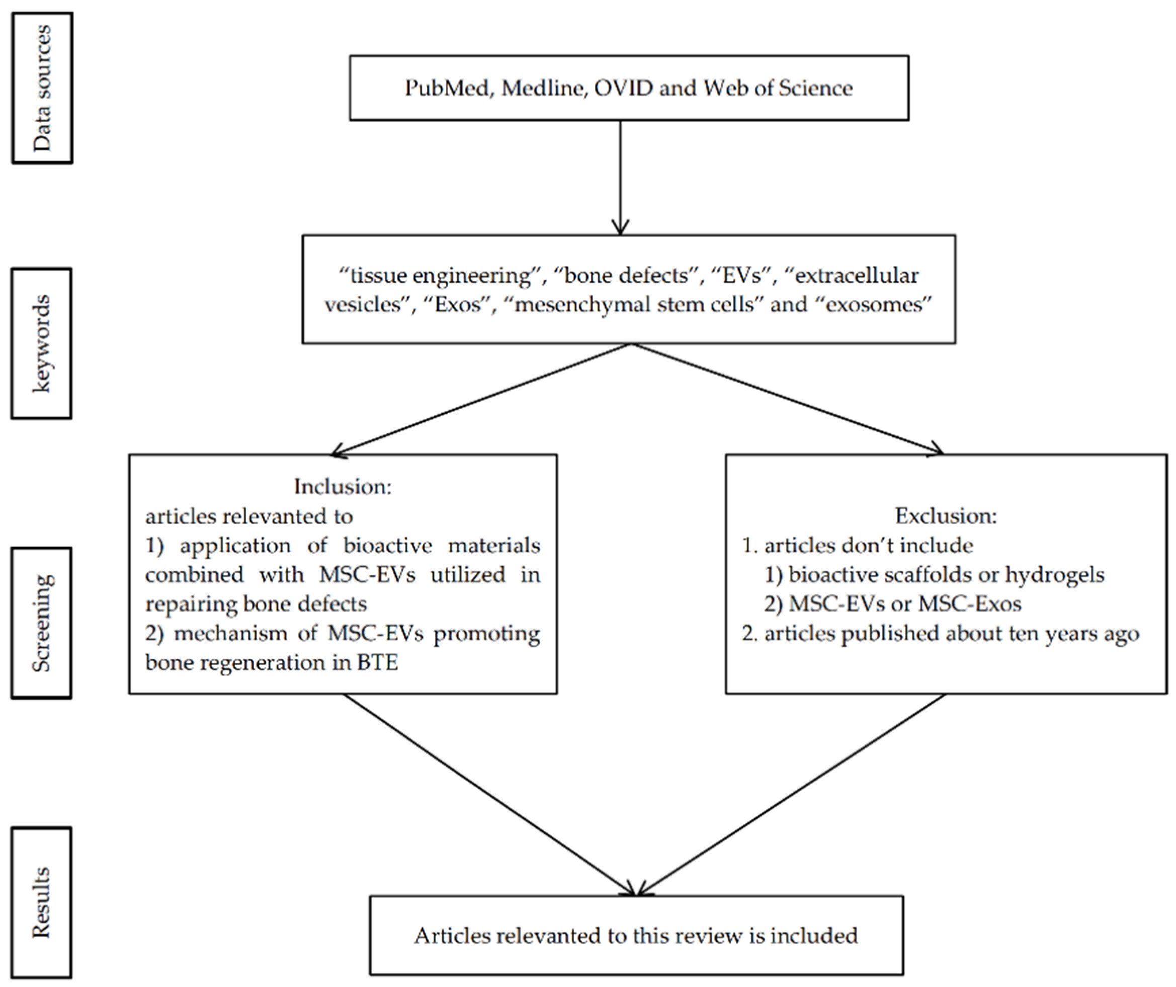
2. Materials and Methods

This paper aims to summarize the advanced application of bioactive materials combined with MSC-EVs utilized in repairing bone defects and the mechanism of MSC-EVs promoting bone regeneration. A literature search was performed in electronic databases, including PubMed, Medline, OVID, and Web of Science, by using the following keywords: “tissue engineering”, “bone defects”, “EVs”, “extracellular vesicles”, “Exos”, “mesenchymal stem cells” and “exosomes”. Documents published in English were selected, and the articles were further screened to identify their relevance to this review (as shown in Figure 1). This narrative review briefly discusses the application of bioactive materials combined with MSC-EVs in repairing bone defects and the underlying mechanism of MSC-EVs to promote bone regeneration, as well as the limits of current studies.

3. Application of MSC-EVs in Bone Defects

3.1. Application of EVs Parent Cells

As key mediators of cell-to-cell communication, Exos play important roles in a variety of biological roles and injury repair processes, such as regulating immune response [30], promoting the repair of traumatic brain injury [31], and stimulating bone tissue regeneration [32]. Studies have shown that in the treatment of bone tissue regeneration, MSC-Exos stimulate osteoblast proliferation and angiogenesis and inhibit osteoclast maturation by delivering endogenous cargo [33]. Since Exos generally have similar therapeutic properties to their parent cells, Exos derived from different MSCs have different effects on bone regeneration and repair [34,35]. Therefore, in the strategy of applying MSC-Exos to bone regeneration, it is important to select an appropriate source of parental cell types [36].
The sources of parental MSCs of Exos mainly include BMSCs [12], ADSCs [13], UCMSCs [14], iPSCs [15], etc. As shown in Table 1, among these cells, BMSCs are the most commonly used cell source for tissue engineering [37]. Numerous studies have shown that BMSC-derived Exos (BMSC-Exos) can stimulate the proliferation and osteogenic differentiation of BMSCs, thereby promoting osteogenesis, angiogenesis, and bone mineralization in critical bone defect models [38]. Compared with BMSCs, human adipose mesenchymal stem cells (hADSCs), mainly derived from adipose tissue, are easier to obtain. hADSCs are widely distributed in the human body and proliferate rapidly, which is an ideal type of MSC for improving Exos production [39]. However, compared with BMSCs, hADSCs may have less osteogenic differentiation potential [40]. Some scholars compared the osteogenic differentiation potential of rat BMSC-Exos and ADSC-derived Exos (ADSC-Exos) in osteogenesis-induced (OI) conditions. The results showed that BMSC-OI-Exos significantly up-regulated the expression of osteogenesis-related genes and proteins, including Col I, Runx2, BSP, OCN, BMPR-IA, and BMPR-II, while ADSC-OI-Exos only up-regulated BSP, BMPR-IA and, BMPR-II, which suggested that ADSC-OI-Exos may have a lower osteoinductive ability compared with BMSC-OI-Exos [41]. In addition, hADSC-derived Exos (hADSC-Exos) have shown great therapeutic potential in the treatment of ischemic diseases, indicating that hADSC-Exos may also play a potential role in bone regeneration by promoting angiogenesis [42]. Human umbilical cord mesenchymal stem cells (hUCMSCs) have also attracted much attention in BTE. hUCMSCs are a primitive population of MSCs obtained from Wharton’s jelly, the main component of the human umbilical cord matrix [43]. hUCMSCs can indirectly promote bone regeneration by inducing angiogenesis rather than directly promoting osteogenic or chondrogenic differentiation. Compared with other MSCs derived from bone marrow or fat, hUCMSCs showed higher pluripotency [44,45] and stronger pro-angiogenesis properties [46]. In addition, hUCMSCs are derived from human postnatal waste tissues and were abundant in source, which is a rich source without ethical and moral controversies, showing greater clinical potential [47]. Similar to hUCMSCs, iPSCs also have no immune rejection or ethical issues and can proliferate indefinitely; however, iPSCs may have tumorigenic risks [48]. In recent years, studies have shown that MSCs derived from iPSCs (iPS-MSCs) have the advantages of both iPSCs and MSCs. iPS-MSCs can maintain their self-renewal ability after 40 passages. Importantly, iPS-MSCs are not tumorigenic [49]. In the treatment of bone defects, iPS-MSCs show strong proliferative and immunomodulatory abilities to promote bone regeneration [15]. Exos derived from iPS-MSCs also have similar properties to their parental cells. Therefore, iPS-MSCs are an excellent cell source for Exos to promote bone regeneration [22]. In addition to the above-mentioned MSCs, some local tissue-derived MSC-Exos also have good application potential. Stem cells from human exfoliated deciduous teeth (SHEDs), which are immature MSCs with multiple differentiation potential, can be easily obtained in a non-invasive way without ethical problems [50]. Compared with BMSCs, SHEDs have a stronger proliferative capacity due to the abundant secretion of growth factors such as fibroblast growth factor 2 (FGF2) and transforming growth factor-β2 (TGF-β2) [51]. Moreover, compared with pulp stem cells, SHEDs have a stronger capacity to promote bone mineralization [52]. SHED-derived Exos, in combination with tricalcium phosphate (β-TCP), have been shown to enhance alveolar bone regeneration by promoting angiogenesis and osteogenesis [53]. In addition to the source of parental MSCs, the therapeutic effect of Exos may also be influenced by some underlying diseases of the donor. A recent study showed that BMSC-Exos obtained from the T1DM rat model was significantly less effective in promoting BMSC osteogenic differentiation and endothelial cell angiogenesis than BMSC-Exos obtained from normal rats [35]. This suggests that Exos from donors with chronic underlying diseases have a poor therapeutic effect, or even the opposite therapeutic effect, which may not be suitable for regenerative medicine [54]. Further research is needed to explore the potential therapeutic effects of donor-derived Exos in disease states.

3.2. Application of MSC-EVs in BTE

The regeneration of critical bone defects caused by bone tumors, injuries, and other bone diseases requires bone grafting to replace the defective bone. EV-mediated BTE therapy has become an attractive bone defect replacement strategy in recent years [55]. Accumulating studies have shown that EV-loaded hydrogels or scaffolds, alone or in combination, can significantly promote local bone regeneration (as shown in Figure 2).

3.2.1. Application of MSC-EVs Combined with Hydrogels

Hydrogels are physically or chemically cross-linked three-dimensional (3D) hydrophilic polymer networks that can adsorb large amounts of water without being dissolved [56]. Biomedical hydrogels are structurally similar to the natural extracellular matrix, which have attracted extensive attention in regenerative medicine due to their properties of tissue-like water content, good biocompatibility, and easy implantation. In regenerative medicine, hydrogels, as an effective carrier, are widely used in drug delivery and active molecule encapsulation matrix for tissue repair, including bone tissue repair [57]. In bone tissue regeneration, biopolymer hydrogels can be injected directly into bone defects and flow over complex and irregular bone surfaces, matching the gaps between implants and randomly shaped defects and increasing the contact area [58]. In addition, the porous structure of hydrogels can continuously release bioactive factors, including EVs/Exos, which is beneficial for repairing bone defects in BTE [58]. Studies have shown that Exos coated in biodegradable hydrogels can still exhibit the desired therapeutic effect [59,60]. Hydrogels provide a 3D matrix for Exos that prevents the dispersion of Exos and maintains its local concentration, which enables controlled release of Exos for sustained efficacy. Hydrogel-released Exos at bone defect sites can reduce Exos consumption and ectopic effects to promote angiogenesis and osteogenesis [61]. Some scholars found gelatin nanoparticle hydrogels combined with hADSC-Exos can accurately transport the hADSC-Exos to the target site and effectively promote bone healing [62]. In addition, the hydrogel 3D microenvironment can also enhance Exos activity and affect the interaction of EV integrin membrane protein between cells and the cell matrix to promote the proliferation and differentiation of osteoblasts and bone angiogenesis in a bone regeneration environment. Yu et al. [4] demonstrated that Exos derived from periodontal ligament stem cells (PDLSCs) encapsulated in a hydrogel 3D microenvironment exhibited enhanced osteoinductive ability and could significantly promote bone defect repair in rats. Another study demonstrated alginate hydrogels combined with EVs showed increased interactions with other cells, cell aggregation, and prolonged long-term viability, which in turn promoted osteogenesis [61]. Therefore, a variety of special properties of hydrogels can be combined with MSC-EVs to effectively treat bone defects.
At present, the commonly used biomedical hydrogels mainly include natural and synthetic polymer hydrogels, such as gelatin, hyaluronic acid hydrogel (HA-gel), chitosan, polyethylene glycol (PEG), etc. [63]. Compared to natural materials, synthetic polymers have basic structural units, well-defined properties, including porosity and degradation time, and known mechanical properties, making synthetic polymer hydrogels more suitable for specific bone tissue regeneration applications [64]. In addition, synthetic polymers are suitable delivery vehicles for EVs in bone defect repair due to their reliable source of material, long shelf life, and low risk of immunogenicity [65]. However, due to their fragile network structure, the mechanical properties, stability, and cell adhesion of traditional hydrogels are usually weak, which hinders their application as bone structural materials [66]. To overcome this limitation, hydrogels are often modified by chemical, physical, and biological methods to prepare high-performance hydrogels with enhanced mechanical properties, stability, cell adhesion, and other properties [63]. Ma et al. [67] used propionic acid (CA) with a catechol group to modify an injectable thermosensitive hydrogel through a covalent bond. The cross-linking network formed by the interaction between the catechol group of CA and the collagen molecules in hydrogels can effectively improve the mechanical properties of hydrogel [68]. In addition, the combination of two hydrogels to prepare new hydrogels is also a common way of hydrogel modification. Previous studies revealed that the combination of a gelatin derivative, gelatin methacrylate, and a PEG derivative, polyethylene glycol diacrylate, can prepare a hydrogel with good cell adhesion and stronger mechanical properties [69]. Hydrogels with good performance can be obtained by various methods; however, how to effectively load bioactive molecules such as EVs/Exos into hydrogels and improve their long-term retention at defect sites is also a challenge in BTE. Currently, the commonly used method is to directly mix Exos with hydrogels [70], which may lead to low loading efficiency, structural destruction of Exos, and hindered osteogenic potential [71]. Recent studies have demonstrated that substances such as peptides can be used to effectively fix EVs/Exos, showing excellent drug delivery ability, which can effectively deliver EVs/Exos [72]. Huang et al. [29] found that integrin membrane proteins in MSC-EVs could combine with collagen and fibronectin-derived peptides secreted by cells. Subsequently, the adhesive peptide (RGD) was added to alginate hydrogels containing EVs overexpressing bone morphogenetic protein 2 (BMP-2) to target cellular integrins. The results showed that EVs could be effectively fixed to hydrogels through the interaction of protein domains on the cell membrane [29]. In another study, some scholars incorporated Exos into an injectable thermosensitive hydrogel by constructing fusion peptides, which also enhanced the retention of Exos and improved the biological activity of Exos [67]. The above results indicated that the addition of bioactive substances such as peptides can enhance the fixation and retention of EVs without impairing the structural integrity and biological activity of EVs. This enhanced fixation and retention may prolong the presence of EVs at healing sites, leading to increased efficiency of delivery as well as functionality. It should be noted that the mechanical properties, gelation time, biocompatibility, and biological functions of hydrogels vary due to different raw materials and synthesis conditions in the practical application of hydrogels [73]. Some studies suggest that chitosan hydrogel has good biocompatibility, hemostasis, and chemical activity [74], while alginate is a biologically neutral material with adjustable mechanical properties and biodegradation [75]. In addition, since the efficacy of MSC-EVs encapsulated in hydrogels on bone regeneration largely depends on the design and function of hydrogels, attention should be paid to the different characteristics of hydrogels when they are used as delivery materials [73]. Zhang et al. [76] prepared an injectable thermosensitive chitosan hydrogel with good biodegradability and biocompatibility, which can improve the stability and retention of human placental MSC-Exos, and effectively enhance bone regeneration by promoting angiogenesis.
In summary, due to its special structure, hydrogels can effectively fill the bone defect area. When combined with EVs and other bioactive molecules, the porous hydrogels can realize the sustained release of EVs. In addition, on the one hand, effective encapsulation of hydrogels can maintain the concentration of EVs and provide a 3D microenvironment similar to the natural extracellular matrix, enhancing the activity of EVs and promoting injury repair; On the other hand, it reduces the possibility of contact with other tissues during transportation, which improves the accuracy and effectiveness of transportation, thereby maximizing the osteogenic effect of EVs. Traditional hydrogels have shortcomings such as insufficient mechanical properties, stability, and adhesion. Recent studies have found that hydrogels can be modified by adding active substances such as peptide sequences to improve their performance and achieve long-term retention of EVs. In addition, different types of hydrogels should be carefully selected to effectively exert their different properties in BTE. However, when applied to critical bone defects with large distances or poor biomechanical environment, it has to be admitted that the poor mechanical properties of hydrogels still cannot satisfy the effective repair of the damage, which makes the development and application of scaffold materials attract extensive attention in BTE.

3.2.2. Application of MSC-EVs Combined with Scaffolds

In the repair of bone defects, bioactive scaffolds are often used as bridging materials to promote bone regeneration [9]. BTE scaffolds are not only bioabsorbable and biodegradable but also can provide temporary mechanical support for the bone at the implant site, with good mechanical properties. In addition, scaffolds have a highly porous and 3D structure that ideally mimics the porosity, pore size, and interconnectedness of native bone. This specific structure promotes cell attachment and proliferation, providing space for the growth and vascularization of new tissue [77,78]. Palma et al. [79] reported the influence of different formulations of bone grafts in providing an adequate scaffold, thus emphasizing the importance of the type of carrier in the three-dimensional distribution of particles and also space provision in new bone formation. The results showed that the lyophilized form carrier created a more homogenous interparticle spacing, allowed a more suitable particle distribution and stabilization, and provided a required space which is crucial for proper cellular and vascular colonization, then promoting a faster bone regeneration with relevant clinical benefits [79]. Similarly, compared with compacted materials, a biocompatible 3D porous scaffold could ensure a uniform spacing and stable distribution of MSC-EVs [80]. The pores or the space provision of scaffolds can assure an adequate environment for growth factors and nutrients, realizing the constant flow of nutrients, cells, and growth factors from the outer portion to the core of the scaffold and promoting bone regeneration [81,82]. In addition, scaffold surface porosity and the related micro- and nano-morphology directly influence cell behavior, stimulating proper communication among the resident cells [80,83,84]. There is no consensus concerning the more appropriate porosity value or pore size currently. However, when mechanical properties are satisfied, over 90% of studies recommended high porosity values, with a wide range of pore sizes from 10 to at least 200 µm [83,85]. In BTE, bioscaffolds with good mechanical properties provide attachment for osteoblasts, which proliferate and differentiate into osteocytes. Subsequently, the implanted scaffold is gradually degraded and replaced by mature osteocytes, and finally, the typical bone structure is restored [86]. In addition, specific surfaces of some osteogenesis-induced scaffolds support osteoblastic cell differentiation and the expression of the osteoblastic phenotype [87]. Studies have shown that the classical β-TCP porous scaffolds have good biocompatibility and are widely used in the clinical application of bone repair and regeneration [88]. However, various scaffolds, including β-TCP scaffolds, have limited repair capacity for critical bone defects due to a lack of osteoinductive activity [89,90]. Only relying on the scaffold itself as a bone graft material cannot achieve satisfactory bone defect repair [77,91]. Accumulated studies have proved that MSC-EVs can effectively enhance the osteoinductive performance of scaffold materials as bioactive molecules. Therefore, as a bioactive material for carrying MSC-EVs, the combined application of bioscaffolds and MSC-EVs has a better regenerative effect in repairing bone defects [22]. Zhang et al. [22] demonstrated for the first time that human iPS-MSC-derived Exos (hiPS-MSC-Exos) can be used in combination with β-TCP to significantly promote osteogenesis in the rat calvarial defect model. Exos loaded on hiPS-MSC-Exos/β-TCP scaffolds are internalized into human bone mesenchymal stem cells (hBMSCs), which can stimulate the proliferation and differentiation of endogenous BMSCs and promote the recruitment of BMSCs to specific sites in bone defects to enhance new bone formation, indicating that Exos can enhance the osteogenic activity of β-TCP, and the potential mechanism of Exos promoting bone regeneration may be the activation of endogenous BMSCs at the bone defect sites. In addition, Xin et al. [92] demonstrated that hiPS-MSC-Exos/β-TCP combined with scaffolds can effectively promote neovascularization in the osteoporosis rat calvarial defect model, and the area of newly formed vessels increases with the increase of hiPSC-MSC-Exos concentration. Furthermore, MSC-Exos from gums or fat in combination with polymer or polymer/calcium silicate composite scaffolds also enhanced bone regeneration [93,94]. All the above results confirm the potential of MSC-EVs combined with scaffolds in repairing bone defects.
There are various types of scaffolds in BTE, including collagen sponge scaffolds [95], β-TCP scaffolds [22], hydroxyapatite (HA) scaffolds [14], calcium sulfate cement scaffolds [96], bioactive glass [41], polycaprolactone (PCL) scaffolds [97], and other innovative synthetic scaffolds [19], which should be selected appropriately according to their different properties when applied to the regeneration of bone defects. For example, compared with other bone substitutes, the translucent collagen sponge is an ideal scaffold for the detection of MSC-Exos in periodontal tissue regeneration and is commonly used as a scaffold material for carrying Exos in periodontal defect models [98]. In addition, native EVs have poor osteoinductive ability. The secretion of EVs and the expression profiles of their intrinsic cargo are influenced by different cellular microenvironments. Different delivery materials or culture conditions can modulate the microenvironment of parental cells and alter the abundance of EV cargo by giving appropriate physical, chemical, and mechanical stimuli, thereby affecting the function of EVs [99,100,101]. Therefore, currently, parental MSCs are often pretreated with hypoxia, chemical agents, or cytokines to obtain EVs with enhanced osteoinductive properties [102,103]. The functional engineered EVs prepared by various methods combined with different scaffold materials can effectively promote the repair of bone defects [104,105]. Liang et al. [106] found that angiogenesis was significantly promoted when dimethyloxalylglycine (DMOG)-pretreated MSC-Exos loaded by classical HA scaffolds in a rat calvarial defect model. DMOG is a small angiogenic molecule that regulates the stability of hypoxia-inducible factor-1α (HIF-1α) by inhibiting proline hydroxylase and can simulate cell hypoxia at normal oxygen levels [107,108]. However, hypoxia can improve the regulatory ability of MSCs to induce angiogenesis, osteogenic differentiation, and anti-apoptosis by activating the expression of HIF-1α, thereby changing the expression profile of MSC-Exo content [109]. Furthermore, preparing genetically modified hBMSCs by constitutively expressing osteogenic-related genes or proteins is also a common strategy to improve the osteoinductive properties of EVs. The functionally engineered EVs prepared by various methods combined with different scaffold materials can effectively promote the repair of bone defects [105,106]. Huang et al. [110] generated functionally engineered EVs with enhanced osteoinductive properties by constitutively expressing BMP-2 in hBMSCs, which showed a good effect on bone defect repair. Moreover, Ying et al. [111] loaded BMSC-Exos carrying mutant HIF-1α (BMSC-Exo-HIF-1α) into β-TCP scaffolds and found that the effect of BMSC-Exo-HIF-1α combined with β-TCP scaffolds in promoting angiogenesis and new bone regeneration was significantly better than that of BMSC-Exos combined with β-TCP scaffolds in the rat calvarial defect model. Although EVs with enhanced osteogenic capacity obtained by various means show good regenerative therapeutic effects, to date, most of the bioactive molecules loaded by carrier systems, including EVs, show explosive release, which may stimulate early bone resorption, leading to brittle bone formation or reduced bone formation, thereby impairing the osteogenic properties of EVs [112]. Therefore, to achieve the slow and sustained release of EVs required for treatment and improve the efficiency of drug administration, many explorations have been carried out. Through the synthesis of degradable copolymer poly(lactic-co-glycolic acid) (PLGA) and metal-organic frameworks (MOFs), Yue et al. [19] developed an Exo-functionalized PLGA/Mg-GA MOF (PLGA/Exo-MG-GA MOF) scaffold with a unique nanostructure that achieved the controlled release of cargo, thereby effectively promoting osteogenesis and angiogenesis and alleviating inflammation. In addition, some scholars have explored the application effect of the combination of synthetic scaffolds and reagents. Qayoom et al. [96] also achieved the controlled release of Exos with long-term effects by using independently synthesized calcium sulfate/nano-hydroxyapatite nano-cement (NC) as the carrier, combined with BMP and zoledronate (ZA), an anti-bone absorption agent. This is attributed to the bionic properties and interaction potential of the NC matrix with BMP and ZA. Previous studies have shown that the explosive release of Exos may be caused by direct adsorption of Exos on the scaffold due to the way of solution infusion. Some scholars believe that the explosive release of Exos may be controlled by surface chemical modification of the scaffold [13,92]. Lin et al. [113] designed a hierarchical mesoporous bioactive glass (MBG) scaffold, which was inherently osteoinductive and could provide structural bioactivity maintenance, which was applied as a carrier for lyophilized Exos in its micron-sized porosities. Further research found that the controlled release of BMSC-Exos and the maintenance of the biological activity were achieved by lyophilized delivery of BMSC-Exos onto graded MBG scaffolds through the sheltering of micropores on the surface of the scaffolds [41]. Recent studies have shown that active lyophilized Exos obtained by adding lyoprotectants may enable more stable and lower-cost long-term preservation of Exos during delivery, although this research has not been widely recognized [114]. The above results demonstrate that controllable release and even long-term preservation of EVs can be achieved by rationally modified scaffolds, thereby effectively improving the bone regeneration effect of EVs.
In summary, loading EVs onto various bioactive scaffold materials is a common strategy for bone defect repair in BTE. However, the osteogenic activity of native EVs may not be sufficient for critical bone defect repair. Engineered EVs, such as pre-treatment of MSCs by genetic engineering, combined with synthetic scaffolds can significantly improve the osteoinductive ability and promote angiogenesis and osteogenesis in bone defect models. In addition, the development of innovative scaffold materials or the application of lyophilized Exos may be a new strategy to achieve effective EV delivery and controlled release to maximize the osteogenic advantages of EVs in BTE. However, it has to be admitted that there are some problems in loading EVs with scaffolds alone: (1) it is still difficult to achieve precise sustained release; (2) the surface environment of the scaffolds may affect the osteogenic properties of EVs; (3) the filling effect and biocompatibility of scaffolds on bone defects are also inferior to hydrogels.

3.2.3. Application of EVs Combined with Hydrogels and Scaffolds

Bioscaffolds with good mechanical and stable properties can fill critical bone defects, while hydrogels with good hydrophilicity can effectively fill complex and irregular bone defects due to their gel-like properties. In addition, hydrogels with good biocompatibility and encapsulation properties have better performance in sustained-release EVs compared to bioscaffolds [56,77,78,115]. Therefore, the combination of hydrogels and scaffolds can not only provide good mechanical stability but also effectively fill bone defects based on irregular defect shapes and encapsulate EVs for controlled release. Currently, the application of EVs loaded by composite bioscaffolds combined with hydrogels is very attractive in bone regenerative medicine [116]. A recent study showed that the incorporation of a novel injectable hydrogel, polyethylene glycol maleate citrate (PEGMC), into β-TCP scaffolds effectively enhanced its composite strength and osteoinductive properties [117]. Subsequently, EVs were loaded into the composite PG/TCP (PEGMC + β-TCP) for application in bone defect repair. It was found that the novel composites loaded with EVs improved the microenvironment of BMSCs and induced high-rate and high-quality bone regeneration by promoting angiogenic activity [117]. Similarly, in another study, the combined use of HA-Gel hydrogel with a customized nano-hydroxyapatite/poly-ε -caprolactone (nHP) scaffold capable of matching the shape and size of the bone defects also significantly enhanced the angiogenic activity of UCMSC-Exos, demonstrating a strong ability to promote bone formation by inducing angiogenesis [14]. In this study, both above-mentioned composite scaffolds showed good biocompatibility. In addition, the customized nHP scaffold showed excellent mechanical support properties, while the HA-Gel hydrogel filled the pore structure of the scaffold and achieved the long-term sustained release of UCMSC-Exos. Moreover, to achieve the sustained release of Exos without compromising its biological activity, Swanson et al. [118] developed a PLGA-PEG-PLGA hydrogel microsphere as an Exos delivery system. Further, Exo-loaded PLGA-PEG-PLGA hydrogel microspheres were filled on poly (L-lactic acid) (PLLA) scaffolds, and it was found that the activity of Exos was not impaired and the repair of rat calvarial defects was significantly promoted. In addition, some studies have found that the new composite material obtained by the combination of solid scaffold materials and hydrogels not only has better mechanical properties but also shows some other unique properties such as self-healing [119]. Self-healing materials can repair themselves in a short time after being damaged, which can prevent the inhibitory effect of soft tissue infiltration on bone regeneration, thereby ultimately promoting bone damage repair [120]. Li et al. [47] successfully prepared a self-healing material (CHA/SF/GCS/DF-PEG) with good mechanical properties and plasticity by using CHA as the main component of the bone graft material, combined with silk fibroin (SF), glycol chitosan (GCS) and difunctionalized polyethylene glycol (DF-PEG). The CHA/SF/GCS/DF-PEG composite material is a promising scaffold for Exos because of its ideal structure and physical properties. The combined application of UCMSC-Exos with this composite material can effectively repair bone defects in rats by increasing BMP-2 and collagen deposition and promoting angiogenesis [47]. In addition, Liu et al. [115] prepared a composite hydrogel system with good adhesion and antibacterial properties modified by zeolitic imidazolate framework-8 (ZIF-8) nanoparticles, which can enhance the stability of the implanting environment after bone transplantation and promote bone repair. As a key member of MOFs, nanoscale ZIF-8, which sustainably releases Zn2+ and plays an active role in osteogenesis, angiogenesis, and antibacterial processes, is an effective modified material in BTE [121,122].
In summary, the combined application of hydrogels and scaffolds can obtain a composite bone graft material with good mechanical properties, biocompatibility, and encapsulation ability, which effectively makes up for the deficiency of hydrogels or scaffolds alone (As shown in Table 2). The combined application of the two materials, on the one hand, can provide the mechanical properties required for bone defect repair and fill critical bone defects with high stability. On the other hand, it can enhance the activity of EVs and make it possible to realize efficient delivery, long-term preservation, and sustained release of EVs, which is also of great significance for the effective repair of bone defects. In addition, the new graft material obtained by combining the hydrogels with the scaffolds also exhibits good self-healing, adhesion, and antibacterial properties, as well as other properties that are beneficial to the repair of bone defects. However, at present, the research on the application of complex composite materials in bone defect repair is insufficient due to the variety and complexity of raw materials. It should be noted that the above-mentioned new composite materials cannot be directly used for the treatment of clinical bone defects due to the high cost and complicated preparation process. Further research is needed to develop a low-cost and high-efficiency composite delivery system to deliver EVs in BTE.

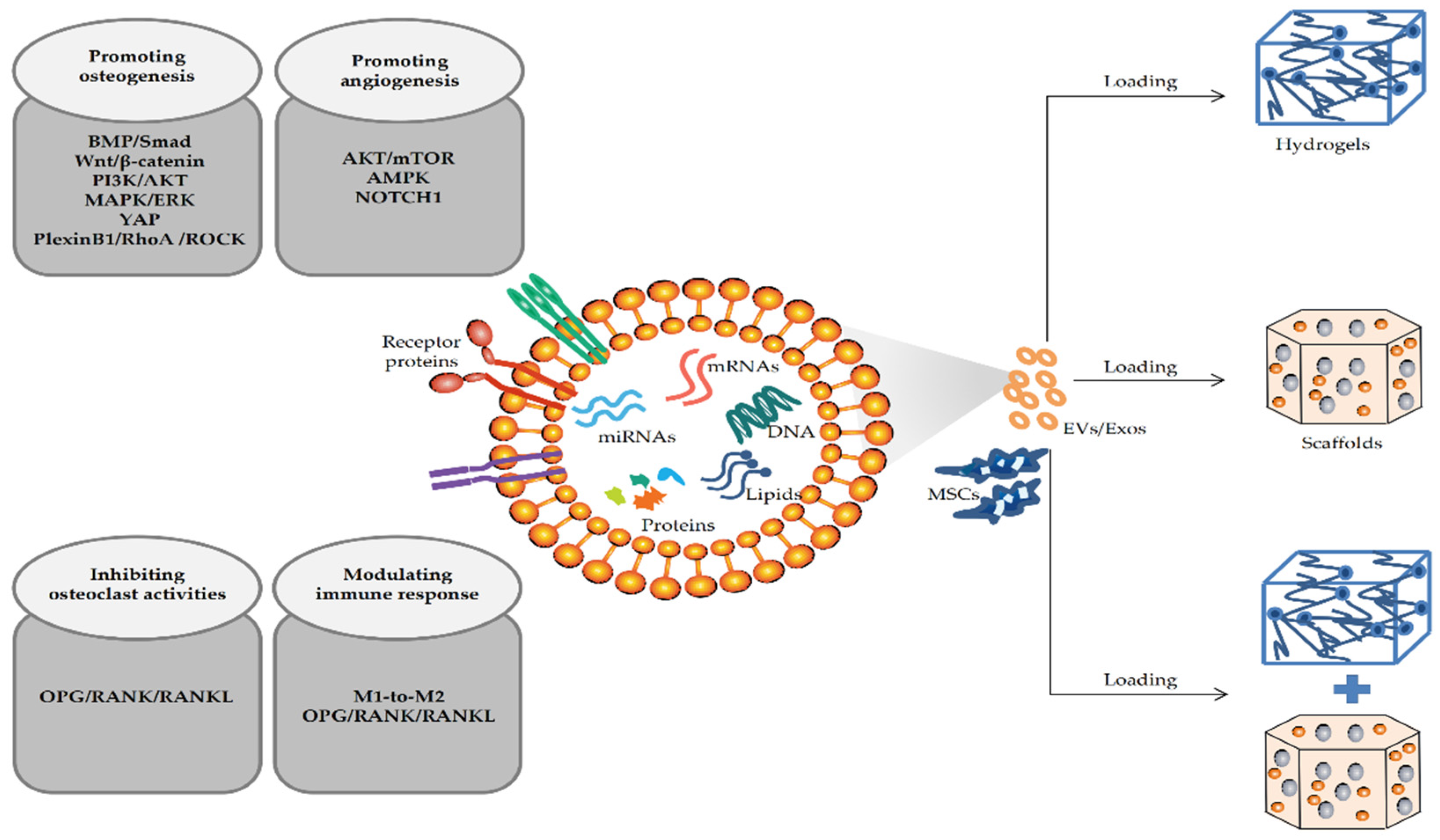
4. Mechanism of MSC-EVs in Repairing Bone Defects

Accumulated studies have shown that MSC-EVs can induce osteogenesis and angiogenesis, regulate immune activity, and inhibit osteoclast activity, ultimately promoting the repair of bone defects by directly transferring their internal cargo and subsequent regulating downstream signaling cascades in target cells [123,124]. It has been reported that MSC-EVs contain a variety of biologically active molecules such as nucleic acids, proteins, and lipids. Among them, miRNAs are one of the most attractive intrinsic cargo of Evs in recent years [125]. miRNAs are small endogenous non-coding RNA, which are one of the main functional components of Exos. Numerous studies have reported that miRNAs play a key role in cell communication and are widely involved in bone metabolism regulation through post-transcriptional modification [126,127,128]. Previous studies have revealed that miRNAs contained in Exos are the main regulators that promote osteogenic differentiation [129]. Wang et al. [130] showed that the ability of T2DM rat-derived BMSC-Exos to promote osteogenic differentiation was significantly impaired, while up-regulating the level of miR-140-3p in Exos could restore its normal osteogenic ability. Further studies revealed that plxnb1 is a direct downstream mRNA target of miR-140-3p, which is closely related to bone formation. Therefore, miR-140-3p may promote bone formation by inhibiting the plexinB1/RhoA/ROCK signaling pathway. In addition, Chen et al. [131] found that hADSC-Exos overexpressing miR-375 could significantly enhance osteogenic differentiation. miR-375, as a positive regulator of osteogenic differentiation of BMSCs, can inhibit insulin-like growth factor binding protein 3 expression and subsequently activate insulin-like growth factor activity, ultimately promoting osteogenic differentiation [132]. Another study found that miR-1260a in Exos was up-regulated after pretreatment with Fe3O4 magnetic nanoparticles and a static magnetic field, which not only promoted osteogenic differentiation but also effectively induced angiogenesis [11]. Previous studies have confirmed that the up-regulation of miR-1260a directly inhibits HDAC7 and COL4A2 expression, while HDAC7 [133] and COL4A2 [134] can inhibit Runx2 activity and angiogenesis, respectively. Therefore, miR-1260a plays a key role in promoting osteogenic differentiation and angiogenesis by targeting HDAC7 and COL4A2. Similarly, another study found that the increased expression of miR-21 within UCMSC-Exos was associated with enhancing angiogenesis in repairing rat cranial defects. Further investigation showed that miR-21 can inhibit NOTCH1, a key pathway for regulating angiogenesis, and then induce the expression of VEGFA and HIF-1α to promote angiogenesis [14,135]. In addition to promoting osteogenesis and angiogenesis, other studies have found that Exo-derived miRNAs can also participate in immune regulation and inhibit osteoclast activity in the process of bone defect repair [62]. However, due to the large number of miRNAs encapsulated in EVs and the possible interaction between different miRNAs to form complex network regulation, the underlying mechanism is still not fully understood. Moreover, in addition to miRNAs, EVs also contain a large number of proteins, lipids, and other types of nucleic acids, such as long non-coding RNAs, circular RNAs, and mRNAs. Therefore, not only the regulatory mechanism of EV-derived miRNAs in bone defect repair needs to be further elucidated, but the role of other intrinsic molecules of EVs also needs to be explored.

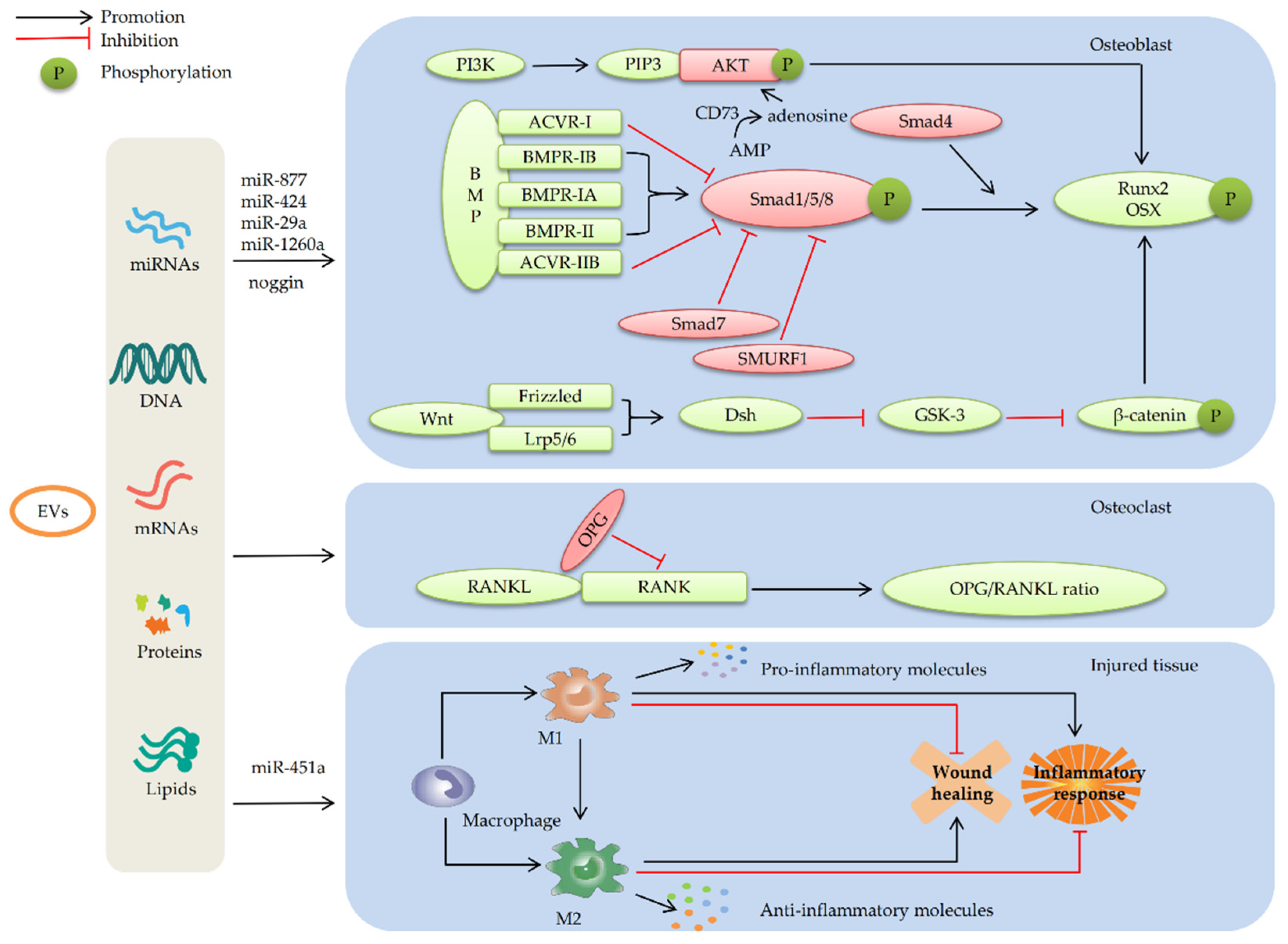
4.1. MSC-EVs Repair Bone Defects by Promoting Osteogenic Differentiation

Enhancing the activity of osteoblasts and inhibiting the activity of osteoclasts are the key factors in bone defect repair. Previous studies have shown that MSC-EVs promote the recruitment of endogenous MSCs to bone defect sites, thereby activating and promoting MSCs proliferation and differentiation and ultimately accelerating bone formation, which involves various signaling pathways such as BMP/Smad, Wnt/β-catenin, and phosphatidylinositol 3-kinase (PI3K)/AKT [136].

4.1.1. MSC-EVs Promote Osteogenic Differentiation by Enhancing the BMP/Smad Signaling Pathway

BMPs, members of the TGF-β superfamily, can promote bone formation by enhancing the osteogenic differentiation of BMSCs [137]. The BMP/Smad signaling pathway is mainly composed of BMPs, BMP receptors, Smad proteins, and other related transcription factors [138,139]. BMPs bind to serine/threonine kinase receptors and transduce signals through Smad-dependent and independent mechanisms [140,141]. Members of the BMP family bind to two distinct type II and type I serine/threonine kinase receptors, both of which are required for signal transduction [142]. BMPs bind to three distinct type II receptors, i.e., BMPR-II, ActR-II, and ActR-IIB. Regarding type I receptors, BMPs bind to three distinct type I receptors, including BMPR-IA, BMPR-IB, and ACVR-I [143,144]. Canonical BMP signaling is initiated when BMPs associate with type I and type II BMP receptors and form a multimeric receptor-ligand complex [145]. Within this complex, the constitutively phosphorylated type II BMP receptors activate type I BMP receptors by phosphorylation, initiating the canonical BMP signaling cascade [146]. Activated type I BMP receptors propagate BMP signals by phosphorylating Smad1/5/8. These Smads complex with Smad4, travel to the nucleus and act as activators and repressors of transcription of target genes, such as Runx2 and OSX, thus regulating osteogenic differentiation [147]. The activation of the BMP/Smad signaling pathway plays an important role in regulating the osteogenic differentiation of MSCs [148]. Recently, numerous studies showed that MSC-EVs promote osteogenic differentiation through the activation of the BMP/Smad signaling pathway, which may be ascribed to multiple MSC-EV-miRNAs [103,110]. Liu et al. [41] found that BMSC-OI-Exos exhibited 8 down-regulated miRNAs, including miR-877, and 16 up-regulated miRNAs (such as miR-328a-5p, miR-31a-5p, let-7a-5p, and let-7c-5p) during the process of promoting osteogenic differentiation in a rat calvarial defect model. Down-regulated miR-877 can up-regulate the expression of target genes involved in the BMP/Smad signaling pathway, such as BMPR-IA, BMPR-IB, and Smad1. Up-regulated miR-328a-5p, miR-31a-5p, let-7a-5p, and let-7c-5p can promote the down-regulation of targeted genes ACVR-I and ACVR-IIB, which inhibit the BMP/Smad signaling pathway [149]. Bioinformatics analysis showed that ACVR-IIB might be an antagonist of BMPR-II, and they are involved in a competitive and synergistic receptor activation network instead of a simple competition relationship between BMPR-II and ACVR-IIB [41]. BMSC-Exo-miRNAs mainly targeted ACVR-IIB/ACVR-I and regulated the competitive balance of BMPR-II/ACVR-IIB toward BMPR-elicited Smad1/5/9 phosphorylation, thus promoting osteogenic differentiation [41]. EVs obtained from different conditions may regulate BMP/Smad signaling pathway through different pathways. Huang et al. [110] generated functionally engineered Evs with enhanced osteoinductive function in vitro and in vivo, which were derived from genetically modified hBMSCs by constitutively expressing BMP-2. A study showed that 5 miRNAs targeting SMAD7 and SMURF1 were demonstrably upregulated in the BMP-2 hBMSC-Evs compared with hBMSC-Evs in vitro. SMURF1 and SMAD7 are well-characterized inhibitors of the BMP signaling pathway [150]. Further studies showed increased expression of miR-424 in the Evs. It has been proved that miR-424 played a role in down-regulating SMURF1 and potentiating BMP-2 signaling [151,152]. Above all, EV-miR-424 may potentiate BMP-2 signaling through down-regulating SMURF1, thereby enhancing the osteoinductive function of Evs. In addition, another study demonstrated that exosome mimetics (Ems) obtained from genetically modified hBMSCs in which expression of noggin, a natural BMP antagonist, was down-regulated could enhance the osteogenic properties of Ems. Further investigation suggested that treatment with Ems from noggin-suppressed hBMSCs with an osteogenic medium significantly decreased the expression of miR-29a, while the expression of BMPR-IA, phosphorylated Smad 1/5/8, and ID1 (a key downstream target of BMP signaling) was detected to significantly increase in MSCs compared to the control group [103]. Previous studies have shown that the knockdown of noggin in MSCs triggers key mediators of BMP signaling (Smad 1/5/8) and osteogenic molecules (Runx2 and OCN) [153]. The above results indicated that the enhanced osteogenic properties of EMs in which noggin was suppressed were mediated by inhibiting the expression of miR-29a to stimulate BMP/Smad signaling [103].
In summary, numerous studies have proved that BMP/Smad is a crucial signaling pathway in the osteogenic differentiation of MSCs. Engineered MSC-EVs can potentiate BMP signaling and promote Smad1/5/8 phosphorylation by altering the expression profile of miRNAs within them. Then, the key downstream targets of BMP signaling are up-regulated, including osteogenic molecules such as Runx2, accelerating osteogenic differentiation of MSCs. The regulation of the BMP/Smad signaling pathway involves the cumulative effect of multiple MSC-EV-miRNAs, such as miR-877 and miR-424 (as shown in Figure 3). However, current investigation regarding the miRNAs related to the positive or negative regulation of the BMP/Smad signaling pathway is still insufficient. In addition, DNA, proteins, lipids, and other active molecules in EVs may also be involved in the regulation of BMP/Smad signaling. Therefore, further studies are expected to explore the key bioactive molecules in EVs that can effectively regulate BMP/Smad signaling to better apply EVs in BTE.

4.1.2. MSC-EVs Promote Osteogenic Differentiation by Regulating the Wnt/β-catenin Signaling Pathway

Coupled bone formation and bone resorption are a guarantee of bone homeostasis. Cumulative evidence demonstrates that Wnt signaling plays a pivotal role in regulating bone homeostasis, and it can be divided into canonical and noncanonical Wnt pathways according to whether β-catenin is involved [154]. Wnts can bind to cell surface receptor LRP5/6 and receptors encoded by the Frizzled gene family. Receptor activation, in turn, somehow activates Dishevelled (Dsh), the most proximal cytosolic component known. In canonical Wnt signaling, Dsh is involved in inhibiting the activation of glycogen synthesis kinase 3 (GSK-3), which is dedicated to the degradation of β-catenin. Inactivation of the GSK-3 allows β-catenin to accumulate within responding cells and to enter the nucleus, where it binds to T cell factor, the transcriptional effector of the Wnt pathway. This complex recruits other proteins to drive transcriptional activation of Wnt target genes [155]. Heo et al. [156] have demonstrated that the Wnt/β-catenin signaling pathway promotes osteogenic differentiation of PDLSCs by activating osteogenic transcription factors such as Runx2. However, recent studies indicate that the Wnt signaling pathway has an opposite dual effect during the process of promoting osteogenic differentiation of MSCs [157]. It was reported that the osteogenic differentiation of MSCs was partially inhibited at a concentration of 100 ng/mL of Wnt3a, whereas treatment with Wnt3a at both 5 and 25 ng/mL resulted in a significant relative increase in osteogenic differentiation, as manifested by increased AP activity and mineralization [158]. Liu et al. [159] demonstrated that the canonical signaling pathway was activated in inflammatory environments while noncanonical Wnt signaling was inhibited, which stimulates PDLSC differentiation and inhibits the terminal differentiation of mature osteoblasts. Therefore, canonical Wnt signaling appears to suppress osteogenic differentiation [160,161]. Further research showed that the expression of Wnt1, Wnt3a, and Wnt10a in inflammatory PDLSCs were significantly decreased after treatment with human PDLSC-derived Exos (hPDLSC-Exos), which contributes to the decrease in β-catenin and the increase in p-GSK3β. p-GSK3β is the most important negative regulator of the Wnt signaling pathway [162]. A study suggested that Exos can rescue the osteogenesis capacity of endogenous stem cells under an inflammatory environment and promote regeneration of alveolar bone by inhibiting the activation of the Wnt/β-catenin pathway [162]. Moreover, the optimal application concentration of hPDLSC-Exos is within a specific threshold [162]. Although numerous studies have shown that the negative effect of canonical Wnt signaling for the osteogenic differentiation of MSCs, there are still studies proving that the BMSC-Exos carrying mutant HIF-1α can activate the Wnt/β-catenin signaling pathway to promote new bone formation [111]. Therefore, the mechanism and effect of the Wnt/β-catenin signaling pathway regulated by MSC-EVs for osteogenic differentiation of MSCs need to be further discussed.
In summary, MSC-Evs can regulate the osteogenic differentiation of MSCs through the Wnt signaling pathway, which has an opposite dual effect on this process. Inhibition or overactivation of Wnt/β-catenin signaling perturbs the osteogenic differentiation of MSCs. The concentrations and activity of Wnt play a significant role in the dual effect of the Wnt signaling pathway. The overactivation of the canonical Wnt signaling at the site of damaged tissue can inhibit osteogenic differentiation of MSCs, which may be involved with the inflammatory environment. Engineered MSC-Evs can promote osteogenesis differentiation by inhibiting the activation of the Wnt/β-catenin signaling pathway. However, it has been shown that MSC-Evs can enhance osteogenesis by activating the canonical Wnt signaling in some studies. In addition, the inhibition of canonical Wnt signaling in the inflammatory environment by MSC-Evs also suggested that Evs may participate in immune regulation via this pathway. However, many issues, such as the optimal concentrations of MSC-Exos to reduce the overactivation of the Wnt/β-catenin pathway and enhance osteogenic differentiation of MSCs, are still not clear. Additionally, the duality of the Wnt signaling pathway in the regulation of osteogenic differentiation still needs to be further explored.

4.1.3. MSC-Evs Promote Osteogenic Differentiation by Activating the PI3K/AKT Signaling Pathway

The PI3K/AKT signaling pathway plays a key role during the process of osteogenic and adipogenic differentiation of BMSCs [163]. As a lipid kinase located in the cytoplasm, PI3K is activated through the stimulation of upstream growth factors, and then PI3K transduces upstream signals from receptors to generate PIP3 [164]. Serving as a critical lipid second messenger, PIP3 recruits protein kinase AKT to the plasma membrane to promote AKT activation and then regulates osteogenic genes [165,166]. In the application of hiPS-MSC-Exos combined with β-TCP scaffolds to promote the repair of rat cranial defects, Zhang et al. [22] found that hiPS-MSC-Exos treatment resulted in a prominent increase in the positive effector genes expression of the PI3K/AKT pathway, such as PDGFA, FGF1/2, FGFR1, COL1A1/2, and BCL2L1. All these genes can positively modulate cell survival, proliferation, migration, osteogenic differentiation, or apoptotic inhibition [167,168,169,170]. The typical negative modulators of PI3K/AKT signaling, including GSK3β and PTEN, were remarkably decreased [167,168,169,170]. The results indicated that the PI3K/AKT signaling in hBMSCs is probably activated after the internalization of Exos, thus stimulating the proliferation and differentiation of hBMSCs. In addition, further investigation showed that the effect of hBMSC-Exos to enhance osteogenic differentiation may be CD73-mediated adenosine receptor activation of pro-survival AKT and MAPK/ERK signaling [136]. ATP is a danger signal released by cells in stress or injury to remove or destroy ‘‘dangers”, which will be subsequently degraded by ectonucleotidases, such as CD39, to ADP and AMP in a short time and finally degraded by CD73 into adenosine, a potent activator of the survival kinases, AKT and ERK [171]. Chew et al. [136] observed that adenosine signaling through phosphorylation of AKT and ERK played a significant role in mediating Exo-induced PDLSC migration and proliferation in a rat periodontal defect model. Compared to ERK, the attenuation of AKT phosphorylation leads to a greater reduction in PDLSC migration and proliferation. AKT, therefore, appears to be a more critical target of MSC-Exos to promote osteogenic differentiation.
In summary, MSC-EVs can promote osteogenic differentiation of MSCs by activating PI3K/AKT and MAPK/ERK signaling pathways after the internalization of EVs. Further research suggests that the enhanced cellular infiltration and proliferation observed during Exo-mediated periodontal regeneration could be attributed at least in part to exosomal CD73-mediated adenosine receptor activation of pro-survival AKT and ERK signaling. AKT is a key mediator in the above process. Moreover, the mediating process may be involved with the cell response to stress or injury. However, the contribution of stress-related mediating mechanisms to the activation of AKT and ERK pathways remains unclear. MSC-EVs cargo is highly diverse and complex and contains growth factors such as IGF and FGF that could potentially activate AKT and/or ERK signaling pathways in PDL cells. The other potential mediating pathways besides CD73-mediated adenosine receptor activation of pro-survival AKT and ERK signaling as well as require further exploration.

4.2. MSC-EVs Repair Bone Defects by Promoting Angiogenesis

Bone is a richly vascularized tissue. The vasculature, as a major transport pathway delivering hormones and growth factors to the bone, provides the bone with oxygen and nutrients and scavenges metabolites, which are critical for bone development and regeneration [172,173]. Furthermore, the blood supply can accelerate bone defect repair by promoting osteoblast migration and bone matrix mineralization, which are critical for bone regeneration in BTE. It has been reported that the major challenge after graft implantation is the maintenance of cell viability in the graft core, which depends on rapid angiogenesis within the tissue-engineered implants [173]. Studies have shown that MSC-EVs play a role in stimulating angiogenesis in bone, which is crucial to accelerating bone regeneration [174]. Researchers proved that BMSC-Exos preconditioned with a low dose of DMOG (DMOG-MSC-Exos) have superior pro-angiogenic properties and can promote bone regeneration in a critical-sized calvarial defect rat model, which may be related to the AKT/mTOR signaling pathway [106]. Compared with the control group, the expression of key components involved with angiogenesis in human umbilical vein endothelial cells (HUVECs) were significantly increased in the DMOG-MSC-Exos group. The molecules that regulated apoptosis were significantly down-regulated such as PTEN, p53, and p21 [106]. PTEN is an endogenous negative regulator of the PI3 kinase signal pathway. On the one hand, PTEN induces the inhibition of AKT kinase and mTOR kinase and inhibits the proliferation of endothelial progenitor cells [175,176]. On the other hand, when the AKT/mTOR pathway is activated, mTOR activation can lead to the down-regulation of PTEN, thus further alleviating its inhibition effect on the AKT/mTOR pathway, which creates a positive feedback loop to facilitate the activation of the pathway [177]. Further analysis showed that the superior pro-angiogenesis ability of DMOG-MSC-Exos was absent after the AKT/mTOR pathway was blocked with the AKT kinase inhibitor MK2206. All results proved that MSC-EVs promoting the angiogenesis of bone defect sites are involved in targeting the AKT/mTOR signaling pathway [106]. In addition, Wu et al. [53] showed that SHED-Exos can up-regulate the expression of angiogenesis-related genes (VEGF-A, KDR, FGF2, and SDF-1), thereby contributing to angiogenesis and osteogenesis in a rat alveolar bone defect model. The study found that adenosine monophosphate-activated protein kinase (AMPK) phosphorylation was increased in both HUVECs and BMSCs after stimulation with SHED-Exos. Additionally, the treatment of compound C, a pharmacological blocker of AMPK, partially reversed the impacts of SHED-Exos on the expression of angiogenesis-related genes (KDR, SDF-1) [53]. The above results suggest that AMPK signaling is activated by SHED-Exos to enhance angiogenesis. AMPK has been shown to exert both positive and negative effects on angiogenesis. The activation of AMPK signaling could attenuate angiogenesis induced by AKT/mTOR and other factors [178]. The specific mechanism of enhancing angiogenesis for EVs via AMPK signaling needs to be further investigated.
In summary, it is demonstrated that MSC-EVs can effectively promote bone regeneration by stimulating angiogenesis in the bone defect area, which may be closely related to the AKT/mTOR and AMPK signaling pathways. Although angiogenesis is critical in bone regeneration, the underlying mechanisms of MSC-EVs activating angiogenesis are not fully understood, and further research is needed. In addition, there are still many problems to be solved in the application of MSC-EVs in BTE: (1) What is the optimal dose of DMOG to treat MSCs for better enhancing angiogenesis of MSC-EVs? (2) What are the specific mechanisms of DMOG and other factors that affect the expression profile of EV cargo? (3) What is the most low-cost way to improve the pro-angiogenic capacity of EVs?

4.3. MSC-EVs Repair Bone Defects by Participating in Immune Regulation

Bone defects caused by trauma and tumors are usually characterized by peripheral inflammation and immune imbalance, which may also lead to implant failure of materials widely used in BTE [179,180]. Previous studies have shown that the immune microenvironment created by immune cells and their metabolites are the critical regulators of the microenvironment for bone regeneration, which has a significant influence on the activity of osteoblasts and osteoclasts and the regulation of bone deposition and strength by regulating the expression of chemokines, growth factors, and inflammatory factors [181,182]. Inflammatory stimulation recruits MSCs that receive pro-inflammatory signals (such as IL-6, IL-17, and TNF-α) to the injured site to improve the local microenvironment, which is conducive to tissue healing [183]. This may be attributed to the anti-inflammatory factors or EVs released by stimulated MSCs to mediate immunoreaction and regulate macrophage polarization [184]. Macrophages are important components of the innate immune system and play an important role in clearing pathogens and regulating inflammatory responses [185,186]. M1 macrophages mainly play a role in pro-inflammatory and immune clearance by secreting pro-inflammatory factors, thereby killing pathogens and tumor cells. However, excessive activation of M1 macrophages causes immune damage [187,188]. M2 macrophages are mainly involved in anti-inflammatory and wound healing by secreting anti-inflammatory and growth factors. Promoting phenotypic polarization of M2 macrophages can effectively promote angiogenesis and bone healing [187,188] (as shown in Figure 3). Numerous studies showed that MSC-EVs can not only promote the proliferation and differentiation of MSCs but also affect the activity of immune cells in a complex internal environment. It is believed that MSC-EVs could stimulate macrophages and other non-stem-like cells to secrete osteogenic growth factors such as BMP-2 and accelerate the healing of injured tissue [189,190]. In addition, it has been widely recognized that osteogenic differentiation can be enhanced by reducing inflammation [191]. Wang et al. [97] constructed a PCL composite scaffold with immunomodulatory potential by combining S-Nitrosoglutathione (GSNO) and MSC-Exos with a PCL scaffold that was surface modified by Poly(dopamine) coating. The results showed that, compared with the control group, Exos combined with GSNO significantly reduced the expression of inflammatory genes via internalization into macrophages. Due to the immunomodulating effects of GSNO by continually releasing nitric oxide (NO), this may be attributed to the regulatory effect of cargo within MSC-Exos or the synergistic effect of both GSNO and MSC-Exos [192]. To further prove whether the bioactive molecules of MSC-Exos play a key role in the process of regulating the immune response, Li et al. [62] analyzed the role of highly expressed miR-451a in ADSC-Exos during the repair of rat cranial defects. Through transfecting miR-451a analogues and inhibitors into macrophages, it was found that miR-451a can directly regulate the expression of macrophage migration inhibitory factor (MIF) by specifically binding to the 3′UTR of MIF mRNA, thereby promoting M1-to-M2 polarization of macrophages [62]. MIF is a multifactorial pro-inflammatory mediator involved in immune regulation [193]. Therefore, the above results demonstrated that the miR-451a derived from MSC-Exos is an effective anti-inflammatory factor. Moreover, it also confirmed that bioactive molecules within MSC-Exos play a vital role in the process of regulating immune response [97,194]. Previous studies have also shown that miRNAs, such as let-7b, or proteins within MSC-Exos pretreated by lipopolysaccharide can reduce inflammation [195]. Further studies demonstrated that MSC-EVs may promote bone regeneration by activating the OPG/RANK/RANKL signaling pathways [190,196]. The bone immunoregulatory pathway OPG/RANK/RANKL participates in bone resorption by regulating osteoclast activity and inflammatory response [197]. RANKL secreted by osteoblasts can activate osteoclasts by binding to RANK [198]. OPG can competitively bind to RANKL to block the RANKL-RANK binding, thereby inhibiting osteoclast activity [199]. Therefore, the balance of bone metabolism depends on the expression ratio of OPG to RANKL (as shown in Figure 3). Shi et al. [195] proved that BMSC-EVs inhibited osteoclast activities by regulating the expression of OPG and RANKL in the therapy of periodontitis rats by combining hydrogels with BMSC-EVs.
In summary, MSC-EVs promote M1-to-M2 polarization of macrophages by delivering their internal cargo, thereby participating in immune regulation in bone defect repair. In addition, MSC-EVs can inhibit osteoclast activity by regulating the OPG/RANK/RANKL signaling pathway, thereby inhibiting bone resorption. Therefore, MSC-EVs may promote bone repair by reducing the inflammatory response and inhibiting the activity of osteoclasts.

5. Future Perspectives

In MSC-EV-mediated BTE, bone graft materials such as scaffolds and hydrogels are often used alone or combined as the bridging structure to repair bone defects. By selecting suitable donor and parent cells, EVs with good osteoinductive potential could be obtained, and the EVs will be loaded onto the ideal bone graft materials to promote bone tissue regeneration at the defect sites. In this paper, we comprehensively reviewed the current status of bioactive materials combined with MSC-EVs utilized in repairing bone defects and the mechanism of MSC-EVs promoting bone regeneration in BTE. Firstly, the combination of hydrogels and scaffolds has achieved some success in terms of the application of MSC-EVs in bone defects. Due to the poor mechanical properties of hydrogels and the low osteoinductive activity and inefficient filling of irregular gaps of scaffolds, the combination of hydrogels and scaffolds is often used to effectively deliver EVs, which is the most common strategy used in BTE currently. Secondly, based on the combined application of hydrogel and scaffold materials, a variety of innovative ways to enhance the delivery efficiency of MSC-EVs have been developed. Owing to most current carriers explosively releasing EVs, it leads to inadequate therapeutic effects and even causes bone resorption. Researchers achieve efficient transport, sustained release, and long-term storage of EVs by developing innovative scaffolds and modifying composite hydrogel scaffolds or combining bone graft materials with tethering peptides and other active substances. Lastly, enhancing the osteoinductive capacity of EVs in various ways is the key factor in promoting bone defect repair in BTE. The limited osteoinduction capacity of EVs is not sufficient to effectively promote bone formation in critical bone defects when used EVs alone. Therefore, EVs are often modified by gene modification, hypoxic environment culture, chemical reagent pretreatment, or combined with other active substances to prepare EVs with enhanced osteoinductive ability. In addition to the specific application of MSC-EVs combined with bioactive materials, numerous studies explored the mechanism of EVs promoting bone defect repair in BTE. Studies found that engineered MSC-EVs may regulate the key signaling pathways of osteogenic differentiation, including BMP/Smad, Wnt/β-catenin, and PI3K/AKT signaling pathways, by altering the levels of miRNAs and other active substances within them. Enhancing the angiogenesis mechanism of MSC-EVs is involved with the activation of AKT/mTOR and AMPK signaling pathways. Moreover, many studies have shown that EVs can participate in immune regulation and reduce inflammatory response by promoting M1-to-M2 polarization of macrophages. Further research proved that it may be associated with the inhibition of osteoclast activities by regulating the OPG/RANK/RANKL signaling pathway, then inhibiting bone resorption and accelerating injury repair (as shown in Figure 2 and Figure 3).
EV-based cell-free bone regeneration strategy can avoid the disadvantages of MSC transplantation, such as ethical issues, immune rejection, and tumor risk, showing infinite application potential. However, it is undeniable that the research on the effect of EVs on bone regeneration is still in the preliminary stage. There are still many issues to be resolved conveniently and reliably, such as the ineffective delivery of EVs, the long-term preservation of EVs, the uncertainty of the optimal treatment dose of EVs, and the unstandardized porosity value or pore size of a biocompatible 3D porous scaffold for bone defects. Therefore, it is necessary to further explore and clarify the key factors to improve the therapeutic effect of EVs in the future. For instance, in the delivery of EVs, future studies need to determine the proportion of components in the composite bone graft materials. Further optimizing the mechanical properties and biological activity of bone graft materials to obtain widely recognized grafts is significant in BTE. In terms of the performance improvement of EVs, as the modified MSC-EVs may produce some unexpected therapeutic effects, it is necessary to carefully evaluate whether there are the disadvantages of high cost, poor pharmacokinetics, low efficiency, and potential side effects. In addition, studies have shown that MSC-EVs are rich in RNAs, proteins, and lipids. Theoretically, EVs are likely to regulate bone formation through the interaction of their various components. However, most studies are limited to the role of miRNAs encapsulated in EVs in BTE, suggesting that the mechanism of EVs in bone regeneration is far from being explored. Therefore, the effective active molecules of different types of MSC-EVs promoting osteogenic differentiation, angiogenesis, inflammatory regulation, and osteoclast inhibition need to be further clarified.

6. Discussion and Conclusions

The strength of this review is that it comprehensively summarized the latest progress in applying MSC-EVs in BTE to promote bone defect repair and the potential mechanism of MSC-EVs promoting bone regeneration, which is of great significance for the currently hot research on bone defect repair. This review systematically and comprehensively describes the latest application progress of MSC-EVs in bone defect repair by summarizing the latest studies, for example, the development and improvement of new hydrogel and scaffold materials and their advantages and disadvantages currently; recent advances and potential risks of EVs delivery by the combination of hydrogel and scaffold materials; and effective advances in improving EVs performance by gene engineering or other methods. In addition, in order to make readers better understand the significance of MSC-EVs in bone defect repair, the potential mechanism of MSC-EVs in BTE was systematically described in detail from the aspects of promoting osteogenesis and angiogenesis, inhibiting osteoclast formation and participating in immune regulation. This is also one of the highlights of this paper, which is of great significance for the application of MSC-EVs in basic research and clinical treatment to promote bone regeneration. In a word, our review provides the latest evidence and views on the effective application, potential risks and solutions of MSC-EVs combined with hydrogels and/or scaffolds and the potential mechanism of MSC-EVs enhancing bone regeneration. However, this paper may also have many limitations. Firstly, this paper focuses on the characteristics and application of MSC-EVs combined with hydrogel and/or scaffold materials for repairing bone defects, while there is little description of the preparation process of bone graft materials in detail, which may hinder the corresponding animal research to some extent. Secondly, this paper mainly compares several common MSC-EVs due to the wide variety of MSCs; therefore, the evidence for the application of EVs parental cell selection may not be sufficient. Thirdly, owing to the limited space of this paper, it is not possible to list and elaborate on all the independent, innovative hydrogel scaffold composites with different characteristics. Therefore, in the future, it is necessary not only to continue to explore the effective application of EVs combined with bioactive carrier materials but also to further clarify the active substances in MSC-EVs and their potential mechanisms for promoting bone regeneration.
In conclusion, although preclinical studies have reported promising results, several enhancements of MSC-EV therapy are required to obtain superior outcomes. Further research on effective delivery, activity maintenance, controlled, sustained release and potential mechanisms of EVs will become key factors for the safe and effective application of EVs in bone defect repair.

Author Contributions

Conceptualization, X.Z. and Z.Z.; methodology, D.W.; writing—original draft preparation, D.W. and W.H.; writing—review and editing, L.G., H.C. and Y.Y.; supervision, X.Z. and Z.Z. All authors have read and agreed to the published version of the manuscript.

Funding

This research received no external funding.

Institutional Review Board Statement

Not applicable.

Informed Consent Statement

Not applicable.

Data Availability Statement

Not applicable.

Conflicts of Interest

The authors declare no conflict of interest.

References

  1. Calciolari, E.; Donos, N.; Mardas, N. Osteoporotic Animal Models of Bone Healing: Advantages and Pitfalls. J. Investig. Surg. Off. J. Acad. Surg. Res. 2017, 30, 342–350. [Google Scholar] [CrossRef] [PubMed]
  2. Falacho, R.I.; Palma, P.J.; Marques, J.A.; Figueiredo, M.H.; Caramelo, F.; Dias, I.; Viegas, C.; Guerra, F. Collagenated Porcine Heterologous Bone Grafts: Histomorphometric Evaluation of Bone Formation Using Different Physical Forms in a Rabbit Cancellous Bone Model. Molecules 2021, 26, 1339. [Google Scholar] [CrossRef] [PubMed]
  3. Shang, F.; Yu, Y.; Liu, S.; Ming, L.; Zhang, Y.; Zhou, Z.; Zhao, J.; Jin, Y. Advancing application of mesenchymal stem cell-based bone tissue regeneration. Bioact. Mater. 2021, 6, 666–683. [Google Scholar] [CrossRef] [PubMed]
  4. Yu, W.; Li, S.; Guan, X.; Zhang, N.; Xie, X.; Zhang, K.; Bai, Y. Higher yield and enhanced therapeutic effects of exosomes derived from MSCs in hydrogel-assisted 3D culture system for bone regeneration. Mater. Sci. Eng. C Mater. Biol. Appl. 2022; Online ahead of print. 112646. [Google Scholar] [CrossRef]
  5. Baldwin, P.; Li, D.J.; Auston, D.A.; Mir, H.S.; Yoon, R.S.; Koval, K.J. Autograft, Allograft, and Bone Graft Substitutes: Clinical Evidence and Indications for Use in the Setting of Orthopaedic Trauma Surgery. J. Orthop. Trauma 2019, 33, 203–213. [Google Scholar] [CrossRef]
  6. Benic, G.I.; Hämmerle, C.H. Horizontal bone augmentation by means of guided bone regeneration. Periodontology 2000 2014, 66, 13–40. [Google Scholar] [CrossRef]
  7. Stahl, A.; Yang, Y.P. Regenerative Approaches for the Treatment of Large Bone Defects. Tissue Eng. Part B Rev. 2021, 27, 539–547. [Google Scholar] [CrossRef]
  8. Behrend, C.; Carmouche, J.; Millhouse, P.W.; Ritter, L.; Moskal, J.; Rubery, P.; Puzas, E. Allogeneic and Autogenous Bone Grafts Are Affected by Historical Donor Environmental Exposure. Clin. Orthop. Relat. Res. 2016, 474, 1405–1409. [Google Scholar] [CrossRef] [Green Version]
  9. Langer, R.; Vacanti, J.P. Tissue engineering. Science 1993, 260, 920–926. [Google Scholar] [CrossRef] [Green Version]
  10. Roseti, L.; Parisi, V.; Petretta, M.; Cavallo, C.; Desando, G.; Bartolotti, I.; Grigolo, B. Scaffolds for Bone Tissue Engineering: State of the art and new perspectives. Mater. Sci. Eng. C Mater. Biol. Appl. 2017, 78, 1246–1262. [Google Scholar] [CrossRef]
  11. Wu, D.; Chang, X.; Tian, J.; Kang, L.; Wu, Y.; Liu, J.; Wu, X.; Huang, Y.; Gao, B.; Wang, H.; et al. Bone mesenchymal stem cells stimulation by magnetic nanoparticles and a static magnetic field: Release of exosomal miR-1260a improves osteogenesis and angiogenesis. J. Nanobiotechnol. 2021, 19, 209. [Google Scholar] [CrossRef]
  12. Qin, Y.; Wang, L.; Gao, Z.; Chen, G.; Zhang, C. Bone marrow stromal/stem cell-derived extracellular vesicles regulate osteoblast activity and differentiation in vitro and promote bone regeneration in vivo. Sci. Rep. 2016, 6, 21961. [Google Scholar] [CrossRef]
  13. Li, W.; Liu, Y.; Zhang, P.; Tang, Y.; Zhou, M.; Jiang, W.; Zhang, X.; Wu, G.; Zhou, Y. Tissue-Engineered Bone Immobilized with Human Adipose Stem Cells-Derived Exosomes Promotes Bone Regeneration. ACS Appl. Mater. Interfaces 2018, 10, 5240–5254. [Google Scholar] [CrossRef]
  14. Zhang, Y.; Xie, Y.; Hao, Z.; Zhou, P.; Wang, P.; Fang, S.; Li, L.; Xu, S.; Xia, Y. Umbilical Mesenchymal Stem Cell-Derived Exosome-Encapsulated Hydrogels Accelerate Bone Repair by Enhancing Angiogenesis. ACS Appl. Mater. Interfaces 2021, 13, 18472–18487. [Google Scholar] [CrossRef]
  15. Fu, Q.L.; Chow, Y.Y.; Sun, S.J.; Zeng, Q.X.; Li, H.B.; Shi, J.B.; Sun, Y.Q.; Wen, W.; Tse, H.F.; Lian, Q.; et al. Mesenchymal stem cells derived from human induced pluripotent stem cells modulate T-cell phenotypes in allergic rhinitis. Allergy 2012, 67, 1215–1222. [Google Scholar] [CrossRef]
  16. Malekpour, K.; Hazrati, A.; Zahar, M.; Markov, A.; Zekiy, A.O.; Navashenaq, J.G.; Roshangar, L.; Ahmadi, M. The Potential Use of Mesenchymal Stem Cells and Their Derived Exosomes for Orthopedic Diseases Treatment. Stem Cell Rev. Rep. 2022, 18, 933–951. [Google Scholar] [CrossRef]
  17. Silva, D.D.D.; Paz, A.; Portinho, C.P.; Lima, E.O.C.; Kliemann, L.M.; Collares, M.V.M. Reconstruction of parietal bone defects with adiposederived mesenchymal stem cells. Experimental study. Acta Cir. Bras. 2021, 35, e351201. [Google Scholar] [CrossRef]
  18. Perez, J.R.; Kouroupis, D.; Li, D.J.; Best, T.M.; Kaplan, L.; Correa, D. Tissue Engineering and Cell-Based Therapies for Fractures and Bone Defects. Front. Bioeng. Biotechnol. 2018, 6, 105. [Google Scholar] [CrossRef] [Green Version]
  19. Kang, Y.; Xu, C.; Meng, L.; Dong, X.; Qi, M.; Jiang, D. Exosome-functionalized magnesium-organic framework-based scaffolds with osteogenic, angiogenic and anti-inflammatory properties for accelerated bone regeneration. Bioact. Mater. 2022, 18, 26–41. [Google Scholar] [CrossRef]
  20. Konala, V.B.; Mamidi, M.K.; Bhonde, R.; Das, A.K.; Pochampally, R.; Pal, R. The current landscape of the mesenchymal stromal cell secretome: A new paradigm for cell-free regeneration. Cytotherapy 2016, 18, 13–24. [Google Scholar] [CrossRef] [Green Version]
  21. Liang, X.; Ding, Y.; Zhang, Y.; Tse, H.F.; Lian, Q. Paracrine mechanisms of mesenchymal stem cell-based therapy: Current status and perspectives. Cell Transplant. 2014, 23, 1045–1059. [Google Scholar] [CrossRef] [Green Version]
  22. Zhang, J.; Liu, X.; Li, H.; Chen, C.; Hu, B.; Niu, X.; Li, Q.; Zhao, B.; Xie, Z.; Wang, Y. Exosomes/tricalcium phosphate combination scaffolds can enhance bone regeneration by activating the PI3K/Akt signaling pathway. Stem Cell Res. Ther. 2016, 7, 136. [Google Scholar] [CrossRef] [Green Version]
  23. Zhou, X.; Cao, H.; Guo, J.; Yuan, Y.; Ni, G. Effects of BMSC-Derived EVs on Bone Metabolism. Pharmaceutics 2022, 14, 1012. [Google Scholar] [CrossRef]
  24. Li, Q.C.; Li, C.; Zhang, W.; Pi, W.; Han, N. Potential Effects of Exosomes and Their MicroRNA Carrier on Osteoporosis. Curr. Pharm. Des. 2022, 12, 550. [Google Scholar] [CrossRef]
  25. Witwer, K.W.; Van Balkom, B.W.M.; Bruno, S.; Choo, A.; Dominici, M.; Gimona, M.; Hill, A.F.; De Kleijn, D.; Koh, M.; Lai, R.C.; et al. Defining mesenchymal stromal cell (MSC)-derived small extracellular vesicles for therapeutic applications. J. Extracell. Vesicles 2019, 8, 1609206. [Google Scholar] [CrossRef] [Green Version]
  26. Jeppesen, D.K.; Fenix, A.M.; Franklin, J.L.; Higginbotham, J.N.; Zhang, Q.; Zimmerman, L.J.; Liebler, D.C.; Ping, J.; Liu, Q.; Evans, R.; et al. Reassessment of Exosome Composition. Cell 2019, 177, 428–445.e418. [Google Scholar] [CrossRef] [Green Version]
  27. Zara, J.N.; Siu, R.K.; Zhang, X.; Shen, J.; Ngo, R.; Lee, M.; Li, W.; Chiang, M.; Chung, J.; Kwak, J.; et al. High doses of bone morphogenetic protein 2 induce structurally abnormal bone and inflammation in vivo. Tissue Eng. Part A 2011, 17, 1389–1399. [Google Scholar] [CrossRef] [Green Version]
  28. Liang, W.; Han, B.; Hai, Y.; Sun, D.; Yin, P. Mechanism of Action of Mesenchymal Stem Cell-Derived Exosomes in the Intervertebral Disc Degeneration Treatment and Bone Repair and Regeneration. Front. Cell Dev. Biol. 2021, 9, 833840. [Google Scholar] [CrossRef]
  29. Huang, C.C.; Kang, M.; Shirazi, S.; Lu, Y.; Cooper, L.F.; Gajendrareddy, P.; Ravindran, S. 3D Encapsulation and tethering of functionally engineered extracellular vesicles to hydrogels. Acta Biomater. 2021, 126, 199–210. [Google Scholar] [CrossRef]
  30. Rahman, M.J.; Regn, D.; Bashratyan, R.; Dai, Y.D. Exosomes released by islet-derived mesenchymal stem cells trigger autoimmune responses in NOD mice. Diabetes 2014, 63, 1008–1020. [Google Scholar] [CrossRef] [Green Version]
  31. Zhang, Y.; Chopp, M.; Meng, Y.; Katakowski, M.; Xin, H.; Mahmood, A.; Xiong, Y. Effect of exosomes derived from multipluripotent mesenchymal stromal cells on functional recovery and neurovascular plasticity in rats after traumatic brain injury. J. Neurosurg. 2015, 122, 856–867. [Google Scholar] [CrossRef] [Green Version]
  32. Yang, X.; Yang, J.; Lei, P.; Wen, T. LncRNA MALAT1 shuttled by bone marrow-derived mesenchymal stem cells-secreted exosomes alleviates osteoporosis through mediating microRNA-34c/SATB2 axis. Aging 2019, 11, 8777–8791. [Google Scholar] [CrossRef]
  33. Qin, Y.; Sun, R.; Wu, C.; Wang, L.; Zhang, C. Exosome: A Novel Approach to Stimulate Bone Regeneration through Regulation of Osteogenesis and Angiogenesis. Int. J. Mol. Sci. 2016, 17, 712. [Google Scholar] [CrossRef]
  34. Tan, S.S.H.; Tjio, C.K.E.; Wong, J.R.Y.; Wong, K.L.; Chew, J.R.J.; Hui, J.H.P.; Toh, W.S. Mesenchymal Stem Cell Exosomes for Cartilage Regeneration: A Systematic Review of Preclinical In Vivo Studies. Tissue Eng. Part B Rev. 2021, 27, 1–13. [Google Scholar] [CrossRef]
  35. Zhu, Y.; Jia, Y.; Wang, Y.; Xu, J.; Chai, Y. Impaired Bone Regenerative Effect of Exosomes Derived from Bone Marrow Mesenchymal Stem Cells in Type 1 Diabetes. Stem Cells Transl. Med. 2019, 8, 593–605. [Google Scholar] [CrossRef] [Green Version]
  36. Behera, J.; Tyagi, N. Exosomes: Mediators of bone diseases, protection, and therapeutics potential. Oncoscience 2018, 5, 181–195. [Google Scholar] [CrossRef] [Green Version]
  37. Garza, J.R.; Campbell, R.E.; Tjoumakaris, F.P.; Freedman, K.B.; Miller, L.S.; Santa Maria, D.; Tucker, B.S. Clinical Efficacy of Intra-articular Mesenchymal Stromal Cells for the Treatment of Knee Osteoarthritis: A Double-Blinded Prospective Randomized Controlled Clinical Trial. Am. J. Sports Med. 2020, 48, 588–598. [Google Scholar] [CrossRef]
  38. Zhang, S.; Wong, K.L.; Ren, X.; Teo, K.Y.W.; Afizah, H.; Choo, A.B.H.; Lai, R.C.; Lim, S.K.; Hui, J.H.P.; Toh, W.S. Mesenchymal Stem Cell Exosomes Promote Functional Osteochondral Repair in a Clinically Relevant Porcine Model. Am. J. Sports Med. 2022, 50, 788–800. [Google Scholar] [CrossRef]
  39. McIntosh, K.; Zvonic, S.; Garrett, S.; Mitchell, J.B.; Floyd, Z.E.; Hammill, L.; Kloster, A.; Di Halvorsen, Y.; Ting, J.P.; Storms, R.W.; et al. The immunogenicity of human adipose-derived cells: Temporal changes in vitro. Stem Cells 2006, 24, 1246–1253. [Google Scholar] [CrossRef]
  40. Li, Q.; Yu, H.; Sun, M.; Yang, P.; Hu, X.; Ao, Y.; Cheng, J. The tissue origin effect of extracellular vesicles on cartilage and bone regeneration. Acta Biomater. 2021, 125, 253–266. [Google Scholar] [CrossRef]
  41. Liu, A.; Lin, D.; Zhao, H.; Chen, L.; Cai, B.; Lin, K.; Shen, S.G. Optimized BMSC-derived osteoinductive exosomes immobilized in hierarchical scaffold via lyophilization for bone repair through Bmpr2/Acvr2b competitive receptor-activated Smad pathway. Biomaterials 2021, 272, 120718. [Google Scholar] [CrossRef]
  42. Hu, G.W.; Li, Q.; Niu, X.; Hu, B.; Liu, J.; Zhou, S.M.; Guo, S.C.; Lang, H.L.; Zhang, C.Q.; Wang, Y.; et al. Exosomes secreted by human-induced pluripotent stem cell-derived mesenchymal stem cells attenuate limb ischemia by promoting angiogenesis in mice. Stem Cell Res. Ther. 2015, 6, 10. [Google Scholar] [CrossRef] [Green Version]
  43. Hendijani, F.; Sadeghi-Aliabadi, H.; Haghjooy Javanmard, S. Comparison of human mesenchymal stem cells isolated by explant culture method from entire umbilical cord and Wharton’s jelly matrix. Cell Tissue Bank. 2014, 15, 555–565. [Google Scholar] [CrossRef]
  44. Fong, C.Y.; Chak, L.L.; Biswas, A.; Tan, J.H.; Gauthaman, K.; Chan, W.K.; Bongso, A. Human Wharton’s jelly stem cells have unique transcriptome profiles compared to human embryonic stem cells and other mesenchymal stem cells. Stem Cell Rev. Rep. 2011, 7, 1–16. [Google Scholar] [CrossRef]
  45. Wang, K.X.; Xu, L.L.; Rui, Y.F.; Huang, S.; Lin, S.E.; Xiong, J.H.; Li, Y.H.; Lee, W.Y.; Li, G. The effects of secretion factors from umbilical cord derived mesenchymal stem cells on osteogenic differentiation of mesenchymal stem cells. PLoS ONE 2015, 10, e0120593. [Google Scholar] [CrossRef] [Green Version]
  46. Ando, Y.; Matsubara, K.; Ishikawa, J.; Fujio, M.; Shohara, R.; Hibi, H.; Ueda, M.; Yamamoto, A. Stem cell-conditioned medium accelerates distraction osteogenesis through multiple regenerative mechanisms. Bone 2014, 61, 82–90. [Google Scholar] [CrossRef]
  47. Wang, L.; Wang, J.; Zhou, X.; Sun, J.; Zhu, B.; Duan, C.; Chen, P.; Guo, X.; Zhang, T.; Guo, H. A New Self-Healing Hydrogel Containing hucMSC-Derived Exosomes Promotes Bone Regeneration. Front. Bioeng. Biotechnol. 2020, 8, 564731. [Google Scholar] [CrossRef]
  48. Jiang, Z.Z.; Liu, Y.M.; Niu, X.; Yin, J.Y.; Hu, B.; Guo, S.C.; Fan, Y.; Wang, Y.; Wang, N.S. Exosomes secreted by human urine-derived stem cells could prevent kidney complications from type I diabetes in rats. Stem Cell Res. Ther. 2016, 7, 24. [Google Scholar] [CrossRef] [Green Version]
  49. Lian, Q.; Zhang, Y.; Zhang, J.; Zhang, H.K.; Wu, X.; Zhang, Y.; Lam, F.F.; Kang, S.; Xia, J.C.; Lai, W.H.; et al. Functional mesenchymal stem cells derived from human induced pluripotent stem cells attenuate limb ischemia in mice. Circulation 2010, 121, 1113–1123. [Google Scholar] [CrossRef] [Green Version]
  50. Martinez Saez, D.; Sasaki, R.T.; Neves, A.D.; da Silva, M.C. Stem Cells from Human Exfoliated Deciduous Teeth: A Growing Literature. Cells Tissues Organs 2016, 202, 269–280. [Google Scholar] [CrossRef]
  51. Nakamura, S.; Yamada, Y.; Katagiri, W.; Sugito, T.; Ito, K.; Ueda, M. Stem cell proliferation pathways comparison between human exfoliated deciduous teeth and dental pulp stem cells by gene expression profile from promising dental pulp. J. Endod. 2009, 35, 1536–1542. [Google Scholar] [CrossRef]
  52. Wang, L.; Zhao, Y.; Shi, S. Interplay between mesenchymal stem cells and lymphocytes: Implications for immunotherapy and tissue regeneration. J. Dent. Res. 2012, 91, 1003–1010. [Google Scholar] [CrossRef] [Green Version]
  53. Wu, J.; Chen, L.; Wang, R.; Song, Z.; Shen, Z.; Zhao, Y.; Huang, S.; Lin, Z. Exosomes Secreted by Stem Cells from Human Exfoliated Deciduous Teeth Promote Alveolar Bone Defect Repair through the Regulation of Angiogenesis and Osteogenesis. ACS Biomater. Sci. Eng. 2019, 5, 3561–3571. [Google Scholar] [CrossRef]
  54. Gómez-Barrena, E.; Rosset, P.; Gebhard, F.; Hernigou, P.; Baldini, N.; Rouard, H.; Sensebé, L.; Gonzalo-Daganzo, R.M.; Giordano, R.; Padilla-Eguiluz, N.; et al. Feasibility and safety of treating non-unions in tibia, femur and humerus with autologous, expanded, bone marrow-derived mesenchymal stromal cells associated with biphasic calcium phosphate biomaterials in a multicentric, non-comparative trial. Biomaterials 2019, 196, 100–108. [Google Scholar] [CrossRef]
  55. Fernandez-Yague, M.A.; Abbah, S.A.; McNamara, L.; Zeugolis, D.I.; Pandit, A.; Biggs, M.J. Biomimetic approaches in bone tissue engineering: Integrating biological and physicomechanical strategies. Adv. Drug Deliv. Rev. 2015, 84, 1–29. [Google Scholar] [CrossRef]
  56. Leijten, J.; Seo, J.; Yue, K.; Santiago, G.T.; Tamayol, A.; Ruiz-Esparza, G.U.; Shin, S.R.; Sharifi, R.; Noshadi, I.; Álvarez, M.M.; et al. Spatially and Temporally Controlled Hydrogels for Tissue Engineering. Mater. Sci. Eng. R Rep. A Rev. J. 2017, 119, 1–35. [Google Scholar] [CrossRef]
  57. Akbari, A.; Jabbari, N.; Sharifi, R.; Ahmadi, M.; Vahhabi, A.; Seyedzadeh, S.J.; Nawaz, M.; Szafert, S.; Mahmoodi, M.; Jabbari, E.; et al. Free and hydrogel encapsulated exosome-based therapies in regenerative medicine. Life Sci. 2020, 249, 117447. [Google Scholar] [CrossRef]
  58. Choi, B.; Kim, S.; Lin, B.; Li, K.; Bezouglaia, O.; Kim, J.; Evseenko, D.; Aghaloo, T.; Lee, M. Visible-light-initiated hydrogels preserving cartilage extracellular signaling for inducing chondrogenesis of mesenchymal stem cells. Acta Biomater. 2015, 12, 30–41. [Google Scholar] [CrossRef]
  59. Liu, B.; Lee, B.W.; Nakanishi, K.; Villasante, A.; Williamson, R.; Metz, J.; Kim, J.; Kanai, M.; Bi, L.; Brown, K.; et al. Cardiac recovery via extended cell-free delivery of extracellular vesicles secreted by cardiomyocytes derived from induced pluripotent stem cells. Nat. Biomed. Eng. 2018, 2, 293–303. [Google Scholar] [CrossRef]
  60. Zhang, Z.; Wang, X.; Wang, Y.; Hao, J. Rapid-Forming and Self-Healing Agarose-Based Hydrogels for Tissue Adhesives and Potential Wound Dressings. Biomacromolecules 2018, 19, 980–988. [Google Scholar] [CrossRef]
  61. Holkar, K.; Kale, V.; Ingavle, G. Hydrogel-Assisted 3D Model to Investigate the Osteoinductive Potential of MC3T3-Derived Extracellular Vesicles. ACS Biomater. Sci. Eng. 2021, 7, 2687–2700. [Google Scholar] [CrossRef]
  62. Li, R.; Li, D.; Wang, H.; Chen, K.; Wang, S.; Xu, J.; Ji, P. Exosomes from adipose-derived stem cells regulate M1/M2 macrophage phenotypic polarization to promote bone healing via miR-451a/MIF. Stem Cell Res. Ther. 2022, 13, 149. [Google Scholar] [CrossRef]
  63. Zhang, T.; Cheng, Q.; Ye, D.; Chang, C. Tunicate cellulose nanocrystals reinforced nanocomposite hydrogels comprised by hybrid cross-linked networks. Carbohydr. Polym. 2017, 169, 139–148. [Google Scholar] [CrossRef]
  64. Pishavar, E.; Luo, H.; Naserifar, M.; Hashemi, M.; Toosi, S.; Atala, A.; Ramakrishna, S.; Behravan, J. Advanced Hydrogels as Exosome Delivery Systems for Osteogenic Differentiation of MSCs: Application in Bone Regeneration. Int. J. Mol. Sci. 2021, 22, 203. [Google Scholar] [CrossRef]
  65. Lee, K.Y.; Alsberg, E.; Mooney, D.J. Degradable and injectable poly(aldehyde guluronate) hydrogels for bone tissue engineering. J. Biomed. Mater. Res. 2001, 56, 228–233. [Google Scholar] [CrossRef] [Green Version]
  66. Sun, T.L.; Kurokawa, T.; Kuroda, S.; Ihsan, A.B.; Akasaki, T.; Sato, K.; Haque, M.A.; Nakajima, T.; Gong, J.P. Physical hydrogels composed of polyampholytes demonstrate high toughness and viscoelasticity. Nat. Mater. 2013, 12, 932–937. [Google Scholar] [CrossRef] [Green Version]
  67. Ma, S.; Wu, J.; Hu, H.; Mu, Y.; Zhang, L.; Zhao, Y.; Bian, X.; Jing, W.; Wei, P.; Zhao, B.; et al. Novel fusion peptides deliver exosomes to modify injectable thermo-sensitive hydrogels for bone regeneration. Mater. Today. Bio 2022, 13, 100195. [Google Scholar] [CrossRef]
  68. Lin, S.; Yang, G.; Jiang, F.; Zhou, M.; Yin, S.; Tang, Y.; Tang, T.; Zhang, Z.; Zhang, W.; Jiang, X. A Magnesium-Enriched 3D Culture System that Mimics the Bone Development Microenvironment for Vascularized Bone Regeneration. Adv. Sci. 2019, 6, 1900209. [Google Scholar] [CrossRef]
  69. Vila, A.; Torras, N.; Castaño, A.G.; García-Díaz, M.; Comelles, J.; Pérez-Berezo, T.; Corregidor, C.; Castaño, Ó.; Engel, E.; Fernández-Majada, V.; et al. Hydrogel co-networks of gelatine methacrylate and poly(ethylene glycol) diacrylate sustain 3D functional in vitro models of intestinal mucosa. Biofabrication 2020, 12, 025008. [Google Scholar] [CrossRef]
  70. Han, C.; Zhou, J.; Liang, C.; Liu, B.; Pan, X.; Zhang, Y.; Wang, Y.; Yan, B.; Xie, W.; Liu, F.; et al. Human umbilical cord mesenchymal stem cell derived exosomes encapsulated in functional peptide hydrogels promote cardiac repair. Biomater. Sci. 2019, 7, 2920–2933. [Google Scholar] [CrossRef]
  71. Wang, J.; Li, W.; Zhang, L.; Ban, L.; Chen, P.; Du, W.; Feng, X.; Liu, B.F. Chemically Edited Exosomes with Dual Ligand Purified by Microfluidic Device for Active Targeted Drug Delivery to Tumor Cells. ACS Appl. Mater. Interfaces 2017, 9, 27441–27452. [Google Scholar] [CrossRef]
  72. Yim, N.; Ryu, S.W.; Choi, K.; Lee, K.R.; Lee, S.; Choi, H.; Kim, J.; Shaker, M.R.; Sun, W.; Park, J.H.; et al. Exosome engineering for efficient intracellular delivery of soluble proteins using optically reversible protein-protein interaction module. Nat. Commun. 2016, 7, 12277. [Google Scholar] [CrossRef]
  73. Zhang, Y.S.; Khademhosseini, A. Advances in engineering hydrogels. Science 2017, 356, eaaf3627. [Google Scholar] [CrossRef]
  74. George, M.; Abraham, T.E. Polyionic hydrocolloids for the intestinal delivery of protein drugs: Alginate and chitosan—A review. J. Control. Release Off. J. Control. Release Soc. 2006, 114, 1–14. [Google Scholar] [CrossRef]
  75. Neves, M.I.; Moroni, L.; Barrias, C.C. Modulating Alginate Hydrogels for Improved Biological Performance as Cellular 3D Microenvironments. Front. Bioeng. Biotechnol. 2020, 8, 665. [Google Scholar] [CrossRef]
  76. Zhang, K.; Zhao, X.; Chen, X.; Wei, Y.; Du, W.; Wang, Y.; Liu, L.; Zhao, W.; Han, Z.; Kong, D.; et al. Enhanced Therapeutic Effects of Mesenchymal Stem Cell-Derived Exosomes with an Injectable Hydrogel for Hindlimb Ischemia Treatment. ACS Appl. Mater. Interfaces 2018, 10, 30081–30091. [Google Scholar] [CrossRef]
  77. El-Rashidy, A.A.; Roether, J.A.; Harhaus, L.; Kneser, U.; Boccaccini, A.R. Regenerating bone with bioactive glass scaffolds: A review of in vivo studies in bone defect models. Acta Biomater. 2017, 62, 1–28. [Google Scholar] [CrossRef]
  78. Loh, Q.L.; Choong, C. Three-dimensional scaffolds for tissue engineering applications: Role of porosity and pore size. Tissue Eng. Part B Rev. 2013, 19, 485–502. [Google Scholar] [CrossRef] [Green Version]
  79. Palma, P.; Matos, S.; Ramos, J.; Figueiredo, M.; Krauser, J. New formulations for space provision and bone regeneration. Biodental Eng. I 2010, 1, 71–76. [Google Scholar]
  80. Langer, R.; Tirrell, D.A. Designing materials for biology and medicine. Nature 2004, 428, 487–492. [Google Scholar] [CrossRef]
  81. Liu, W.C.; Chen, S.; Zheng, L.; Qin, L. Angiogenesis Assays for the Evaluation of Angiogenic Properties of Orthopaedic Biomaterials—A General Review. Adv. Healthc. Mater. 2017, 6, 434. [Google Scholar] [CrossRef]
  82. Chen, Y.; Chen, S.; Kawazoe, N.; Chen, G. Promoted Angiogenesis and Osteogenesis by Dexamethasone-loaded Calcium Phosphate Nanoparticles/Collagen Composite Scaffolds with Microgroove Networks. Sci. Rep. 2018, 8, 14143. [Google Scholar] [CrossRef]
  83. Boyan, B.D.; Bonewald, L.F.; Paschalis, E.P.; Lohmann, C.H.; Rosser, J.; Cochran, D.L.; Dean, D.D.; Schwartz, Z.; Boskey, A.L. Osteoblast-mediated mineral deposition in culture is dependent on surface microtopography. Calcif. Tissue Int. 2002, 71, 519–529. [Google Scholar] [CrossRef]
  84. Washburn, N.R.; Yamada, K.M.; Simon, C.G., Jr.; Kennedy, S.B.; Amis, E.J. High-throughput investigation of osteoblast response to polymer crystallinity: Influence of nanometer-scale roughness on proliferation. Biomaterials 2004, 25, 1215–1224. [Google Scholar] [CrossRef]
  85. Perez, R.A.; Mestres, G. Role of pore size and morphology in musculo-skeletal tissue regeneration. Mater. Sci. Eng. C Mater. Biol. Appl. 2016, 61, 922–939. [Google Scholar] [CrossRef]
  86. Ohgushi, H.; Dohi, Y.; Tamai, S.; Tabata, S. Osteogenic differentiation of marrow stromal stem cells in porous hydroxyapatite ceramics. J. Biomed. Mater. Res. 1993, 27, 1401–1407. [Google Scholar] [CrossRef]
  87. Ripamonti, U.; Crooks, J.; Khoali, L.; Roden, L. The induction of bone formation by coral-derived calcium carbonate/hydroxyapatite constructs. Biomaterials 2009, 30, 1428–1439. [Google Scholar] [CrossRef]
  88. Katagiri, W.; Osugi, M.; Kawai, T.; Hibi, H. First-in-human study and clinical case reports of the alveolar bone regeneration with the secretome from human mesenchymal stem cells. Head Face Med. 2016, 12, 5. [Google Scholar] [CrossRef] [Green Version]
  89. Zhang, J.; Guan, J.; Zhang, C.; Wang, H.; Huang, W.; Guo, S.; Niu, X.; Xie, Z.; Wang, Y. Bioactive borate glass promotes the repair of radius segmental bone defects by enhancing the osteogenic differentiation of BMSCs. Biomed. Mater. 2015, 10, 065011. [Google Scholar] [CrossRef]
  90. Han, P.; Xu, M.; Chang, J.; Chakravorty, N.; Wu, C.; Xiao, Y. Lithium release from β-tricalcium phosphate inducing cementogenic and osteogenic differentiation of both hPDLCs and hBMSCs. Biomater. Sci. 2014, 2, 1230–1243. [Google Scholar] [CrossRef]
  91. Khaled, E.G.; Saleh, M.; Hindocha, S.; Griffin, M.; Khan, W.S. Tissue engineering for bone production- stem cells, gene therapy and scaffolds. Open Orthop. J. 2011, 5 (Suppl. 2), 289–295. [Google Scholar] [CrossRef] [Green Version]
  92. Qi, X.; Zhang, J.; Yuan, H.; Xu, Z.; Li, Q.; Niu, X.; Hu, B.; Wang, Y.; Li, X. Exosomes Secreted by Human-Induced Pluripotent Stem Cell-Derived Mesenchymal Stem Cells Repair Critical-Sized Bone Defects through Enhanced Angiogenesis and Osteogenesis in Osteoporotic Rats. Int. J. Biol. Sci. 2016, 12, 836–849. [Google Scholar] [CrossRef] [Green Version]
  93. Diomede, F.; Gugliandolo, A.; Cardelli, P.; Merciaro, I.; Ettorre, V.; Traini, T.; Bedini, R.; Scionti, D.; Bramanti, A.; Nanci, A.; et al. Three-dimensional printed PLA scaffold and human gingival stem cell-derived extracellular vesicles: A new tool for bone defect repair. Stem Cell Res. Ther. 2018, 9, 104. [Google Scholar] [CrossRef] [Green Version]
  94. Gandolfi, M.G.; Gardin, C.; Zamparini, F.; Ferroni, L.; Esposti, M.D.; Parchi, G.; Ercan, B.; Manzoli, L.; Fava, F.; Fabbri, P.; et al. Mineral-Doped Poly(L-lactide) Acid Scaffolds Enriched with Exosomes Improve Osteogenic Commitment of Human Adipose-Derived Mesenchymal Stem Cells. Nanomaterials 2020, 10, 432. [Google Scholar] [CrossRef] [Green Version]
  95. Takeuchi, R.; Katagiri, W.; Endo, S.; Kobayashi, T. Exosomes from conditioned media of bone marrow-derived mesenchymal stem cells promote bone regeneration by enhancing angiogenesis. PLoS ONE 2019, 14, e0225472. [Google Scholar] [CrossRef]
  96. Qayoom, I.; Teotia, A.K.; Kumar, A. Nanohydroxyapatite Based Ceramic Carrier Promotes Bone Formation in a Femoral Neck Canal Defect in Osteoporotic Rats. Biomacromolecules 2020, 21, 328–337. [Google Scholar] [CrossRef]
  97. Wang, X.; Ao, J.; Lu, H.; Zhao, Q.; Ma, Y.; Zhang, J.; Ren, H.; Zhang, Y. Osteoimmune Modulation and Guided Osteogenesis Promoted by Barrier Membranes Incorporated with S-Nitrosoglutathione (GSNO) and Mesenchymal Stem Cell-Derived Exosomes. Int. J. Nanomed. 2020, 15, 3483–3496. [Google Scholar] [CrossRef]
  98. Shirakata, Y.; Miron, R.J.; Nakamura, T.; Sena, K.; Shinohara, Y.; Horai, N.; Bosshardt, D.D.; Noguchi, K.; Sculean, A. Effects of EMD liquid (Osteogain) on periodontal healing in class III furcation defects in monkeys. J. Clin. Periodontol. 2017, 44, 298–307. [Google Scholar] [CrossRef]
  99. Patel, D.B.; Gray, K.M.; Santharam, Y.; Lamichhane, T.N.; Stroka, K.M.; Jay, S.M. Impact of cell culture parameters on production and vascularization bioactivity of mesenchymal stem cell-derived extracellular vesicles. Bioeng. Transl. Med. 2017, 2, 170–179. [Google Scholar] [CrossRef] [Green Version]
  100. Liu, L.; Liu, Y.; Feng, C.; Chang, J.; Fu, R.; Wu, T.; Yu, F.; Wang, X.; Xia, L.; Wu, C.; et al. Lithium-containing biomaterials stimulate bone marrow stromal cell-derived exosomal miR-130a secretion to promote angiogenesis. Biomaterials 2019, 192, 523–536. [Google Scholar] [CrossRef]
  101. Moreno Madrid, A.P.; Vrech, S.M.; Sanchez, M.A.; Rodriguez, A.P. Advances in additive manufacturing for bone tissue engineering scaffolds. Mater. Sci. Eng. C Mater. Biol. Appl. 2019, 100, 631–644. [Google Scholar] [CrossRef]
  102. Alvarez-Erviti, L.; Seow, Y.; Yin, H.; Betts, C.; Lakhal, S.; Wood, M.J. Delivery of siRNA to the mouse brain by systemic injection of targeted exosomes. Nat. Biotechnol. 2011, 29, 341–345. [Google Scholar] [CrossRef]
  103. Fan, J.; Lee, C.S.; Kim, S.; Chen, C.; Aghaloo, T.; Lee, M. Generation of Small RNA-Modulated Exosome Mimetics for Bone Regeneration. ACS Nano 2020, 14, 11973–11984. [Google Scholar] [CrossRef]
  104. Yin, K.; Zhu, R.; Wang, S.; Zhao, R.C. Low-Level Laser Effect on Proliferation, Migration, and Antiapoptosis of Mesenchymal Stem Cells. Stem Cells Dev. 2017, 26, 762–775. [Google Scholar] [CrossRef]
  105. Hu, C.; Li, L. Preconditioning influences mesenchymal stem cell properties in vitro and in vivo. J. Cell. Mol. Med. 2018, 22, 1428–1442. [Google Scholar] [CrossRef] [Green Version]
  106. Liang, B.; Liang, J.M.; Ding, J.N.; Xu, J.; Xu, J.G.; Chai, Y.M. Dimethyloxaloylglycine-stimulated human bone marrow mesenchymal stem cell-derived exosomes enhance bone regeneration through angiogenesis by targeting the AKT/mTOR pathway. Stem Cell Res. Ther. 2019, 10, 335. [Google Scholar] [CrossRef] [Green Version]
  107. An, S.Y.; Han, J.; Lim, H.J.; Park, S.Y.; Kim, J.H.; Do, B.R.; Kim, J.H. Valproic acid promotes differentiation of hepatocyte-like cells from whole human umbilical cord-derived mesenchymal stem cells. Tissue Cell 2014, 46, 127–135. [Google Scholar] [CrossRef]
  108. Ge, T.; Yu, Q.; Liu, W.; Cong, L.; Liu, L.; Wang, Y.; Zhou, L.; Lin, D. Characterization of bone marrow-derived mesenchymal stem cells from dimethyloxallyl glycine-preconditioned mice: Evaluation of the feasibility of dimethyloxallyl glycine as a mobilization agent. Mol. Med. Rep. 2016, 13, 3498–3506. [Google Scholar] [CrossRef] [Green Version]
  109. Crisostomo, P.R.; Wang, Y.; Markel, T.A.; Wang, M.; Lahm, T.; Meldrum, D.R. Human mesenchymal stem cells stimulated by TNF-alpha, LPS, or hypoxia produce growth factors by an NF kappa B- but not JNK-dependent mechanism. Am. J. Physiol. Cell Physiol. 2008, 294, C675–C682. [Google Scholar] [CrossRef]
  110. Huang, C.C.; Kang, M.; Lu, Y.; Shirazi, S.; Diaz, J.I.; Cooper, L.F.; Gajendrareddy, P.; Ravindran, S. Functionally engineered extracellular vesicles improve bone regeneration. Acta Biomater. 2020, 109, 182–194. [Google Scholar] [CrossRef]
  111. Ying, C.; Wang, R.; Wang, Z.; Tao, J.; Yin, W.; Zhang, J.; Yi, C.; Qi, X.; Han, D. BMSC-Exosomes Carry Mutant HIF-1α for Improving Angiogenesis and Osteogenesis in Critical-Sized Calvarial Defects. Front. Bioeng. Biotechnol. 2020, 8, 565561. [Google Scholar] [CrossRef] [PubMed]
  112. Kaneko, H.; Arakawa, T.; Mano, H.; Kaneda, T.; Ogasawara, A.; Nakagawa, M.; Toyama, Y.; Yabe, Y.; Kumegawa, M.; Hakeda, Y. Direct stimulation of osteoclastic bone resorption by bone morphogenetic protein (BMP)-2 and expression of BMP receptors in mature osteoclasts. Bone 2000, 27, 479–486. [Google Scholar] [CrossRef]
  113. Lin, D.; Chai, Y.; Ma, Y.; Duan, B.; Yuan, Y.; Liu, C. Rapid initiation of guided bone regeneration driven by spatiotemporal delivery of IL-8 and BMP-2 from hierarchical MBG-based scaffold. Biomaterials 2019, 196, 122–137. [Google Scholar] [CrossRef] [PubMed]
  114. Bari, E.; Perteghella, S.; Catenacci, L.; Sorlini, M.; Croce, S.; Mantelli, M.; Avanzini, M.A.; Sorrenti, M.; Torre, M.L. Freeze-dried and GMP-compliant pharmaceuticals containing exosomes for acellular mesenchymal stromal cell immunomodulant therapy. Nanomedicine 2019, 14, 753–765. [Google Scholar] [CrossRef] [PubMed]
  115. Liu, Y.; Zhu, Z.; Pei, X.; Zhang, X.; Cheng, X.; Hu, S.; Gao, X.; Wang, J.; Chen, J.; Wan, Q. ZIF-8-Modified Multifunctional Bone-Adhesive Hydrogels Promoting Angiogenesis and Osteogenesis for Bone Regeneration. ACS Appl. Mater. Interfaces 2020, 12, 36978–36995. [Google Scholar] [CrossRef]
  116. Delawi, D.; Kruyt, M.C.; Huipin, Y.; Vincken, K.L.; de Bruijn, J.D.; Oner, F.C.; Dhert, W.J. Comparing autograft, allograft, and tricalcium phosphate ceramic in a goat instrumented posterolateral fusion model. Tissue Eng. Part C Methods 2013, 19, 821–828. [Google Scholar] [CrossRef]
  117. Zhang, B.; Huang, J.; Liu, J.; Lin, F.; Ding, Z.; Xu, J. Injectable composite hydrogel promotes osteogenesis and angiogenesis in spinal fusion by optimizing the bone marrow mesenchymal stem cell microenvironment and exosomes secretion. Mater. Sci. Eng. C Mater. Biol. Appl. 2021, 123, 111782. [Google Scholar] [CrossRef]
  118. Swanson, W.B.; Zhang, Z.; Xiu, K.; Gong, T.; Eberle, M.; Wang, Z.; Ma, P.X. Scaffolds with controlled release of pro-mineralization exosomes to promote craniofacial bone healing without cell transplantation. Acta Biomater. 2020, 118, 215–232. [Google Scholar] [CrossRef]
  119. Yang, J.; Han, C.R.; Duan, J.F.; Xu, F.; Sun, R.C. Mechanical and viscoelastic properties of cellulose nanocrystals reinforced poly(ethylene glycol) nanocomposite hydrogels. ACS Appl. Mater. Interfaces 2013, 5, 3199–3207. [Google Scholar] [CrossRef]
  120. Chen, R.; Zhu, C.; Xu, L.; Gu, Y.; Ren, S.; Bai, H.; Zhou, Q.; Liu, X.; Lu, S.; Bi, X.; et al. An injectable peptide hydrogel with excellent self-healing ability to continuously release salvianolic acid B for myocardial infarction. Biomaterials 2021, 274, 120855. [Google Scholar] [CrossRef]
  121. Zheng, H.; Zhang, Y.; Liu, L.; Wan, W.; Guo, P.; Nyström, A.M.; Zou, X. One-pot Synthesis of Metal-Organic Frameworks with Encapsulated Target Molecules and Their Applications for Controlled Drug Delivery. J. Am. Chem. Soc. 2016, 138, 962–968. [Google Scholar] [CrossRef]
  122. Zhang, X.; Chen, J.; Pei, X.; Wang, J.; Wan, Q.; Jiang, S.; Huang, C.; Pei, X. Enhanced Osseointegration of Porous Titanium Modified with Zeolitic Imidazolate Framework-8. ACS Appl. Mater. Interfaces 2017, 9, 25171–25183. [Google Scholar] [CrossRef]
  123. Xie, Y.; Chen, Y.; Zhang, L.; Ge, W.; Tang, P. The roles of bone-derived exosomes and exosomal microRNAs in regulating bone remodelling. J. Cell. Mol. Med. 2017, 21, 1033–1041. [Google Scholar] [CrossRef]
  124. Gao, M.; Gao, W.; Papadimitriou, J.M.; Zhang, C.; Gao, J.; Zheng, M. Exosomes-the enigmatic regulators of bone homeostasis. Bone Res. 2018, 6, 36. [Google Scholar] [CrossRef]
  125. Toh, W.S.; Lai, R.C.; Hui, J.H.P.; Lim, S.K. MSC exosome as a cell-free MSC therapy for cartilage regeneration: Implications for osteoarthritis treatment. Semin. Cell Dev. Biol. 2017, 67, 56–64. [Google Scholar] [CrossRef]
  126. Ha, M.; Kim, V.N. Regulation of microRNA biogenesis. Nat. Rev. Mol. Cell Biol. 2014, 15, 509–524. [Google Scholar] [CrossRef]
  127. Feng, Q.; Zheng, S.; Zheng, J. The emerging role of microRNAs in bone remodeling and its therapeutic implications for osteoporosis. Biosci. Rep. 2018, 38, BSR. [Google Scholar] [CrossRef] [Green Version]
  128. Zhou, X.; Cao, H.; Wang, M.; Zou, J.; Wu, W. Moderate-intensity treadmill running relieves motion-induced post-traumatic osteoarthritis mice by up-regulating the expression of lncRNA H19. Biomed. Eng. Online 2021, 20, 111. [Google Scholar] [CrossRef]
  129. Xu, J.F.; Yang, G.H.; Pan, X.H.; Zhang, S.J.; Zhao, C.; Qiu, B.S.; Gu, H.F.; Hong, J.F.; Cao, L.; Chen, Y.; et al. Altered microRNA expression profile in exosomes during osteogenic differentiation of human bone marrow-derived mesenchymal stem cells. PLoS ONE 2014, 9, e114627. [Google Scholar] [CrossRef]
  130. Wang, N.; Liu, X.; Tang, Z.; Wei, X.; Dong, H.; Liu, Y.; Wu, H.; Wu, Z.; Li, X.; Ma, X.; et al. Increased BMSC exosomal miR-140-3p alleviates bone degradation and promotes bone restoration by targeting Plxnb1 in diabetic rats. J. Nanobiotechnol. 2022, 20, 97. [Google Scholar] [CrossRef]
  131. Chen, S.; Tang, Y.; Liu, Y.; Zhang, P.; Lv, L.; Zhang, X.; Jia, L.; Zhou, Y. Exosomes derived from miR-375-overexpressing human adipose mesenchymal stem cells promote bone regeneration. Cell Prolif. 2019, 52, e12669. [Google Scholar] [CrossRef] [Green Version]
  132. Conover, C.A. Insulin-like growth factor-binding proteins and bone metabolism. Am. J. Physiol. Endocrinol. Metab. 2008, 294, E10–E14. [Google Scholar] [CrossRef] [PubMed]
  133. Jensen, E.D.; Schroeder, T.M.; Bailey, J.; Gopalakrishnan, R.; Westendorf, J.J. Histone deacetylase 7 associates with Runx2 and represses its activity during osteoblast maturation in a deacetylation-independent manner. J. Bone Miner. Res. Off. J. Am. Soc. Bone Miner. Res. 2008, 23, 361–372. [Google Scholar] [CrossRef] [PubMed] [Green Version]
  134. Okada, M.; Yamawaki, H. A current perspective of canstatin, a fragment of type IV collagen alpha 2 chain. J. Pharmacol. Sci. 2019, 139, 59–64. [Google Scholar] [CrossRef]
  135. Harrington, L.S.; Sainson, R.C.; Williams, C.K.; Taylor, J.M.; Shi, W.; Li, J.L.; Harris, A.L. Regulation of multiple angiogenic pathways by Dll4 and Notch in human umbilical vein endothelial cells. Microvasc. Res. 2008, 75, 144–154. [Google Scholar] [CrossRef] [PubMed] [Green Version]
  136. Chew, J.R.J.; Chuah, S.J.; Teo, K.Y.W.; Zhang, S.; Lai, R.C.; Fu, J.H.; Lim, L.P.; Lim, S.K.; Toh, W.S. Mesenchymal stem cell exosomes enhance periodontal ligament cell functions and promote periodontal regeneration. Acta Biomater. 2019, 89, 252–264. [Google Scholar] [CrossRef]
  137. Tang, Z.; Wang, Z.; Qing, F.; Ni, Y.; Fan, Y.; Tan, Y.; Zhang, X. Bone morphogenetic protein Smads signaling in mesenchymal stem cells affected by osteoinductive calcium phosphate ceramics. J. Biomed. Mater. Res. Part A 2015, 103, 1001–1010. [Google Scholar] [CrossRef]
  138. Ko, C.H.; Chan, R.L.; Siu, W.S.; Shum, W.T.; Leung, P.C.; Zhang, L.; Cho, C.H. Deteriorating effect on bone metabolism and microstructure by passive cigarette smoking through dual actions on osteoblast and osteoclast. Calcif. Tissue Int. 2015, 96, 389–400. [Google Scholar] [CrossRef]
  139. Nguyen, A.; Scott, M.A.; Dry, S.M.; James, A.W. Roles of bone morphogenetic protein signaling in osteosarcoma. Int. Orthop. 2014, 38, 2313–2322. [Google Scholar] [CrossRef]
  140. Carreira, A.C.; Lojudice, F.H.; Halcsik, E.; Navarro, R.D.; Sogayar, M.C.; Granjeiro, J.M. Bone morphogenetic proteins: Facts, challenges, and future perspectives. J. Dent. Res. 2014, 93, 335–345. [Google Scholar] [CrossRef]
  141. Mi, M.; Jin, H.; Wang, B.; Yukata, K.; Sheu, T.J.; Ke, Q.H.; Tong, P.; Im, H.J.; Xiao, G.; Chen, D. Chondrocyte BMP2 signaling plays an essential role in bone fracture healing. Gene 2013, 512, 211–218. [Google Scholar] [CrossRef] [PubMed] [Green Version]
  142. Miyazono, K.; Maeda, S.; Imamura, T. BMP receptor signaling: Transcriptional targets, regulation of signals, and signaling cross-talk. Cytokine Growth Factor Rev. 2005, 16, 251–263. [Google Scholar] [CrossRef] [PubMed]
  143. Gamer, L.W.; Tsuji, K.; Cox, K.; Capelo, L.P.; Lowery, J.; Beppu, H.; Rosen, V. BMPR-II is dispensable for formation of the limb skeleton. Genesis 2011, 49, 719–724. [Google Scholar] [CrossRef] [PubMed]
  144. Watanabe, H.; Shionyu, M.; Kimura, T.; Kimata, K.; Watanabe, H. Splicing factor 3b subunit 4 binds BMPR-IA and inhibits osteochondral cell differentiation. J. Biol. Chem. 2007, 282, 20728–20738. [Google Scholar] [CrossRef] [Green Version]
  145. Shi, Y.; Massagué, J. Mechanisms of TGF-beta signaling from cell membrane to the nucleus. Cell 2003, 113, 685–700. [Google Scholar] [CrossRef] [Green Version]
  146. Kawabata, M.; Imamura, T.; Miyazono, K. Signal transduction by bone morphogenetic proteins. Cytokine Growth Factor Rev. 1998, 9, 49–61. [Google Scholar] [CrossRef]
  147. Choi, Y.H.; Kim, Y.J.; Jeong, H.M.; Jin, Y.H.; Yeo, C.Y.; Lee, K.Y. Akt enhances Runx2 protein stability by regulating Smurf2 function during osteoblast differentiation. FEBS J. 2014, 281, 3656–3666. [Google Scholar] [CrossRef] [Green Version]
  148. Takimoto, A.; Kawatsu, M.; Yoshimoto, Y.; Kawamoto, T.; Seiryu, M.; Takano-Yamamoto, T.; Hiraki, Y.; Shukunami, C. Scleraxis and osterix antagonistically regulate tensile force-responsive remodeling of the periodontal ligament and alveolar bone. Development 2015, 142, 787–796. [Google Scholar] [CrossRef] [Green Version]
  149. Olsen, O.E.; Wader, K.F.; Hella, H.; Mylin, A.K.; Turesson, I.; Nesthus, I.; Waage, A.; Sundan, A.; Holien, T. Activin A inhibits BMP-signaling by binding ACVR2A and ACVR2B. Cell Commun. Signal. CCS 2015, 13, 27. [Google Scholar] [CrossRef] [Green Version]
  150. Murakami, G.; Watabe, T.; Takaoka, K.; Miyazono, K.; Imamura, T. Cooperative inhibition of bone morphogenetic protein signaling by Smurf1 and inhibitory Smads. Mol. Biol. Cell 2003, 14, 2809–2817. [Google Scholar] [CrossRef]
  151. Lu, L.; Wu, M.; Lu, Y.; Zhao, Z.; Liu, T.; Fu, W.; Li, W. MicroRNA-424 regulates cisplatin resistance of gastric cancer by targeting SMURF1 based on GEO database and primary validation in human gastric cancer tissues. OncoTargets Ther. 2019, 12, 7623–7636. [Google Scholar] [CrossRef] [PubMed] [Green Version]
  152. Baptista, R.; Marques, C.; Catarino, S.; Enguita, F.J.; Costa, M.C.; Matafome, P.; Zuzarte, M.; Castro, G.; Reis, A.; Monteiro, P.; et al. MicroRNA-424(322) as a new marker of disease progression in pulmonary arterial hypertension and its role in right ventricular hypertrophy by targeting SMURF1. Cardiovasc. Res. 2018, 114, 53–64. [Google Scholar] [CrossRef] [PubMed]
  153. Fan, J.; Im, C.S.; Guo, M.; Cui, Z.K.; Fartash, A.; Kim, S.; Patel, N.; Bezouglaia, O.; Wu, B.M.; Wang, C.Y.; et al. Enhanced Osteogenesis of Adipose-Derived Stem Cells by Regulating Bone Morphogenetic Protein Signaling Antagonists and Agonists. Stem Cells Transl. Med. 2016, 5, 539–551. [Google Scholar] [CrossRef]
  154. Zhong, Z.; Ethen, N.J.; Williams, B.O. WNT signaling in bone development and homeostasis. Wiley Interdiscip. Rev. Dev. Biol. 2014, 3, 489–500. [Google Scholar] [CrossRef]
  155. Bejsovec, A. Wnt pathway activation: New relations and locations. Cell 2005, 120, 11–14. [Google Scholar] [CrossRef] [Green Version]
  156. Heo, J.S.; Lee, S.Y.; Lee, J.C. Wnt/β-catenin signaling enhances osteoblastogenic differentiation from human periodontal ligament fibroblasts. Mol. Cells 2010, 30, 449–454. [Google Scholar] [CrossRef]
  157. Zhou, X.; Cao, H.; Yuan, Y.; Wu, W. Biochemical Signals Mediate the Crosstalk between Cartilage and Bone in Osteoarthritis. BioMed Res. Int. 2020, 2020, 5720360. [Google Scholar] [CrossRef]
  158. Liu, G.; Vijayakumar, S.; Grumolato, L.; Arroyave, R.; Qiao, H.; Akiri, G.; Aaronson, S.A. Canonical Wnts function as potent regulators of osteogenesis by human mesenchymal stem cells. J. Cell Biol. 2009, 185, 67–75. [Google Scholar] [CrossRef]
  159. Liu, N.; Shi, S.; Deng, M.; Tang, L.; Zhang, G.; Liu, N.; Ding, B.; Liu, W.; Liu, Y.; Shi, H.; et al. High levels of β-catenin signaling reduce osteogenic differentiation of stem cells in inflammatory microenvironments through inhibition of the noncanonical Wnt pathway. J. Bone Miner. Res. Off. J. Am. Soc. Bone Miner. Res. 2011, 26, 2082–2095. [Google Scholar] [CrossRef]
  160. Tomokiyo, A.; Wada, N.; Maeda, H. Periodontal Ligament Stem Cells: Regenerative Potency in Periodontium. Stem Cells Dev. 2019, 28, 974–985. [Google Scholar] [CrossRef]
  161. Akiyama, K.; Chen, C.; Gronthos, S.; Shi, S. Lineage differentiation of mesenchymal stem cells from dental pulp, apical papilla, and periodontal ligament. Methods Mol. Biol. 2012, 887, 111–121. [Google Scholar] [CrossRef] [PubMed]
  162. Lei, F.; Li, M.; Lin, T.; Zhou, H.; Wang, F.; Su, X. Treatment of inflammatory bone loss in periodontitis by stem cell-derived exosomes. Acta Biomater. 2022, 141, 333–343. [Google Scholar] [CrossRef] [PubMed]
  163. Huang, X.; Huang, D.; Zhu, T.; Yu, X.; Xu, K.; Li, H.; Qu, H.; Zhou, Z.; Cheng, K.; Wen, W.; et al. Sustained zinc release in cooperation with CaP scaffold promoted bone regeneration via directing stem cell fate and triggering a pro-healing immune stimuli. J. Nanobiotechnol. 2021, 19, 207. [Google Scholar] [CrossRef] [PubMed]
  164. Sakaguchi, K.; Katagiri, W.; Osugi, M.; Kawai, T.; Sugimura-Wakayama, Y.; Hibi, H. Periodontal tissue regeneration using the cytokine cocktail mimicking secretomes in the conditioned media from human mesenchymal stem cells. Biochem. Biophys. Res. Commun. 2017, 484, 100–106. [Google Scholar] [CrossRef]
  165. Fu, H.; Jin, C.; Zhu, Q.; Liu, T.; Ke, B.; Li, A.; Zhang, T. Dysregulated expressions of PTEN, NF-κB, WWP2, p53 and c-Myc in different subtypes of B cell lymphoma and reactive follicular hyperplasia. Am. J. Transl. Res. 2019, 11, 1092–1101. [Google Scholar]
  166. Sandova, V.; Pavlasova, G.M.; Seda, V.; Cerna, K.A.; Sharma, S.; Palusova, V.; Brychtova, Y.; Pospisilova, S.; Fernandes, S.M.; Panovska, A.; et al. IL4-STAT6 signaling induces CD20 in chronic lymphocytic leukemia and this axis is repressed by PI3Kδ inhibitor idelalisib. Haematologica 2021, 106, 2995–2999. [Google Scholar] [CrossRef]
  167. Debiais, F.; Lefèvre, G.; Lemonnier, J.; Le Mée, S.; Lasmoles, F.; Mascarelli, F.; Marie, P.J. Fibroblast growth factor-2 induces osteoblast survival through a phosphatidylinositol 3-kinase-dependent, -beta-catenin-independent signaling pathway. Exp. Cell Res. 2004, 297, 235–246. [Google Scholar] [CrossRef]
  168. Yokota, J.; Chosa, N.; Sawada, S.; Okubo, N.; Takahashi, N.; Hasegawa, T.; Kondo, H.; Ishisaki, A. PDGF-induced PI3K-mediated signaling enhances the TGF-β-induced osteogenic differentiation of human mesenchymal stem cells in a TGF-β-activated MEK-dependent manner. Int. J. Mol. Med. 2014, 33, 534–542. [Google Scholar] [CrossRef] [Green Version]
  169. Tsai, K.S.; Kao, S.Y.; Wang, C.Y.; Wang, Y.J.; Wang, J.P.; Hung, S.C. Type I collagen promotes proliferation and osteogenesis of human mesenchymal stem cells via activation of ERK and Akt pathways. J. Biomed. Mater. Res. Part A 2010, 94, 673–682. [Google Scholar] [CrossRef]
  170. Zhou, F.; Yang, Y.; Xing, D. Bcl-2 and Bcl-xL play important roles in the crosstalk between autophagy and apoptosis. FEBS J. 2011, 278, 403–413. [Google Scholar] [CrossRef]
  171. Arslan, F.; Lai, R.C.; Smeets, M.B.; Akeroyd, L.; Choo, A.; Aguor, E.N.; Timmers, L.; van Rijen, H.V.; Doevendans, P.A.; Pasterkamp, G.; et al. Mesenchymal stem cell-derived exosomes increase ATP levels, decrease oxidative stress and activate PI3K/Akt pathway to enhance myocardial viability and prevent adverse remodeling after myocardial ischemia/reperfusion injury. Stem Cell Res. 2013, 10, 301–312. [Google Scholar] [CrossRef] [Green Version]
  172. Filipowska, J.; Tomaszewski, K.A.; Niedźwiedzki, Ł.; Walocha, J.A.; Niedźwiedzki, T. The role of vasculature in bone development, regeneration and proper systemic functioning. Angiogenesis 2017, 20, 291–302. [Google Scholar] [CrossRef] [PubMed] [Green Version]
  173. Grosso, A.; Burger, M.G.; Lunger, A.; Schaefer, D.J.; Banfi, A.; Di Maggio, N. It Takes Two to Tango: Coupling of Angiogenesis and Osteogenesis for Bone Regeneration. Front. Bioeng. Biotechnol. 2017, 5, 68. [Google Scholar] [CrossRef] [PubMed] [Green Version]
  174. Liu, X.; Li, Q.; Niu, X.; Hu, B.; Chen, S.; Song, W.; Ding, J.; Zhang, C.; Wang, Y. Exosomes Secreted from Human-Induced Pluripotent Stem Cell-Derived Mesenchymal Stem Cells Prevent Osteonecrosis of the Femoral Head by Promoting Angiogenesis. Int. J. Biol. Sci. 2017, 13, 232–244. [Google Scholar] [CrossRef] [PubMed]
  175. Simons, M.; Raposo, G. Exosomes-vesicular carriers for intercellular communication. Curr. Opin. Cell Biol. 2009, 21, 575–581. [Google Scholar] [CrossRef]
  176. Song, M.S.; Salmena, L.; Pandolfi, P.P. The functions and regulation of the PTEN tumour suppressor. Nat. Rev. Mol. Cell Biol. 2012, 13, 283–296. [Google Scholar] [CrossRef]
  177. Maity, S.; Das, F.; Ghosh-Choudhury, N.; Kasinath, B.S.; Ghosh Choudhury, G. High glucose increases miR-214 to power a feedback loop involving PTEN and the Akt/mTORC1 signaling axis. FEBS Lett. 2019, 593, 2261–2272. [Google Scholar] [CrossRef]
  178. Li, Y.; Sun, R.; Zou, J.; Ying, Y.; Luo, Z. Dual Roles of the AMP-Activated Protein Kinase Pathway in Angiogenesis. Cells 2019, 8, 752. [Google Scholar] [CrossRef] [Green Version]
  179. Terashima, A.; Takayanagi, H. The role of bone cells in immune regulation during the course of infection. Semin. Immunopathol. 2019, 41, 619–626. [Google Scholar] [CrossRef]
  180. Guder, C.; Gravius, S.; Burger, C.; Wirtz, D.C.; Schildberg, F.A. Osteoimmunology: A Current Update of the Interplay Between Bone and the Immune System. Front. Immunol. 2020, 11, 58. [Google Scholar] [CrossRef] [Green Version]
  181. Loi, F.; Córdova, L.A.; Pajarinen, J.; Lin, T.H.; Yao, Z.; Goodman, S.B. Inflammation, fracture and bone repair. Bone 2016, 86, 119–130. [Google Scholar] [CrossRef] [PubMed] [Green Version]
  182. Chen, Z.; Chen, L.; Liu, R.; Lin, Y.; Chen, S.; Lu, S.; Lin, Z.; Chen, Z.; Wu, C.; Xiao, Y. The osteoimmunomodulatory property of a barrier collagen membrane and its manipulation via coating nanometer-sized bioactive glass to improve guided bone regeneration. Biomater. Sci. 2018, 6, 1007–1019. [Google Scholar] [CrossRef] [PubMed]
  183. Zhang, G.; Nakamura, Y.; Wang, X.; Hu, Q.; Suggs, L.J.; Zhang, J. Controlled release of stromal cell-derived factor-1 alpha in situ increases c-kit+ cell homing to the infarcted heart. Tissue Eng. 2007, 13, 2063–2071. [Google Scholar] [CrossRef]
  184. Liu, H.; Li, D.; Zhang, Y.; Li, M. Inflammation, mesenchymal stem cells and bone regeneration. Histochem. Cell Biol. 2018, 149, 393–404. [Google Scholar] [CrossRef] [PubMed]
  185. Bozec, A.; Soulat, D. Latest perspectives on macrophages in bone homeostasis. Pflug. Arch. Eur. J. Physiol. 2017, 469, 517–525. [Google Scholar] [CrossRef] [PubMed]
  186. Sinder, B.P.; Pettit, A.R.; McCauley, L.K. Macrophages: Their Emerging Roles in Bone. J. Bone Miner. Res. Off. J. Am. Soc. Bone Miner. Res. 2015, 30, 2140–2149. [Google Scholar] [CrossRef] [Green Version]
  187. Spiller, K.L.; Nassiri, S.; Witherel, C.E.; Anfang, R.R.; Ng, J.; Nakazawa, K.R.; Yu, T.; Vunjak-Novakovic, G. Sequential delivery of immunomodulatory cytokines to facilitate the M1-to-M2 transition of macrophages and enhance vascularization of bone scaffolds. Biomaterials 2015, 37, 194–207. [Google Scholar] [CrossRef] [Green Version]
  188. Wasnik, S.; Rundle, C.H.; Baylink, D.J.; Yazdi, M.S.; Carreon, E.E.; Xu, Y.; Qin, X.; Lau, K.W.; Tang, X. 1,25-Dihydroxyvitamin D suppresses M1 macrophages and promotes M2 differentiation at bone injury sites. JCI Insight 2018, 3, e98773. [Google Scholar] [CrossRef] [Green Version]
  189. Wang, X.; Thomsen, P. Mesenchymal stem cell-derived small extracellular vesicles and bone regeneration. Basic Clin Pharm. Toxicol 2021, 128, 18–36. [Google Scholar] [CrossRef]
  190. Liu, L.; Guo, S.; Shi, W.; Liu, Q.; Huo, F.; Wu, Y.; Tian, W. Bone Marrow Mesenchymal Stem Cell-Derived Small Extracellular Vesicles Promote Periodontal Regeneration. Tissue Eng. Part A 2021, 27, 962–976. [Google Scholar] [CrossRef]
  191. Sadowska, J.M.; Ginebra, M.P. Inflammation and biomaterials: Role of the immune response in bone regeneration by inorganic scaffolds. J. Mater. Chem. B 2020, 8, 9404–9427. [Google Scholar] [CrossRef]
  192. Ferguson, S.W.; Wang, J.; Lee, C.J.; Liu, M.; Neelamegham, S.; Canty, J.M.; Nguyen, J. The microRNA regulatory landscape of MSC-derived exosomes: A systems view. Sci. Rep. 2018, 8, 1419. [Google Scholar] [CrossRef] [PubMed]
  193. Barbosa de Souza Rizzo, M.; Brasilino de Carvalho, M.; Kim, E.J.; Rendon, B.E.; Noe, J.T.; Darlene Wise, A.; Mitchell, R.A. Oral squamous carcinoma cells promote macrophage polarization in an MIF-dependent manner. QJM Mon. J. Assoc. Physicians 2018, 111, 769–778. [Google Scholar] [CrossRef] [PubMed]
  194. Phinney, D.G.; Pittenger, M.F. Concise Review: MSC-Derived Exosomes for Cell-Free Therapy. Stem Cells 2017, 35, 851–858. [Google Scholar] [CrossRef] [PubMed] [Green Version]
  195. Shi, W.; Guo, S.; Liu, L.; Liu, Q.; Huo, F.; Ding, Y.; Tian, W. Small Extracellular Vesicles from Lipopolysaccharide-Preconditioned Dental Follicle Cells Promote Periodontal Regeneration in an Inflammatory Microenvironment. ACS Biomater. Sci. Eng. 2020, 6, 5797–5810. [Google Scholar] [CrossRef]
  196. Zhang, Y.; Xiong, Y.; Chen, X.; Chen, C.; Zhu, Z.; Li, L. Therapeutic effect of bone marrow mesenchymal stem cells pretreated with acetylsalicylic acid on experimental periodontitis in rats. Int. Immunopharmacol. 2018, 54, 320–328. [Google Scholar] [CrossRef]
  197. Liu, Y.; Zhang, X.; Sun, X.; Wang, X.; Zhang, C.; Zheng, S. Abnormal bone remodelling activity of dental follicle cells from a cleidocranial dysplasia patient. Oral Dis. 2018, 24, 1270–1281. [Google Scholar] [CrossRef]
  198. Harada, S.; Takahashi, N. Control of bone resorption by RANKL-RANK system. Clin. Calcium 2011, 21, 1121–1130. [Google Scholar]
  199. Belibasakis, G.N.; Bostanci, N. The RANKL-OPG system in clinical periodontology. J. Clin. Periodontol. 2012, 39, 239–248. [Google Scholar] [CrossRef] [Green Version]
Figure 1. The flowchart for literature screening.
Membranes 12 00716 g001
Figure 2. Mechanisms of MSC-EVs/MSC-Exos promoting the repair of bone defects in BTE.
Membranes 12 00716 g002
Figure 3. The main mechanism of EVs to improve the activity of osteoblasts, inhibit the activity of osteoclasts and reduce inflammation through the intrinsic cargo within EVs.
Membranes 12 00716 g003
Table 1. Comparison of application characteristics and osteogenesis mechanism of common EVs parent cells.
Parent CellsApplication CharacteristicsFunctionsReferences
BMSCsEffective osteogenesis
Easy to obtain
Most widely used
Osteogenesis differentiation[37,38]
hADSCsEasy to obtain
Rapid multiplication; the best choice to increase yield
The most widely distributed in humans
Poor osteogenic ability and require additional substances to induce osteogenesis
Osteogenesis differentiation[39,40,41,42]
hUCMSCsHigher pluripotency
Strongest angiogenic properties
Source from the waste organization, abundant sources
There are no ethical and moral disputes
Higher clinical potential
Angiogenesis[43,44,45,46,47]
iPS-MSCsThere are advantages both of iPSCs and MSCs
Unlimited growth and self-renewal
No longer tumorigenic
There are no ethical and moral disputes
Stronger proliferation capacity and immune regulation function
Osteogenic differentiation and angiogenesis[15,22,48,49]
SHEDsMultiple differentiation potential
Non-invasive means to obtain (easy access)
There are no ethical issues
Rich in growth factors such as FGF2, TGF-β2
Stronger proliferative capacity
Osteogenic differentiation and angiogenesis[50,51,52,53]
Table 2. Summary of the application of different carrier materials in BTE.
MaterialsAdvantagesDisadvantagesCommon TypesApplicationReferences
HydrogelsSimilar to the 3D environment in vivo
Effectively encapsulate EVs to maintain local concentrations and enhance EVs performance
Effectively fill irregular defect environment
Release EVs slowly and sustainably
Targeted transport, reducing loss and ectopic effect
Good biocompatibility and chemical activity
Poor mechanical properties
Poor stability
Inadequate adhesion of cell
Failure of long-term retained of EVs
Natural materials,
(Gelatin; HA-Gel; chitosan)
Synthetic polymers,
(PEG)High-performance composite hydrogels,
(modified injectable thermosensitive hydrogels;
composite hydrogels with enhanced mechanical properties)
Enhancing the performance of hydrogels(modifying hydrogels;
combination application of different hydrogels)
Improving the transport efficiency of EVs
(adding fixed peptides;
construction of fusion polypeptides)
[29,57,58,59,60,61,62,63,64,65,66,67,72]
ScaffoldsThe 3D pore structure is similar to natural bone and provides space for the growth and vascularization of new tissue
Good mechanical properties
Absorbable and biodegradable
Specific inducible surface stimuli enhance the activity of EVs
Failure of EVs Slow releasing
Risk of missing the target
Unable to provide similar living environments in vivo
Poor effect of filling irregular voids
Classical scaffold materials
(collagen sponge, bone cement scaffold, BG;
β-TCP, HA scaffolds;
polymer scaffolds)
Innovative synthetic scaffolds
Enhancing the activity of EVs
(preconditioning MSCs;inducing the expression of osteogenic related genes or proteins;
combined with small molecule drugs and inducible factors such as siRNAs (externally and externally loaded))
Realizing the slow and sustained release of EVs(innovative synthetic scaffolds;
scaffold materials combined with other materials;scaffold materials that provides EVs lyophilization protection)
[19,22,77,78,88,89,90,92,98,102,103,104,105,106,111,113]
Hydrogels + ScaffoldsEffectively encapsulate EVs and enhance EVs activity
Sustain and slow release of EVs
Effective and efficient delivery of EVs
Good effect of filling bone defects
Stable mechanical properties
Good biocompatibility
Long-term retained of EVs
The synthesis of composite materials is complicated
The quality of application varies
Hydrogels filling into scaffold materials
(HA-Gel hydrogels combined with nHP scaffolds;
PLGA-PEG-PLGA gel microspheres combined with PLLA scaffolds)
Forming new composite materials
(omposite material PG/TCP;
Self-healing composites)
Various new composite materials with good mechanical properties, such as self-healing, stability, adhesion and antibacterial abilities, were obtained[47,56,77,78,115,117,118,119]
Publisher’s Note: MDPI stays neutral with regard to jurisdictional claims in published maps and institutional affiliations.

