Article

The Evaluation of DHPMs as Biotoxic Agents on Pathogen Bacterial Membranes

1
Institute of Chemistry, Jan Kochanowski University, Uniwersytecka 7, 25-406 Kielce, Poland
2
Department of Animal Nutrition, The Kielanowski Institute of Animal Physiology and Nutrition, Polish Academy of Sciences, Instytucka 3, 05-110 Jabłonna, Poland
3
Institute of Organic Chemistry, Polish Academy of Sciences, Kasprzaka 44/52, 01-224 Warsaw, Poland
4
Department of Physical Chemistry, Medical University of Bialystok, Kilińskiego 1 Str., 15-089 Białystok, Poland
5
Centre for Modern Interdisciplinary Technologies, Nicolaus Copernicus University in Torun, ul. Wileńska 4, 87-100 Toruń, Poland
*
Authors to whom correspondence should be addressed.
Membranes 2022, 12(2), 238; https://doi.org/10.3390/membranes12020238
Submission received: 19 January 2022 / Revised: 8 February 2022 / Accepted: 15 February 2022 / Published: 18 February 2022
(This article belongs to the Special Issue Biocompatible Membranes for Bone Regeneration)

Abstract

:
Herein, we present biological studies on 3,4-dihydropyrimidin-2(1H)-ones (DHPMs) obtained via Biginelli reaction catalyzed by NH4Cl under solvent-free conditions. Until now, DHPMs have not been tested for biological activity against pathogenic E. coli strains. We tested 16 newly synthesized DHPMs as antimicrobial agents on model E. coli strains (K12 and R2–R4). Preliminary cellular studies using MIC and MBC tests and digestion of Fpg after modification of bacterial DNA suggest that these compounds may have greater potential as antibacterial agents than typically used antibiotics, such as ciprofloxacin (ci), bleomycin (b) and cloxacillin (cl). The described compounds are highly specific for pathogenic E. coli strains based on the model strains used and may be engaged in the future as new substitutes for commonly used antibiotics in clinical and nosocomial infections in the pandemic era.

Graphical Abstract

1. Introduction

3,4-Dihydropyrimidin-2(1H)-ones (DHPMs) are heterocycles with a pyrimidine moiety in their structure, synthesized via multicomponent Biginelli reaction [1,2] under various reaction conditions [3,4,5]. These compounds are of great importance in microbiology and medicinal chemistry due to their specific biological and pharmacological activities [6,7]. Monastrol as a specific compound is a protagonist of the DHPM class. Research has revealed that its action as an inhibitor on human kinesin Eg5 leads to apoptosis by mitotic spindle arrest [8,9,10,11]. Reported data show other possible targets for these molecules, such as centrin [12], calcium channels [13] and topoisomerase I [14]. Pharmacological properties are reported to include anticancer [15] anti-inflammatory [16], antihypertensive [17], antibacterial [18], antifungal [19], antiviral [20], antiparasitic [21], antithyroid [22], antimuscarinic [23], antidiabetic [24] and hypolipidemic [25] activities. DHPMs play an antagonistic/inhibitory role against acetylcholinesterase [26], urease [27], calcium channel modulation [28,29] and GABA agonism [30]. The general structure of these compounds is depicted in Figure 1 [31]. However, despite many literature reports, the pharmacological and biological properties of DHPMs are still being discovered in practical clinical biochemistry and microbiological engineering; therefore, there is still a need for additional research on their cytotoxic effects on selected hospital bacterial strains causing diseases associated with blood infections, such as sepsis. There is still a need for further in vivo studies to better delineate the pharmacological potential of this class of substances. Here, we present an attempt to answer this question. Until now, such compounds have not been tested for biological activities against pathogenic Escherichia coli strains, so there is a need to clarify their role.

2. Materials and Methods

2.1. Microorganisms and Media

Bacterial strains were obtained as a donation from Prof. Jolanta Łukasiewicz (Polish Academy of Sciences, Wrocław, Poland).

2.2. Agarose-Native Gel Electrophoresis

Basic protocol for agarose-native gel electrophoresis in horizontal mode is as follows, unless otherwise indicated. Agarose was dissolved at 1% in hot 0.1 M His/0.1 M MES buffer at pH 6.1 and cast onto a flat bed with a comb in the center position. After loading the sample, electrophoresis was run at room temperature for 30 min or an indicated period under a constant voltage of 100 V. Electrophoresis was run on an ATTO-AE6500 apparatus by ATTO (Tokyo, Japan).

2.3. Statistical Analysis

All experimental data were presented as the means ± standard error of the mean (SEM) of a minimum of three independent experiments for BeWo cells (n = 3). In the case of villous explants, each experiment was performed on five independent cultures of human placenta explants (n = 5). Normality was checked by the Shapiro–Wilk test. One-way ANOVA was used to determine differences among more than two treatment groups, and the Tukey test was used post hoc Prism 8 Software by GraphPad (La Jolla, CA, USA). Statistical significance is indicated by different letters (p < 0.05): the same letters indicate no significant difference, with a < b < c < d < e < f or * p < 0.05, ** p < 0.1 and *** p < 0.01.

2.4. Experimental Chemistry

All reagents used in this work were purchased from Sigma-Aldrich (Sant Louis, MO, USA). Melting points were measured on an Electrothermal 9200 apparatus (Cole-Parmer, Staffordshire, UK) and are uncorrected. 1H NMR, 13C NMR and 19F NMR spectra were recorded in solutions (DMSO-d6) on a Varian 400 spectrometer (Agilent, Santa Clara, CA, USA). Elemental analysis was carried out on a vario MICRO cube elemental Analyzer Elementar, (Langenselbold, Germany). FTIR spectra were recorded with a Nicolet 380 FT-IR spectrophotometer (Thermo Fisher Scientific, Waltham, MA, USA) in the region of 4000–400 cm−1 using the KBr discs method. HR-ESI-MS spectra were recorded on a BrukermicrOTOF-Q II (Bruker, Billerica, MA, USA) with electrospray ionization (ESI). Analytical thin-layer chromatography (TLC) was carried out on silica gel 60 F254 (Merck, Darmstadt, Germany), and various developing systems were applied. Compounds were detected with 254 nm UV light (Lamp UV Consort 2 × 15 W, 254 NM, VL215-C). Column chromatography was performed on silica gel (Kiselgel 60, 230–400 mesh Merck, Darmstadt, Germany) with acetone/n-hexane 1:2 (v/v).

3. Microorganisms and Media

Similar research is presented in [32,33,34,35,36,37,38,39,40]. The newly synthesized compounds presented in our earlier manuscripts were described as new and innovative broad-spectrum antimicrobial drugs more potent than the antibiotics commonly used in nosocomial infections. All analyzed compounds belong to the compounds referred to as peptidomimetics, with a structure and function similar to that of peptides. Therefore, the analysis of new compounds from this group is extremely important in hospital or clinical infections [1,2,3,4,5,6,7,8,9,10,11,12,13,14,15,16,17,18,19,20,21,22,23,24,25,26,27,28,29,30,31,32,33,34]. On the basis of the obtained results, including MIC and MBC tests, it was found that the analyzed compounds significantly affect the defragmentation of the membrane and the structure of the cell wall of bacteria containing LPSs of various lengths [32,33,34,35,36,37,38,39,40]. Additionally, studies were carried out related to the effects of the oxidative stress generated in the cell under the influence (modification) of the analyzed compounds on the damage and modification of bacterial DNA after digestion with the Fpg enzyme (Labjot, New England Biolabs, UK). The obtained values of oxidative damage after digestion with Fpg protein were compared with the modifications to bacterial DNA after treatment with antibiotics, such as ciprofloxacin, bleomycin and cloxacillin [32,33,34,35,36,37,38,39,40]. The presented research shows that 3,4-dihydropyrimidin-2(1H)-ones (DHPMs) could be used in the future as potential “candidates” for new drugs in relation to the analyzed antibiotics [1,2,3,4,5,6,7,8,9,10,11,12,13,14,15,16,17,18,19,20,21,22,23,24,25,26,27,28,29,30,31,32,33,34,35,36,37,38,39,40,41,42,43,44,45].

4. Experimental Section

4.1. General Procedure for the Synthesis of P-fluorophenyl-substituted 3,4-dihydropyrimidine-2 (1H)-one Derivatives 46

A mixture of 3.7 mmol of 4-fluorobenzaldehyde or benzaldehyde, 5.6 mmol of urea or thiourea, 3.7 mmol acetoacetate and 1.5 mmol NH4Cl was heated at 100 °C and stirred for 3 h. After the completion of the reaction, as indicated by TLC (EtOAc/n-hexane, 1:2, v/v), 10 mL of diethyl ether was added to the reaction mixture. The precipitate was filtered and dried in vacuo. The crude product was purified by crystallization from ethanol. The crude products, 4d and 4e, were purified by column chromatography on silica gel using acetone/n-hexane 1:2 (v/v) as an eluent.

4.2. Product 4a: Ethyl 4-(4-fluorophenyl)-6-methyl-1,2,3,4-tetrahydro-2-oxopyrimidine-5-carboxylate

The crude product was purified by crystallization from ethanol to obtain a suitable product in the form of colorless crystals; mp. 179–180 °C [Lit mp. 181–183 °C] [46]; 1H NMR (400 MHz, DMSO-d6) δ 9.17 (s, 1H), 7.70 (d, J = 2.9 Hz, 1H), 7.24 (dd, J = 8.5, 5.5 Hz, 2H), 7.12 (t, J = 8.7 Hz, 2H), 5.13 (d, J = 3.3 Hz, 1H), 4.07–e3.87 (m, 2H), 2.23 (s, 3H), 1.07 (t, J = 7.1 Hz, 3H); 13C NMR (100 MHz, DMSO-d6) δ 165.7, 152.4, 148.9, 141.6, 128.7, 128.6, 115.6, 115.4, 99.6, 59.6, 53.8, 18.2, 14.5; 19F NMR (376 MHz, DMSO-d6) δ −115.47. 1H- and 13CNMR data were in accordance with those reported in the literature [47]; IR (cm−1): 3236, 2977, 1704, 1651, 1461, 1219, 1083, 816, 794, 605; ESI-TOF HR: m/z calculated for C14H15FN2O3: 557.2212 [2M+H]+, found 557.2233; elemental analysis calculated for C14H15FN2O3: C, 60.43%; H, 5.43%; N, 10.07%, found C, 59.72%; H, 5.44%; N, 10,06.

4.3. Product 4b: Methyl 4-(4-fluorophenyl)-6-methyl-1,2,3,4-tetrahydro-2-oxopyrimidine-5-carboxylate

The crude product was purified by crystallization from ethanol to obtain a suitable product in the form of colorless crystals; mp. 195–196 °C [Lit mp.191–192 °C] [48]; 1H NMR (400 MHz, DMSO-d6) δ 9.28–9.06 (m, 1H), 7.72 (d, J = 3.5 Hz, 1H), 7.31–7.21 (m, 2H), 7.12 (t, J = 8.9 Hz, 2H), 5.13 (d, J = 3.4 Hz, 1H), 3.51 (s, 3H), 2.24 (s, 3H); 13C NMR (100 MHz, DMSO-d6) δ 166.2, 152.4, 149.2, 141.4, 128.6, 128.5, 115.7, 115.5, 99.4, 53.6, 51.2, 18.2; 19F NMR (376 MHz, DMSO-d6) δ -115.4. 1H- and 13CNMR data were in accordance with those reported in the literature [49]; IR (cm−1): 3327, 2952, 1697, 1667, 1414, 1324, 1240, 1094, 825, 780, 646; ESI-TOF HR: m/z calculated for C13H13FN2O3: 551.1718 [2M+Na]+, found 551.1691; elemental analysis calculated for C13H13FN2O3: C, 59.09%; H, 4.96%; N, 10.60%, found C 59.12%; H, 4.59%; N, 10.63%.

4.4. Product 4c: iso-Butyl 4-(4-fluorophenyl)-6-methyl-1,2,3,4-tetrahydro-2-oxopyrimidine-5-carboxylate

The crude product was purified by crystallization from ethanol to obtain a suitable product in the form of white crystals; mp. 164–165 °C, 1H NMR (400 MHz, DMSO-d6) δ 9.22 (s, 1H), 7.71 (s, 1H), 7.24 (dd, J = 8.5, 5.7 Hz, 2H), 7.13 (dd, J = 9.9, 7.7 Hz, 2H), 5.14 (d, J = 3.4 Hz, 1H), 3.82–3.61 (m, 2H), 2.26 (s, 3H), 1.73 (dt, J = 13.2, 6.6 Hz, 1H), 0.72 (dd, J = 6.8, 3.6 Hz, 6H); 13C NMR (100 MHz, DMSO-d6) δ 165.7, 152.3, 149.4, 128.7, 128.6, 115.7, 115.4, 99.2, 69.7, 31.1, 27.6, 19.3, 19.2, 18.2; 19F NMR (376 MHz, DMSO-d6) δ-115.43; IR (cm−1): 3315, 2962, 1704, 1682, 1643, 1507, 1467, 1379, 1223, 1092, 841, 796, 662; ESI-TOF HR: m/z calculated for C16H19FN2O3: 635.2657 [2M+Na]+, found 635.2644; elemental analysis calculated for C16H19FN2O3: C, 62.73%; H, 6.25%; N, 9.14%, found C, 62.23%; H, 6.26%; N, 9.17%.

4.5. Product 4d: Ethyl 4-(4-fluorophenyl)-6-propyl-1,2,3,4-tetrahydro-2-oxopyrimidine-5-carboxylate

The crude product was purified by column chromatography with silica gel (n-hexane/acetone; 2:1; v/v) to afford the corresponding product as colorless crystals; mp. 152–153 °C, 1H NMR (400 MHz, DMSO-d6) δ 9.23–9.06 (m, 1H), 7.72–7.64 (m, 1H), 7.24 (dd, J = 8.6, 5.6 Hz, 2H), 7.12 (t, J = 8.9 Hz, 2H), 5.13 (d, J = 3.4 Hz, 1H), 3.97 (qd, J = 7.1, 1.6 Hz, 2H), 2.61 (td, J = 7.4, 4.3 Hz, 2H), 1.54 (q, J = 7.5 Hz, 2H), 1.07 (t, J = 7.1 Hz, 3H), 0.89 (t, J = 7.3 Hz, 3H); 13C NMR (100 MHz, DMSO-d6) δ 165.6, 152.6, 152.4, 128.7, 128.9, 115.7, 115.6, 99.3, 59.0, 53.9, 31.1, 22.8, 14.5, 14.0; 19F NMR (376 MHz, DMSO-d6) δ −115.2; IR (cm−1): 3441, 2966, 1698, 1644, 1508, 1456, 1232, 1095, 836, 773; ESI-TOF HR: m/z calculated for C16H19FN2O3: 305.1301 [M+H]+, found 305.1301; elemental analysis calculated for C16H19FN2O3: C, 62.73%; H, 6.25%; N, 9.14%, found 62.74%; H, 6.22%; N, 9,17%.

4.6. Product 4e: Ethyl 4-(4-fluorophenyl)-6-phenyl-1,2,3,4-tetrahydro-2-oxopyrimidine-5-carboxylate

The crude product was purified by column chromatography with silica gel (n-hexane/acetone; 2:1; v/v) to afford the corresponding product as colorless crystals; mp. 168–169 °C [Lit mp. 168–170 °C] [50]; 1H NMR (400 MHz, DMSO-d6) δ 9.28 (d, J = 2.0 Hz, 1H), 7.82 (dd, J = 3.7, 1.9 Hz, 1H), 7.47–7.33 (m, 5H), 7.33–7.25 (m, 2H), 7.19 (t, J = 8.8 Hz, 2H), 5.23 (d, J = 3.4 Hz, 1H), 3.69 (q, J = 7.1 Hz, 2H), 0.70 (t, J = 7.1 Hz, 3H); 13C NMR (101 MHz, DMSO-d6) δ 165.5, 152.4, 149.5, 141.1, 135.4, 129.3, 128.8, 128.8, 128.7, 128.1, 115.8, 115.6, 100.7, 59.5, 53.9, 13.8; 19F NMR (376 MHz, DMSO-d6) δ -115.2. 1H- and 13CNMR data were in accordance with those reported in the literature [50]; IR (cm−1): 3350, 2987, 1698, 1659, 1506, 1450, 1370, 1299, 1084, 836, 762, 693; ESI-TOF HR: m/z calculated for C19H17FN2O3: 363.1772 [M+Na]+, found 363.1775; elemental analysis calculated for C19H17FN2O3: C, 67.05%; H, 5.03%; N, 8.23%, found C 67.03%; H, 5.06%; N, 8.26%.

4.7. Product 4h: 4-(4-fluorophenyl)-1,2,3,4-tetrahydro-6-methyl-2-oxo-N-phenylpyrimi-dinecarboxamide

The crude product was purified by crystallization from ethanol to obtain a suitable product in the form of white crystals; mp. 218–219 °C; 1H NMR (400 MHz, DMSO-d6) δ 9.51 (s, 1H), 8.71 (d, J = 2.0 Hz, 1H), 7.58–7.49 (m, 3H), 7.32–7.26 (m, 2H), 7.22 (dd, J = 8.5, 7.3 Hz, 2H), 7.17–7.10 (m, 2H), 7.01–6.95 (m, 1H), 5.38 (d, J = 2.9 Hz, 1H), 2.03 (s, 3H); 13C NMR (100 MHz, DMSO-d6) δ 165.7, 152.9, 140.9, 139.6, 139.02 128.9, 128.8, 128.7, 123.5, 120.1, 115.7, 115.5, 105.7, 54.9, 31.1, 17.5; 19F NMR (376 MHz, DMSO-d6) δ −115.4. 1H- and 13CNMR data were in accordance with those reported in the literature [51]; IR (cm−1): 3277, 2986, 1702, 1675, 1629, 1509, 1439, 1321, 1244, 1096, 841, 755, 692; ESI-TOF HR: m/z calculated for C18H16FN3O2: 326.0787 [M+H]+, found 326.0791; elemental analysis calculated for C18H16FN3O2: C, 66.45%; H, 4.96%; N, 12.92%, found C, 66.28%; H, 4.52%; N, 13.01%.

4.8. Product 4i: 4-(4-fluorophenyl)-1,2,3,4-tetrahydro-6-methyl-2-oxopyrimidine-5-carbo-xamide

The crude product was purified by crystallization from ethanol to obtain a suitable product in the form of white crystals; mp. 197–198 °C; 1H NMR (400 MHz, DMSO-d6) δ 8.58 (d, J = 2.0 Hz, 1H), 7.49 (d, J = 2.4 Hz, 1H), 7.27 (dd, J = 8.6, 5.6 Hz, 2H), 7.12 (t, J = 8.9 Hz, 2H), 6.88 (s, 2H), 5.22 (d, J = 3.0 Hz, 1H), 2.05 (s, 3H); 13C NMR (100 MHz, DMSO-d6) δ 168.5, 153.0, 141.0, 141.0, 139.4, 128.9, 128.8, 115.5, 115.3, 104.7, 54.5, 17.5; 19F NMR (376 MHz, DMSO-d6) δ −115.6; IR (cm−1): 3243, 2936, 1700, 1682, 1636, 1507, 1457, 1214, 1094, 836, 798, 647; elemental analysis calculated for C12H12FN3O2: C, 58.06%; H, 4.67%; N, 16.93%, found C, 58.03%; H, 4.65%; N, 16.95%.

4.9. Product 5a: Ethyl 4-(4-fluorophenyl)-6-methyl-1,2,3,4-tetrahydro-2-thioxopyrimi-dine-5-carboxylate

The crude product was purified by crystallization from ethanol to obtain a suitable product in the form of yellow crystals; mp. 191–192 °C [Lit mp. 190–192 °C] [52]; 1H NMR (400 MHz, DMSO-d6) δ 10.31 (s, 1H), 9.61 (s, 1H), 7.39–7.09 (m, 4H), 5.16 (d, J = 3.7 Hz, 1H), 3.99 (dd, J = 7.1, 2.4 Hz, 2H), 2.28 (s, 3H), 1.08 (t, J = 7.1 Hz, 3H); 13C NMR (100 MHz, DMSO-d6) δ 174.3, 165.7, 152.4, 148.9, 141.6, 128.7, 128.6, 115.6, 115.4, 99.6, 59.6, 53.8, 18.2, 14.5; 19F NMR (376 MHz, DMSO-d6) δ −114.7. 1H- and 13CNMR data were in accordance with those reported in the literature [53]; IR (cm−1): 3326, 2985, 1682, 1603, 1574, 1467, 1334, 1283, 1176, 1118, 1029, 854, 759, 649; ESI-TOF HR: m/z calculated for C14H15FN2O2S: 295.0916 [M+H]+, found 295.0947; elemental analysis calculated for C14H15FN2O2S: C, 57.13%; H, 5.14%; N, 9.52%; S, 10.89%, found C, 57.17%; H, 5.13%; N, 9.58%; S, 10.79%.

4.10. Product 5b: Methyl 4-(4-fluorophenyl)-6-methyl-1,2,3,4-tetrahydro-2-thioxopyrimi-dine-5-carboxylate

The crude product was purified by crystallization from ethanol to obtain a suitable product in the form of yellow crystals; mp. 189–190 °C [Lit mp. 191–193 °C] [54], 1H NMR (400 MHz, DMSO-d6) δ 10.34 (s, 1H), 9.63 (dd, J = 3.8, 1.9 Hz, 1H), 7.35–7.09 (m, 4H), 5.16 (d, J = 3.8 Hz, 1H), 3.54 (s, 3H), 2.28 (s, 3H); 13C NMR (100 MHz, DMSO-d6) δ 174.7, 166.0, 163.2, 160.8, 145.9, 140.0, 140.01, 128.8, 128.7, 115.9, 115.7, 100.8, 53.7, 51.5, 17.6; 19F NMR (376 MHz, DMSO-d6) δ −114.6. 1H- and 13CNMR data were in accordance with those reported in the literature. [55]; IR (cm−1): 3309, 2998, 1669, 1574, 1470, 1385, 1200, 1117, 1094, 838, 767, 651; ESI-TOF HR: m/z calculated for C13H13FN2O2S: 303.0579 [M+Na]+, found 303.0572; elemental analysis calculated for C13H13FN2O2S: C, 55.70%; H, 4.67%; N, 9.99%; S, 11.44%, found C, 55.74%; H, 4.65%; N, 10.09%; S, 11.41%.

4.11. Product 5e: Ethyl 4-(4-fluorophenyl)-6-phenyl-1,2,3,4-tetrahydro-2-thioxopyrimi-dine-5-carboxylate

The crude product was purified by crystallization from ethanol to obtain a suitable product in the form of yellow crystals; mp. 204–205 °C; 1H NMR (400 MHz, DMSO-d6) δ 9.73 (s, 1H), 7.43–7.17 (m, 9H), 5.26 (s, 1H), 3.73 (q, J = 7.1 Hz, 2H), 0.71 (t, J = 7.1 Hz, 3H); 13C NMR (100 MHz, DMSO-d6) δ 174.9, 165.3, 146.3, 139.7, 134.3, 129.6, 129.1, 128.9, 128.8, 128.16, 116.0, 115.8, 102.2, 59., 53.9, 13.7; 19F NMR (376 MHz, DMSO-d6) δ −114.5. 1H- and 13CNMR data were in accordance with those reported in the literature [56]; IR (cm−1): 3297, 2983, 1672, 1570, 1464, 1333, 1201, 1135, 1118, 1029, 835, 769, 693; ESI-TOF HR: m/z calculated for C19H17FN2O2S: 713.2068 [2M+H]+, found 713.2074; elemental analysis calculated for C19H17FN2O2S: C, 64.03%; H, 4.81%; N, 7.86%; S, 9.00%, found C, 64.05%; H, 4.84%; N, 7.91%; S, 9.06%.

4.12. Product 5g: Ethyl 4-(4-fluorophenyl)-6-(3-methylphenyl)-1,2,3,4-tetrahydro-2-thi-oxopyrimi-dine-5-carboxylate

The crude product was purified by crystallization from ethanol to obtain a suitable product in the form of yellow crystals; mp. 196–197 °C; 1H NMR (400 MHz, DMSO-d6) δ 9.71 (s, 1H), 7.46–7.33 (m, 2H), 7.33–7.19 (m, 4H), 7.19–6.89 (m, 3H), 5.24 (s, 1H), 3.74 (qd, J = 7.0, 2.6 Hz, 2H), 2.31 (s, 3H), 0.73 (t, J = 7.1 Hz, 3H); 13C NMR (100 MHz, DMSO-d6) δ 175.0, 165.3, 146.4, 137.2, 134.2, 130.2, 129.5, 128.9, 128.8, 128.0, 126.3, 116.0, 115.8, 102.0, 59.9, 53.9, 21.2, 13.7; 19F NMR (376 MHz, DMSO-d6) δ −114.5; IR (cm−1): 3304, 2979, 1674, 1567, 1462, 1332, 1181, 1132, 1095, 856, 768, 698; ESI-TOF HR: m/z calculated for C20H19FN2O2: 741.2381 [2M+H]+, found 741.2386; elemental analysis calculated for C20H19FN2O2: C, 64.85%; H, 5.17%; N, 7.56%; S, 8.66%, found C, 64.83%; H, 5.11%; N, 7.51%; S, 8.68%.

4.13. Product 5h: Ethyl 4-(4-fluorophenyl)-6-(4-methylphenyl)-1,2,3,4-tetrahydro-2-thioxo-pyrimidine-5-carboxylate

The crude product was purified by crystallization from ethanol to obtain a suitable product in the form of yellow crystals; mp. 198–199 °C; 1H NMR (400 MHz, DMSO-d6) δ 9.72 (s, 1H), 7.38 (dd, J = 8.6, 5.6 Hz, 2H), 7.28–7.14 (m, 6H), 5.24 (s, 1H), 3.75 (q, J = 7.1 Hz, 2H), 2.33 (s, 3H), 0.75 (t, J = 7.1 Hz, 3H); 13C NMR (100 MHz, DMSO-d6) δ 175.0, 165.3, 146.4, 139.2, 131.3, 129.1, 128.9, 128.8, 128.6, 116.0, 115.8, 101.9, 59.9, 53.8, 21.4, 13.8; 19F NMR (376 MHz, DMSO-d6) δ −114.6; IR (cm−1): 3292, 2993, 1668, 1570, 1455, 1334, 1201, 1135, 1051, 821, 754, 641; ESI-TOF HR: m/z calculated for C20H19FN2O2S: 741.2381 [2M+H]+, found 741.2418; elemental analysis calculated for C20H19FN2O2S: C, 64.85%; H, 5.17%; N, 7.56%; S, 8.66%, found C, 64.83%; H, 5.18%; N, 7.55%; S, 8.68%.

4.14. Product 5j: 4-(4-fluorophenyl)-1,2,3,4-tetrahydro-6-methyl-N-phenyl-2-thioxo-pyrimidine-5-carboxamide

The crude product was purified by crystallization from ethanol to obtain a suitable product in the form of yellow crystals; mp. 182–183 °C; 1H NMR (400 MHz, DMSO-d6) δ 9.98 (s, 1H), 9.69 (s, 1H), 9.40 (d, J = 2.8 Hz, 1H), 7.59–7.45 (m, 2H), 7.33–7.11 (m, 6H), 7.04–6.94 (m, 1H), 5.46–5.28 (m, 1H), 2.05 (s, 3H); 13C NMR (101 MHz, DMSO-d6) δ 174.5, 165.3, 139.3, 136.1, 129.0, 128.9, 128.9, 123.8, 120.1, 115.9, 115.7, 107.5, 54.9, 16.9; 19F NMR (376 MHz, DMSO-d6) δ −114.7. 1H- and 13CNMR data were in accordance with those reported in the literature. [57] IR (cm−1): 3262, 2951, 1682, 1631, 1585, 1484, 1338, 1208, 1162, 842, 750, 689; ESI-TOF HR: m/z calculated for C18H16FN3OS: 364.2380 [M+Na]+, found 364.2382; elemental analysis calculated for C18H16FN3OS: C, 63.32%; H, 4.72%; N, 12.31%; S, 9.39%, found C, 63.29%; H, 4.75%; N, 12.30%; S, 9.41%.

4.15. Product 5k: 4-(4-fluorophenyl)-1,2,3,4-tetrahydro-6-methyl-2-thioxopyrimidine-5-carboxamide

The crude product was purified by crystallization from ethanol to obtain a suitable product in the form of yellow crystals; mp. 138–139 °C; 1H NMR (400 MHz, DMSO-d6) δ 9.83 (d, J = 1.9 Hz, 1H), 9.33 (d, J = 1.5 Hz, 1H), 7.25 (dd, J = 8.6, 5.7 Hz, 2H), 7.15 (t, J = 8.9 Hz, 2H), 5.25 (d, J = 3.4 Hz, 1H), 2.06 (s, 3H); 13C NMR (100 MHz, DMSO-d6) δ 174.3, 168.2, 139.7, 136.4, 129.0, 129.0, 115.7, 115.5, 106.6, 54.5, 31.1, 16.9; 19F NMR (376 MHz, DMSO-d6) δ −114.9; IR (cm−1): 3370, 2983, 1681, 1609, 1506, 1479, 1210, 1186, 1097, 841, 687, 597; ESI-TOF HR: m/z calculated for C12H12FN3OS: 288.1402 [M+Na]+, found 288.1402; elemental analysis calculated for C12H12FN3OS: C, 54.32%; H, 4.56%; N, 15.84%; S, 12.09%, found C, 54.33%; H, 4.58%; N, 15.82%; S, 12.11%.

4.16. Product 6a: Ethyl 1,2,3,4-tetrahydro-6-methyl-2-oxo-4-phenylpyrimidine-5-carboxy-late

The crude product was purified by crystallization from ethanol to obtain a suitable product in the form of yellow crystals; mp. 209–210 °C [Lit mp. 211–213 °C] [58]; 1H NMR (400 MHz, DMSO-d6) δ 9.18–9.06 (m, 1H), 7.68 (d, J = 2.8 Hz, 1H), 7.34–7.13 (m, 5H), 5.13 (d, J = 3.4 Hz, 1H), 3.96 (dd, J = 7.0, 1.1 Hz, 2H), 2.23 (s, 3H), 1.07 (t, J = 7.1 Hz, 3H); 13C NMR (100 MHz, DMSO-d6) δ 165.8, 148.7, 145.3, 128.8, 127.7, 126.6, 99.7, 59.6, 54.4, 18.2, 14.5. 1H- and 13CNMR data were in accordance with those reported in the literature [59]. IR (cm−1): 3245, 2978, 1700, 1647, 1465, 1313, 1220, 1090, 817, 782, 698; ESI-TOF HR: m/z calculated for C14H16N2O3: 261.0302 [M+H]+, found 261.0303; elemental analysis calculated for C14H16N2O3: C, 64.60%; H, 6.20%; N, 10.76%, found C, 64.65%; H, 6.17%; N, 10.79%.

4.17. Product 6b: Ethyl 1,2,3,4-tetrahydro-6-methyl-2-thioxo-4-phenylpyrimidine-5-carbo-xylate

The crude product was purified by crystallization from ethanol to obtain a suitable product in the form of yellow crystals; mp. 214–215 °C [Lit mp. 211–212 °C] [60], 1H NMR (400 MHz, DMSO-d6) δ 10.28 (s, 1H), 9.60 (s, 1H), 7.42–7.16 (m, 5H), 5.16 (d, J = 3.8 Hz, 1H), 3.99 (q, J = 7.0 Hz, 2H), 2.27 (s, 3H), 1.08 (t, J = 7.1 Hz, 3H); 13C NMR (100 MHz, DMSO-d6) δ 174.7, 165.5, 145.4, 143.9, 129.0, 128.1, 126.8, 101.2, 60.0, 54.5, 17.6, 14.4. 1H- and 13CNMR data were in accordance with those reported in the literature [61], IR (cm−1): 3328, 2978, 1670, 1574, 1465, 1370; 1283, 1176, 1118, 1027, 833, 760, 692; ESI-TOF HR: m/z calculated for C14H16N2O2S: 277.0417 [M+H]+, found 277.0419; elemental analysis calculated C14H16N2O2S: C, 60.86%; H, 5.84%; N, 10.14%; S, 11.61%, found C, 60.81%; H, 5.85%; N, 10.15%; S, 11.63%.

5. Results and Discussion

5.1. Chemistry

Recently, we showed that δ-lactones show high biological activity in terms of action on bacterial lipopolysaccharide (LPS) in model strains of Escherichia coli K12 (without LPS in its structure) and R2–R4 (LPSs of different lengths in its structure) [41]. Therefore, we were interested in whether the same effect of fluorine presented in the structure of the investigated DHPMs would modulate their antimicrobial activity, as was observed for δ-lactones. For this purpose, it was necessary to develop a method to provides DHPMs containing fluorine in their structure. A series of target DHPM 4s were prepared via Biginelli reaction and catalyzed by ammonium chloride under solvent-free conditions (Figure 2). The desired DHPMs were obtained with the yields ranging from 56% to 82% (Table 1). The developed method turned out to also be effective for the synthesis of thioxopyrimidine derivative 5j, which was provided with up to 76% of isolated yield. In order to verify the impact of the fluorine atom on biological activity, DHPMs without fluorine substituents (6a and 6b) were obtained (Table 1).
The structures of all obtained compounds were confirmed using NMR and mass spectroscopy. The analytical data of all synthesized DHPMs are presented in the Experimental Section.

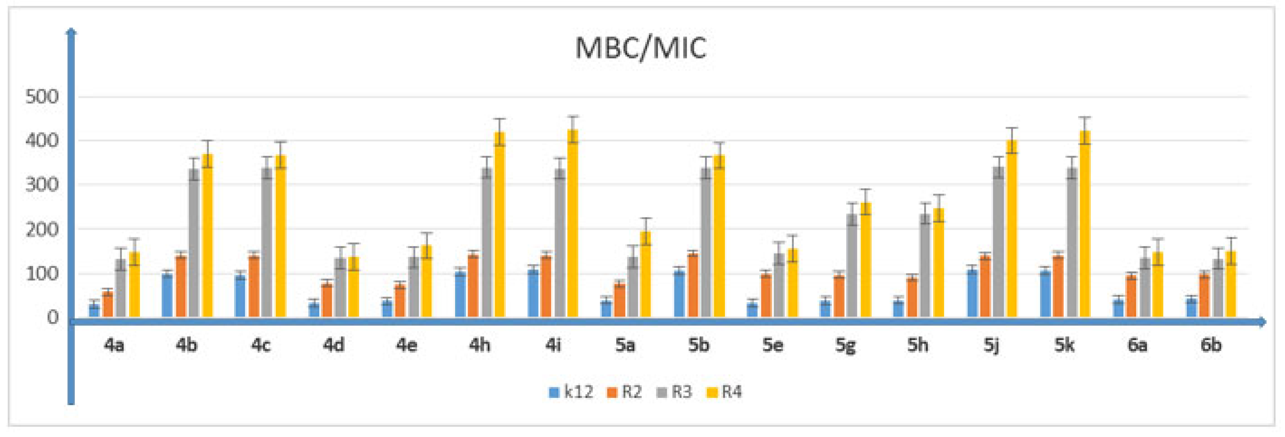
5.2. Cytotoxic Studies of the Library of Peptidomimetics

Analyzed DHPMs had an inhibitory effect on each studied bacterial model. Varied inhibitory activities were noted, depending on the nature of the substituent R1 in the pyrimidine ring of tested compounds 4 and 5. Interestingly, DHPM derivatives 4b, 4c, 4h, 4i, 5b, 5g, 5h, 5j and 5k exhibited selectivity towards the K12 and R2 strains. The presence of more lipophilic groups at the R1 position of tested compounds 4d and 4e resulted in increased inhibitory activity against the R3 strain. It is clearly visible that the structure of the tested compounds is closely related to their inhibitory activity. The R3 and R4 strains turned out to be less sensitive to tested compounds. The 4-fluorine group introduced into the aromatic ring did not have such a pronounced effect on the increase in activity of the tested compounds, as was the case of the δ-lactones tested previously [41]. MIC values for each model E. coli R2–R4 and K12 strains were visible on all analyzed microplates after the addition of the microbial growth index (resazurin)(Table 2).
In all analyzed plates where model strains K12 and R2–R4 of E. coli bacteria were treated with the analyzed compounds, MIC and MBC test values were observed for all 16 compounds but at different levels. Bacterial strains R3 and R4 were more susceptible to modification with these compounds (visible dilutions of 10–2, corresponding to concentrations of 0.0025 µM) than strains K12 and R2 (visible dilutions of 10–6, corresponding to concentrations of 0.2 µM). Strain R4 was the most sensitive of all strains, possibly due to having the longest lipopolysaccharide chain length. Based on the values in both types of tests and after the color-change analysis, the compounds 4b, 4c, 4h, 4i, 5b, 5g, 5h, 5j and 5k were used for further analyses. In all analyzed cases, MBC values were approximately 40 times higher than MIC values (Figure 3). Modification of functional groups in the tested compounds significantly changed the MBC/MIC ratios, (Figure 3, Figure 4 and Figure 5 and Table 1).

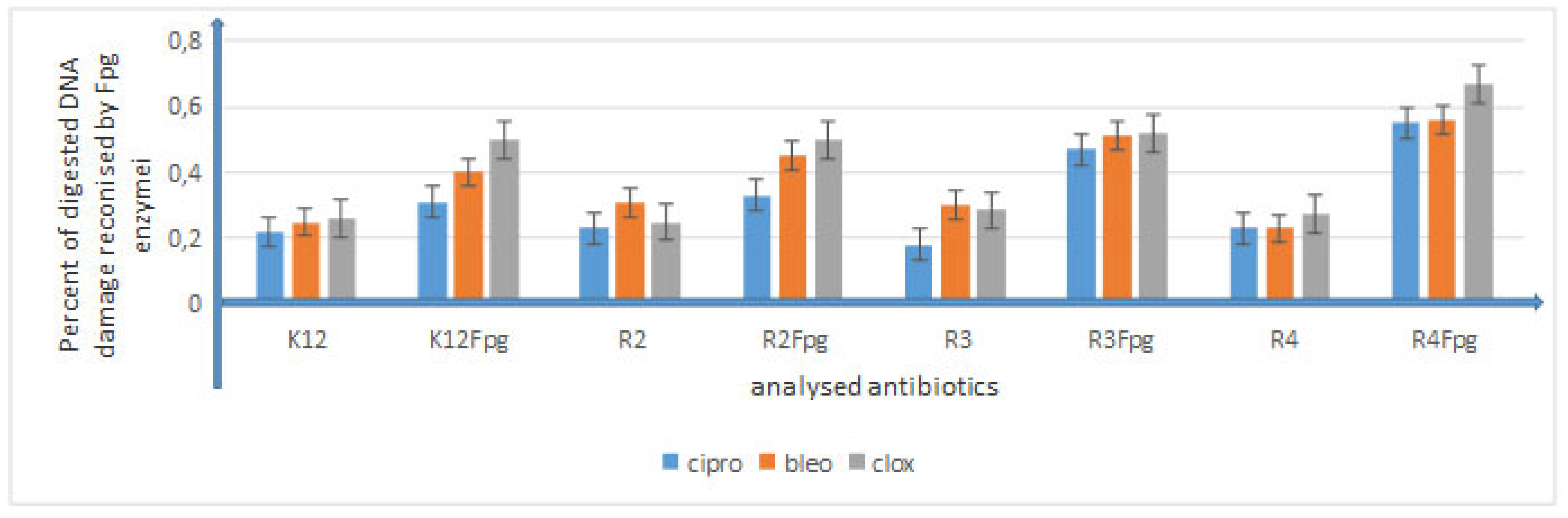
5.3. Analysis of Bacterial DNA Isolated from E. coli R2–R4 Strains Modified with 3,4-dihydropyrimidin-2(1H)-ones (DHPMs)

The obtained MIC values, as well as those obtained in our previous studies with the various types of analyzed compounds [37,38,39,40,41,42], indicate that 3,4-dihydropyrimidin-2(1H)-ones (DHPMs) also have a toxic effect on the analyzed model bacterial strains K12 and R2–R4 due to the type of substituent in the aromatic rings and the appropriate length of the alkyl chain with the specified type of substituent. Selected compounds 4b, 4c, 4h, 4i, 5b, 5g, 5h, 5j and 5k were used to modify model E. coli strains and digest them with Fpg protein from the group of repair glycosylases, which is a marker of oxidative stress [37,38,39,40,41,42]. We wanted to observe the effect of modification on the size and location of the oxidative damage (base type) in the DNA chain, which is a substantial reduction in the three forms of bacterial DNA: ccc, oc and linear form, as observed in previous studies [37,38,39,40,41,42]. The results of bacterial DNA modified with 3,4-dihydropyrimidin-2(1H)-ones (DHPMs) (Figure 6; with the action of Fpg) showed that all analyzed compounds with different alkyl chain lengths and substituents containing a hydroxyl group or an amino group together with substituents containing bromine and chlorine can strongly change the topology of the plasmid form, even after digestion with Fpg protein (Supplemental Materials S1 and S2) and are highly toxic to it, similar to the observations of previous studies [37,38,39,40,41,42]. The obtained results were also statistically significant at the level of p < 0.05. (Figure 6).
The obtained results indicate that these compounds can also potentially be used as “substitutes for” commonly used antibiotics (Figure 7 and Figure 8, Supplementary Materials Figure S3).
Large modifications of plasmid DNA were observed for compounds 4b, 4c, 4h, 4i, 5b, 5g, 5h, 5j and 5k among all analyzed compounds. Modifications with antibiotics were smaller and not as clear as in the case of the analyzed DHPMs. The sensitivity of E. coli strains to the cytotoxic effect of the compounds used and after Fpg protein digestion was as follows: R4 > R2 > R3 > K12; this effect was very similar to that observed in our previous studies [37,38,39,40,41,42]. This indicates a very high cytotoxicity of the analyzed DHPMs towards bacterial DNA, probably resulting from the modification of the components of the bacterial membrane and the LPS contained in it, which may induce specific enzymes from the group of topoisomerases and helicases, destabilizing the structure of the exposed DNA bases. The stabilization of the complex that regulates these enzymes is perhaps necessary for cell survival. Blocking these enzymes inhibits DNA replication and rewriting, which can affect its total amount.

6. Conclusions

The analysis of the presented compounds through a complex cycle of their synthesis reactions may constitute a potential source of innovative, cheap substitutes for antibiotics against various types of bacterial microorganisms (LPS). We focused on the structure–activity relationship of compounds with the 3,4-dihydropyrimidin-2(1H)-one scaffold. The obtained results show a strong influence of the activity of all 16 analyzed compounds on the values of MIC and MBC, as well as MBC/MIC for various strains of E. coli: R2–R4 and K12. Based on the analysis of the above studies, nine compounds (4b, 4c, 4h, 4i, 5b, 5g, 5h, 5j and 5k) were selected for further research. The repair activity using the Fpg-glycosylase protein of the BER pathway (base excision repair) was then compared, which is in accordance with our research hypothesis. The above results are very important for research on the mechanism of cytotoxic action of new compounds as innovative and safe drugs based on 3,4-dihydropyrimidin-2(1H)-one (DHPM) derivatives, which may lead to the destruction of the bacterial cell membrane by changing its surface charge and may play an important role in changing its electrokinetic potential, expressing the reversal of burdens. A particular effect was observed for the mentioned nine select compounds, which showed certain MIC values and MBC/MIC ratios. Compounds 5b, 5j and 5k showed superselectivity in all analyzed bacterial strains, even differentiating cytotoxicity in the K12 strain. The described compounds can be highly specific for pathogenic E. coli strains on the basis of the model strains used. In the future, cytotoxicity studies should also be carried out using various cell lines and cultures to assess the biocompatibility of the tested compounds with active peptidomimetics.

Supplementary Materials

The following are available online at https://www.mdpi.com/article/10.3390/membranes12020238/s1. Figure S1: Examples of MIC and MBC on microplates with different concentration of studied compounds (mg·L−1). Figure S2: Example of an agarose gel electrophoresis separation of isolated plasmids DNA from R4 strains modified with selected DHPM derivatives. Figure S3: Example of an agarose gel electrophoresis separation of isolated plasmids DNA from R4 strains modified with antibiotics: cloxacillin, ciprofloxacin, and bleomycin digested (or not) with repair enzymes Fpg. Figure S4: 1H NMR (400 MHz, DMSO-d6) of 4a. Figure S5: 13C NMR (100 MHz, DMSO-d6) of 4a. Figure S6: 19F NMR (376 MHz, DMSO-d6) of 4a. Figure S7: 1H NMR (400 MHz, DMSO-d6) of 4b. Figure S8: 13C NMR (100 MHz, DMSO-d6) of 4b. Figure S9: 19F NMR (376 MHz, DMSO-d6) of 4b. Figure S10: 1H NMR (400 MHz, DMSO-d6) of 4c. Figure S11: 13C NMR (100 MHz, DMSO-d6) of 4c. Figure S12: 19F NMR (376 MHz, DMSO-d6) of 4c. Figure S13: 1H NMR (400 MHz, DMSO-d6) of 4d. Figure S14. 13C NMR (100 MHz, DMSO-d6) of 4d. Figure S15: 19F NMR (376 MHz, DMSO-d6) of 4d. Figure S16: 1H NMR (400 MHz, DMSO-d6) of 4e. Figure S17: 13C NMR (100 MHz, DMSO-d6) of 4e. Figure S18: 19F NMR (376 MHz, DMSO-d6) of 4e. Figure S19: 1H NMR (400 MHz, DMSO-d6) of 4h. Figure S20: 13C NMR (100 MHz, DMSO-d6) of 4h. Figure S21: 19F NMR (376 MHz, DMSO-d6) of 4h. Figure S22: 1H NMR (400 MHz, DMSO-d6) of 4i. Figure S23: 13C NMR (100 MHz, DMSO-d6) of 4i. Figure S24: 19F NMR (376 MHz, DMSO-d6) of 4i. Figure S25: 1H NMR (400 MHz, DMSO-d6) of 5a. Figure S26: 13C NMR (100 MHz, DMSO-d6) of 5a. Figure S27: 19F NMR (376 MHz, DMSO-d6) of 5a. Figure S28: 1H NMR (400 MHz, DMSO-d6) of 5b. Figure S29: 13C NMR (100 MHz, DMSO-d6) of 5b. Figure S30: 19F NMR (376 MHz, DMSO-d6) of 5b. Figure S31: 1H NMR (400 MHz, DMSO-d6) of 5e. Figure S32: 13C NMR (100 MHz, DMSO-d6) of 5e. Figure S33: 19F NMR (376 MHz, DMSO-d6) of 5e. Figure S34: 1H NMR (400 MHz, DMSO-d6) of 5g. Figure S35: 13C NMR (100 MHz, DMSO-d6) of 5g. Figure S36: 19F NMR (376 MHz, DMSO-d6) of 5g. Figure S37: 1H NMR (400 MHz, DMSO-d6) of 5h. Figure S38: 13C NMR (100 MHz, DMSO-d6) of 5h. Figure S39: 19F NMR (376 MHz, DMSO-d6) of 5h. Figure S40: 1H NMR (400 MHz, DMSO-d6) of 5j. Figure S41: 13C NMR (100 MHz, DMSO-d6) of 5j. Figure S42: 19F NMR (376 MHz, DMSO-d6) of 5j. Figure S43: 1H NMR (400 MHz, DMSO-d6) of 5k. Figure S44: 13C NMR (100 MHz, DMSO-d6) of 5k. Figure S45: 19F NMR (376 MHz, DMSO-d6) of 5k. Figure S46: 1H NMR (400 MHz, DMSO-d6) of 6a. Figure S47: 13C NMR (100 MHz, DMSO-d6) of 6a. Figure S48: 1H NMR (400 MHz, DMSO-d6) of 6b. Figure S49: 13C NMR (100 MHz, DMSO-d6) of 6b.

Author Contributions

Conceptualization or design of the work, B.G., P.K., D.K. and R.O.; methodology, P.K., D.K., A.B., J.M., K.K., A.W. and R.O.; synthesis of all compounds, B.G., D.K., A.B. and J.M.; software, P.K. and A.W.; validation, B.G., P.K., D.K., A.B., J.M., K.K., A.W. and R.O.; formal analysis, B.G., P.K., D.K. and R.O.; investigation, B.G. and P.K.; interpretation of data for the work, B.G., P.K. and D.K.; drafting the work, P.K. and D.K.; critical revision for important intellectual content, B.G., P.K., D.K., A.B., J.M. and K.K.; resources, P.K., R.O. and K.K.; data curation, B.G., P.K. and D.K.; writing—original draft preparation, B.G., P.K. and D.K.; writing—review and editing, P.K.; visualization, P.K.; supervision, B.G., R.O., D.K. and P.K.; project administration, B.G., R.O. and P.K.; funding acquisition, K.K. All authors have read and agreed to the published version of the manuscript.

Funding

This work was supported by a grant from the Medical University of Białystok: SUB/2/DN/22/001/2201 and by the National Science Center, Poland, project OPUS No. 2019/33/B/ST4/01118.

Institutional Review Board Statement

Not applicable.

Informed Consent Statement

Not applicable.

Data Availability Statement

The data presented in this study are available on request from the corresponding author.

Acknowledgments

The authors thank Jolanta Łukasiewicz from Ludwik Hirszfeld Institute of Immunology and Experimental Therapy (Polish Academy of Sciences) for providing the strains of E. coli.

Conflicts of Interest

The authors declare no conflict of interest.

Abbreviations

MICminimum inhibitory concentration
MBCminimum bactericidal concentration
Ocopen circle
Ccccovalently closed circle
BER base excision repair
Fpg DNAformamidopyrimidine glycosylase

References

  1. Deepa; Yadav, G.D.; Aalam, M.J.; Chaudhary, P.; Singh, S. Synthesis of Dihydropyrimidinones (DHPMs) and Hexahydro Xanthene Catalyzed by 1,4-Diazabicyclo [2.2.2] Octane Triflate Under Solvent-Free Condition. Curr. Org. Synth. 2019, 16, 776–786. [Google Scholar] [CrossRef] [PubMed]
  2. Matos, L.H.S.; Masson, F.T.; Simeoni, L.A.; Homem-De-Mello, M. Biological activity of dihydropyrimidinone (DHPM) derivatives: A systematic review. Eur. J. Med. Chem. 2018, 143, 1779–1789. [Google Scholar] [CrossRef] [PubMed]
  3. Biginelli, P. The urea-aldehyde derivatives of acetoacetic esters. Gazz. Chim. Ital. 1893, 23, 360–416. [Google Scholar]
  4. Rogerio, K.; Vitório, F.; Kümmerle, A.; Graebin, C. Reações multicomponentes: Um breve histórico e a versatilidade destas reações na síntese de moléculas bioativas. Rev. Virtual Quim. 2016, 8, 1934–1962. [Google Scholar] [CrossRef]
  5. Sweet, F.; Fissekis, J.D. Synthesis of 3,4-dihydro-2(1H)-pyrimidinones and the mechanism of the Biginelli reaction. J. Am. Chem. Soc. 1973, 95, 8741–8749. [Google Scholar] [CrossRef]
  6. Kappe, C.O. A Reexamination of the Mechanism of the Biginelli Dihydropyrimidine Synthesis. Support for an N-Acyliminium Ion Intermediate1. J. Org. Chem. 1997, 62, 7201–7204. [Google Scholar] [CrossRef] [PubMed]
  7. Ramos, L.M.; Guido, B.C.; Nobrega, C.C.; Corrêa, J.R.; Silva, R.G.; de Oliveira, H.C.B.; Gomes, A.F.; Gozzo, F.C.; Neto, B.A.D. The Biginelli Reaction with an Imidazolium-Tagged Recyclable Iron Catalyst: Kinetics, Mechanism, and Antitumoral Activity. Chem. Eur. J. 2013, 19, 4156–4168. [Google Scholar] [CrossRef] [PubMed]
  8. Mayer, T.U.; Kapoor, T.M.; Haggarty, S.J.; King, R.W.; Schreiber, S.L.; Mitchison, T.J. Small Molecule Inhibitor of Mitotic Spindle Bipolarity Identified in a Phenotype-Based Screen. Science 1999, 286, 971–974. [Google Scholar] [CrossRef] [PubMed] [Green Version]
  9. Kappe, C. Biologically active dihydropyrimidones of the Biginelli-type—A literature survey. Eur. J. Med. Chem. 2000, 35, 1043–1052. [Google Scholar] [CrossRef]
  10. Kapoor, T.M.; Mayer, T.; Coughlin, M.L.; Mitchison, T.J. Probing Spindle Assembly Mechanisms with Monastrol, a Small Molecule Inhibitor of the Mitotic Kinesin, Eg5. J. Cell Biol. 2000, 150, 975–988. [Google Scholar] [CrossRef]
  11. Alrazi, I.M.D.; Sadakane, K.; Maruta, S. Novel photochromic inhibitor for mitotic kinesin Eg5 which forms multiple isomerization states. J. Biochem. 2021, 170, 229–237. [Google Scholar] [CrossRef] [PubMed]
  12. DeBonis, S.; Simorre, J.P.; Crevel, I.; Lebeau, L.; Skoufias, D.A.; Blangy, A.; Ebel, C.; Gans, P.; Cross, R.; Hackney, D.D.; et al. Interaction of the mitotic inhibitor monastrol with human kinesin Eg5. Biochemistry 2003, 42, 338–349. [Google Scholar] [CrossRef] [PubMed] [Green Version]
  13. Kapoor, T.M.; Mayer, T.U.; Desai, A.; Maddox, P.; Salmon, E.D.; Schreiber, S.L.; Mitchison, T.J. Investigating the inhibition of bipolar spindle formation by monastrol, a small molecule kinesin inhibitor. Mol. Biol. Cell 1999, 10, 128A. [Google Scholar]
  14. Duan, L.; Wang, T.-Q.; Bian, W.; Liu, W.; Sun, Y.; Yang, B.-S. Centrin: Another target of monastrol, an inhibitor of mitotic spindle. Spectrochim. Acta Part A Mol. Biomol. Spectrosc. 2015, 137, 1086–1091. [Google Scholar] [CrossRef] [PubMed]
  15. Abassi, Y.A.; Xi, B.; Zhang, W.; Ye, P.; Kirstein, S.L.; Gaylord, M.R.; Feinstein, S.C.; Wang, X.; Xu, X. Kinetic Cell-Based Morphological Screening: Prediction of Mechanism of Compound Action and Off-Target Effects. Chem. Biol. 2009, 16, 712–723. [Google Scholar] [CrossRef] [Green Version]
  16. Zhu, L.; Cheng, P.; Lei, N.; Yao, J.; Sheng, C.; Zhuang, C.; Guo, W.; Liu, W.; Zhang, Y.; Dong, G.; et al. Synthesis and Biological Evaluation of Novel Homocamptothecins Conjugating with Dihydropyrimidine Derivatives as Potent Topoisomerase I Inhibitors. Arch. Pharm. 2011, 344, 726–734. [Google Scholar] [CrossRef]
  17. Abdou, A.M.; Botros, S.; Hassan, R.A.; Kamel, M.M.; Taber, D.F.; Taher, A.T. Useful four-carbon synthons en route to monastrol analogs. Tetrahedron 2015, 71, 139–146. [Google Scholar] [CrossRef]
  18. Bahekar, S.S.; Shinde, D.B. Synthesis and anti-inflammatory activity of some [4,6-(4-substituted aryl)-2-thioxo-1,2,3,4-tetrahydro-pyrimidin-5-yl]-acetic acid derivatives. Bioorg. Med. Chem. Lett. 2004, 14, 1733–1736. [Google Scholar] [CrossRef]
  19. Bryzgalov, A.O.; Dolgikh, M.P.; Sorokina, I.V.; Tolstikova, T.G.; Sedova, V.F.; Shkurko, O.P. Antiarrhythmic activity of 4,6-di(het)aryl-5-nitro-3,4-dihydropyrimidin-(1H)-2-ones and its effects on arterial pressure in rats. Bioorg. Med. Chem. Lett. 2006, 16, 1418–1420. [Google Scholar] [CrossRef]
  20. Akhaja, T.N.; Raval, J. 1,3-dihydro-2H-indol-2-ones derivatives: Design, Synthesis, in vitro antibacterial, antifungal and antitubercular study. Eur. J. Med. Chem. 2011, 46, 5573–5579. [Google Scholar] [CrossRef]
  21. Chhillar, A.K.; Arya, P.; Mukherjee, C.; Kumar, P.; Yadav, Y.; Sharma, A.K.; Yadav, V.; Gupta, J.; Dabur, R.; Jha, H.N.; et al. Microwave-assisted synthesis of antimicrobial dihydropyridines and tetrahydropyrimidin-2-ones: Novel compounds against aspergillosis. Bioorg. Med. Chem. 2006, 14, 973–981. [Google Scholar] [CrossRef]
  22. Kim, J.; Ok, T.; Park, C.; So, W.; Jo, M.; Kim, Y.; Seo, M.; Lee, D.; Jo, S.; Ko, Y.; et al. A novel 3,4-dihydropyrimidin-2(1H)-one: HIV-1 replication inhibitors with improved metabolic stability. Bioorg. Med. Chem. Lett. 2012, 22, 2522–2526. [Google Scholar] [CrossRef] [PubMed]
  23. Singh, N.; Kaur, J.; Kumar, P.; Gupta, S.; Singh, N.; Ghosal, A.; Dutta, A.; Kumar, A.; Tripathi, R.; Siddiqi, M.I.; et al. An orally effective dihydropyrimidone (DHPM) analogue induces apoptosis-like cell death in clinical isolates of Leishmania donovani overexpressing pteridine reductase 1. Parasitol. Res. 2009, 105, 1317–1325. [Google Scholar] [CrossRef] [PubMed] [Green Version]
  24. Lacotte, P.; Buisson, D.-A.; Ambroise, Y. Synthesis, evaluation and absolute configuration assignment of novel dihydropyrimidin-2-ones as picomolar sodium iodide symporter inhibitors. Eur. J. Med. Chem. 2013, 62, 722–727. [Google Scholar] [CrossRef]
  25. Acharya, B.N.; Rao, G.B.D.; Kumar, D.; Kumar, P.; Kaushik, M.P. Design, synthesis, and evaluation of dihydropyrimidinone (DHPM) based muscarinic receptor antagonist. Med. Chem. Res. 2014, 24, 1763–1775. [Google Scholar] [CrossRef]
  26. Dhumaskar, K.L.; Meena, S.N.; Ghadi, S.C.; Tilve, S.G. Graphite catalyzed solvent free synthesis of dihydropyrimidin-2(1H)-ones/thiones and their antidiabetic activity. Bioorg. Med. Chem. Lett. 2014, 24, 2897–2899. [Google Scholar] [CrossRef]
  27. Blackburn, C.; Guan, B.; Brown, J.; Cullis, C.; Condon, S.M.; Jenkins, T.J.; Peluso, S.; Ye, Y.; Gimeno, R.E.; Punreddy, S.; et al. Identification and characterization of 4-aryl-3,4-dihydropyrimidin-2(1H)-ones as inhibitors of the fatty acid transporter FATP4. Bioorg. Med. Chem. Lett. 2006, 16, 3504–3509. [Google Scholar] [CrossRef]
  28. Ahmad, S.; Iftikhar, F.; Ullah, F.; Sadiq, A.; Rashid, U. Rational design and synthesis of dihydropyrimidine based dual binding site acetylcholinesterase inhibitors. Bioorg. Chem. 2016, 69, 91–101. [Google Scholar] [CrossRef] [PubMed]
  29. Khan, A.; Hashim, J.; Arshad, N.; Khan, I.; Siddiqui, N.; Wadood, A.; Ali, M.; Arshad, F.; Khan, K.M.; Choudhary, M.I. Dihydropyrimidine based hydrazine dihydrochloride derivatives as potent urease inhibitors. Bioorg. Chem. 2016, 64, 85–96. [Google Scholar] [CrossRef]
  30. Atwal, K.S.; Swanson, B.N.; Unger, S.E.; Floyd, D.M.; Moreland, S.; Hedberg, A.; O’Reilly, B.C. Dihydropyrimidine calcium channel blockers. 3. 3-Carbamoyl-4-aryl-1,2,3,4-tetrahydro-6-methyl-5-pyrimidinecarboxylic acid esters as orally effective antihypertensive agents. J. Med. Chem. 1991, 34, 806–811. [Google Scholar] [CrossRef]
  31. Chikhale, R.; Thorat, S.; Pant, A.; Jadhav, A.; Thatipamula, K.C.; Bansode, R.; Bhargavi, G.; Karodia, N.; Rajasekharan, M.; Paradkar, A.; et al. Design, synthesis and pharmacological evaluation of pyrimidobenzothiazole-3-carboxylate derivatives as selective L-type calcium channel blockers. Bioorg. Med. Chem. 2015, 23, 6689–6713. [Google Scholar] [CrossRef]
  32. Lewis, R.W.; Mabry, J.; Polisar, J.G.; Eagen, K.; Ganem, B.; Hess, G.P. Dihydropyrimidinone Positive Modulation of δ-Subunit-Containing γ-Aminobutyric Acid Type A Receptors, Including an Epilepsy-Linked Mutant Variant. Biochemistry 2010, 49, 4841–4851. [Google Scholar] [CrossRef] [PubMed] [Green Version]
  33. de Fátima, Â.; Braga, T.C.; Neto, L.; Terra, B.S.; Oliveira, B.; da Silva, D.L.; Modolo, L. A mini-review on Biginelli adducts with notable pharmacological properties. J. Adv. Res. 2015, 6, 363–373. [Google Scholar] [CrossRef] [Green Version]
  34. Prokopcová, H.; Dallinger, D.; Uray, G.; Kaan, H.Y.K.; Ulaganathan, V.; Kozielski, F.; Laggner, C.; Kappe, C.O. Structure-Activity Relationships and Molecular Docking of Novel Dihydropyrimidine-Based Mitotic Eg5 Inhibitors. ChemMedChem 2010, 5, 1760–1769. [Google Scholar] [CrossRef] [PubMed]
  35. Holla, B.; Rao, B.; Sarojini, B.; Akberali, P. One pot synthesis of thiazolodihydropyrimidinones and evaluation of their anticancer activity. Eur. J. Med. Chem. 2004, 39, 777–783. [Google Scholar] [CrossRef] [PubMed]
  36. Treptow, T.G.; Figueiró, F.; Jandrey, E.H.F.; Battastini, A.M.; Salbego, C.G.; Hoppe, J.B.; Taborda, P.S.; Rosa, S.B.; Piovesan, L.A.; Doca, C.; et al. Novel hybrid DHPM-fatty acids: Synthesis and activity against glioma cell growth in vitro. Eur. J. Med. Chem. 2015, 95, 552–562. [Google Scholar] [CrossRef] [PubMed]
  37. Kowalczyk, P.; Trzepizur, D.; Szymczak, M.; Skiba, G.; Kramkowski, K.; Ostaszewski, R. 1,2-Diarylethanols—A New Class of Compounds That Are Toxic to E. coli K12, R2–R4 Strains. Materials 2021, 14, 1025. [Google Scholar] [CrossRef] [PubMed]
  38. Kowalczyk, P.; Madej, A.; Szymczak, M.; Ostaszewski, R. α-Amidoamids as New Replacements of Antibiotics—Research on the Chosen K12, R2–R4 E. coli Strains. Materials 2020, 13, 5169. [Google Scholar] [CrossRef] [PubMed]
  39. Kowalczyk, P.; Borkowski, A.; Czerwonka, G.; Cłapa, T.; Cieśla, J.; Misiewicz, A.; Borowiec, M.; Szala, M. The microbial toxicity of quaternary ammonium ionic liquids is dependent on the type of lipopolysaccharide. J. Mol. Liq. 2018, 266, 540–547. [Google Scholar] [CrossRef]
  40. Borkowski, A.; Kowalczyk, P.; Czerwonka, G.; Cieśla, J.; Cłapa, T.; Misiewicz, A.; Szala, M.; Drabik, M. Interaction of quaternary ammonium ionic liquids with bacterial membranes—Studies with Escherichia coli R1–R4-type lipopolysaccharides. J. Mol. Liq. 2017, 246, 282–289. [Google Scholar] [CrossRef]
  41. Kowalczyk, P.; Gawdzik, B.; Trzepizur, D.; Szymczak, M.; Skiba, G.; Raj, S.; Kramkowski, K.; Lizut, R.; Ostaszewski, R. δ-Lactones—A New Class of Compounds That Are Toxic to E. coli K12 and R2–R4 Strains. Materials 2021, 14, 2956. [Google Scholar] [CrossRef] [PubMed]
  42. Kowalczyk, P.; Wilk, M.; Parul, P.; Szymczak, M.; Kramkowski, K.; Raj, S.; Skiba, G.; Sulejczak, D.; Kleczkowska, P.; Ostaszewski, R. The Synthesis and Evaluation of Aminocoumarin Peptidomimetics as Cytotoxic Agents on Model Bacterial E. coli Strains. Materials 2021, 14, 5725. [Google Scholar] [CrossRef]
  43. Dissanayake, D.; Wijewardana, T.; Gunawardena, G.; Poxton, I. Distribution of lipopolysaccharide core types among avian pathogenic Escherichia coli in relation to the major phylogenetic groups. Vet. Microbiol. 2008, 132, 355–363. [Google Scholar] [CrossRef]
  44. Maciejewska, A.; Kaszowska, M.; Jachymek, W.; Lugowski, C.; Lukasiewicz, J. Lipopolysaccharide-linked Enterobacterial Common Antigen (ECALPS) Occurs in Rough Strains of Escherichia coli R1, R2, and R4. Int. J. Mol. Sci. 2020, 21, 6038. [Google Scholar] [CrossRef] [PubMed]
  45. Prost, M.E.; Prost, R. Basic parameters of evaluation of the effectiveness of antibiotic therapy. OphthaTherapy 2017, 4, 233–236. [Google Scholar] [CrossRef]
  46. Albadi, J.; Mansournezhad, A.; Baghernehad, M.; Frozan, N. A Green Recyclable Poly(4-vinylpyridine)-Supported Copper Iodide Nanoparticles Catalyst for the Multicomponent Synthesis of 3,4-dihydropyrimidin-2(1H)-ones/thiones. J. Korean Chem. Soc. 2013, 57, 169–171. [Google Scholar] [CrossRef]
  47. Li, Y.; Ma, J.; Liu, Z.; Jin, D.; Jiao, G.; Guo, Y.; Wang, Q.; Zhou, J.; Sun, R. Fabrication of porous ultrathin carbon nitride nanosheet catalysts with enhanced photocatalytic activity for N- and O-heterocyclic compound synthesis. New J. Chem. 2020, 45, 365–372. [Google Scholar] [CrossRef]
  48. Nagarajan, S.; Shaikh, T.; Kandasamy, E. Synthesis of 1-alkyl triazolium triflate room temperature ionic liquids and their catalytic studies in multi-component Biginelli reaction. J. Chem. Sci. 2015, 127, 1539–1545. [Google Scholar] [CrossRef] [Green Version]
  49. Paudyal, M.P.; Wang, M.; Siitonen, J.H.; Hu, Y.; Yousufuddin, M.; Shen, H.C.; Falck, J.R.; Kürti, L. Intramolecular N-Me and N-H aminoetherification for the synthesis of N-unprotected 3-amino-O-heterocycles. Org. Biomol. Chem. 2021, 19, 557–560. [Google Scholar] [CrossRef]
  50. Han, B.; Han, R.-F.; Ren, Y.-W.; Duan, X.-Y.; Xu, Y.-C.; Zhang, W. Efficient aerobic oxidative dehydrogenation of dihydropyrimidinones and dihydropyrimidines. Tetrahedron 2011, 67, 5615–5620. [Google Scholar] [CrossRef]
  51. Kurmach, M.N.; Ryabitskiy, A.B.; Britsun, V.N. 2-Acylthioacetamides in the Biginelli Reaction. Chem. Heterocycl. Compd. 2014, 49, 1770–1776. [Google Scholar] [CrossRef]
  52. Braga, T.C.; Silva, T.F.; Maciel, T.M.S.; da Silva, E.C.D.; da Silva-Júnior, E.F.; Modolo, L.V.; Figueiredo, I.M.; Santos, J.C.C.; de Aquino, T.M.; de Fátima, Â. Ionic liquid-assisted synthesis of dihydropyrimidin(thi)one Biginelli adducts and investigation of their mechanism of urease inhibition. New J. Chem. 2019, 43, 15187–15200. [Google Scholar] [CrossRef]
  53. Bais, J.; Benedetti, F.; Berti, F.; Cerminara, I.; Drioli, S.; Funicello, M.; Regini, G.; Vidali, M.; Felluga, F. One Pot Synthesis of Micromolar BACE-1 Inhibitors Based on the Dihydropyrimidinone Scaffold and Their Thia and Imino Analogues. Molecules 2020, 25, 4152. [Google Scholar] [CrossRef] [PubMed]
  54. Zolfagharinia, S.; Koukabi, N.; Kolvari, E. A unique opportunity for the utilization of glass wastes as a resource for catalytic applications: Toward a cleaner environment. RSC Adv. 2016, 6, 113844–113858. [Google Scholar] [CrossRef]
  55. Mohamadpour, F.; Lashkari, M.; Maghsoodlou, M.T.; Heydari, R. Phthalic acid: A green, biodegradable and environmentally bening nature di-functional Brønsted acid catalyst for the one-pot synthesis of 3, 4-dihydropyrimidin-2-(1H)-one derivatives and substituted dihydro-2-oxypyroles. J. Chil. Chem. Soc. 2018, 63, 3811–3818. [Google Scholar] [CrossRef] [Green Version]
  56. Sari, O.; Roy, V.; Métifiot, M.; Marchand, C.; Pommier, Y.; Bourg, S.; Bonnet, P.; Schinazi, R.F.; Agrofoglio, L.A. Synthesis of dihydropyrimidine α,γ-diketobutanoic acid derivatives targeting HIV integrase. Eur. J. Med. Chem. 2015, 104, 127–138. [Google Scholar] [CrossRef] [Green Version]
  57. Zhou, B.; Li, X.; Li, Y.; Xu, Y.; Zhang, Z.; Zhou, M.; Zhang, X.; Liu, Z.; Zhou, J.; Cao, C.; et al. Discovery and Development of Thiazolo[3,2-a]pyrimidinone Derivatives as General Inhibitors of Bcl-2 Family Proteins. ChemMedChem 2011, 6, 904–921. [Google Scholar] [CrossRef] [PubMed]
  58. Wang, J.-H.; Tang, G.-M.; Yan, S.-C.; Wang, Y.-T.; Zhan, S.-J.; Zhang, E.; Sun, Y.; Jiang, Y.; Cui, Y.-Z. Cobalt-based metal coordination polymers with 4,4′-bipyridinyl groups: Highly efficient catalysis for one-pot synthesis of 3,4-dihydropyrimidin-2(1H)-ones under solvent-free conditions. Appl. Organomet. Chem. 2016, 30, 1009–1021. [Google Scholar] [CrossRef]
  59. Zafar, A.M.; Qureshi, S.; Khan, M.N.; Azad, M.; Munawar, M.A.; Khan, M.A. Amino Acids Catalyzed Biginelli Protocols. Asian J. Chem. 2013, 25, 3244–3246. [Google Scholar] [CrossRef]
  60. Godugu, K.; Yadala, V.D.S.; Pinjari, M.K.M.; Gundala, T.R.; Sanapareddy, L.R.; Nallagondu, C.G.R. Natural dolomitic limestone-catalyzed synthesis of benzimidazoles, dihydropyrimidinones, and highly substituted pyridines under ultrasound irradiation. Beilstein J. Org. Chem. 2020, 16, 1881–1900. [Google Scholar] [CrossRef]
  61. Khatri, C.K.; Potadar, S.M.; Chaturbhuj, G. A reactant promoted solvent free synthesis of 3,4-dihydropyrimidin-2(1H)-thione analogues using ammonium thiocyanate. Tetrahedron Lett. 2017, 58, 1778–1780. [Google Scholar] [CrossRef]
Figure 1. Pharmaceutically relevant 3,4-Dihydropyrimidin-2(1H)-ones (DHPMs) based on [1,2,3].
Membranes 12 00238 g001
Figure 2. Synthesis of the studied DHPMs.
Membranes 12 00238 g002
Figure 3. Minimum inhibitory concentration (MIC) of 3,4-dihydropyrimidin-2(1H)-ones (DHPMs) in model bacterial strains. The x-axis features 3,4-dihydropyrimidin-2(1H)-ones (DHPMs) used sequentially. The y-axis shows the MIC value in µg/mL−1. Investigated strains of E. coli: K12 as control (blue), R2 strain (orange), R3 strain (gray) and R4 strain (yellow). The y-axis shows the MBC value in µg/mL−1. The order in which the compounds were applied to the plate is shown in Supplementary Materials Figure S1.
Membranes 12 00238 g003
Figure 4. Minimum bactericidal concentration (MBC) of 3,4-dihydropyrimidin-2(1H)-ones (DHPMs) in model bacterial strains. The x-axis features 3,4-dihydropyrimidin-2(1H)-ones (DHPMs) used sequentially. The y-axis shows the MIC value in µg/mL−1. Investigated strains of E. coli: K12 as control (blue), R2 strain (orange), R3 strain (gray) and R4 strain (yellow). The y-axis shows the MBC value in µg/mL−1. The order in which the compounds were applied to the plate is shown in Supplementary Materials Figure S1.
Membranes 12 00238 g004
Figure 5. The ratio of MBC/MIC of 3,4-dihydropyrimidin-2(1H)-ones (DHPMs) in model bacterial strains. The x-axis features 3,4-dihydropyrimidin-2(1H)-ones (DHPMs) used sequentially. The y-axis shows the MIC value in µg/mL−1. Investigated strains of E. coli: K12 as control (blue), R2 strain (orange), R3 strain (gray) and R4 strain (yellow). The y-axis shows the MBC value in µg/mL−1. The order in which the compounds were applied to the plate is shown in Supplementary Materials Figure S1.
Membranes 12 00238 g005
Figure 6. Percentage of plasmid DNA recognized by Fpg enzyme (y-axis) with model bacterial strains K12 and R2–R4 (x-axis). Compounds 4b, 4c, 4h, 4i, 5b, 5g, 5h, 5j and 5k were statistically significant at * p < 0.05 (see Table 1).
Membranes 12 00238 g006
Figure 7. Examples of MIC with model bacterial strains K12, R2, R3 and R4 for study of the mechanisms of antibiotics ciprofloxacin and bleomycin. The x-axis features antibiotics used sequentially. The y-axis features the MIC value in µg/mL−1.
Membranes 12 00238 g007
Figure 8. Percentage of bacterial DNA recognized by Fpg enzyme in model bacterial strains after ciprofloxacin, bleomycin and cloxacillin treatment. The compounds were statistically significant at p < 0.05.
Membranes 12 00238 g008
Table 1. Yield of DHPMs 4, 5 and 6.
EntryCompoundXR1R2R3Yield
14aOMeOEtF67%
24bOMeOMeF56%
34cOMeOi-BuF63%
44dOPrOEtF69%
54eOPhOEtF82%
64hOMeNHPhF67%
74iOMeNH2F57%
85aSMeOEtF54%
95bSMeOMeF55%
105eSPhOEtF54%
115gSPh(3-Me)OEtF58%
125hSPh(4-Me) OEtF63%
135jSMeNHPhF76%
145kSMeNH2F61%
156aOMeOEtH70%
166bSMeOEtH59%
Table 2. Statistical analysis of analyzed compounds by MIC, MBC and MBC/MIC; * p < 0.05, ** p < 0.01 and *** p < 0.001.
No. of Samples4b4c4h4i5b,5g5h5j,5kType of Test
K12***********MIC
R2***********MIC
R3***********MIC
R4***********MIC
K12***********MBC
R2************MBC
R3************MBC
R4************MBC
K12***********MBC/MIC
R2***********MBC/MIC
R3***********MBC/MIC
R4***********MBC/MIC
Publisher’s Note: MDPI stays neutral with regard to jurisdictional claims in published maps and institutional affiliations.

