Article

Comparison of NH3 and N2O Plasma Treatments on Bi2O3 Sensing Membranes Applied in an Electrolyte–Insulator–Semiconductor Structure

1
Department of Electronic Engineering, Chang Gung University, 259 Wen-Hwa 1st Road, Kwei-Shan District, Taoyuan City 333, Taiwan
2
Kidney Research Center, Department of Nephrology, Chang Gung Memorial Hospital, Chang Gung University, No. 5 Fuxing Street, Guishan District, Taoyuan City 333, Taiwan
3
Department of Electronic Engineering, Ming Chi University of Technology, 284 Gungjuan Road, Taishan District, New Taipei City 243, Taiwan
4
Department of Applied Materials and Optoelectronic Engineering, National Chi Nan University, Puli, Nantou 545, Taiwan
5
Department of Electro-Optical Engineering, Minghsin University of Science and Technology, No. 1, Xinxing Road, Xinfeng, Hsinchu 30401, Taiwan
*
Authors to whom correspondence should be addressed.
Membranes 2022, 12(2), 188; https://doi.org/10.3390/membranes12020188
Submission received: 30 December 2021 / Revised: 24 January 2022 / Accepted: 24 January 2022 / Published: 5 February 2022
(This article belongs to the Section Membrane Applications)

Abstract

:
In this study, bismuth trioxide (Bi2O3) membranes in an electrolyte–insulator–semiconductor (EIS) structure were fabricated with pH sensing capability. To optimize the sensing performance, the membranes were treated with two types of plasma—NH3 and N2O. To investigate the material property improvements, multiple material characterizations were conducted. Material analysis results indicate that plasma treatments with appropriate time could enhance the crystallization, remove the silicate and facilitate crystallizations. Owing to the material optimizations, the pH sensing capability could be greatly boosted. NH3 or N2O plasma treated-Bi2O3 membranes could reach the pH sensitivity around 60 mV/pH and show promise for future biomedical applications.

1. Introduction

Fifty years ago, the first ion-sensitive field-effect transistor (ISFET) was invented by Bergveld in 1970 [1,2]. Following the invention, semiconductor-based ion sensing technology [1,2,3] has been developed since the late 20th century. Among various types of ion sensing semiconductor devices, electrolyte–insulator–semiconductor (EIS) sensors with rapid response, high reliability and simple structure have been intensively studied [4]. Because of low capacitance and poor electrochemical properties, SiO2 has been replaced by various oxides to improve the membranes properties [5]. Recently, Ta2O5 [6], WO3 [7], and La2O3 [8] have emerged as novel membrane materials [9]. However, to further boost the membrane sensing performance, novel materials and new treatments are worthwhile to be explored. Bismuth trioxide (Bi2O3) with a bandgap around 2.5 eV has been utilized as photocatalyst [10], super capacitors, and gas sensor materials [11]. However, Bi2O3-based pH sensing membranes [12] have not been clearly reported, yet. Furthermore, to enhance the sensing capability, Bi2O3 membranes were treated with two types of plasma- NH3 and N2O [13,14]. To investigate the improvement of the treatment, multiple material analysis techniques including X-ray diffraction (XRD), X-ray photoelectron spectroscopy (XPS), atomic force microscopy (AFM), and secondary ion mass spectrometry (SIMS) were performed. Material analysis results indicate that Bi2O3 membranes treated with NH3 plasma for 3 min and N2O plasma for 1 min had strong crystallization, silicate suppression, high grainization, and effective nitrogen passivation. Moreover, the pH sensing measurements [15] indicate that Bi2O3 membranes treated in these plasma treatment conditions had high pH sensitivity around 60 mV/pH and high linearity close to 100%. Hysteresis and drift [16] rate evaluation also reveal that the membrane treated in these conditions had the lowest hysteresis voltage and the smallest drift rate. NH3 and N2O plasma treatments could incorporate the nitrogen atoms into the deep part of the devices to fix the defects and eliminate the silicates.
According to previous reports [17,18], plasma treatment can eliminate the silicate layer because, because silicates can have chemical reaction and transform from SiOx with dangling bonds to Si–O–Si bonds. The atoms in the plasma such as F or N can facilitate the combination of the silicon or oxygen dangling bonds to form well-crystallized Si–O–Si [19]. Therefore, silicate can be reduced, and near-perfect crystals can replace the silicate. The effective electric field across the membranes can be enhanced so the sensing capability can be improved [20,21]. Moreover, the nitrogen incorporation in the bulks may form NH2 bond to strengthen the chemical bonds and improve the material properties [22]. On the other hand, plasma treatment of the sensing film could cause an EIS structure sensitive to H+ ions [23] because the increase of the metal ions produced on the surface sites to decrease the diffusion capacitance in the solution and enhance the sensitivity. Furthermore, based on the Gouy–Chapman–Stern model [24,25], the sensing parameter β is proportional to the density of surface states, as shown in (1), where Ns is the number of surface sites per unit surface area and CDL is the double layer capacitance. Therefore, the sensing capability can be enhanced by NH3 and N2O plasma treatments.
β = 2 q 2 Ns K a K b KTC DL
Owing to high sensitivity, fair linearity, stable response, Bi2O3-based EIS membranes [26] with NH3 or N2O plasma treatments show promise for future industrial biomedical sensing [27] applications.

2. Experimental

To prepare the EIS sensor with Bi2O3 sensing film, 3.95 g of bismuth nitrate (Bi(NO₃)₃·5H₂O) was dissolved in 20 mL of nitric acid with the solution concentration of 1 M. The sol-gel solution was dropped onto the cleansed p-Si substrate. Then, a Bi2O3 film formed on it, and then the plasma treatments were performed. The samples were treated with NH3 and N2O plasma at 100 W RF power and 500 mTorr processing pressure for 1 min, 3 min, and 6 min, respectively. Then, an Al film with a thickness of 300 nm was deposited on the back of the silicon wafer and the silicone glue was used to define the sensing window, and the device was glued to be fixed on a PCB board. Finally, AB glue was used for packaging to prevent oxidation. The device structure is illustrated in Figure 1.

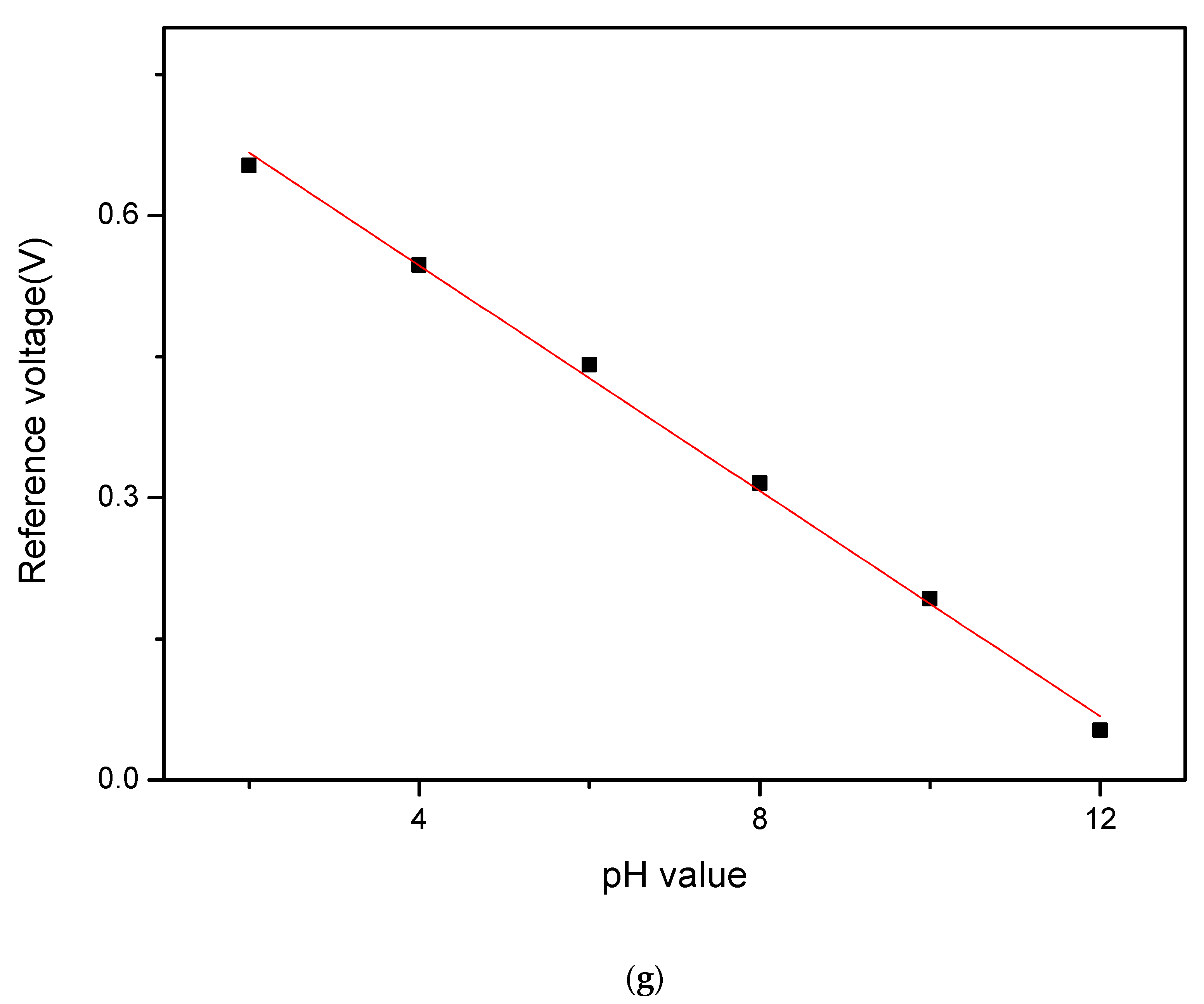
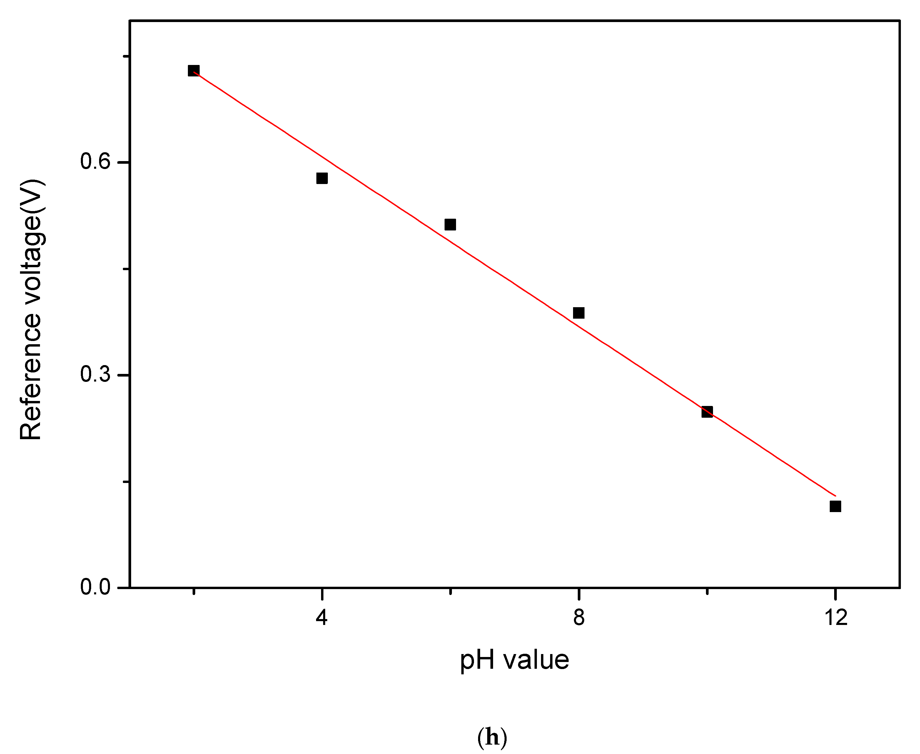
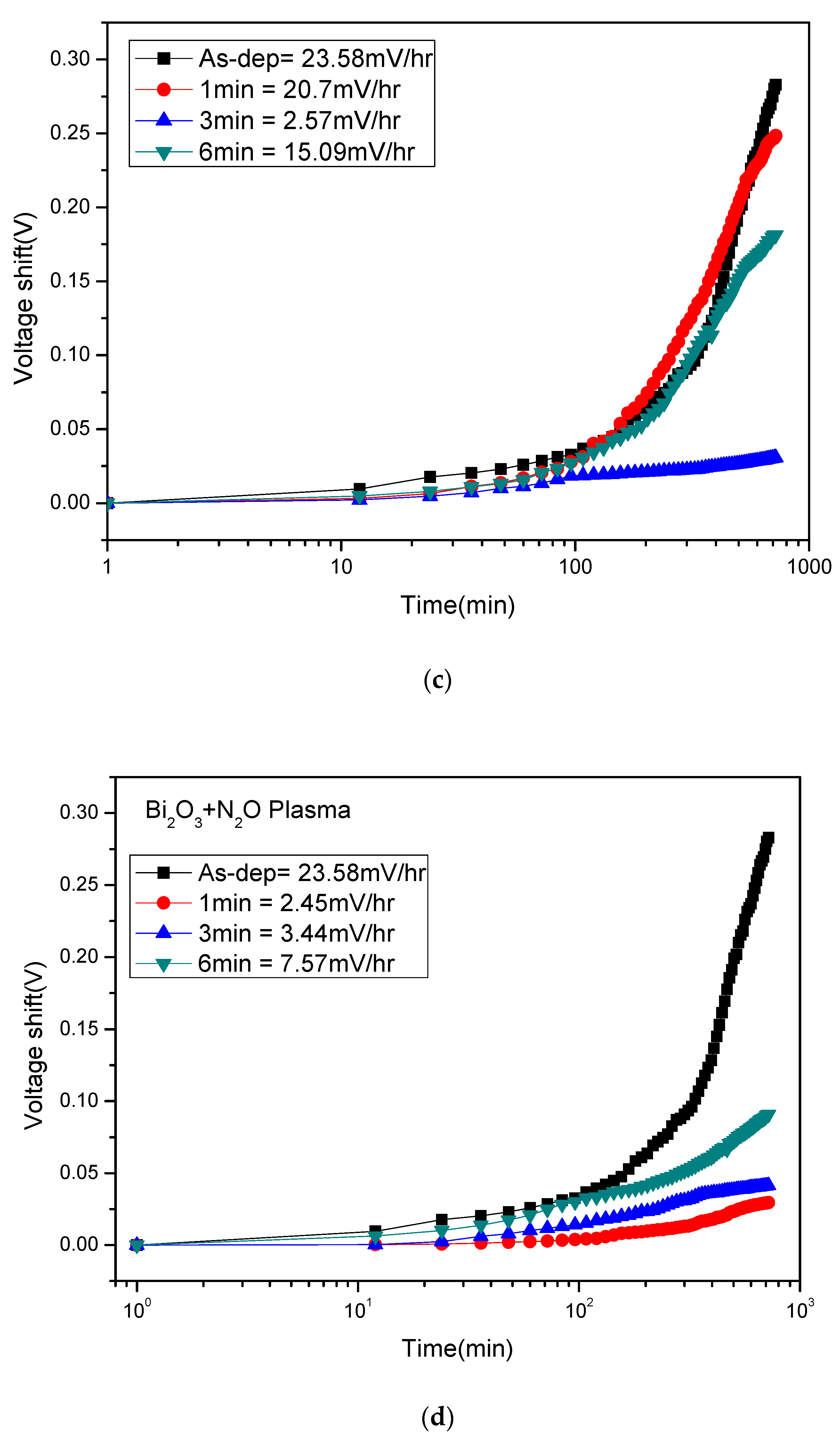
3. Results and Discussion

To examine the crystalline structures of the membranes, XRD was used. Figure 2a shows the X-ray diffraction patterns of Bi2O3 film after NH3 plasma treatments for various times. The two diffraction peaks BiO (012) and Bi2O3 (200) are located at 27.8° and 32.6°, respectively. The as-deposited sample shows a peak of BiO. After NH3 plasma treatment for 3 min, the sample shows the strongest peak of Bi2O3 (200) among all the samples.
By contrast, Figure 2b shows the X-ray diffraction analysis of the Bi2O3 film after N2O plasma treatments for various times. The diffraction peaks BiO (012) and Bi2O3 (200) are located at 27.8° and 32.6°, respectively. The as-deposited sample also shows a peak of BiO. After N2O plasma treatment for 1 min, the sample shows the strongest Bi2O3 (200) peak among all the samples. With the increase of the plasma treatment time, the intensity of the Bi2O3 (200) peak gradually decreased.
Furthermore, XPS analysis was used to study the chemical bonding state of the Bi2O3 sensor film after NH3 and N2O plasma treatments. The O1s spectra of the samples after NH3 plasma treatments is shown in Figure 3a. The as-deposited and annealed samples have 4 peak fitting curves, namely SiO2 (531.8 eV), silicate (531.4 eV), oxygen defect (530.3 eV), and Bi-O (529 eV). After the NH3 plasma treatment, the oxygen defects and slicates were significantly reduced. Because NH3 plasma treatment could dope N into the film to improve dangling bonds and strain bonds, the sensing properties were improved.
The O1s spectra of the samples after N2O plasma treatment is shown in Figure 3b. The as-deposited and annealed samples have three peak fitting curves, namely, silicate (531.4 eV), oxygen defect (530.3 eV), and Bi-O (529 eV). After N2O plasma treatment, the oxygen defects and silicates were significantly suppressed. Since N2O plasma treatment could incorporate N into the film, mitigate dangling bonds and strain bonds, and strengthen the film structure, the sensing behaviors could be improved. Results indicate that N atoms in the plasma could transform SiOx with dangling bonds to form well-crystallized Si–O–Si.
Figure 4a–d shows the atomic force microscope (AFM) images of the Bi2O3 film after NH3 plasma treatments for various times, and Figure 4e–h shows the atomic force microscope (AFM) images of the Bi2O3 film after N2O plasma treatments for various times. The root mean square (Rms) roughness of the sample without plasma treatment and of the samples after NH3 plasma treatment for 1, 3, and 6 min were 1.31, 5.32, 15.56, and 11.33 nm, respectively. The root mean square (Rms) roughness of the sample without plasma treatment and of the samples after N2O plasma treatment for 1, 3, and 6 min were 1.31, 3.9, 3.83, and 3.21 nm, respectively. After 3 min of NH3 plasma treatment of 1 min of N2O plasma treatment, the Bi2O3 film has the largest Rms value. The incorporation of N can passivate the defects improve the crystalline structure, and strengthen the grainization, thereby increasing the surface sites and improving the sensing characteristics.
To examine the surface morphologies, Figure 5a–f shows the field emission scanning electron microscope (FESEM) images of the deposited Bi2O3 film and the Bi2O3 film after NH3 and N2O plasma treatments. After 1 min of NH3 plasma treatment, irregular crystals with uneven distribution were produced on the surface. After 3 min of plasma treatment, the crystals became denser. After 6 min of plasma treatment, the crystals became sparsely distributed again. Therefore, the NH3 plasma treatment in 3 min had the best material properties. (FESEM) images of the deposited Bi2O3 film and the Bi2O3 film before and after N2O plasma treatment for 1 min are shown in Figure 5e,f. After N2O plasma treatments for 1 min, the crystallization of the film became obvious. Due to the incorporation of N, the dangling bonds and strain bonds in the film can be fixed, and the crystalline structure could be strengthened, so the sensing could be improved.
In addition, the images of the two types of plasma treatments are compared. It can be found that the uniformity of the film after N2O plasma treatments is more uniform than that of the Bi2O3 film after NH3 plasma treatment. Therefore, Bi2O3 film maybe more stable after N2O plasma treatments than NH3 plasma treatments.
Figure 6a,b shows the SIMS of the samples with NH3 and N2O plasma treatment for various time. It can be seen that as the plasma treatment time increased, the thickness of the sensing film decreased. As the film after plasma treatment would be etched with the increase of the plasma treatment time, the sensing characteristics were slightly reduced. On the other hand, nitrogen atoms can be introduced into the Bi2O3/Si interface by NH3 and N2O plasma treatment as shown in the two SIMS profiles. These accumulated nitrogen atoms can passivate the defects of the interface. Since the incorporation of N can improve the dangling bonds and strain bonds of the film, the sensing performance is improved.
As the two plasma treatments are compared, it can be found that the amount of nitrogen incorporated after the N2O plasma treatment was relatively stable, and the nitrogen content only slightly decreased with the increase of the plasma time. Therefore, the N2O plasma treatment had relatively stable sensing characteristics.
To assess the pH sensing behaviors, C–V curves of Bi2O3 after different NH3 and N2O plasma treatment conditions were measured. Figure 7a–d shows C–V curves of Bi2O3 after NH3 plasma for various times. The sensitivity value without NH3 plasma treatment was 42.66 mV/pH, and the linearity was 94.483%. After NH3 plasma treatment for 1 min, 3 min and 6 min, the sensitivity values became 30.87, 59.84 and 40.83 mV/pH, respectively, and the linearity values became 84.45%, 99.25% and 97.17%. As for the sensitivity among the Bi2O3 samples with different NH3 plasma time, it can be found that the Bi2O3 sensor film had the highest sensitivity after NH3 plasma treatment for the 3 min sample. Consistent with the FESEM images, the film treated in this condition produced densely arranged and layered crystals, which produced a larger contact area and increased sensitivity. After 3 min of NH3 plasma treatment, there were small and dense Bi2O crystals, thereby increasing the sensitivity of the sensing film.
Figure 7e–h shows the C–V curve of Bi2O3 after different N2O plasma treatment time. After N2O plasma treatment for 1 min, 3 min and 6 min, the sensitivity were 60.43, 59.91 and 59.8 mV/pH, respectively. The linearity was 99.82%, 98.97% and 99.64%. As for the sensitivity of Bi2O3 under different N2O plasma time, it can be found that the sensitivity of the Bi2O3 film after N2O plasma for various times and the samples in all the conditions were improved, consistent with FESEM images of the uniform distributed crystals under various plasma treatment conditions. After N2O plasma treatment, surface defects could be passivated, and dangling bonds and strain bonds can be fixed. Therefore, plasma treatments could improve the material properties and enhance the crystalline structure and grainization effect, and thereby increasing sensitivity.
To investigate the reliability of the membranes, Figure 8a shows the hysteresis voltage of the Bi2O3 sensing film after NH3 and N2O plasma treatment. The Bi2O3 sensing film without plasma had a hysteresis voltage of 22.49 mV, and the hysteresis voltage after 1, 3, and 6 min of NH3 plasma were 24.18, 3.31, and 16.13 mV, respectively. As for the Bi2O3 sensing film treated by NH3 plasma at different times, the NH3 plasma treatment for 3 min shows the lowest hysteresis voltage. Since the incorporation of N with NH3 plasma treatment can passivate the defects, thereby inhibiting the diffusion of reactive ions and delaying the reference voltage response. Combined with the XPS analysis, it can be seen that the 3 min NH3 plasma had the least oxygen vacancies, and the Bi-O bond was the strongest. After calculation, it can be known that the 3 min NH3 plasma has the lowest Bi2+ content, so the sensor film shows low hysteresis voltage.
The hysteresis voltage of the Bi2O3 sensing film after N2O plasma is shown in Figure 8b. The hysteresis voltage of the Bi2O3 sensing film without plasma was 22.49 mV, and the hysteresis voltage after 1, 3, and 6 min of N2O plasma were 2.31, 3.01, and 4.87 mV, respectively. It was observed that after 1 min of N2O plasma the membrane had a lower hysteresis voltage compared with all the other samples. As the plasma treatment time increased, the hysteresis voltage gradually increased because the crystals gradually became smaller, which caused the hysteresis voltage to rise.
Furthermore, Figure 8c,d shows the drift coefficient of the Bi2O3 sensing film after NH3 and N2O plasma, respectively. The drift coefficient is an important parameter describing the long-term stability of the sensor. In order to sense the long-term reliability of the film, we placed the Bi2O3 sensing film treated with plasma treatments in a pH7 solution for 12 h to obtain the drift rate of the sensing film. The drift rate of the Bi2O3 sensing film without plasma was 23.58 mV/hr, and the drift rate of the sensing film with NH3 plasma after 1, 3, and 6 min were 20.7, 2.57, 15.09 mV/hr. It can be seen that the sensing film after 3 min of NH3 plasma had the lowest drift rate. This is because NH3 plasma could effectively passivate the defects, which allow ions to adhere, thereby inhibiting the diffusion of reactive ions and varying the reference voltage response. Therefore, the drift rate was reduced. Figure 8d shows the drift coefficient of the Bi2O3 sensing film after N2O plasma. The drift rate of Bi2O3 sensing film without plasma was 23.58 mV/hr, and the sensing film after N2O plasma for 1, 3, and 6 min were 2.45, 3.44, and 7.57 mV/hr. The sensing film of N2O plasma had the lowest drift rate in the sample with N2O plasma for 1 min.

4. Conclusions

Bi2O3 EIS sensing membranes in EIS structures were fabricated. To boost the sensing performance, NH3 and N2O plasma treatment were performed on the membranes. The results indicated that the sample treated with NH3 plasma for 3 min and the sample with N2O plasma treatment for 1 min had higher sensitivity than all the other conditions. Multiple material characterizations confirmed the enhancement of crystallization, and the removal of the defects may cause the improvements of the sensing behaviors owing to nitrogen passivation in the device. The plasma treatments could cause N atoms to incorporate into the bulks and the silicate could be transformed to well-crystallized films. Furthermore, plasma treatment could enhance grainization, which increased the density of the sensing surface sites, thereby boosting the sensing behaviors. Therefore, NH3 or N2O plasma treated-Bi2O3 membranes could reach the pH sensitivity around 60 mV/pH and show promise for future biomedical applications.

Author Contributions

Conceptualization, C.-H.K. and H.C.; methodology K.-L.C., C.-H.K. and H.C.; data curation, K.-L.C.; writing—original draft preparation, Y.-S.C., L.S.H., S.-M.C. and H.C.; writing—review and editing, M.-H.L., M.-L.L. and S.-M.C.; visualization, H.C.; supervision, M.-H.L., M.-L.L., C.-H.K. and H.C.; project administration, H.C.; funding acquisition, M.-L.L. and H.C. All authors have read and agreed to the published version of the manuscript.

Funding

This research was funded by the Ministry of Science and Technology (MOST), Taiwan, grant number “110-2221-E-260-006-”, 110-2221-E-182-032, 110-2222-E-159 -002 -MY2 and the APC was funded by National Chi Nan University and the Chang Gung Medical Foundation grant CMRP program (Assistance Agreement CMRPD2J0092 and BMRPA00).

Institutional Review Board Statement

Not applicable.

Data Availability Statement

Not applicable.

Acknowledgments

This work was supported by Ministry of Science and Technology, Taiwan, under the contract of MOST 107-2221-E-260-015-MY3.

Conflicts of Interest

There are no conflict of interest to declare. The funders had no role in the design of the study; in the collection, analyses, or interpretation of data; in the writing of the manuscript, or in the decision to publish the result.

References

  1. Bergveld, P. Development of an ion-sensitive solid-state device for neurophysiological measurements. IEEE Trans. Biomed. Eng. 1970, BME-17, 70–71. [Google Scholar] [CrossRef] [PubMed]
  2. Jeon, J.-H.; Cho, W.-J. High-performance extended-gate ion-sensitive field-effect transistors with multi-gate structure for transparent, flexible, and wearable biosensors. Sci. Technol. Adv. Mater. 2020, 21, 371–378. [Google Scholar] [CrossRef] [PubMed]
  3. Karmakar, A.; Wang, J.; Prinzie, J.; De Smedt, V.; Leroux, P. A Review of Semiconductor Based Ionising Radiation Sensors Used in Harsh Radiation Environments and Their Applications. Radiation 2021, 1, 18. [Google Scholar] [CrossRef]
  4. Al-Khalqi, E.M.; Hamid, M.A.A.; Shamsudin, R.; Al-Hardan, N.H.; Jalar, A.; Keng, L.K. Zinc Oxide Nanorod Electrolyte–Insulator–Semiconductor Sensor for Enhanced 2-Methoxyethanol Selectivity. IEEE Sens. J. 2020, 21, 6234–6240. [Google Scholar] [CrossRef]
  5. Lee, K.H.; Chu, J.Y.; Kim, A.R.; Yoo, D.J. Effect of functionalized SiO2 toward proton conductivity of composite membranes for PEMFC application. Int. J. Energy Res. 2019, 43, 5333–5345. [Google Scholar] [CrossRef]
  6. Samsudin, N.; Ferdaous, M.; Shahahmadi, S.; Mustafa, S.; Akhtaruzzaman, M.; Sopian, K.; Chelvanathan, P.; Amin, N. Deposition and characterization of RF-sputtered-Ta2O5 thin films for O2 reduction reaction in polymer electrolyte membrane fuel cells (PEMFC). Optik 2018, 170, 295–303. [Google Scholar] [CrossRef]
  7. Martins, A.S.; Lachgar, A.; Zanoni, M.V.B. Sandwich Nylon/stainless-steel/WO3 membrane for the photoelectrocatalytic removal of Reactive Red 120 dye applied in a flow reactor. Sep. Purif. Technol. 2020, 237, 116338. [Google Scholar] [CrossRef]
  8. Meng, D.; Zhao, Q.; Pan, X. Preparation of La2O3 by ion-exchange membrane electrolysis of LaCl3 aqueous solution. J. Rare Earths 2019, 37, 1009–1014. [Google Scholar] [CrossRef]
  9. Dmitrenko, M.; Penkova, A.; Atta, R.; Zolotarev, A.; Plisko, T.; Mazur, A.; Solovyev, N.; Ermakov, S. The development and study of novel membrane materials based on polyphenylene isophthalamide-Pluronic F127 composite. Mater. Des. 2019, 165, 107596. [Google Scholar] [CrossRef]
  10. Jiang, L.; Yuan, X.; Zeng, G.; Liang, J.; Chen, X.; Yu, H.; Wang, H.; Wu, Z.; Zhang, J.; Xiong, T. In-situ synthesis of direct solid-state dual Z-scheme WO3/g-C3N4/Bi2O3 photocatalyst for the degradation of refractory pollutant. Appl. Catal. B Environ. 2018, 227, 376–385. [Google Scholar] [CrossRef]
  11. Nundy, S.; Eom, T.-y.; Song, K.-Y.; Park, J.-S.; Lee, H.-J. Hydrothermal synthesis of mesoporous ZnO microspheres as NOX gas sensor materials—Calcination effects on microstructure and sensing performance. Ceram. Int. 2020, 46, 19354–19364. [Google Scholar] [CrossRef]
  12. Guo, X.; Zhao, X.; Liu, J.; He, W. Synthesis of Bismuth Doped Yttria Stabilized Zirconia Electrolyte and Study of Ionic Conductivity. 2021. Available online: https://assets.researchsquare.com/files/rs-479437/v1_covered.pdf?c=1631865568 (accessed on 29 December 2021).
  13. Hu, X.; Zhu, X.; Wu, X.; Cai, Y.; Tu, X. Plasma-enhanced NH3 synthesis over activated carbon-based catalysts: Effect of active metal phase. Plasma Processes Polym. 2020, 17, 2000072. [Google Scholar] [CrossRef]
  14. Jõgi, I.; Erme, K.; Levoll, E.; Raud, J.; Stamate, E. Plasma and catalyst for the oxidation of NOx. Plasma Sources Sci. Technol. 2018, 27, 035001. [Google Scholar] [CrossRef] [Green Version]
  15. Ghoneim, M.; Nguyen, A.; Dereje, N.; Huang, J.; Moore, G.; Murzynowski, P.; Dagdeviren, C. Recent progress in electrochemical pH-sensing materials and configurations for biomedical applications. Chem. Rev. 2019, 119, 5248–5297. [Google Scholar] [CrossRef] [PubMed]
  16. Paredes-Madrid, L.; Fonseca, J.; Matute, A.; Gutiérrez Velásquez, E.I.; Palacio, C.A. Self-compensated driving circuit for reducing drift and hysteresis in Force Sensing Resistors. Electronics 2018, 7, 146. [Google Scholar] [CrossRef] [Green Version]
  17. Lai, C.S.; Wu, W.C.; Chao, T.S.; Chen, J.H.; Wang, J.C.; Tay, L.-L.; Rowell, N. Suppression of interfacial reaction for HfO2 on silicon by pre-CF4 plasma treatment. Appl. Phys. Lett. 2006, 89, 072904. [Google Scholar] [CrossRef] [Green Version]
  18. Wong, H.; Zhou, J.; Zhang, J.; Jin, H.; Kakushima, K.; Iwai, H. The interfaces of lanthanum oxide-based subnanometer EOT gate dielectrics. Nanoscale Res. Lett. 2014, 9, 472. [Google Scholar] [CrossRef] [Green Version]
  19. Li, D.; Cui, X.; Du, M.; Zhou, Y.; Lan, F. Effect of Combined Hydrophilic Activation on Interface Characteristics of Si/Si Wafer Direct Bonding. Processes 2021, 9, 1599. [Google Scholar] [CrossRef]
  20. Plawsky, J.; Gill, W.; Jain, A.; Rogojevic, S. Nanoporous dielectric films: Fundamental property relations and microelectronics applications. In Interlayer Dielectrics for Semiconductor Technologies; Elsevier: Amsterdam, The Netherlands, 2003; pp. 261–325. [Google Scholar]
  21. Tin, N.P. Electrolyte-gated organic field effect transistor with functionalized lipid monolayer for novel sensors. Appl. Phys. Express 2020, 13, 011005. [Google Scholar]
  22. Gohain, M.B.; Pawar, R.R.; Karki, S.; Hazarika, A.; Hazarika, S.; Ingole, P.G. Development of thin film nanocomposite membrane incorporated with mesoporous synthetic hectorite and MSH@ UiO-66-NH2 nanoparticles for efficient targeted feeds separation, and antibacterial performance. J. Membr. Sci. 2020, 609, 118212. [Google Scholar] [CrossRef]
  23. Lin, C.F.; Kao, C.H.; Lin, C.Y.; Chen, K.L.; Lin, Y.H. NH3 Plasma-Treated Magnesium Doped Zinc Oxide in Biomedical Sensors with Electrolyte–Insulator–Semiconductor (EIS) Structure for Urea and Glucose Applications. Nanomaterials 2020, 10, 583. [Google Scholar] [CrossRef] [Green Version]
  24. Oldham, K.B. A Gouy–Chapman–Stern model of the double layer at a (metal)/(ionic liquid) interface. J. Electroanal. Chem. 2008, 613, 131–138. [Google Scholar] [CrossRef]
  25. Allagui, A.; Benaoum, H.; Olendski, O. On the Gouy–Chapman–Stern model of the electrical double-layer structure with a generalized Boltzmann factor. Phys. A Stat. Mech. Appl. 2021, 582, 126252. [Google Scholar] [CrossRef]
  26. Klamminger, K. Installation and Optimisation of a Test Stand for Solid Oxide Fuel Cells and Solid Oxide Electrolyser Cells. Mater’s Thesis, University of Leoben, Leoben, Austria, 2018. [Google Scholar]
  27. Riley, A.; Nica, E. Internet of Things-based Smart Healthcare Systems and Wireless Biomedical Sensing Devices in Monitoring, Detection, and Prevention of COVID-19. Am. J. Med. Res. 2021, 8, 51–64. [Google Scholar]
Figure 1. A Bi2O3-based membrane in an EIS structure with plasma treatments.
Membranes 12 00188 g001
Figure 2. XRD patterns of the Bi2O3 film after (a) NH3 and (b) N2O plasma treatments for various time.
Membranes 12 00188 g002aMembranes 12 00188 g002b
Figure 3. The O1s XPS spectra of the Bi2O3 film after (a) NH3 and (b) N2O plasma for various treatment time.
Membranes 12 00188 g003aMembranes 12 00188 g003b
Figure 4. Three-dimensional (3D)-AFM images of Bi2O3 film after NH3 plasma treatment for (a) 0 min RMS:1.31 nm; (b) 1 min NH3 plasma RMS: 5.32 nm; (c) 3 min NH3 plasma RMS: 15.56 nm; (d) 6 min NH3 plasma RMS: 11.33 nm. Three-dimensional (3D)-AFM of Bi2O3 film after different N2O plasma treatment for (e) 0 min RMS:1.31 nm; (f) 1 min N2O plasma RMS: 3.9 nm; (g) 3 min N2O plasma RMS: 3.83 nm; (h) 6 min N2O plasma RMS: 3.21 nm.
Membranes 12 00188 g004aMembranes 12 00188 g004b
Figure 5. FESEM of Bi2O3 film after different NH3 and N2O plasma treatment times: (a) As-dep; (b) 1 min NH3 plasma; (c) 3 min NH3 plasma; (d) 6 min NH3 plasma; (e) As-dep; (f) 1 min N2O plasma.
Membranes 12 00188 g005
Figure 6. SIMS analysis for Bi2O3 film after: (a) NH3 plasma treatments; (b) N2O plasma treatments for various time.
Membranes 12 00188 g006
Figure 7. C–V curves of Bi2O3 sensing membrane with NH3 plasma for (a) as-dep, (b) 1 min, (c) 3 min, (d) 6 min NH3 plasma treatment. C–V curves of the Bi2O3 sensing membrane with N2O plasma for (e) as-dep, (f) 1 min, (g) 3 min, (h) 6 min N2O plasma treatment.
Membranes 12 00188 g007aMembranes 12 00188 g007bMembranes 12 00188 g007cMembranes 12 00188 g007dMembranes 12 00188 g007eMembranes 12 00188 g007fMembranes 12 00188 g007gMembranes 12 00188 g007hMembranes 12 00188 g007iMembranes 12 00188 g007jMembranes 12 00188 g007k
Figure 8. (a) Hysteresis voltage of the Bi2O3 sensing membrane after NH3 plasma treatment during the pH loop of 747107. (b) Hysteresis voltage of the Bi2O3 sensing membrane after N2O plasma treatment during the pH loop of 747107. (c) Drift voltage of the Bi2O3 sensing membrane after NH3 plasma treatment, then dipped in pH 7 buffer solution for 12 h. (d) Drift voltage of the Bi2O3 sensing membrane after N2O plasma treatment, then dipped in pH 7 buffer solution for 12 h.
Membranes 12 00188 g008aMembranes 12 00188 g008b
Publisher’s Note: MDPI stays neutral with regard to jurisdictional claims in published maps and institutional affiliations.

