Article

Evaluation of a New Extracorporeal CO2 Removal Device in an Experimental Setting

1
Pediatric Intensive Care Unit, Bambino Gesù Children’s Hospital, IRCCS, 00165 Rome, Italy
2
Department of Intensive Care, Hôpital Erasme, Université Libre de Bruxelles (ULB), 1050 Brussels, Belgium
3
Anestesia e Rianimazione II Cardiopolmonare, Foundation IRCCS, Policlinico San Matteo, 27100 Pavia, Italy
4
Heart & Vascular Centre, Maastricht University Medical Centre, 6229 Maastricht, The Netherlands
5
ECMO Centre Karolinska, Karolinska University Hospital, 17164 Stockholm, Sweden
6
Department of Physiology and Pharmacology, Karolinska Institutet, 17177 Stockholm, Sweden
7
Internal Medicine II, University of Regensburg, 93053 Regensburg, Germany
*
Author to whom correspondence should be addressed.
Membranes 2021, 11(1), 8; https://doi.org/10.3390/membranes11010008
Submission received: 14 October 2020 / Revised: 4 December 2020 / Accepted: 21 December 2020 / Published: 23 December 2020
(This article belongs to the Special Issue Advances in Extracorporeal Membrane Oxygenation)

Abstract

:
Background: Ultra-protective lung ventilation in acute respiratory distress syndrome or early weaning and/or avoidance of mechanical ventilation in decompensated chronic obstructive pulmonary disease may be facilitated by the use of extracorporeal CO2 removal (ECCO2R). We tested the CO2 removal performance of a new ECCO2R (CO2RESET) device in an experimental animal model. Methods: Three healthy pigs were mechanically ventilated and connected to the CO2RESET device (surface area = 1.8 m2, EUROSETS S.r.l., Medolla, Italy). Respiratory settings were adjusted to induce respiratory acidosis with the adjunct of an external source of pure CO2 (target pre membrane lung venous PCO2 (PpreCO2): 80–120 mmHg). The amount of CO2 removed (VCO2, mL/min) by the membrane lung was assessed directly by the ECCO2R device. Results: Before the initiation of ECCO2R, the median PpreCO2 was 102.50 (95.30–118.20) mmHg. Using fixed incremental steps of the sweep gas flow and maintaining a fixed blood flow of 600 mL/min, VCO2 progressively increased from 0 mL/min (gas flow of 0 mL/min) to 170.00 (160.00–200.00) mL/min at a gas flow of 10 L/min. In particular, a high increase of VCO2 was observed increasing the gas flow from 0 to 2 L/min, then, VCO2 tended to progressively achieve a steady-state for higher gas flows. No animal or pump complications were observed. Conclusions: Medium-flow ECCO2R devices with a blood flow of 600 mL/min and a high surface membrane lung (1.8 m2) provided a high VCO2 using moderate sweep gas flows (i.e., >2 L/min) in an experimental swine models with healthy lungs.

1. Introduction

Significant advancements have been done to understand the feasibility and safety of extracorporeal CO2 removal (ECCO2R) in patients with hypoxemic and/or hypercapnic respiratory failure [1,2,3,4]. ECCO2R has been used either to reduce the main components of the mechanical power (i.e., respiratory rate, driving pressure, flow rate and/or positive end expiratory pressure), which may potentially cause ventilator-induced lung injury (VILI) in patients with severe acute respiratory distress syndrome (ARDS) [5], or, to avoid endotracheal intubation and invasive mechanical ventilation in patients failing non-invasive ventilation for acute exacerbations of chronic obstructive pulmonary disease (COPD) or of end-stage respiratory disease awaiting for a lung transplant [3,4,5,6,7]. For these purposes, several devices have been developed (i.e., pumpless arterio-venous or pump-driven veno-venous circuits), using a varying range of blood flow (i.e., from 200 to 1800 mL/min), sweep gas flow and different sizes (m2) of membrane lungs. Further, to increase CO2 removal with minimal blood flow, hybrid techniques have been developed. These techniques, using an acidified dialysate and a hemofilter, may increase the amount of H+ in the blood and consequently the CO2 content upstream of the membrane lung. So far, hybrid techniques (regional acidification) may increase the amount of CO2 removed; however, their complexity has limited their clinical use [8,9].
Recently, a multicenter pilot study conducted in patients with moderate ARDS showed that high-flow CO2 removal devices (i.e., blood flow around 800–1000 mL/min) had fewer hemorrhagic complications and hemolysis than low-flow (i.e., blood flow around 400 mL/min) devices, with a significantly better reduction of PaCO2 [2]. Animal data, instead, were controversial. Duscio et al. [10] reported very high CO2 removal (171 mL/min) using a low-flow device (400 mL/min), while Karagiannidis et al. [11,12] showed that only high blood flow rates (>900 mL/min) and adequate membrane lungs (surface area > 1 m2) can effectively correct severe respiratory acidosis. However, both studies converge on the point that the sweep gas flow can increase CO2 removal only when high blood flow rates are used.
With the present study conducted in healthy pigs, we aimed to describe the CO2 removal performance and operational characteristics of a new medium-flow ECCO2R device, which has been created specifically for CO2 removal using a fixed amount of blood flow rate, a membrane lung of 1.8 m2 and different sweep gas flow rates.

2. Methods

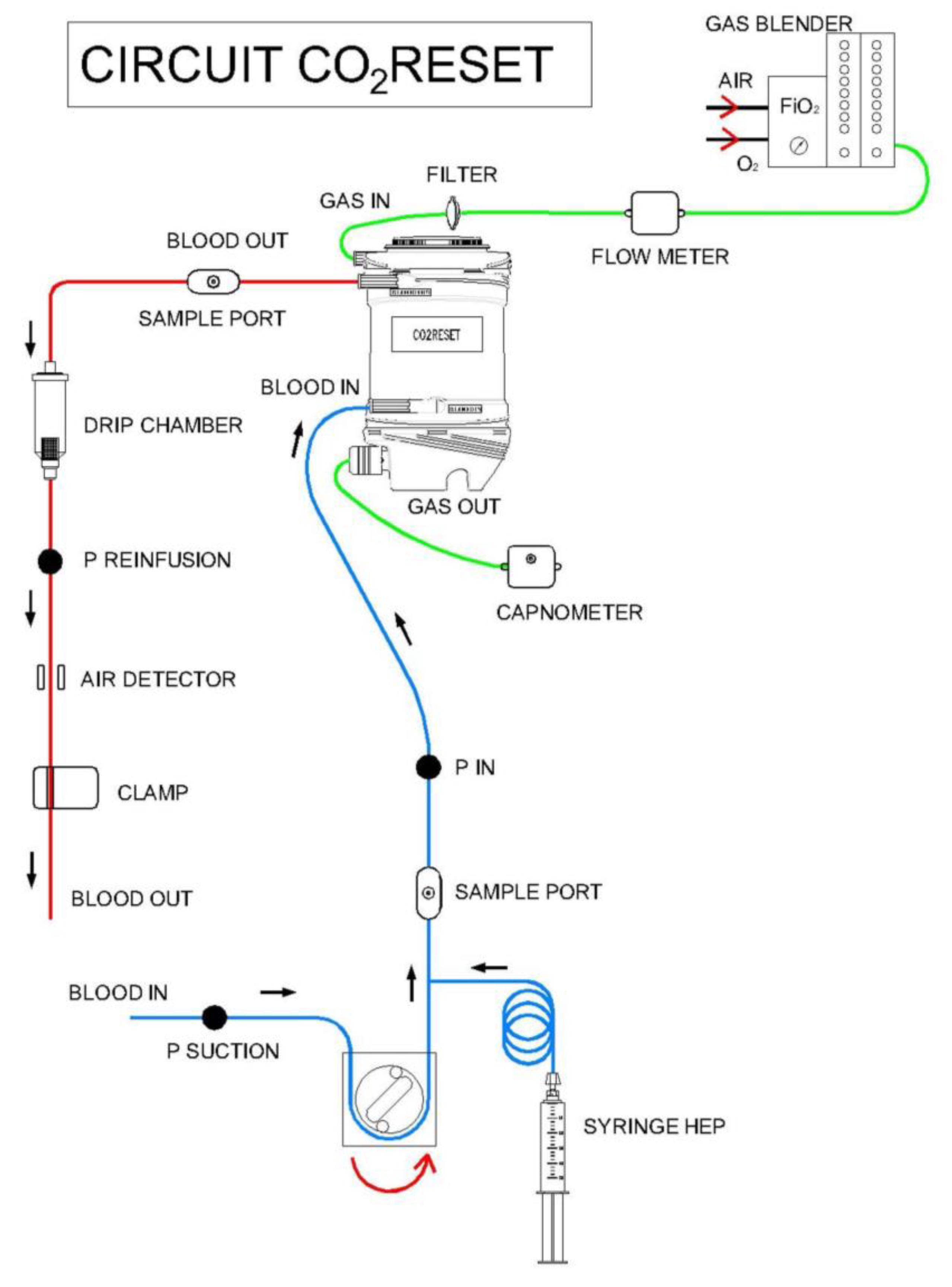
2.1. Extracorporeal CO2 Removal Technique

Medium-flow veno-venous ECCO2R was performed using the CO2RESET device (EUROSETS S.r.l., Medolla, MO, Italy). This device, driven by roller pumps, incorporates both a hemoperfusion membrane and a phosphorylcoline-coated polymethylpentene hollow fiber membrane lung (surface area = 1.8 m2), without an integrated heat exchanger. The membrane lung may be connected either to a sweep gas source of pure oxygen or to a mixture of air/oxygen to provide CO2 removal. The CO2RESET circuit is customized for single-use and can be connected to a wide range of cannulas. In our animal model, the hemoperfusion membrane was not incorporated and the device was used specifically for CO2 removal (Figure 1). The CO2RESET circuit is customized to receive only ¼ connectors and connects to either with dual lumen cannulas (13, 16, 19 French) or two single cannulas.

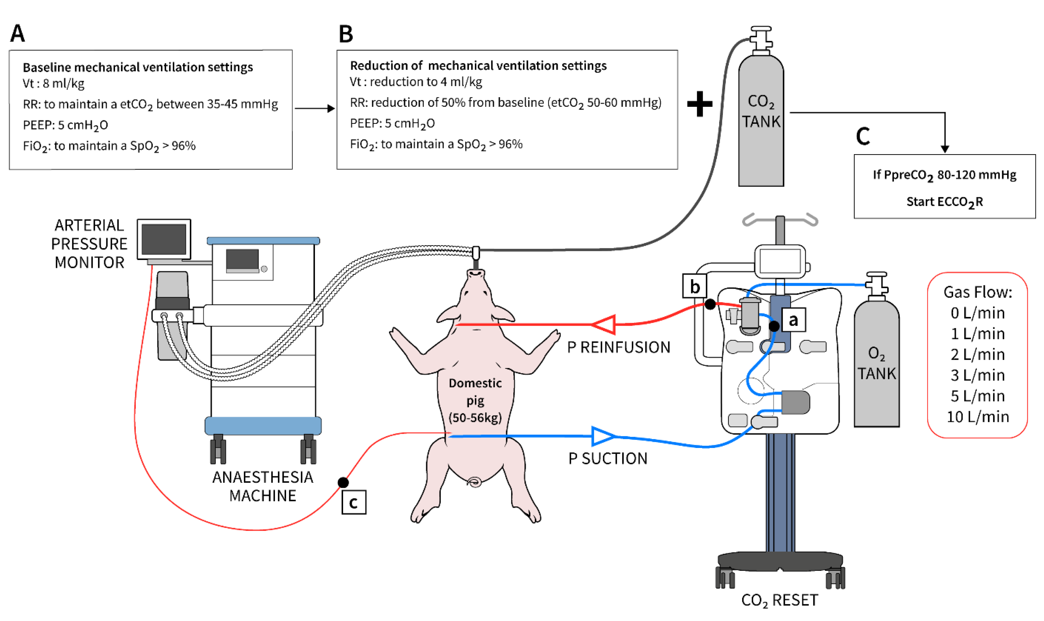
2.2. Animal Preparation

The Institutional Review Board of the Free University of Brussels (Belgium) approved the experimental protocol (number of Ethical Committee approval: 731N). On the day of the experiment, the animal (swine, Sus Scrofa Domesticus) was fasted for 12 h with free access to water. Anesthesia was initiated with a combined intramuscular injection of midazolam (1 mg/kg, Mylan, Auckland New Zeland and ketamine (100 mg/kg, Dechra, Lille, Belgium) administered in the neck and placed in supine position. The animal was monitored with a continuous electrocardiogram, and a peripheral vein (18-gauge) was inserted to provide a continuous infusion of sufentanil citrate (3 µg/kg, Janssen, Beerse, Belgium). A femoral 4.5 French (Fr) arterial catheter (Vygon, Ecouen, France) was inserted in the femoral artery and connected to a pressure transducer (True Wave, Edwards, CA, USA) for invasive arterial pressure monitoring and blood gas analysis (BGA). After a sequential intravenous injection of 1 mg atropine sulfate (Sterop, Anderlecht, Belgium), 3 µg/kg of sufentanil citrate and 1.2 mg/kg of rocuronium (Esmeron, MSD, Kenilworth, NJ, USA), an 8 mm endotracheal tube (Medtronic, Minneapolis, MN, USA) was placed and mechanical ventilation was started in controlled volume mode (Primus, Drägerwerk AG and Co, Frankfurt, Germany) with a tidal volume of 8 mL/kg, 5 cmH2O of positive end-expiratory pressure (PEEP), fraction of inspired oxygen (FiO2) of 1.0 and an inspiratory to expiratory time ratio of 1 to 2. A 1% mixture of inspired sevofluorane (Sevoflo, Abbott, Abbott Park, IL, USA) was started to achieve an expiratory percentage between 1.2 to 1.6%. Mechanical power of the respiratory system was calculated according to the validated formulas [13].
Ventilation parameters were subsequently adjusted to ensure an end tidal CO2 between 35 and 45 mmHg and SpO2 > 96%, using the minimally required FiO2. A continuous infusion of rocuronium (2–4 mg/kg/h) and sufentanil citrate (3.5 µg/kg/h) was maintained, and balanced crystalloids (PLASMA-LYTE, Baxter, Lessines, Belgium) were administered at a rate of 300–500 mL/h. A 14 Fr Foley catheter was surgically inserted to measure urine output thorough a midline incision in the lower abdomen, and the parietal layers were sutured separately. Under ultrasound guidance, a 5 Fr triple lumen central venous catheter (Arrow International, Reading, PA, USA) was placed in the right internal jugular vein and drug infusion was transferred to the distal line. For veno-venous ECCO2R, a 12 Fr multistage drainage cannula (REVAS, Free Life Medical GmbH, Aachen, Germany) was inserted in the left femoral vein and a 10 Fr return cannula (REVAS, Free Life Medical GmbH, Aachen, Germany) was inserted in the internal left jugular.
At this point, a continuous infusion of propofol (Propovet 2%, Abbott, NJ, USA) at the dose of 2–3 mg/kg was started, allowing for a progressive decrease until complete arrest of the anesthetic gas flow. The respiratory system was then opened with the removal of the soda lime absorber (Drägersorb Free, Drägerwerk AG and Co, Frankfurt, Germany). At the end of the preparation, the animal was proned and stabilized on the surgical table. For anticoagulation, heparin infusion was adjusted to achieve an activate clotting time (ACT) between 180 and 220 s, monitored via a dedicated point of care device (i-STAT, Kaolin ACT, Abbott Park, IL, USA). The estimated CO2 production in pigs at rest is about 200–250 mL/min [14], which is comparable to an adult human.

2.3. Study Design and Experiment Procedure

Considering the high reproducibility of the study and the request to avoid unnecessary use of animals from the Institutional Review Board for Animal Care (IRBAC), three healthy pigs were included in this study. After oral intubation and induction of respiratory acidosis by reducing mechanical ventilation settings, the pig was connected to the veno-venous ECCO2R device (CO2RESET, EUROSETS S.r.l., Medolla, MO, Italy).
Respiratory acidosis was induced with a 50% reduction of both the baseline tidal volume (from 8 to 4 mL/kg) and the respiratory frequency, to achieve an end tidal CO2 (etCO2) between 50 and 60 mmHg (Figure 2). To ensure a pre-membrane lung CO2 (PpreCO2) between 80–120 mmHg, an external supplementation of 1 L/min CO2 was provided to the inspiratory side of the ventilator circuit [15,16]. FiO2 was adjusted to maintain a SpO2 > 96%. A pre-membrane lung blood gas analysis (BGA) to control the achievement of this target and an arterial BGA from the animal were undertaken before starting each step of experiment. At this time, the veno-venous ECCO2R device was connected with a fixed blood flow rate of 600 mL/min and a sweep gas flow of 0 L/min (i.e., no CO2 removal capacity). All the experiments started with PpreCO2 between 80 and 120 mmHg. The experiment included six steps from 0 to 1, 2, 3, 5 and 10 L/min of sweep gas flow, respectively. Blood flow was fixed at 600 mL/min during all the steps. Each step of sweep gas flow was maintained for 30 min, and at the end, a post-membrane lung BGA and an arterial BGA from the animal were sampled and CO2 elimination (VCO2) was collected. At the end of each step, the sweep gas flows were brought to 0 L/min until the animal reached a PpreCO2 between 80 and 120 mmHg. Body temperature was maintained stable at 37 °C during the study using a warming blanket (Bair Hugger 3M, Zwijndrecht, Belgium). VCO2 was calculated directly from the device (multiplying the specific sweep gas flow for the partial pressure of the CO2 exhaled from the membrane lung) and provided in BTPS (body temperature, pressure, water vapor saturated). Operational characteristics of the ECCO2R device, including access, return and pressure drop across the membrane lung, were recorded at each step. Experiments were performed in each pig in a standardized fashion. At the end of the experiment, the animal was sacrificed by injection of 80 mEq of KCl under deep sedation.

2.4. Statistical Analysis
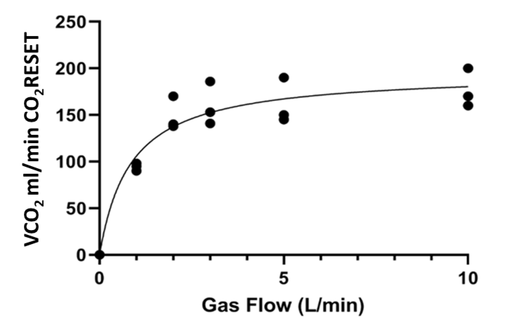
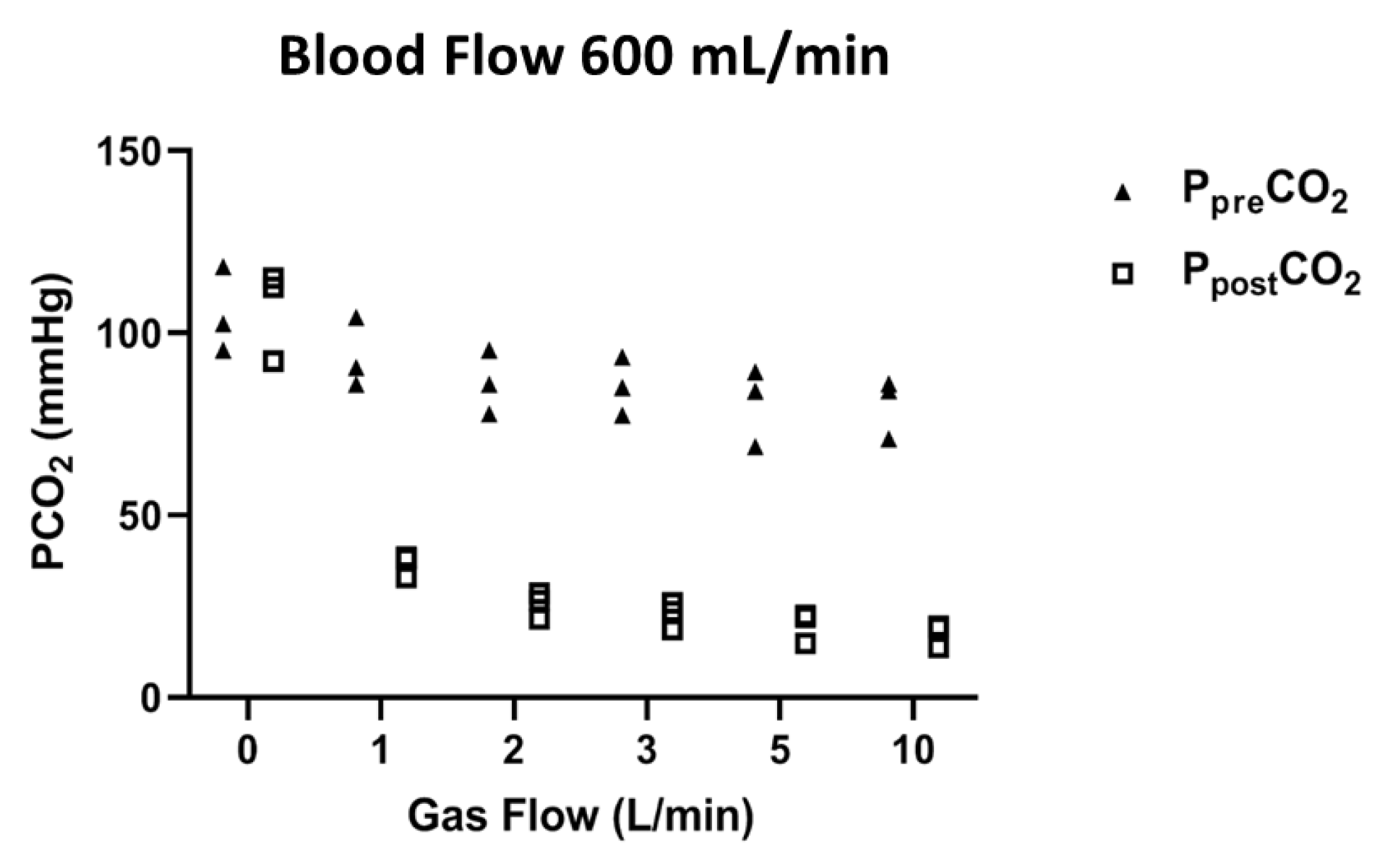
Data are expressed as median and inter-quantile ranges. Descriptive statistical analysis for non-parametric data was performed with Wilcoxon and Friedman test with Dunn’s multiple comparisons using GraphPad Prism 8 (San Diego, CA, USA). A p value < 0.05 was defined as statistically significant.

3. Results

Total duration of experiment was 6.30 (6.00–7.00) hours. Baseline characteristics of the studied animals (Table 1) were weight 54.00 (50.00–56.00) kg; static compliance of the respiratory system 34.00 (28.00–36.00) cmH2O/mL; driving pressure 13.00 (11.00–15.00) cmH2O; respiratory rate 18.00 (17.00–20.00) breaths/minute; minute ventilation 7.74 (6.80–9.00) L/min; PEEP 5.00 (5.00–5.00) cmH2O; mechanical power of the respiratory system 12.18 (10.00–13.25) Joule/minute; PaO2/FiO2 480.00 (460.00–512.00); pH 7.44 (7.35–7.48) and PaCO2 43.00 (39.00–44.00) mmHg. After the reduction of the ventilator settings and before the initiation of ECCO2R (gas flow 0 L/min), we observed (Table 1) a decrease of static compliance (28.00 (20.00–30.00) cmH2O/mL), driving pressure (8.00 (7.00–10.00) cmH2O), respiratory rate (9.00 (8.00–10.00) breaths/minute), minute ventilation (1.80 (1.72–2.24) L/min); mechanical power (1.91 (1.66–2.15) Joule/minute), pH (7.19 (7.16–7.25)) and PaO2/FiO2 (400.00 (380.00–450.00)); (p = 0.25 for all vs baseline); an increase of FiO2 (0.35 (0.25–0.40)); and p = 0.25 vs. baseline.
At the beginning of the experiments, PpreCO2 was 102.50 (95.30–118.20) mmHg and animal PaCO2 was 99.50 (88.10–105.00) mmHg. VCO2 progressively increased with an hyperbolic shape from 0 mL/min (gas flow: 0 L/min) to 90.00 (88.00–93.00) mL/min (gas flow: 1 L/min), 140.00 (138.00–170.00) mL/min (gas flow: 2 L/min), 153.00 (141.00–186.00) mL/min (gas flow: 3 L/min), 150.00 (145.00–190.00) mL/min (gas flow: 5 L/min) and 170 (160.00–200.00) mL/min (gas flow: 10 L/min); p < 0.001, (Figure 3 and Figures S1–S3). VCO2 did not significantly increase during each step of increase of the sweep gas flow (p > 0.99, respectively, with Dunn’s multiple comparisons test). At the end of each step, animal PaCO2 was 71.80 (67.90–84.20) with 1 L/min gas flow, 69.50 (58.80–80.00) with 2 L/min gas flow, 68.50 (56.80–75.60) with 3 L/min, 66.70 (52.60–74.80) with 5 L/min and 66.00 (56.50–69.10) with 10 L/min (p = 0.25 respectively vs. baseline).
PpreCO2 and PpostCO2 at each step of the experiment are shown in Figure 4. Decrease of PpostCO2 at each step of the experiment was not significant (p > 0.99, p = 0.25, p = 0.25, p = 0.25, p = 0.25, p = 0.25, respectively). Operational characteristics of the ECCO2R device are reported in Table 2. During the experiments, the animals did not report any problem of bleeding or clotting. Pressure drops across membrane lung were 32.00 (30.00–34.00) mmHg. Median platelet count, fibrinogen and D-dimer levels remained stable during all the experiments (213.00 (185.00–230.00) cells/mm3, 180.00 (150.00–210.00) mg/dL and 215.00 (180.00–350.00) ng/mL, respectively). Levels of free plasma hemoglobin were undetectable (within normal < 50 mg/dL).

4. Discussion

The main finding of the present porcine study is that the use of a high-surface membrane lung may substantially impact CO2 elimination with a stepwise increase of the sweep gas flow rate despite using a fixed relatively blood flow rate (i.e., 600 mL/min). Pressure drops using these settings were within normal ranges. Although the experiment was conducted in a small cohort of healthy animals and with a limited time, no complications or technical issues were reported during the short observational period.
From a technical point of view, CO2 elimination may be manipulated at the bedside by modifying the blood flow rate, the sweep gas flow or the size of the membrane lung, or by acidifying the blood before the membrane lung (increase of PpreCO2). Animal data conflict with one another. Karagiannidis et al. [11,12] demonstrated, using a porcine model, that a blood flow rate > 1 L/min with a membrane lung with a surface area > 0.8 m2 may remove the 50% of total CO2 production and correct severe respiratory acidosis. Contrarily, Duscio et al. [10] reported a very high VCO2 (171 mL/min) with a low blood flow (400 mL/min) and a high surface membrane lung (1.8 m2).
Our results are in between the previous findings of Karagiannidis et al. [11,12] and Duscio et al. [10]. In our porcine model, using a fixed blood flow rate of 600 mL/min and a high surface membrane lung (1.8 m2), we reported a CO2 elimination of 170.00 (160.00–200.00) mL/min using 10 L/min of sweep gas starting with a PpreCO2 around 102.50 (95.30–118.20). Furthermore, CO2 elimination progressively increased when increasing the gas flow from 0 to 2 L/min and reached a plateau at sweep gas flows higher than 3–4 L/min, in line with other previous studies [11,16].
Using this configuration, our VCO2 was similar to the ones reported by Karagiannidis et al. [11], which used the same range of PpreCO2, higher blood flow rates (1 L/min) and smaller membrane lungs (0.8–1.3 m2), similar to the ones of Duscio et al. [10], which used a lower blood flood (400 mL/min) with a very high membrane surface area (1.8 m2), and higher than the ones reported by Hospach et al. [15], which used a blood flow of 600 mL/min with the PrismaLung (Baxter, Lessines, Belgium) and the A.L.ONE (EUROSETS S.r.l., Medolla, MO, Italy) membrane lungs (0.80 and 1.35 m2, respectively).
The use of low/medium-flow ECCO2R devices with this setting may have some advantages compared to high-flow ECCO2R devices. First, low/medium-flow ECCO2R are generally driven by roller pumps and have been developed specifically to remove CO2 [15]. High-flow devices are generally centrifugal pumps designed for higher blood flows and used for extracorporeal membrane oxygenation (ECMO). When these ECMO pumps are adapted to work at a lower blood flow rates and with smaller membrane lungs (neonatal or pediatric), these “adjustments” are not free of risks and may induce platelets activation and/or destruction as well as hemolysis, due to the reduction of the hydraulic efficiency and the increase of both pump recirculation rate and shear stress [17]. Second, circuit priming is fast and similar to the ones used for renal replacement therapy (RRT); furthermore, it does not require dedicated specialists as for ECMO (i.e., perfusionists). Third, low/medium-flow ECCO2R devices can integrate other organ support techniques (i.e., RRT) to promptly manage the patients with both acute respiratory failure and acute kidney injury/fluid overload. Together with these potential advantages, some challenges exist for low/medium-flow ECCO2R devices. First, to provide adequate level of VCO2 using a low blood flow, they require a large membrane surface area [10]. The interaction between a large membrane surface area and a low blood flow may induce the development of areas of blood stagnation, increasing the risk of thrombotic complications (“circuit” diffuse intravascular coagulation) and secondary hemolysis. Second, low/medium-flow ECCO2R devices require an external heating system to maintain body temperature within normal ranges.
This study presents some limitations. First, due to the ethical concerns raised by our IRBAC, few animals have been included; thus, our data cannot be directly transferred to humans with acute respiratory distress syndrome (ARDS) or decompensated COPD. Further, the blood and the interstitial fluid account only for less than 20% of the total human CO2 stores that can be mobilized within 48 h [18]. Thus, adding CO2 with an external source for a limited amount of time is not the most accurate strategy to increase CO2 storages [18]. However, we adopted this approach, previously used by other authors [15,16] to rapidly increase PpreCO2, avoiding the reduction of tidal volume (<4 mL/kg) and respiratory rate to unsafe levels that could have required an adjustment of the ventilator settings.
Second, only one fixed blood flow (600 mL/min) was used in our experiments, although the CO2 elimination is known to increase with the increase of blood flow; the operating range of CO2RESET is wider with a maximum of 800 mL/min. However, our purpose was to maintain a blood flow that was comparable with the ones described in other studies [15,17]. Of note, we preferred not to use lower blood flows (i.e., 400 mL/min) [10], to avoid the use of higher ACT (300 sec) for anticoagulation [1].
Third, our study has been designed to maintain a wide range of high PpreCO2 and consequently provides high VCO2. This wide range, even though not common when using ECCO2R to prevent VILI in ARDS patients, has also been chosen to test this new ECCO2R device in extreme clinical situations such as near fatal asthma and acute exacerbation of COPD or end-stage lung diseases awaiting a lung transplant [7].
Fourth, even though we did not report any mechanical complications (bleeding, clotting, air embolism, circuit failure or hemolysis) during the three experiments, we cannot draw any clinical conclusions since the duration of our experiments was limited.

5. Conclusions

Medium-flow ECCO2R devices with a blood flow of 600 mL/min and a high surface membrane lung (1.8 m2) provided high VCO2 using moderate sweep gas flows (i.e., >2 L/min) in an experimental swine model with healthy lungs. Future clinical data from these devices would provide further information on this approach in a human setting.

Supplementary Materials

The following are available online at https://www.mdpi.com/2077-0375/11/1/8/s1, Figure S1: VCO2 removal at different gas flow (Pig 1), Table S1: Pig 1: main physiologic variables at baseline (Time 1) and at the beginning of the experiment (Time 2), Figure S2: VCO2 removal at different gas flow (Pig 2), Table S2: Pig 2: main physiologic variables at baseline (Time 1) and at the beginning of the experiment (Time 2), Figure S3: VCO2 removal at different gas flow (Pig 3), Table S3: Pig 3: main physiologic variables at baseline (Time 1) and at the beginning of the experiment (Time 2).

Author Contributions

F.S.T., F.A. and F.S. performed the animal experiments. M.B., F.S.T. and M.D.N. conceived and designed the experimental layout; all authors evaluated and analyzed the data, supported the writing of the manuscript and studied and discussed the literature and experimental results. All authors have read and agreed to the published version of the manuscript.

Funding

EUROSETS srl provided the ECCO2R device and cannula to support the study.

Acknowledgments

We are thankful for Roxanne Ziman, MSc, medical illustrator from the University of Toronto, for the support provided to develop Figure 2.

Conflicts of Interest

Matteo Di Nardo, Mirko Belliato, Roberto Lorusso, Lars Mikael Broman, Maximilian Malfertheiner and Fabio Silvio Taccone are on the Advisory Board of EUROSETS srl.

References

  1. Combes, A.; Fanelli, V.; Pham, T.; Ranieri, V.M.; On behalf of the European Society of Intensive Care Medicine Trials Group and the “Strategy of Ultra-Protective Lung Ventilation with Extracorporeal CO2 Removal for New-Onset Moderate to Severe ARDS” (SUPERNOVA) Investigators. Feasibility and safety of extracorporeal CO2 removal to enhance protective ventilation in acute respiratory distress syndrome: The SUPERNOVA study. Intensiv. Care Med. 2019, 45, 592–600. [Google Scholar] [CrossRef] [PubMed] [Green Version]
  2. Combes, A.; Tonetti, T.; Fanelli, V.; Pham, T.; Pesenti, A.; Mancebo, J.; Brodie, D.; Ranieri, V.M. Efficacy and safety of lower versus higher CO2 extraction devices to allow ultraprotective ventilation: Secondary analysis of the SUPERNOVA study. Thorax 2019, 74, 1179–1181. [Google Scholar] [CrossRef] [PubMed]
  3. Del Sorbo, L.; Pisani, L.; Filippini, C.; Fanelli, V.; Fasano, L.; Terragni, P.; Dell’Amore, A.; Urbino, R.; Mascia, L.; Evangelista, A.; et al. Extracorporeal Co2 Removal in Hypercapnic Patients at Risk of Noninvasive Ventilation Failure. Crit. Care Med. 2015, 43, 120–127. [Google Scholar] [CrossRef] [PubMed]
  4. Morelli, A.; Del Sorbo, L.; Pesenti, A.; Ranieri, V.M.; Fan, E. Extracorporeal carbon dioxide removal (ECCO2R) in patients with acute respiratory failure. Intensiv. Care Med. 2017, 43, 519–530. [Google Scholar] [CrossRef] [PubMed]
  5. Schmidt, M.; Jaber, S.; Zogheib, E.; Godet, T.; Capellier, G.; Combes, A. Feasibility and safety of low-flow extracorporeal CO2 removal managed with a renal replacement platform to enhance lung-protective ventilation of patients with mild-to-moderate ARDS. Crit. Care 2018, 22, 122. [Google Scholar] [CrossRef] [PubMed] [Green Version]
  6. Braune, S.; Sieweke, A.; Brettner, F.; Staudinger, T.; Joannidis, M.; Verbrugge, S.; Frings, D.; Nierhaus, A.; Wegscheider, K.; Kluge, S. The feasibility and safety of extracorporeal carbon dioxide removal to avoid intubation in patients with COPD unresponsive to noninvasive ventilation for acute hypercapnic respiratory failure (ECLAIR study): Multicentre case–control study. Intensiv. Care Med. 2016, 42, 1437–1444. [Google Scholar] [CrossRef] [PubMed]
  7. Pisani, L.; Polastri, M.; Pacilli, A.M.G.; Nava, S. Extracorporeal Lung Support for Hypercapnic Ventilatory Failure. Respir. Care 2018, 63, 1174–1179. [Google Scholar] [CrossRef] [PubMed]
  8. Zanella, A.; Castagna, L.; Salerno, D.; Scaravilli, V.; Deab, S.A.E.A.E.S.; Magni, F.; Giani, M.; Mazzola, S.; Albertini, M.; Patroniti, N.; et al. Respiratory Electrodialysis. A Novel, Highly Efficient Extracorporeal CO2Removal Technique. Am. J. Respir. Crit. Care Med. 2015, 192, 719–726. [Google Scholar] [CrossRef] [PubMed] [Green Version]
  9. Cove, M.E.; Vu, L.H.; Ring, T.; Federspiel, W.J.; Kellum, J.A. Respiratory Dialysis-A Novel Low Bicarbonate Dialysate to Provide Extracorporeal CO2 Removal. Crit. Care Med. 2020, 48, e592–e598. [Google Scholar] [CrossRef] [PubMed]
  10. Duscio, E.; Cipulli, F.; Vasques, F.; Collino, F.; Rapetti, F.; Romitti, F.; Behnemann, T.; Niewenhuys, J.; Tonetti, T.; Pasticci, I.; et al. Extracorporeal CO2 Removal. Crit. Care Med. 2019, 47, 33–40. [Google Scholar] [CrossRef] [PubMed]
  11. Karagiannidis, C.; Strassmann, S.; Brodie, D.; Ritter, P.; Larsson, A.; Borchardt, R.; Windisch, W. Impact of membrane lung surface area and blood flow on extracorporeal CO2 removal during severe respiratory acidosis. Intensiv. Care Med. Exp. 2017, 5, 1–13. [Google Scholar] [CrossRef] [PubMed] [Green Version]
  12. Strassmann, S.; Merten, M.; Schäfer, S.; De Moll, J.; Brodie, D.; Larsson, A.; Windisch, W.; Karagiannidis, C. Impact of sweep gas flow on extracorporeal CO2 removal (ECCO2R). Intensiv. Care Med. Exp. 2019, 7, 17. [Google Scholar] [CrossRef] [PubMed]
  13. Tonetti, T.; Vasques, F.; Rapetti, F.; Maiolo, G.; Collino, F.; Romitti, F.; Camporota, L.; Cressoni, M.; Cadringher, P.; Quintel, M.; et al. Driving pressure and mechanical power: New targets for VILI prevention. Ann. Transl. Med. 2017, 5, 286. [Google Scholar] [CrossRef] [PubMed] [Green Version]
  14. Van Milgen, J.; Noblet, J.; Dubois, S.; Bernier, J.-F. Dynamic aspects of oxygen consumption and carbon dioxide production in swine. Br. J. Nutr. 1997, 78, 397–410. [Google Scholar] [CrossRef] [PubMed] [Green Version]
  15. Hospach, I.; Goldstein, J.; Harenski, K.; Laffey, J.G.; Pouchoulin, D.; Raible, M.; Votteler, S.; Storr, M. In vitro characterization of PrismaLung+: A novel ECCO2R device. Intensiv. Care Med. Exp. 2020, 8, 1–18. [Google Scholar] [CrossRef] [PubMed]
  16. Barrett, N.A.; Hart, N.; Camporota, L. In vivo carbon dioxide clearance of a low-flow extracorporeal carbon dioxide removal circuit in patients with acute exacerbations of chronic obstructive pulmonary disease. Perfusion 2020, 35, 436–441. [Google Scholar] [CrossRef] [PubMed]
  17. Gross-Hardt, S.; Hesselmann, F.; Arens, J.; Steinseifer, U.; Vercaemst, L.; Windisch, W.; Brodie, D.; Karagiannidis, C. Low-flow assessment of current ECMO/ECCO2R rotary blood pumps and the potential effect on hemocompatibility. Crit. Care 2019, 23, 348. [Google Scholar] [CrossRef] [PubMed] [Green Version]
  18. Giosa, L.; Busana, M.; Bonifazi, M.; Romitti, F.; Vassalli, F.; Pasticci, I.; Macrì, M.M.; D’Albo, R.; Collino, F.; Gatta, A.; et al. Mobilizing Carbon Dioxide Stores: An Experimental Study. Am. J. Respir. Crit. Care Med. 2020. [Google Scholar] [CrossRef] [PubMed]
Figure 1. Schematic representation of the CO2RESET device. P = pressure.
Membranes 11 00008 g001
Figure 2. Diagram showing the experiment steps (AC). a: sampling site pre-membrane lung; b: sampling site post-membrane lung; and c: arterial sampling site.
Membranes 11 00008 g002
Figure 3. The relationship between sweep gas flow rate and the median CO2 elimination (VCO2) measured by the CO2RESET device with a fixed blood flow rate of 600 mL/min.
Membranes 11 00008 g003
Figure 4. Median pre and post-membrane PCO2 under different sweep gas flow conditions (0, 1, 2, 3, 5 and 10 L/min) and a fixed blood flow rate of 600 mL/min.
Membranes 11 00008 g004
Table 1. Main physiologic variables at baseline (Time 1) and at the beginning of the experiment (Time 2).
Time 1Time 2
Respiratory rate (breaths/min)18 (17–20)9 (8–10)
Tidal volume-pig (mL)430.00 (400.00–450.00)216.00 (200.00–224.00)
Minute ventilation (L/min)7.74 (6.80–9.00)1.80 (1.72–2.24)
Positive end-expiratory pressure (cmH2O)5.00 (5.00–5.00)5.00 (5.00–5.00)
Compliance respiratory system (cmH2O)34.00 (28.00–36.00)28.00 (20.00–30.00)
Respiratory system mechanical power (J/min)12.18 (10.00–13.25)1.91 (1.66–2.15)
Heart rate (beats/min)73 (65–85)77 (70–81)
Central venous pressure (mmHg)8.00 (6.00–10.00)9.00 (6.00–10.00)
Mean systemic arterial pressure (mmHg)92 (88–100)93 (87–99)
Arterial lactates (mmol/L)1.20 (0.90–1.60)1.35 (0.88–1.55)
Table 2. Operational characteristics of CO2RESET in the three treated pigs.
CharacteristicsValue
Access pressure (mmHg)−7.00 (−10.50–−3.50)
Pre-membrane pressure (mmHg)40.00 (46.75–69.50)
Post-membrane pressure (mmHg)17.00 (15.75–32.00)
Δ pressure (mmHg)32.00 (30.00–34.00)
Activate Clotting Time187.00 (184.00–191.00)
Publisher’s Note: MDPI stays neutral with regard to jurisdictional claims in published maps and institutional affiliations.

