Article

Fusion Cytokines IL-7-Linker-IL-15 Promote Mycobacterium Tuberculosis Subunit Vaccine to Induce Central Memory like T Cell-Mediated Immunity

1
Gansu Provincial Key Laboratory of Evidence Based Medicine and Clinical Translation & Lanzhou Center for Tuberculosis Research, School of Basic Medical Sciences, Lanzhou University, Lanzhou 730000, China
2
Institute of Pathogen Biology, School of Basic Medical Sciences, Lanzhou University, Lanzhou 730000, China
*
Author to whom correspondence should be addressed.
Vaccines 2020, 8(4), 715; https://doi.org/10.3390/vaccines8040715
Submission received: 31 October 2020 / Revised: 25 November 2020 / Accepted: 28 November 2020 / Published: 1 December 2020
(This article belongs to the Special Issue Vaccines and Other Antibody Therapies Against Bacterial Infection)

Abstract

:
Tuberculosis (TB), caused by Mycobacterium tuberculosis (M. tuberculosis), is among the most serious infectious diseases worldwide. Adjuvanted protein subunit vaccines have been demonstrated as a kind of promising novel vaccine. This study proposed to investigate whether cytokines interliukine-7 (IL-7) and interliukine-15 (IL-15) help TB subunit vaccines induce long-term cell-mediated immune responses, which are required for vaccination against TB. In this study, mice were immunized with the M. tuberculosis protein subunit vaccines combined with adnovirus-mediated cytokines IL-7, IL-15, IL-7-IL-15, and IL-7-Linker-IL-15 at 0, 2, and 4 weeks, respectively. Twenty weeks after the last immunization, the long-term immune responses, especially the central memory-like T cells (TCM like cell)-mediated immune responses, were determined with the methods of cultured IFN-γ-ELISPOT, expanded secondary immune responses, cell proliferation, and protective efficacy against Mycobacterium bovis Bacilli Calmette-Guerin (BCG) challenge, etc. The results showed that the group of vaccine + rAd-IL-7-Linker-IL-15 induced a stronger long-term antigen-specific TCM like cells-mediated immune responses and had higher protective efficacy against BCG challenge than the vaccine + rAd-vector control group, the vaccine + rAd-IL-7 and the vaccine + rAd-IL-15 groups. This study indicated that rAd-IL-7-Linker-IL-15 improved the TB subunit vaccine’s efficacy by augmenting TCM like cells and provided long-term protective efficacy against Mycobacteria.

1. Introduction

Tuberculosis (TB), mainly caused by Mycobacterium tuberculosis (M. tuberculosis), ranks as the first leading cause of death from a single infectious disease worldwide [1]. The effective way to prevent and control the infectious disease is vaccination. A potentially successful M. tuberculosis vaccine should have the ability to induce more long-term antigen-specific immune memory cells, which could expand rapidly when it encounters the same pathogen. Currently, attenuated Mycobacterium bovis Bacilli Calmette-Guerin (BCG) is the only approved TB vaccine in clinic. However, it provides variable protection against TB [2,3]. There are reports that BCG vaccination mainly induces shorter-term effector memory T cells (TEM) rather than long-lived central memory T cells (TCM) [4,5]. Thus, it is urgently required to develop novel vaccines for inducing enough TCM to produce long-term protection against M. tuberculosis [6].
The protein subunit vaccine is a kind of promising TB vaccine. So far, there are at least 14 TB vaccines in clinical trials, including four protein subunit vaccines, such as ID93 + GLA-SE, H1/H56 + IC31, H4 + IC31, and M72 + AS01E [7]. Protein subunit vaccines require an adjuvant to induce a stronger immune response. Adjuvants can have an effect on the strength and duration of immune responses [8]. We have developed an adjuvant composed of N,N’-dimethyl-N,N’-dioctadecylammonium bromide (DDA), and polyinosinic-polycytidylic acid [Poly (I:C)] (DP for short), which could help the TB subunit vaccine induce strong Th1-type cell-mediated immunity [9]. Considering that an effective TB vaccine is needed to induce long-term immune memory mediated by TCM rather than TEM [10,11], there is also need to survey how to promote the development of TCM during vaccination.
IL-7 and IL-15 are both members of the γ-chain cytokine family, which share γ-chains of IL-2R for signal transduction and play a role in immune memory. Some studies showed that IL-7 and IL-15 were essential for development and maintenance of memory T cells [12,13,14]. IL-7 could regulate T cell homeostasis and enhance the survival of memory T cells [15,16]. While IL-15 could promote the differentiation of CD8+ memory T cells [17]. IL-7 and IL-15 had been proved to enhance the immune memory induced by many vaccines, such as vaccines against Toxoplasma gondii [18,19], HIV-1 vaccine [20], and BCG against M. tuberculosis [21]. However, it is still uncertain if both IL-7 and IL-15 promote the development of TCM and enhance the protective efficacy of the vaccines. For example, it was reported that the administration of IL-15 increased antigen-specific CD8+ memory T cells after BCG infection, but the protective efficacy against M. tuberculosis was not improved [22]. Thus, it is necessary to explore the immune memory character induced by IL-7 and IL-15 combined with vaccines. In our study, we co-administrated M. tuberculosis subunit vaccine ESAT6-Ag85B-MPT64<190-198>-Mtb8.4-Rv2626c (LT70 for short) [23] and Mtb10.4-HspX (MH for short) [24] in DP adjuvant, which showed stronger protective efficacy in mice, with rAd-IL-7, rAd-IL-15, rAd-IL-7-IL-15 and rAd-IL-7-Linker-IL-15 to investigate the properties of T cell immune.

2. Materials and Methods

2.1. Ethics Statement

Mice procedures were performed in accordance with the guidelines of China Council on Animal Care and Use. Animal license numbers SCXK (Gan) 2018-0002. The experiments were performed under isoflurane anesthesia to be made to minimize suffering.

2.2. Mice

Briefly, female 6–8-week old C57BL/6 mice were obtained from animal center of Lanzhou University (Lanzhou, China). All mice were maintained in special pathogen-free conditions in Lanzhou University and received free access to water and food throughout the study.

2.3. Preparation of Subunit Vaccines and Single Mycobacterial Antigens

The fusion proteins LT70 [23] and MH [24] were purified as previously described. Briefly, the plasmid encoding LT70 and MH were cloned into pET30(+) expression vector, respectively. Then, they were transformed into the Escherichia coli BL21 strain (DE3) for fusion proteins LT70 and MH from supernatant. Single mycobacterial proteins ESAT6, Ag85B, Rv2626c, and HspX were produced by Ni-NTA His column (Novagen) as previously indicated [25]. The purified LT70 and MH (10 μg/dose, respectively) were emulsified in adjuvant of DDA (250 μg/dose) and [Poly (I:C)] (50 μg/dose) with PBS in a volume of 200 μL for protein subunit vaccine (vaccine for short).

2.4. Construction of rAd-IL-7, rAd-IL-15, rAd-IL-7-IL-15, rAd-IL-7-Linker-IL-15 and rAd-Vector

The mouse IL-7 and IL-15 gene sequences were sub-cloned into shuttle plasmid pDC316, respectively. Subsequently, pDC316, pDC316-IL-7, pDC316-IL-15, pDC316-IL-7-IL-15, and pDC316-IL-7-Linker-IL-15, which the linker (Gly-Gly-Gly-Ser)3 [26] was connected between IL-7 and IL-15, combined with pBHGlox△E1, 3Cre adenovirus expression vector co-transfected into HEK293 cells by homologous recombination. The recombinant viruses of rAd-IL-7, rAd-IL-15, rAd-IL-7-IL-15, rAd-IL-7-Linker-IL-15, and the recombined adenoviral vector (rAd-vector), were verified with PCR analysis and cytopathic effect (CPE). The amplification of the recombinant virus was done in HEK293 cells. Adenoviral titers were determined as previously described [27].

2.5. Vaccine Immunization

Mice were divided into eight groups: The non-vaccinated mice received PBS; The vaccinated mice were topically immunized with vaccine, either co-administration of rAd-vector, rAd-IL-7, rAd-IL-15, rAd-IL-7-IL-15, rAd-IL-7-Linker-IL-15 or rAd-IL-7 + rAd-IL-15, respectively. For the group of PBS, mice were inoculated with PBS in a total volume of 200 μL/dose subcutaneously once at 0 week. For the other groups, vaccinations were performed at weeks 0, 2 and 4, respectively. Mice were immunized by the vaccine subcutaneously in a total volume of 200 μL/dose/mice on one side of the groin. At 30 min after protein vaccine immunization, 5 × 10⁶ PFU/100 μL/mice of rAd-vector, rAd-IL-7, rAd-IL-15, rAd-IL-7-IL-15, rAd-IL-7-Linker-IL-15, or rAd-IL-7 + rAd-IL-15, respectively were injected at the same site.

2.6. Cultured IFN-γ ELISPOT assay In Vitro

Cultured IFN-γ ELISPOT assay was done as described previously [28]. Briefly, firstly lymphocytes (2 × 10⁶ cells/mL/well) were stimulated with a cocktail of ESAT-6, Ag85B, Rv2626c and HspX (2 μg/mL of each protein) in a 24-well plate. Media was replaced containing 100 U/mL IL-2 at days 3 and 7. At day 9, cultured lymphocytes were harvested. Then, cultured cells (1 × 10⁶ cultured cells/well) in the presence of additional antigen presenting cells (APCs) isolated freshly from C57BL/6 mice spleen at the ratio of 10:1 were restimulated with the same antigens and incubated in anti-mouse IFN-γ capture-mAb coated 96-well ELISPOT tech Company Limited, Shenzhen, China) either a cocktail of ESAT-6, Ag85B, Rv2626c, PPD or medium alone for an additional 20 h in the standard ELISPOT assay as the manufacturer’s protocols [28].

2.7. EdU Incorporation and Proliferation Assay

Lymphocytes (5 × 10⁶ cells/well) were stimulated with mixed antigens of ESAT-6, Ag85B, Rv2626c and HspX (2 μg/mL of each protein) in 24-well plates and at days 3 after antigen stimulation with EdU at a final concentration of 30 μM were added to the cells for another 4 days. At days 7, cells were harvested and treated with Click-iT reaction buffer (Cat. no. C10425, Click-iT™ EdU Flow Cytometry Assay Kit, Invitrogen™, Carlsbad, CA, USA) according to the manufacturer’s instructions. Then, cells were stained with anti-CD4-PE (RM4-5, eBioscience, San Diego, CA, USA) and anti-CD8-APC (53-6.7, BD, USA) [28]. Finally, samples were detected by flow cytometry.

2.8. IFN-γ Secretion Following Twice-Stimulation with Antigens

IFN-γ secretion following twice-stimulation with antigens in vivo and in vitro sequentially was performed [28]. Firstly, mice were stimulated with BCG (Danish 1331, 1 × 10⁶ CFU/dose) by intraperitoneal injection (i.p.) at 20 weeks after the final immunization. Nine days later, lymphocytes were isolated and stimulated with mixed antigens of ESAT-6, Ag85B, Rv2626c and HspX (2 μg/mL of each protein) for 4 h in vitro. Then, cells were incubated for 5–6 h with BD GolgiPlug™ (including brefeldin A, BD, USA) at 37 °C. Subsequently, cells were stained with anti-CD4-FITC (RM4-5, BD, USA) and anti-CD8-PerCP-Cy5.5 (53-6.7, BD, USA) at 4 °C for 30 min. Later, cells were permeabilized (Cytofix/Cytoperm kit, BD, USA) and intracellular cytokine (ICC) staining of anti-IFN-γ (XMG1.2, BD, USA) was performed at 4 °C for 30 min as previous reported [29]. All samples were run on ACEA NoveCyte.

2.9. TB10.44-12 Pentamer Staining

Mice were treated with BCG (Danish 1331, 1 × 10⁶ CFU/dose) by i.p. After 9 days, lymphocytes were isolated and stained with TB10.44-12 pentamer-PE (Pro5® MHC Class I Pentamers, Pro Immune, Pro5® MHC Class I Pentamers, Pro Immune) for TB10.4 specific CD8+ memory T cells at 22 °C for 10 min. Subsequently, cells were stained with anti-CD3-FITC (145-2C11, BD, USA) and anti-CD8-APC (53-6.7, BD, USA) at 4 °C for 30 min. Finally, samples were detected by flow cytometry.

2.10. BCG Challenge and Enumeration of Bacteria-Load

Mice were challenged with BCG (Danish 1331) at 1 × 10⁷ CFU/dose by intranasal route (i.n.). At 20 days later, bacterial colony forming units (CFU) in the lungs and spleens were detected. The homogenates of lungs and spleens were plated at 10-fold serial dilutions on Middlebrook 7H11 medium (BD, NJ, USA) and incubated at 37 °C for 3 weeks. Finally, CFU was enumerated.

2.11. Statistical Analysis

The results were presented as means ± SD. Comparisons were analyzed by one-way ANOVA and SPSS17.0 software. Data were considered as statistically significant at p < 0.05.

3. Results

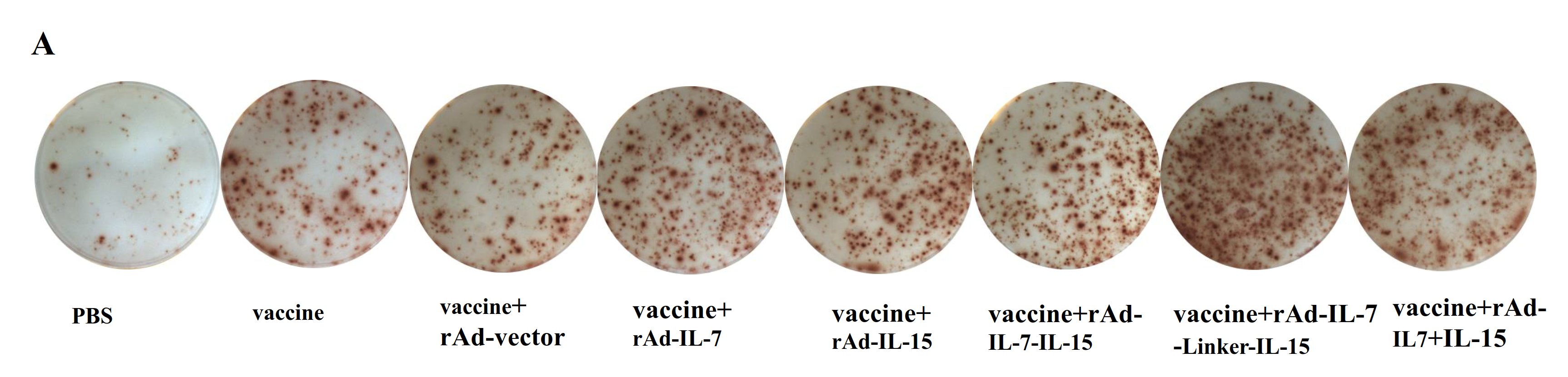
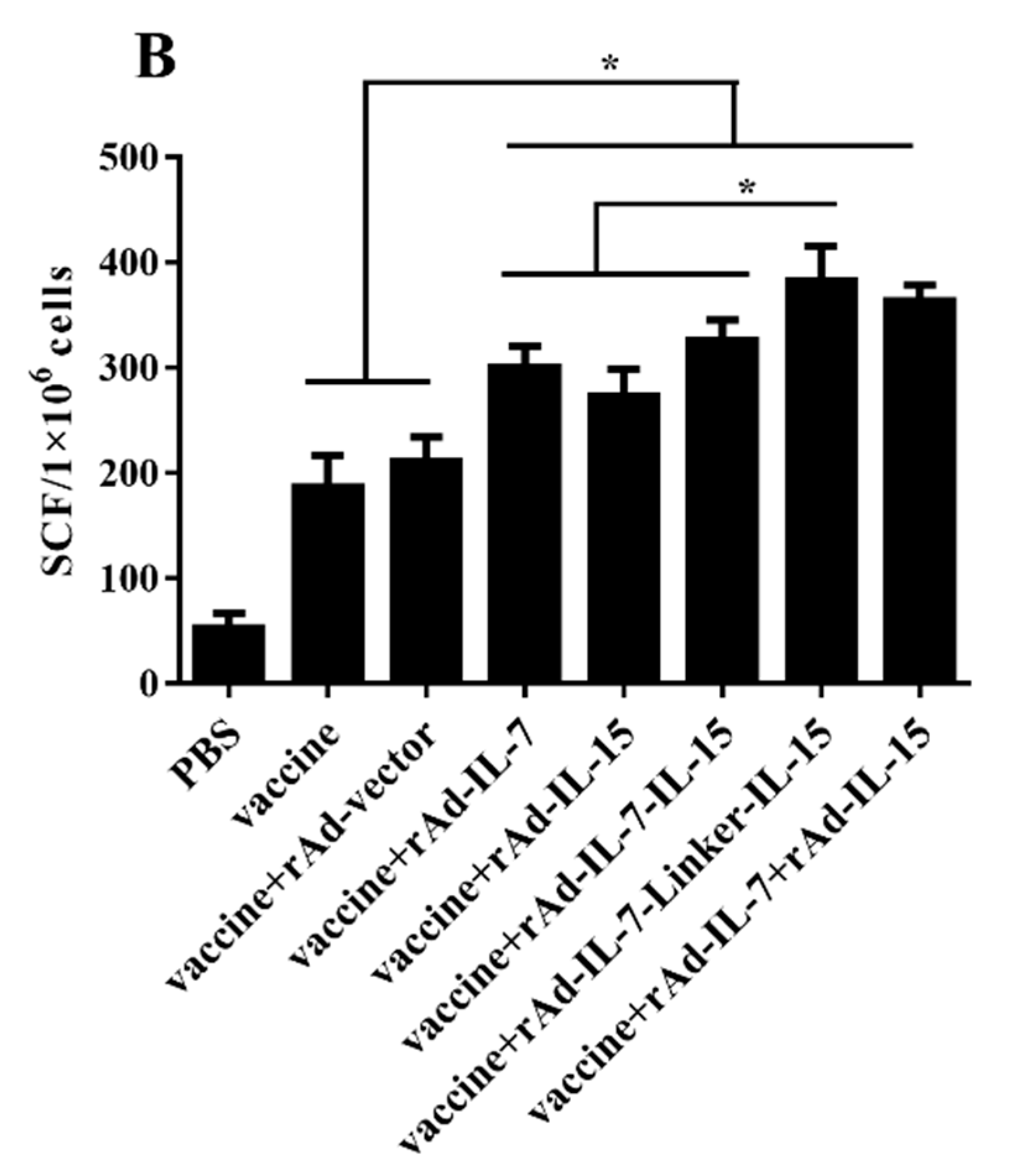
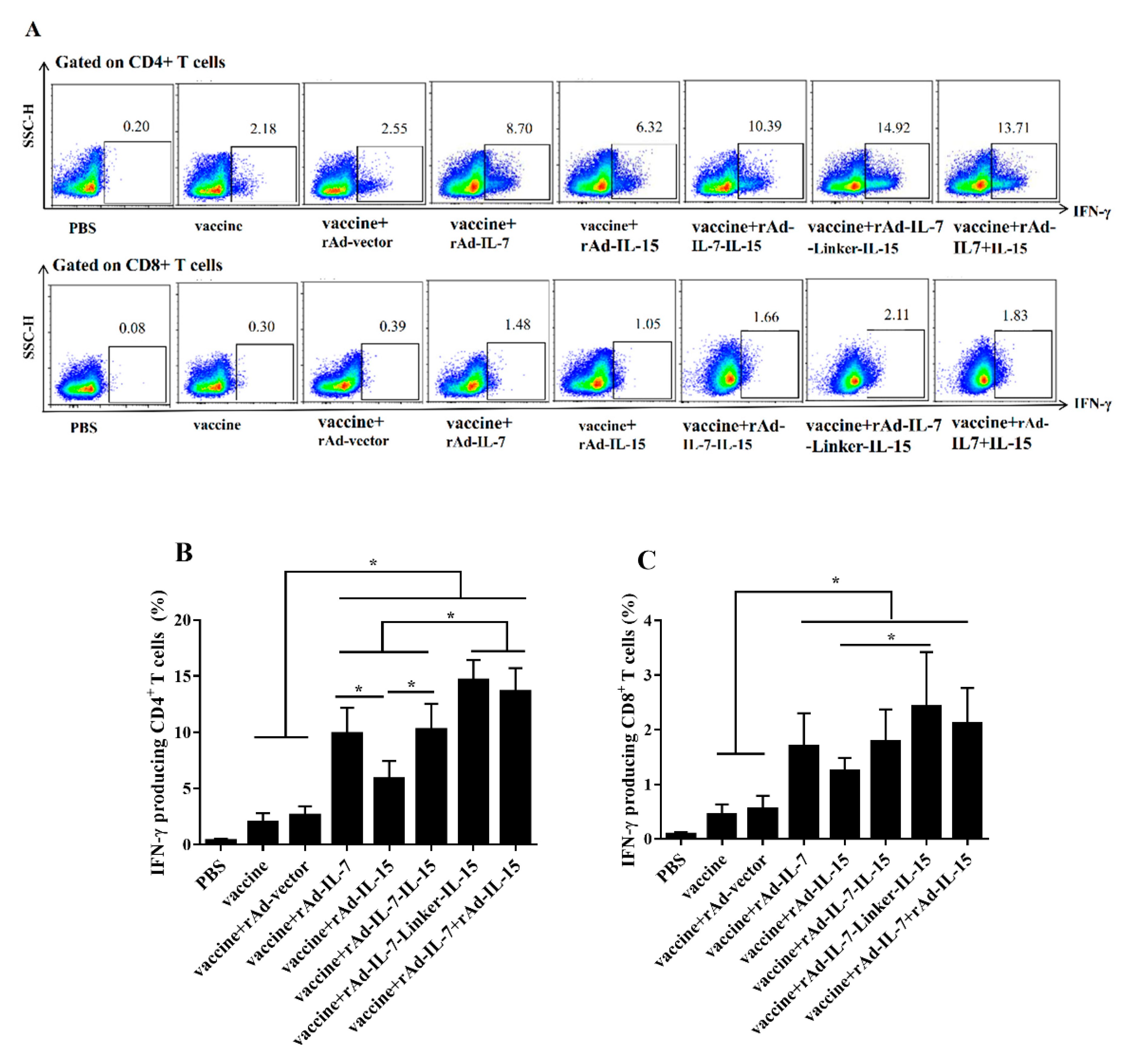
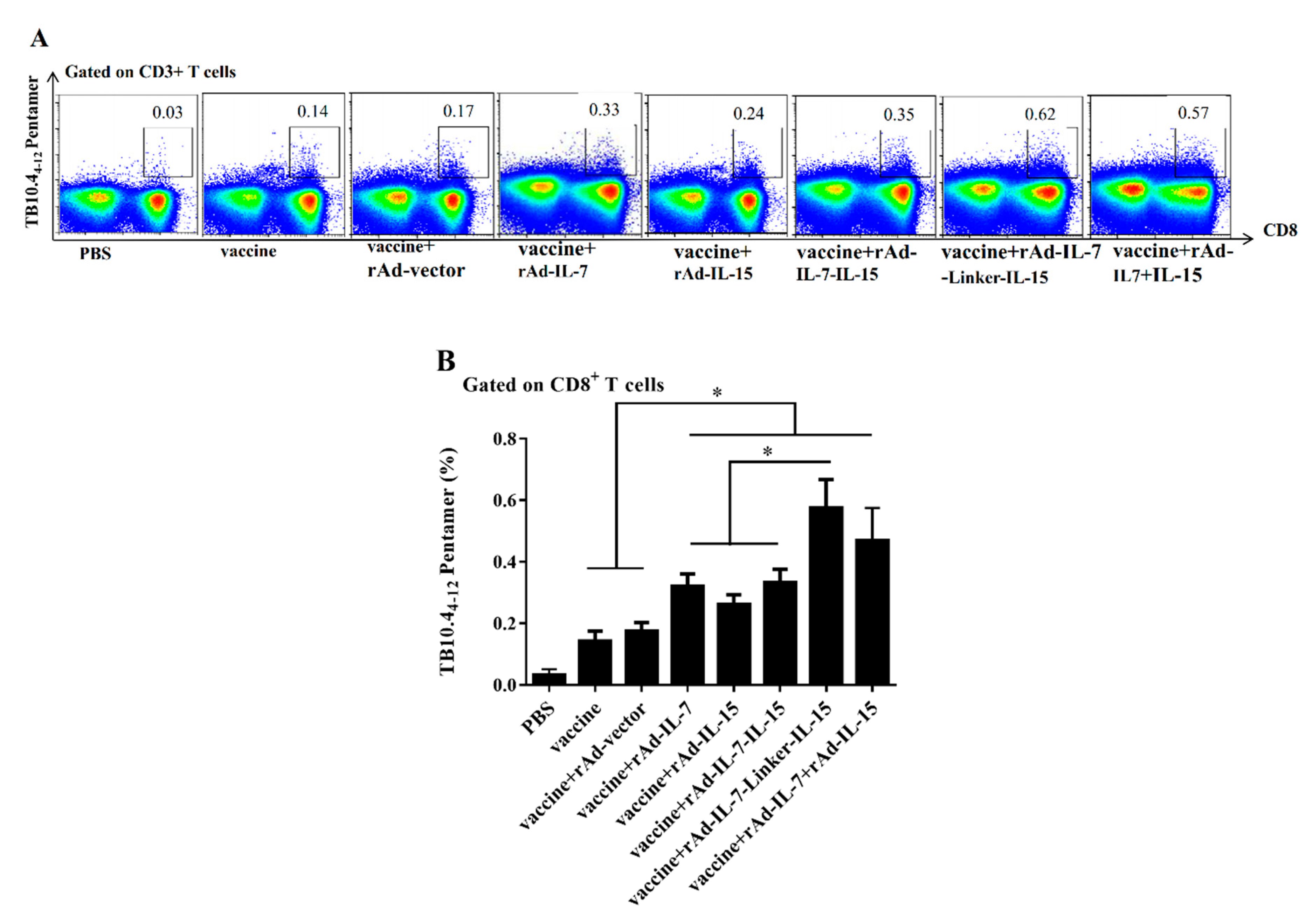
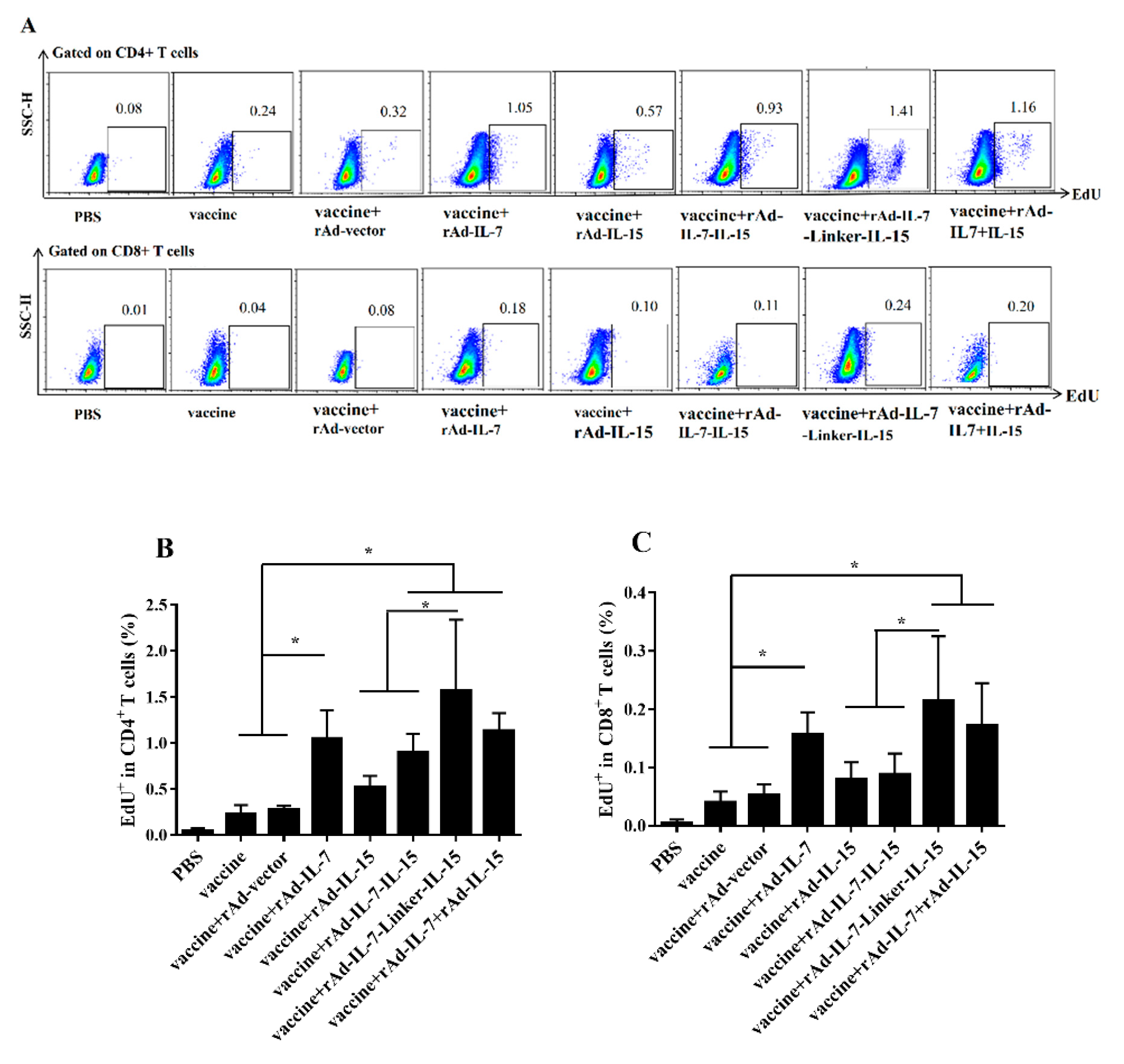
3.1. rAd-IL-7-Linker-IL-15 Enhanced Quality and Quantity of TCM Like Cells

To observe TCM like cells-mediated immune responses induced by different cytokines, we performed cultured IFN-γ ELISPOT assay, IFN-γ secretion by ICC following twice-stimulation in vivo and in vitro, and the number of expanded TB10.4-specific CD8+ memory T cells at 20 weeks after the final immunization. In cultured ELISPOT assay, the results showed that the groups of vaccine + rAd-IL-7 (299.3 ± 20.9 SFC/1 × 106 cells), vaccine + rAd-IL-15 (273.3 ± 25.2 SFC/1 × 106 cells), vaccine + rAd-IL-7-IL-15 (325.6 ± 19.7 SFC/1 × 106 cells), vaccine + rAd-IL-7-Linker-IL-15 (382.2 ± 32.9S FC/1 × 106 cells) and vaccine + rAd-IL-7 + rAd-IL-15 (361.4 ± 17.2 SFC/1 × 106 cells) induced a larger increase of long-term antigen-specific IFN-γ producing cells, which is the character of TCM like cells [30], than the control groups of vaccine alone (186.0 ± 30.5 SFC/1 × 106 cells) and vaccine + rAd-vector (211.6 ± 22.4 SFC/1 × 106 cells). In the meantime, the group of vaccine + rAd-IL-7-Linker-IL-15 enhanced long-term antigen-specific IFN-γ producing cells, compared with the groups of vaccine + rAd-IL-7, and vaccine + rAd-IL-15 and vaccine + rAd-IL-7-IL-15. However, the group of vaccine + rAd-IL-7-Linker-IL-15 didn’t demonstrate an obvious difference with the group of vaccine + rAd-IL-7 + rAd-IL-15 (Figure 1).
Meanwhile, according to the principle of long-term cultured ELISPOT assay, we detected antigen specific TCM like cells by IFN-γ secretion following twice-stimulation in vivo and in vitro sequentially [28]. Firstly, mice were injected with BCG by i.p at 20 weeks after the final immunization. Secondly, lymphocytes were isolated after 9 days later and stimulated for 4 h with mixed antigens in vitro. Then, ICC was performed. The data indicated that the vaccine + rAd-IL-7-Linker-IL-15 group induced higher frequency of IFN-γ on CD4+ T cells (14.64 ± 1.79%) than the groups of vaccine + rAd-IL-7 (9.87 ± 0.79%), vaccine + rAd-IL-15 (5.84 ± 1.62%), vaccine + rAd-IL-7-IL-15 (10.22 ± 2.34%), vaccine alone (2.02 ± 0.79%) and vaccine + rAd-vector control (2.63 ± 0.77%). The groups of vaccine + rAd-IL-7, vaccine + rAd-IL-15, vaccine + rAd-IL-7-IL-15 and vaccine + rAd-IL-7 + rAd-IL-15 produced higher levels of IFN-γ secretion than that of the control groups of vaccine alone and vaccine + rAd-vector. Moreover, the vaccine + rAd-IL-7 group had an increase of IFN-γ by 4.03% compared with the vaccine + rAd-IL-15 group (Figure 2B). For CD8+ T cells, the groups of vaccine + rAd-IL-7, vaccine + rAd-IL-15, vaccine + rAd-IL-7-IL-15, vaccine + rAd-IL-7-Linker-IL-15 and vaccine + rAd-IL-7 + rAd-IL-15 induced significantly more TCM like cells immune responses compared to the control groups of vaccine and vaccine + rAd-vector. The frequency of CD8+ IFN-γ+ cells in the vaccine + rAd-IL-7-Linker-IL-15 group increased 1.18% compared with the group of vaccine + rAd-IL-15 (Figure 2C).
Mice were injected with BCG by i.p at 20 weeks after the final immunization. After 9 days, the number of TB10.4-specific CD8+ memory T cells were evaluated by TB10.44-12 pentamer, which was the same principle with IFN-γ secretion following twice-stimulation in vivo and in vitro. The results showed that the frequency of TB10.4-specific CD8+ TCM like cells in the group of vaccine + rAd-IL-7-Linker-IL-15 was highest and had a significant increase compared with the groups of vaccine + rAd-IL-7, vaccine + rAd-IL-7-IL-15, vaccine + rAd-IL-15, vaccine + rAd-vector and vaccine. The groups of vaccine + rAd-IL-7, vaccine + rAd-IL-15 and vaccine + rAd-IL-7 + rAd-IL-15 induced more TB10.4-specific CD8+ TCM like cells than the control groups of vaccine + rAd-vector and vaccine. There was no obvious difference between the groups of vaccine + rAd-IL-7-Linker-IL-15 and vaccine + rAd-IL-7 + rAd-IL-15 (Figure 3).

3.2. rAd-IL-7-Linker-IL-15 Induced Higher Proliferation Capability of TCM Like Cells

To evaluate proliferation of TCM like cells induced by different cytokines, at 20 weeks after the last immunization, we performed EdU proliferation assay [28]. For CD4+ T cells, the groups of vaccine + rAd-IL-7, vaccine + rAd-IL-7-IL-15, vaccine + rAd-IL-7-Linker-IL-15 and vaccine + rAd-IL-7 + rAd-IL-15 showed an enhanced incorporation compared with the control groups of vaccine and vaccine + rAd-vector; The frequency of EdU+ T cells in the vaccine + rAd-IL-7-Linker-IL-15 group was highest, which was obviously higher than that of the vaccine + rAd-IL-15 group and vaccine + rAd-IL-7-IL-15; There was no significant difference among the groups of vaccine + rAd-IL-15, vaccine and vaccine + rAd-vector (Figure 4A,B). For CD8+ T cells, the tendency was consistent with CD4+ T cells, the groups of vaccine + rAd-IL-7, vaccine + rAd-IL-7-Linker-IL-15 and vaccine + rAd-IL-7 + rAd-IL-15 promoted the frequency of CD8+ T cells incorporated with EdU compares with that of the vaccine and vaccine + rAd-vector control groups (Figure 4A,C). In conclusion, the vaccine + rAd-IL-7-Linker-IL-15 group induced the strongest capability of proliferation on CD4+ T cells and CD8+ T cells among these groups. The group of the vaccine + rAd-IL-7 showed stronger capability of proliferation on CD4+ T cells and CD8+ T cells than the control groups of vaccine and vaccine + rAd-vector. The group of the vaccine + rAd-IL-15 showed no obvious difference compared with the control groups of vaccine and vaccine + rAd-vector.

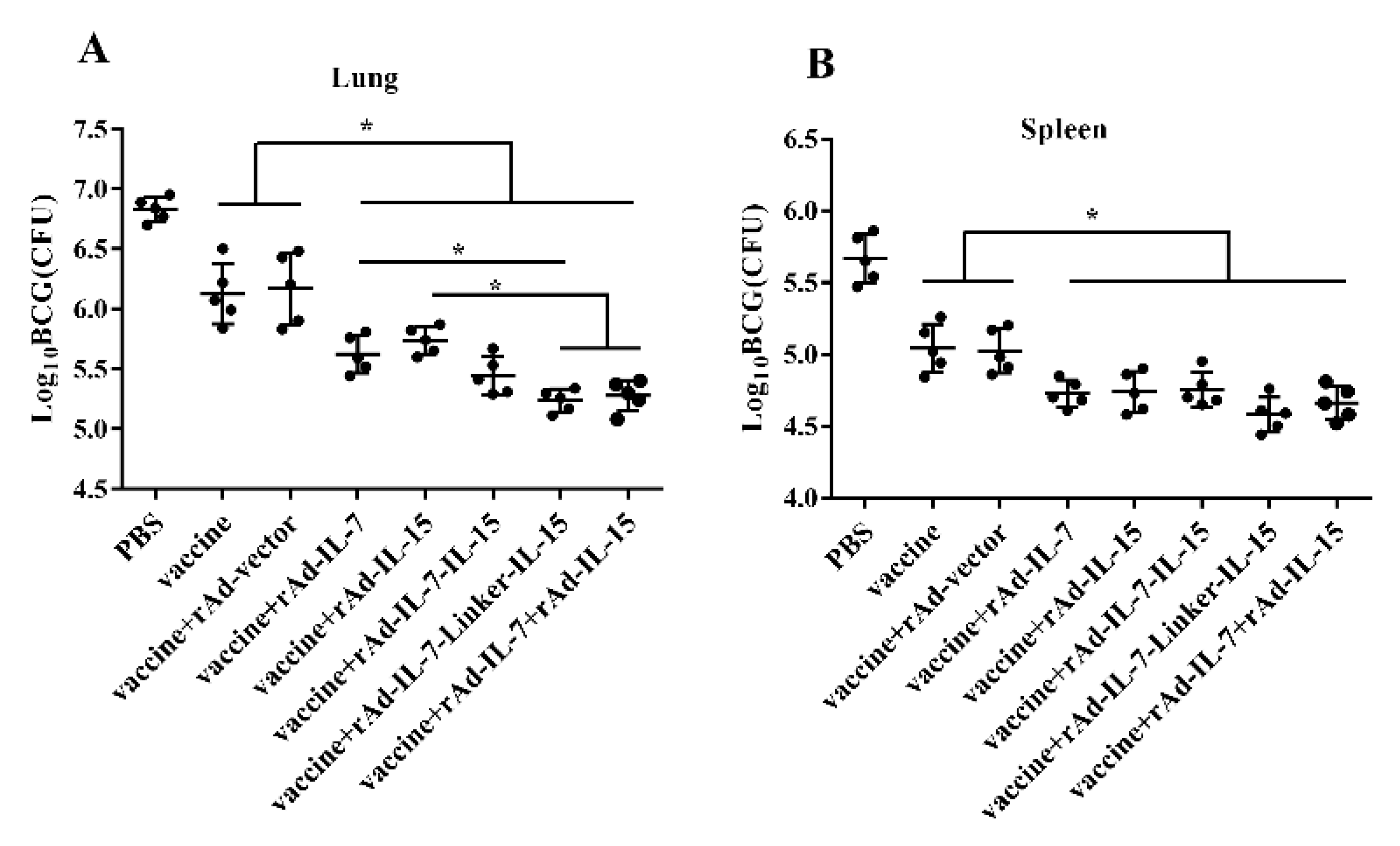
3.3. rAd-IL-7-Linker-IL-15 Promoted the Protective Efficacy of Vaccine

To identify the protective efficacy of vaccine associated with different cytokines, we examined CFU of the lungs and spleens after Mycobacterium bovis BCG challenge. At 24 weeks after the last immunization, mice were challenged with BCG. At three weeks post-challenge, CFU of the lungs and spleens was measured. Against BCG infection, the results showed that, CFU of the lungs in the groups of vaccine + rAd-IL-7, vaccine + rAd-IL-15, vaccine + rAd-IL-7-IL-15, vaccine + rAd-IL-7-Linker-IL-15 and vaccine + rAd-IL-7 + rAd-IL-15 all had a significant reduction compared with the control groups of vaccine and vaccine + rAd-vector; Moreover, the groups of vaccine + rAd-IL-7-Linker-IL-15 and vaccine + rAd-IL-7 + rAd-IL-15 had the least bacteria load, vaccine + rAd-IL-7-Linker-IL-15 declining approximately 0.38log10 CFU compared with the group of vaccine + rAd-IL-7 and 0.50log10 CFU compared with the group of vaccine + rAd-IL-15 (Figure 5A). In the spleen, the bacterial load in the groups of vaccine + rAd-IL-7 (4.73 ± 0.95log10 CFU), vaccine + rAd-IL-15 (4.74 ± 0.14log10 CFU), vaccine + rAd-IL-7-IL-15 (4.75 ± 0.12log10 CFU), vaccine + rAd-IL-7-Linker-IL-15 (4.58 ± 0.12log10 CFU) and vaccine + rAd-IL-7 + rAd-IL-15 (4.66 ± 0.12log10 CFU) were significantly lower than the control groups of vaccine (5.04 ± 0.17log10 CFU) and vaccine + rAd-vector (5.02 ± 0.15log10 CFU). However, there was no obvious difference among the groups of vaccine + rAd-IL-7-Linker-IL-15, vaccine + rAd-IL-7, vaccine + rAd-IL-15, vaccine + rAd-IL-7-IL-15 and vaccine + rAd-IL-7 + rAd-IL-15 (Figure 5B). These results indicated that IL-7-Linker-IL-15 promoted vaccine produce stronger immune memory with higher protective efficacy than IL-7 and IL-15.

4. Discussion

The ideal tuberculosis vaccines should be able to induce more TCM to provide a long-term protection against TB. Currently, there are a few measures to prolong the protection of vaccines against TB. It is reported that low dose of antigen favors the induction of TCM while high dose of antigen mainly induces TEM or effective T cells [31,32]. It is also demonstrated that prolonging boosting intervals can induce a stronger booster response and enhanced long-term protective efficacy against M. tuberculosis [28,33]. Metformin could expand memory-like antigen-inexperienced CD8+ T cells and enhance protective efficacy against M. tuberculosis challenge [34]. Some studies have demonstrated that IL-7 and IL-15 can promote formation and homeostasis of memory T cells [35,36]. In our study, we explored TCM like cells-mediated immunity induced by recombinant adenovirus encoding cytokines IL-7, IL-15, IL-7-IL-15, and IL-7-Linker-IL-15 combined with Mycobacterium tuberculosis subunit vaccine. We found that IL-7-Linker-IL-15 increased the quantity of TCM like cells and enhanced proliferation capability compared with IL-7, IL-15, and IL-7-IL-15. Consistent with these, IL-7-Linker-IL-15 helped the vaccine produce higher protective efficacy against BCG than IL-7 and IL-15.
IL-7 plays crucial roles in both development of naïve T cells and expanding clonotypically diverse CD4+ and CD8+ memory T cells populations [12]. Our study showed that rAd-IL-7 promoted vaccine to induce more CD4+ TCM like cells than the vaccine + rAd-IL-15 group, increasing the secretion of IFN-γ and proliferative capability following the repeated stimulation with same antigens in several days. For CD8+ T cells, the vaccine + rAd-IL-7 group increased TCM like cells compared with the control groups of vaccine and vaccine + rAd-vector, with expanded number of TB10.4-specific CD8+ memory T cells and higher IFN-γ secretion following twice antigen stimulation, but the vaccine + rAd-IL-15 group didn’t. Taken together, this study showed that rAd-IL-7 resulted in significant increases in TCM like cells compared with rAd-IL-15.
IL-15 plays a complicated effect on development of TCM like cells, which may be related to the strength of IL-15 signaling. It is well-known that T cell development depends on IL-15β receptor, also known as CD122. Weak CD122 signaling supports TCM development, while stronger CD122 signaling supports the development of TEM. Moreover, high CD122 signaling mainly promotes generation of short lived terminally differentiated effector T cells [37]. Our experiment showed that IL-15 have a weak effect on inducing TCM like cells-mediated immune responses. On one hand, IL-15 contributes to maintaining the homeostasis of memory T cells [38,39]. On the other hand, IL-15 selectively promoted the proliferation of TEM rather than TCM [40].
It is interesting to point out that rAd-IL-7-Linker-IL-15, in which IL-7 and IL-15 is connected by a 12-amino acids linker (Gly-Gly-Gly-Ser)3, promote TB subunit vaccine to induce stronger long-term immune responses than rAd-IL-7-IL-15 and single rAd-IL-7 and rAd-IL-15. It has been demonstrated that this linker can minimize the refolding problems of the two fused chains, such as incorrect domain pairing or aggregation, and improves the stability of the structure. Consequently, it is beneficial for IL-7 and IL-15 to play a part in regulating the development of memory T cells [26,41,42]. Our study showed that rAd-IL-7-Linker-IL-15 promoted formation and maintenance TCM like cells and improved proliferative capability of TCM like cells, which resulted in stronger protective efficacy against BCG. Moreover, the results indicated that connection of IL -7 and IL-15 by the linker had a synergetic effect that had shown promising ability to produce unheralded biological effects to augment the TCM like cells-mediated immune responses.

5. Conclusions

For the first time, our study demonstrates that supplementation of TB protein-subunit vaccine with rAd- IL-7-Linker-IL-15 would induce more TCM like cells and improve its protective efficacy against M. tuberculosis. Meanwhile, IL-7 and IL-15 have been applied for the treatment of tumors in clinical trials and they were proved safe for patients [43,44,45]. However, IL-7 and IL-15 haven’t been used as adjuvants for the vaccine to healthy individuals in clinic. Therefore, fusion cytokine IL-7-Linker-IL-15 developed as an adjuvant need to be explored for triggering a stronger long-term cellular immune response against tuberculosis.

Author Contributions

B.Z. and C.B. conceived and designed the research; C.B., L.Z., J.T., J.H. (Juanjuan He), J.H. (Jiangyuan Han) and H.N. performed the research; B.Z. and C.B. wrote the first draft of the article and revised it. All authors have read and agreed to the published version of the manuscript.

Funding

This research was supported by National Major Science and Technology Projects of China [2018ZX10302302-002-003]; and National Natural Science Foundation of China [31470895].

Conflicts of Interest

The authors declare no conflict of interest.

Abbreviations

TB—Tuberculosis
M. tuberculosis—Mycobacterium tuberculosis
BCG—Mycobacterium bovis Bacilli Calmette-Guerin
TCM like cells—central memory-like T cells
TCM—central memory T cells
TEM—effector memory T cells
DDA—dioctadecylammonium bromide
Poly (I:C)—polyinosinic-polycytidylic acid
IL—Interleukin
LT70-ESAT6-Ag85B-MPT64<190-198>-Mtb8.4-Rv2626c
MH—Mtb10.4-HspX
PBS—Phosphate-buffered saline
rAd—recombined adenoviral
CPE—cytopathic effect
i.p.—intraperitoneal injection
APCs—antigen presenting cells
Linker—(Gly-Gly-Gly-Ser)3

References

  1. WHO. Global Tuberculosis Report 2020. Available online: https://apps.who.int/iris/bitstream/handle/10665/336069/9789240013131-eng.pdf (accessed on 22 November 2020).
  2. Andersen, P.; Doherty, T.M. The success and failure of BCG—Implications for a novel tuberculosis vaccine. Nat. Rev. Microbiol. 2005, 3, 656–662. [Google Scholar] [CrossRef]
  3. Barker, L.F.; Leadman, A.E.; Clagett, B. The challenges of developing new tuberculosis vaccines. Health Aff. 2011, 30, 1073–1079. [Google Scholar] [CrossRef] [Green Version]
  4. Orme, I.M. The Achilles heel of BCG. Tuberculosis 2010, 90, 329–332. [Google Scholar] [CrossRef] [PubMed]
  5. Kaveh, D.A.; Garcia-Pelayo, M.C.; Hogarth, P.J. Persistent BCG bacilli perpetuate CD4 T effector memory and optimal protection against tuberculosis. Vaccine 2014, 32, 6911–6918. [Google Scholar] [CrossRef] [PubMed] [Green Version]
  6. Youngblood, B.; Hale, J.S.; Ahmed, R. T-cell memory differentiation: Insights from transcriptional signatures and epigenetics. Immunology 2013, 139, 277–284. [Google Scholar] [CrossRef] [PubMed]
  7. Voss, G.; Casimiro, D.; Neyrolles, O.; Williams, A.; Kaufmann, S.H.E.; McShane, H.; Hatherill, M.; Fletcher, H.A. Progress and challenges in TB vaccine development. F1000Research 2018, 7, 199. [Google Scholar] [CrossRef] [PubMed] [Green Version]
  8. Tregoning, J.S.; Russell, R.F.; Kinnear, E. Adjuvanted influenza vaccines. Hum. Vaccines Immunother. 2018, 14, 550–564. [Google Scholar] [CrossRef] [Green Version]
  9. Liu, X.; Da, Z.; Wang, Y.; Niu, H.; Li, R.; Yu, H.; He, S.; Guo, M.; Luo, Y.; Ma, X.; et al. A novel liposome adjuvant DPC mediates Mycobacterium tuberculosis subunit vaccine well to induce cell-mediated immunity and high protective efficacy in mice. Vaccine 2016, 34, 1370–1378. [Google Scholar] [CrossRef]
  10. Vogelzang, A.; Perdomo, C.; Zedler, U.; Kuhlmann, S.; Hurwitz, R.; Gengenbacher, M.; Kaufmann, S.H. Central memory CD4+ T cells are responsible for the recombinant Bacillus Calmette-Guerin DeltaureC::hly vaccine’s superior protection against tuberculosis. J. Infect. Dis. 2014, 210, 1928–1937. [Google Scholar] [CrossRef]
  11. Lindenstrom, T.; Knudsen, N.P.H.; Agger, E.M.; Andersen, P. Control of Chronic Mycobacterium tuberculosis Infection by CD4 KLRG1(-) IL-2-Secreting Central Memory Cells. J. Immunol. 2013, 190, 6311–6319. [Google Scholar] [CrossRef] [Green Version]
  12. Okoye, A.A.; Rohankhedkar, M.; Konfe, A.L.; Abana, C.O.; Reyes, M.D.; Clock, J.A.; Duell, D.M.; Sylwester, A.W.; Sammader, P.; Legasse, A.W.; et al. Effect of IL-7 Therapy on Naive and Memory T Cell Homeostasis in Aged Rhesus Macaques. J. Immunol. 2015, 195, 4292–4305. [Google Scholar] [CrossRef] [PubMed] [Green Version]
  13. Melchionda, F.; Fry, T.J.; Milliron, M.J.; McKirdy, M.A.; Tagaya, Y.; Mackall, C.L. Adjuvant IL-7 or IL-15 overcomes immunodominance and improves survival of the CD8+ memory cell pool. J. Clin. Investig. 2005, 115, 1177–1187. [Google Scholar] [CrossRef] [PubMed] [Green Version]
  14. Purton, J.F.; Martin, C.E.; Surh, C.D. Enhancing T cell memory: IL-7 as an adjuvant to boost memory T-cell generation. Immunol. Cell Biol. 2008, 86, 385–386. [Google Scholar] [CrossRef] [PubMed]
  15. Yeon, S.M.; Halim, L.; Chandele, A.; Perry, C.J.; Kim, S.H.; Kim, S.U.; Byun, Y.; Yuk, S.H.; Kaech, S.M.; Jung, Y.W. IL-7 plays a critical role for the homeostasis of allergen-specific memory CD4 T cells in the lung and airways. Sci. Rep. 2017, 7, 11155. [Google Scholar] [CrossRef] [Green Version]
  16. Knop, L.; Deiser, K.; Bank, U.; Witte, A.; Mohr, J.; Philipsen, L.; Fehling, H.J.; Muller, A.J.; Kalinke, U.; Schuler, T. IL-7 derived from lymph node fibroblastic reticular cells is dispensable for naive T cell homeostasis but crucial for central memory T cell survival. Eur. J. Immunol. 2020, 50, 846–857. [Google Scholar] [CrossRef] [Green Version]
  17. DeGottardi, M.Q.; Okoye, A.A.; Vaidya, M.; Talla, A.; Konfe, A.L.; Reyes, M.D.; Clock, J.A.; Duell, D.M.; Legasse, A.W.; Sabnis, A.; et al. Effect of Anti-IL-15 Administration on T Cell and NK Cell Homeostasis in Rhesus Macaques. J. Immunol. 2016, 197, 1183–1198. [Google Scholar] [CrossRef] [Green Version]
  18. Chen, J.; Li, Z.Y.; Petersen, E.; Liu, W.G.; Zhu, X.Q. Co-administration of interleukins 7 and 15 with DNA vaccine improves protective immunity against Toxoplasma gondii. Exp. Parasitol. 2016, 162, 18–23. [Google Scholar] [CrossRef]
  19. Gao, Q.; Zhang, N.Z.; Zhang, F.K.; Wang, M.; Hu, L.Y.; Zhu, X.Q. Immune response and protective effect against chronic Toxoplasma gondii infection induced by vaccination with a DNA vaccine encoding profilin. BMC Infect. Dis. 2018, 18, 117. [Google Scholar] [CrossRef] [Green Version]
  20. Calarota, S.A.; Dai, A.; Trocio, J.N.; Weiner, D.B.; Lori, F.; Lisziewicz, J. IL-15 as memory T-cell adjuvant for topical HIV-1 DermaVir vaccine. Vaccine 2008, 26, 5188–5195. [Google Scholar] [CrossRef] [Green Version]
  21. Singh, V.; Gowthaman, U.; Jain, S.; Parihar, P.; Banskar, S.; Gupta, P.; Gupta, U.D.; Agrewala, J.N. Coadministration of interleukins 7 and 15 with bacille Calmette-Guerin mounts enduring T cell memory response against Mycobacterium tuberculosis. J. Infect. Dis. 2010, 202, 480–489. [Google Scholar] [CrossRef] [Green Version]
  22. Tang, C.; Yamada, H.; Shibata, K.; Yoshida, S.; Wajjwalku, W.; Yoshikai, Y. IL-15 protects antigen-specific CD8+ T cell contraction after Mycobacterium bovis bacillus Calmette-Guerin infection. J. Leukoc. Biol. 2009, 86, 187–194. [Google Scholar] [CrossRef] [PubMed] [Green Version]
  23. Liu, X.; Peng, J.; Hu, L.; Luo, Y.; Niu, H.; Bai, C.; Wang, Q.; Li, F.; Yu, H.; Wang, B.; et al. A multistage mycobacterium tuberculosis subunit vaccine LT70 including latency antigen Rv2626c induces long-term protection against tuberculosis. Hum. Vaccines Immunother. 2016, 12, 1670–1677. [Google Scholar] [CrossRef] [PubMed] [Green Version]
  24. Niu, H.; Hu, L.; Li, Q.; Da, Z.; Wang, B.; Tang, K.; Xin, Q.; Yu, H.; Zhang, Y.; Wang, Y.; et al. Construction and evaluation of a multistage Mycobacterium tuberculosis subunit vaccine candidate Mtb10.4-HspX. Vaccine 2011, 29, 9451–9458. [Google Scholar] [CrossRef] [PubMed]
  25. Xin, Q.; Niu, H.; Li, Z.; Zhang, G.; Hu, L.; Wang, B.; Li, J.; Yu, H.; Liu, W.; Wang, Y.; et al. Subunit vaccine consisting of multi-stage antigens has high protective efficacy against Mycobacterium tuberculosis infection in mice. PLoS ONE 2013, 8, e72745. [Google Scholar] [CrossRef]
  26. Huston, J.S.; Levinson, D.; Mudgett-Hunter, M.; Tai, M.S.; Novotny, J.; Margolies, M.N.; Ridge, R.J.; Bruccoleri, R.E.; Haber, E.; Crea, R.; et al. Protein engineering of antibody binding sites: Recovery of specific activity in an anti-digoxin single-chain Fv analogue produced in Escherichia coli. Proc. Natl. Acad. Sci. USA 1988, 85, 5879–5883. [Google Scholar] [CrossRef] [Green Version]
  27. Darling, A.J.; Boose, J.A.; Spaltro, J. Virus assay methods: Accuracy and validation. Biol. J. Int. Assoc. Biol. Stand. 1998, 26, 105–110. [Google Scholar] [CrossRef]
  28. Bai, C.; He, J.; Niu, H.; Hu, L.; Luo, Y.; Liu, X.; Peng, L.; Zhu, B. Prolonged intervals during Mycobacterium tuberculosis subunit vaccine boosting contributes to eliciting immunity mediated by central memory-like T cells. Tuberculosis 2018, 110, 104–111. [Google Scholar] [CrossRef]
  29. Billeskov, R.; Vingsbo-Lundberg, C.; Andersen, P.; Dietrich, J. Induction of CD8 T cells against a novel epitope in TB10.4: Correlation with mycobacterial virulence and the presence of a functional region of difference-1. J. Immunol. 2007, 179, 3973–3981. [Google Scholar] [CrossRef]
  30. Calarota, S.A.; Baldanti, F. Enumeration and characterization of human memory T cells by enzyme-linked immunospot assays. Clin. Dev. Immunol. 2013, 2013, 637649. [Google Scholar] [CrossRef] [Green Version]
  31. Aagaard, C.; Hoang, T.; Dietrich, J.; Cardona, P.J.; Izzo, A.; Dolganov, G.; Schoolnik, G.K.; Cassidy, J.P.; Billeskov, R.; Andersen, P. A multistage tuberculosis vaccine that confers efficient protection before and after exposure. Nat. Med. 2011, 17, 189–194. [Google Scholar] [CrossRef]
  32. Niu, H.; Peng, J.; Bai, C.; Liu, X.; Hu, L.; Luo, Y.; Wang, B.; Zhang, Y.; Chen, J.; Yu, H.; et al. Multi-Stage Tuberculosis Subunit Vaccine Candidate LT69 Provides High Protection against Mycobacterium tuberculosis Infection in Mice. PLoS ONE 2015, 10, e0130641. [Google Scholar] [CrossRef] [PubMed] [Green Version]
  33. Rouanet, C.; Debrie, A.S.; Lecher, S.; Locht, C. Subcutaneous boosting with heparin binding haemagglutinin increases BCG-induced protection against tuberculosis. Microbes Infect. 2009, 11, 995–1001. [Google Scholar] [CrossRef] [PubMed]
  34. Bohme, J.; Martinez, N.; Li, S.; Lee, A.; Marzuki, M.; Tizazu, A.M.; Ackart, D.; Frenkel, J.H.; Todd, A.; Lachmandas, E.; et al. Metformin enhances anti-mycobacterial responses by educating CD8+ T-cell immunometabolic circuits. Nat. Commun. 2020, 11, 5225. [Google Scholar] [CrossRef] [PubMed]
  35. Lenz, D.C.; Kurz, S.K.; Lemmens, E.; Schoenberger, S.P.; Sprent, J.; Oldstone, M.B.; Homann, D. IL-7 regulates basal homeostatic proliferation of antiviral CD4+ T cell memory. Proc. Natl. Acad. Sci. USA 2004, 101, 9357–9362. [Google Scholar] [CrossRef] [PubMed] [Green Version]
  36. Lugli, E.; Goldman, C.K.; Perera, L.P.; Smedley, J.; Pung, R.; Yovandich, J.L.; Creekmore, S.P.; Waldmann, T.A.; Roederer, M. Transient and persistent effects of IL-15 on lymphocyte homeostasis in nonhuman primates. Blood 2010, 116, 3238–3248. [Google Scholar] [CrossRef]
  37. Castro, I.; Yu, A.; Dee, M.J.; Malek, T.R. The basis of distinctive IL-2- and IL-15-dependent signaling: Weak CD122-dependent signaling favors CD8+ T central-memory cell survival but not T effector-memory cell development. J. Immunol. 2011, 187, 5170–5182. [Google Scholar] [CrossRef] [Green Version]
  38. Ku, C.C.; Murakami, M.; Sakamoto, A.; Kappler, J.; Marrack, P. Control of homeostasis of CD8+ memory T cells by opposing cytokines. Science 2000, 288, 675–678. [Google Scholar] [CrossRef]
  39. Surh, C.D.; Sprent, J. Homeostasis of naive and memory T cells. Immunity 2008, 29, 848–862. [Google Scholar] [CrossRef] [Green Version]
  40. Boyman, O.; Letourneau, S.; Krieg, C.; Sprent, J. Homeostatic proliferation and survival of naive and memory T cells. Eur. J. Immunol. 2009, 39, 2088–2094. [Google Scholar] [CrossRef]
  41. Reddy, S.T.; Ge, X.; Miklos, A.E.; Hughes, R.A.; Kang, S.H.; Hoi, K.H.; Chrysostomou, C.; Hunicke-Smith, S.P.; Iverson, B.L.; Tucker, P.W.; et al. Monoclonal antibodies isolated without screening by analyzing the variable-gene repertoire of plasma cells. Nat. Biotechnol. 2010, 28, 965–969. [Google Scholar] [CrossRef]
  42. Alfthan, K.; Takkinen, K.; Sizmann, D.; Soderlund, H.; Teeri, T.T. Properties of a single-chain antibody containing different linker peptides. Protein Eng. 1995, 8, 725–731. [Google Scholar] [CrossRef] [PubMed]
  43. Brown, V.I.; Hulitt, J.; Fish, J.; Sheen, C.; Bruno, M.; Xu, Q.; Carroll, M.; Fang, J.; Teachey, D.; Grupp, S.A. Thymic stromal-derived lymphopoietin induces proliferation of pre-B leukemia and antagonizes mTOR inhibitors, suggesting a role for interleukin-7Ralpha signaling. Cancer Res. 2007, 67, 9963–9970. [Google Scholar] [CrossRef] [PubMed] [Green Version]
  44. Wagner, J.; Pfannenstiel, V.; Waldmann, A.; Bergs, J.W.J.; Brill, B.; Huenecke, S.; Klingebiel, T.; Rodel, F.; Buchholz, C.J.; Wels, W.S.; et al. A Two-Phase Expansion Protocol Combining Interleukin (IL)-15 and IL-21 Improves Natural Killer Cell Proliferation and Cytotoxicity against Rhabdomyosarcoma. Front. Immunol. 2017, 8, 676. [Google Scholar] [CrossRef] [PubMed] [Green Version]
  45. Conlon, K.C.; Lugli, E.; Welles, H.C.; Rosenberg, S.A.; Fojo, A.T.; Morris, J.C.; Fleisher, T.A.; Dubois, S.P.; Perera, L.P.; Stewart, D.M.; et al. Redistribution, hyperproliferation, activation of natural killer cells and CD8 T cells, and cytokine production during first-in-human clinical trial of recombinant human interleukin-15 in patients with cancer. J. Clin. Oncol. 2015, 33, 74–82. [Google Scholar] [CrossRef]
Figure 1. Cultured ELISPOT assay for antigen specific TCM like cells. At 20 weeks after the last vaccination, lymphocytes of mice were cultured with or without a cocktail of antigens ESAT-6, Ag85B, Rv2626c and HspX for 9 days. Then, cells were re-stimulated with the same antigens for 20 h. (A) Representative images of IFN-γ ELISPOT wells from long-term cultured ELISPOT assays at 5× magnification. (B) Results of long-term cultured ELISPOT responses assay. Data were presented as means ± SD from groups of 4 mice. * p < 0.05.
Vaccines 08 00715 g001aVaccines 08 00715 g001b
Figure 2. IFN-γ secretion following twice stimulation with antigens in spleens. At 20 weeks after the last immunization, mice were injected with BCG (1 × 10⁶ CFU) by i.p for 9 days. Then, lymphocytes of spleens were isolated and stimulated with a cocktail of antigens ESAT-6, Ag85B, Rv2626c and HspX (2 μg/mL) in vitro. Secretion of IFN-γ was determined by flow cytometry. (A) The representative results of every group; (B) CD4+ T cells secreting IFN-γ; (C) CD8+ T cells secreting IFN-γ. Data collected were presented as means ± SD from 5 mice per group. * p < 0.05.
Vaccines 08 00715 g002
Figure 3. Number of expanded TB10.4-specific CD8+ T cells in spleens. Mice were injected with BCG by i.p. at 20 weeks after the final immunization. After 9 days, the number of TB10.4-specific CD8+ TCM like cells was evaluated by TB10.44-12 pentamer. (A) The representative results of every group; (B) Frequency percentages of TB10.4-specific CD8+ TCM like cells. Data collected were presented as means ± SD from 5 mice per group. * p < 0.05.
Vaccines 08 00715 g003
Figure 4. Proliferative capability of lymphocytes. For proliferation assay, 20 weeks after the last vaccination, lymphocytes (5 × 10⁶ cells/well) were stimulated with a cocktail of antigens ESAT-6, Ag85B, Rv2626c and HspX (2 μg/mL) for 7 days in 24-well plates. Three days after antigen stimulation, EdU was added at a final concentration of 30 μM and cells were cultured for another 4 days. At the 7th day, proliferative cells were detected by flow cytometry. (A) The representative results of every group. (B) Frequency percentages of EdU+ in CD4+ T cells; (C) Frequency percentages of EdU+ in CD8+ T cells. Results are presented as means ± SD from groups of 5 mice. * p < 0.05.
Vaccines 08 00715 g004
Figure 5. The protective efficacy of vaccines against BCG infection in mice. At 24 weeks after the last vaccination, mice were challenged with Mycobacterium bovis BCG 1 × 10⁷ CFU/100 μL/mice by nasal. 3 weeks post-challenge, (A) mice were euthanized and the bacterial burden (CFU) was measured in lungs; (B) mice were euthanized and the bacterial burden (CFU) was measured in spleens. Data are presented as log10 CFU ± SD from groups of 5 mice. * p < 0.05.
Vaccines 08 00715 g005
Publisher’s Note: MDPI stays neutral with regard to jurisdictional claims in published maps and institutional affiliations.

