Review

Next-Generation TB Vaccines: Progress, Challenges, and Prospects

1
Beijing Key Laboratory of New Techniques of Tuberculosis Diagnosis and Treatment, Senior Department of Tuberculosis, Eighth Medical Center of Chinese PLA General Hospital, Beijing 100091, China
2
Hebei North University, Zhangjiakou 075000, China
*
Author to whom correspondence should be addressed.
Vaccines 2023, 11(8), 1304; https://doi.org/10.3390/vaccines11081304
Submission received: 1 July 2023 / Revised: 28 July 2023 / Accepted: 28 July 2023 / Published: 31 July 2023
(This article belongs to the Special Issue Tuberculosis Diagnostic, Treatments and Vaccines Research)

Abstract

:
Tuberculosis (TB), caused by Mycobacterium tuberculosis (MTB), is a prevalent global infectious disease and a leading cause of mortality worldwide. Currently, the only available vaccine for TB prevention is Bacillus Calmette–Guérin (BCG). However, BCG demonstrates limited efficacy, particularly in adults. Efforts to develop effective TB vaccines have been ongoing for nearly a century. In this review, we have examined the current obstacles in TB vaccine research and emphasized the significance of understanding the interaction mechanism between MTB and hosts in order to provide new avenues for research and establish a solid foundation for the development of novel vaccines. We have also assessed various TB vaccine candidates, including inactivated vaccines, attenuated live vaccines, subunit vaccines, viral vector vaccines, DNA vaccines, and the emerging mRNA vaccines as well as virus-like particle (VLP)-based vaccines, which are currently in preclinical stages or clinical trials. Furthermore, we have discussed the challenges and opportunities associated with developing different types of TB vaccines and outlined future directions for TB vaccine research, aiming to expedite the development of effective vaccines. This comprehensive review offers a summary of the progress made in the field of novel TB vaccines.

1. Introduction

Tuberculosis (TB) is an ancient disease widely distributed across the world and is the leading cause of death from bacterial infections [1]. In 17–18th century Europe, TB was known as the “white plague”, with almost 100% of Europeans infected and 25% of them dying from the disease [2]. In 1882, Robert Koch, a German microbiologist, deciphered Mycobacterium tuberculosis (MTB) as the etiological factor responsible for TB, thereby opening a new chapter in the prevention, diagnosis, and treatment of TB [3]. With the continuous development of anti-TB drugs and improvements in sanitation and living conditions, the incidence and mortality of TB decreased significantly. However, since the 1990s, the global TB epidemic has worsened due to factors such as the emergence of drug-resistant strains of MTB, the use of immunosuppressive agents, drug addiction, poverty, and population movements [4]. TB has become a leading re-emerging infectious disease and a serious global health problem, especially in developing countries.
Despite the effectiveness of anti-TB drugs, eradicating TB still faces many challenges. On 27 October 2022, the World Health Organization (WHO) Global TB Report showed that approximately one-quarter of the world’s population is infected with MTB [4]. In 2021, the estimated global number of new TB cases was 10.6 million, a 4.5% increase from 2020, and the number of deaths from TB was 1.6 million, a 6.7% increase from 2020 [5]. China is one of the countries with a high burden of TB, with an estimated number of TB cases only second to India and Indonesia, accounting for 7.4% of the global TB incidence [5]. In 2015, the WHO introduced the “End TB Strategy”, a comprehensive plan that outlines ambitious targets for the period of 2020–2035. These objectives include a significant reduction of 90% in the incidence of TB and a remarkable decrease of 95% in TB-related mortality compared to the 2015 levels by the year 2035 [6].
Vaccination stands as the most economically efficient method for the prevention and management of TB, serving as a crucial approach towards realizing the WHO’s Global End TB Strategy by 2035. One of the most successful measures in this regard is the Bacillus Calmette–Guérin (BCG) vaccine, which has been widely deployed worldwide for 140 years since its inception [7]. Although BCG provides significant protection against severe TB in infants and young children, such as disseminated tuberculosis and meningeal tuberculosis [8], its protection against adult pulmonary tuberculosis (PTB) is limited, with varying efficacy and no effective protection against primary or latent TB infection caused by MTB [9,10,11]. Currently, approximately one-quarter of the world’s population has latent TB infection (LTBI), which has no clinical symptoms, and about 90% of LTBI patients do not progress to TB disease. However, this latent state is a potential source of active TB and a major obstacle to TB elimination [12]. Therefore, there is an urgent need to develop more effective TB vaccines to prevent and control both latent and active infections caused by MTB.
With the rapid development of immunology and molecular biology, some new vaccines have entered clinical trial stages and have shown certain safety and protective efficacy. However, designing an ideal TB vaccine still faces many challenges, such as unclear pathogenic mechanisms of MTB, difficulty in screening for specific antigens, lack of ideal adjuvants, and the limitations of animal models. This article provides a review, analysis, and discussion of the immunological mechanisms of MTB–host interactions and the current status of new TB vaccine research in clinical trials. Challenges faced by TB vaccine research and future development directions are also discussed to provide a new perspective for the future development on TB vaccines.

2. Infection and Immunity to MTB

The main route of transmission of MTB is through airborne particles, which enter the human body through respiration and initiate the infection of alveolar macrophages. Previous studies have shown that approximately 5% of infected individuals can completely clear MTB from their bodies, approximately 5–10% will develop active TB disease, and nearly 90% of infected individuals remain in a LTBI state [13]. This indicates that after MTB infection, only a small proportion of individuals have an immune system that can effectively recognize, monitor, and clear MTB, while the vast majority of individuals are in an LTBI state. Thus, the interaction between MTB and its host is extremely complex, and the occurrence, development, and outcome of TB are not only related to the virulence of MTB, but also closely related to the immunity of TB patients. Genetic differences lead to certain immune defects or abnormalities in TB patients, which affect the ability of the body to resist MTB infection [14,15]. After MTB infection, a series of immune responses are induced, mainly including innate immunity (also known as non-specific immunity) and adaptive immunity (also known as specific immunity) (Figure 1). In this article, we will focus on the innate and adaptive immune responses induced by MTB and provide new insights for the research on new TB vaccines.

2.1. Innate Immune Responses Induced by MTB

Innate immune response plays a crucial role in the non-specific defense against MTB, and it is responsible for initiating adaptive immune response which specifically targets MTB [16]. Therefore, innate immunity serves as the first line of defense against MTB and is of utmost importance. The evolutionarily formed innate immune system is responsible for the defense function of innate immunity, which includes barrier, cellular, and molecular components. To resist MTB invasion, the host’s innate immune system relies on tissue barriers such as the mucosal barrier, physical and chemical barriers [17,18]. Concurrently, various components of the innate immune system, including macrophages, neutrophils, natural killer (NK) cells, dendritic cells (DCs), natural killer T (NKT) cells, and γδT cells, contribute to the generation of immune responses that combat TB infection [19,20]. In addition, the host exerts its anti-tuberculosis immune response and immune regulation function through immune molecules such as cytokines IFN-γ, IL-12, IL-1β, MIP-1α/CCL3, chemotactic factors, complement molecules, antimicrobial peptides, lysozyme, and βdefensins [21,22,23].
Macrophages play a key role in the protective immune response against MTB by their potent phagocytic and bactericidal ability [24]. Macrophages possess the ability to detect the pathogen-associated molecular patterns (PAMPs) exhibited by MTB via specific pattern recognition receptors (PRRs) like C-type lectin receptors (CLRs), Toll-like receptors (TLRs), retinoic acid-inducible gene I (RIG-I)-like helicase receptors (RLRs), and nucleotide-binding oligomerization domain (NOD)-like receptors (NLRs) [25]. Subsequently, upon the recognition of MTB, macrophages employ a range of mechanisms, including autophagy, phagocytosis, apoptosis, generation of reactive oxygen species (ROS), and inflammasome activation, to eliminate MTB from the system [26]. The interaction between PAMPs and PRRs induces macrophages to express various inflammatory cytokines, including interferon-gamma (IFN-γ), interleukin-12 (IL-12), IL-1β, and macrophage inflammatory protein-1α (MIP-1α/CCL3) [27]. These cytokines recruit neutrophils, T lymphocytes, and monocytes to the site of infection, forming granulomas around infected cells to restrict bacterial spread [28].
Structurally, granulomas are tightly organized aggregates with macrophages and other immune cells, including neutrophils, DCs, and NK cells, around the site of infection. DCs play a critical role in the anti-mycobacterial infection by being the most efficient antigen-presenting cells (APCs). DCs not only present TB antigens to initial T cells but also connect innate immunity and adaptive immunity. Depending on the developmental stage of DC cells, they can be divided into immature DCs (iDCs) and mature DCs (mDCs) [29]. iDCs do not have the ability to secrete cytokines, but they have a strong antigen uptake ability. After antigen uptake, they gradually migrate to lymph nodes and present the antigen to effector T cells, thereby initiating adaptive immune response [30]. mDCs have the ability to produce cytokines and stimulate T cell differentiation, thus activating T cell-mediated adaptive immune responses [31].
In addition, NK cells and neutrophils also participate in the early innate immune response to the mycobacterial infection by producing non-specific cytokines and chemokines [32,33,34]. After the body is infected with MTB, NK cells can respond quickly. NK cells are not restricted by MHC and antibodies and can directly kill MTB inside and outside cells by producing cytolytic granules containing perforin, granulysin, and granzymes [35]. NK cells can mediate the response of CD8+ T effector cells by secreting cytokines such as IFN-γ. IFN-γ is the main cytokine secreted by NK cells and can enhance the antigen presentation ability of APCs by promoting the expression of MHC class molecules in monocytes and macrophages, thus participating in the control of MTB infection during the granuloma stage after MTB is recognized [36,37]. After MTB is recognized, neutrophils quickly arrive at the site of infection, and then activate and secrete a large number of chemokines, including ILs, tumor necrosis factor alpha (TNF-α), monokine induced by IFN-inducible T cell alpha chemoattractant (I-TAC), macrophage inflammatory protein-1alpha (MIP-1α), monocyte chemoattractant protein (MCP), IFN-γ-inducible protein-10 (IP-10/CXCL10), and monokine induced by IFN-γ (MIG, CXCL9) [38,39,40]. These chemokines can selectively recruit immune cells, including Th1 T cells, Th17 T cells, neutrophils, DCs, and NK cells to the site of infection. Moreover, the chemokines secreted by neutrophils can further enhance the recruitment of similar cells [41].

2.2. Adaptive Immune Responses Induced by MTB

Upon stimulation by MTB antigens, antigen-specific T/B lymphocytes undergo self-activation, proliferation, and differentiation into effector cells, generating a series of biological bactericidal effects. According to the different types of cells and mechanisms involved in the immune response, the adaptive immune response can be divided into two types: B cell-mediated humoral immune response and T cell-mediated cellular immune response.

2.2.1. CD4+ T Cells and Their Differentiation and Balance

CD4+ T cells are crucial effector cells in the host’s response to MTB infection [42]. Initially, native CD4+ T lymphocytes (Th0 cells) lack effector T cell function and only produce low levels of IL-2, while failing to produce IFN-γ, IL-4, or IL-5. DC cells take up MTB antigen and present it mainly through MHC II to Th0 cells. Activated Th0 cells differentiate into different types of helper T cells under the regulation of different cytokines, including Th1, Th2, Th17, regulatory T cells (Treg cells), etc. [43] (Figure 2). DC cell-secreted IL-12 and IFN-γ can induce Th0 cells to differentiate into Th1 cells, while in the microenvironment of IL-2 and IL-4, Th0 cells differentiate into Th2 cells [44]. Th1 and Th2 cells are both effector T lymphocytes and play important roles in host resistance to MTB infection. During MTB infection, Th1 cells predominantly release crucial effector molecules like IFN-γ, IL-2, and TNF-α, which play a vital role in activating the macrophage system to effectively control and eliminate MTB [45]. These mechanisms involve the use of autophagy, reactive oxygen and nitrogen intermediates, antimicrobial peptides, the initiation of downstream antimicrobial pathways, and lysosomal enzymes to combat the infection [46,47]. Although Th1 plays a major role in intracellular pathogen infection, the role of Th2 in combating MTB infection is also important. Th2 cells mainly secrete cytokines such as IL-4, IL-5, IL-10, etc., assist in the activation and proliferation of B cells, and participate in the regulation of humoral immunity [22,48,49,50]. Th1 and Th2 cells are regulatory cells that regulate each other through the cytokines they secrete, and they are in a dynamic balance state and play an important role in maintaining the balance of the immune response (Figure 2). The imbalance between Th1 and Th2 cells is also considered an important cause of TB. TB is not due to the lack of an effective Th1 response, but rather an excessive shift towards a Th2 response, with Th2 cytokines suppressing the action of Th1 cytokines [51].
Similarly, under the action of cytokines such as IL-6, IL-21, IL-23, and TGF-β, Th0 cells can differentiate into Th17 cells. Th17 cells, a novel subset of CD4+ effector T cells, have recently been discovered within the human body. These cells exhibit the ability to secrete various effector cytokines, including IL-17, IL-17F, IL-21, and IL-22. By producing such cytokines, Th17 cells facilitate the recruitment of neutrophils and IFN-γ-positive CD4+ T cells to the site of infection, subsequently playing a crucial role in combating MTB infection [52]. Interestingly, the recruitment function of Th17 cells cannot persist at all times. If its activity is too strong, Th1 and Th2 cells counteract the effects of Th17 cells by secreting cytokines such as IFN-γ and IL-4, thus maintaining a dynamic equilibrium of the immune response (Figure 2).
Th0 cells can differentiate into Treg cells in response to IL-10 and TGF-β, in addition to differentiating into Th1, Th2, and Th17 cells. Both in vitro and in vivo, Treg cells exhibit immunosuppressive functions, characterized by the high expression of Foxp3, CD45RO, CTLA-4, CCR4, mTGF-β, GITR, and CD62L, as well as a low expression of CD45RA and CD127 (IL-7R), and a memory-like phenotype upon activation. Previous research has demonstrated higher frequencies of Treg cells and lower frequencies of IFN-γ-positive T cells in elderly tuberculosis patients, with Treg cell expansion closely related to MTB loads [53]. Human cohort studies have reported the lowest frequencies of Treg cells in healthy individuals, followed by those with LTBI, and the highest frequencies in ATB patients [54,55]. An increase in CD4+CD25highFoxP3+ cells is associated with the damage to Th1 responses and decreased in vitro microbial killing activity observed in both LTBI and TB cohorts [56].
Interestingly, Treg and Th17 cells also possess a reciprocal inhibitory and balancing relationship. TGF-β can induce the expression of Foxp3 or RORγT in Th0 cells, and the expression of IL-6 plays a critical role in determining whether Th0 cells differentiate towards Treg or Th17 cells (Figure 2) [57]. In the presence of IL-6, the balance tilts towards Th17 differentiation, whereas, in its absence, differentiation towards Treg cells predominates [58]. FoxP3 inhibits the function of RORγT, and prior studies have found that CD4+CD25+Foxp3+CD39+ Treg cells play a role in suppressing pathogenic Th17 cells [59,60]. Additionally, recent studies suggest that Treg cells can suppress pro-inflammatory Th1 and Th17 cells by expressing coinhibitory molecule T cell Ig and ITIM domain (TIGIT) [61].

2.2.2. CD8+ T Cells and B Cells

Although the critical role of CD4+ T cells in controlling MTB infection is well established, the roles of CD8+ T cells and B cells are increasingly being recognized. After phagocytizing MTB, dendritic cells present antigens to CD8+ T cells through MHC class I molecules, activating them into cytotoxic T lymphocytes (CTLs). CTLs can lyse macrophages that have lost their immune activity by releasing perforin and granulysin. As a result, MTB loses its protection and can be eliminated by activated macrophages and NK cells [30,62,63,64]. Additionally, activated CTLs can secrete cytokines such as IFN-γ, stimulating monocytes and macrophages to produce reactive oxygen species (ROS) and reactive nitrogen intermediates (RNIs) that induce direct cytotoxicity [65,66].
T lymphocytes play a critical role in mediating adaptive immunity to clear MTB. However, recent studies have also revealed the importance of humoral immunity in preventing MTB infection [18]. It is certain that humoral immunity plays a significant role in acquired immune responses. It involves the generation of antigen-specific antibodies against invading pathogens or vaccine antigens. In the case of TB, research indicates that B cells and humoral immunity can indeed modulate the immune response [67]. This suggests that B cells and the production of antibodies may have a role in the immune defense against TB infection. Activated B cells can regulate the inflammatory response by producing IL-10 and stimulate the Th1 response by producing IL-12, IFN-γ, and TNF-α [68]. Furthermore, B cells can mediate cytotoxicity through the production of antibodies [69].

3. The Clinical Pipeline of TB Vaccines

Advancements in the scientific understanding of the genetic system, proteomics, and immune mechanisms against MTB have accelerated the development of safer and more effective new TB vaccines [70]. The ideal vaccine strategy for TB should include three components: the prevention of primary infection and disease after exposure, the prevention of the reactivation of latent infection, and immunotherapeutic adjuvant to standard TB treatment for patient recovery [71]. The new TB vaccines currently undergoing clinical trials include inactivated vaccines, live attenuated vaccines, recombinant BCG vaccines, subunit vaccines, viral vector vaccines, and DNA vaccines [72,73,74]. Inactivated vaccines are primarily used for TB treatment; live attenuated vaccines are targeted for the initial immunization of newborns or the prevention of TB in adolescents and adults; subunit vaccines are mainly used for pre- and post-exposure prophylaxis against MTB; while viral vector vaccines aim to enhance the immune effect of BCG [75]. This review provides an overview of the development progress and obstacles facing 19 candidate TB vaccines currently in clinical pipelines (Figure 3).

3.1. Inactivated TB Vaccines

Inactivated vaccines use either the entire or fragmented, lysed forms of MTB to induce an immune response against a variety of MTB antigens. Inactivated vaccines have long been used for the prevention and treatment of TB. These vaccines induce both Th1 cell-mediated and humoral immune responses, defending against extracellular MTB infections, and have shown good immunotherapeutic effects in controlling TB [76,77]. The disadvantages of inactivated vaccines include short immunization time, short immunization period, and a requirement for multiple doses. However, inactivated vaccines have advantages in terms of stability, safety, and production, making them a rapidly developing vaccine type [23]. Currently, inactivated vaccines undergoing clinical trials include MIP, RUTI, Vaccae, and DAR-901 (Table 1).

3.1.1. MIP

Mycobacterium indicus pranii (MIP) is a non-pathogenic, fast-growing strain of non-tuberculosis mycobacteria (NTM) [106]. MIP was initially widely used as an immunomodulator for leprosy, and it was later found to prevent TB in mice [107] and guinea pigs [108]. Additionally, MIP has been shown to be safe in TB retreatment patients [96] and can contribute to protective immune response by activating NF-KB through TLR-4 signaling and inducing macrophages infected with MTB to secrete proinflammatory cytokines and NO [109]. A clinical trial conducted in Ghatampur, Kanpur (India), assessed the protective effect of MIP (Mycobacterium indicus pranii) in a rural population of approximately 28,948 individuals residing in 272 villages. The results revealed a substantial reduction in the incidence of PTB in the vaccinated group compared to the placebo group. This reduction was observed 10–13 years after vaccination, and the statistical analysis showed a p-value of 0.002 and a chi-square value of 11.604 [110]. Additionally, it has been reported that TB patients who received DOTS along with MIP demonstrated a faster recovery in terms of sputum clearance compared to those who received only DOTS treatment [111,112]. This indicates that the inclusion of MIP in the treatment regimen enhanced the therapeutic efficacy of DOTS in TB patients. A phase III randomized, double-blind, placebo-controlled, multicenter clinical trial (NCT00265226) sponsored by the Ministry of Science and Technology, Govt. of India and Cadila Pharmaceuticals Ltd., India evaluated the therapeutic efficacy and safety of MIP in category II TB patients in India [96]. It was observed that the sputum smear conversion rate in MIP treated patients and placebo-treated patients did not show any significant difference after two weeks with rates of 53.35% and 48.72%, respectively. However, after four weeks of treatment, the sputum culture conversion rate in the MIP group (67.1%) was significantly higher than that in the placebo group (57%), indicating that MIP has the ability to clear the bacteria [96]. Although local reactions were significantly higher in the MIP group than in the placebo group, they quickly self-resolved and disappeared. These results demonstrate that MIP has good safety and has the potential to clear MTB in TB patients.

3.1.2. RUTI Vaccine

The RUTI vaccine is a therapeutic vaccine designed to combat TB infection. In a collaborative effort between Archivel Farma S.L. (Badalona, Spain) and Parexel (Glendale, CA, USA), a vaccine has been developed that incorporates purified and liposomal cellular fragments of MTB bacilli. These bacilli are cultured under stress conditions to simulate the environment within granulomas, thereby inducing the production of latency-specific antigens that are typically hidden from the immune system [113]. This vaccine can provide a strong and effective immune response against both replicating and non-replicating bacteria [114]. Experimental models involving mice, guinea pigs, goats, and mini-pigs have demonstrated RUTI’s efficacy in controlling LTBI after brief chemotherapy [98,115,116]. Based on these satisfactory outcomes, a randomized, double-blinded, and placebo-controlled phase I clinical trial (NCT00546273) was conducted in healthy volunteers in 2007 to evaluate the safety and immunogenicity of four doses (5 µg, 25 µg, 100 µg, and 200 µg) of RUTI. The trial confirmed that RUTI vaccination at different doses caused only twitching symptoms at the injection site in volunteers, and two individuals (2/16) experienced sterile granulomatous inflammation, possibly related to a non-active compound used in the RUTI vaccine product [97]. Additionally, this clinical trial proved that the RUTI vaccine can elicit specific humoral and cell-mediated immune responses in volunteers [97]. As the first clinical trial evaluating RUTI vaccine, the results of this phase I trial provided a foundation for the subsequent clinical trials of the RUTI vaccine. In the year when the results of this phase I clinical trial were published (2010), a phase II clinical trial of the RUTI vaccine (NCT01136161) was launched in three South African regions to assess the safety, tolerability, and immunogenicity of three RUTI doses (5 µg, 25 µg, and 50 µg) in HIV-positive (n = 47) and negative (n = 48) LTBI individuals. In 2014, the outcomes of the phase II clinical trial were released, showcasing the findings regarding adverse reactions in individuals receiving the RUTI vaccine. The most frequently observed adverse reactions among the RUTI recipients were related to local injection site responses. These included erythema (91/92), sterile abscess (6/6), induration (94/92), swelling (74/83), local nodules (46/25), local pain (66/75), ulcer (20/11), nasopharyngitis (20/5), and headache (17/22) [98]. It is important to note that the majority of these reactions were mild in nature and well tolerated by the participants [98]. Furthermore, the results indicated that HIV-negative patients who received 5 µg or 25 µg RUTI showed good immune responses and a slight increase in immune response intensity after the second vaccination, while HIV-positive patients showed a similar polyantigenic profile of immune response after the first vaccination of 25 µg or 50 µg RUTI, and no significant increase in immune response was observed after the second vaccination [98]. These results suggest that the immune responses induced by the RUTI vaccine differ between HIV-negative and positive populations, possibly due to impaired CD4+ T lymphocytes in HIV-positive individuals.
Although the results of the above two clinical trials have proven the acceptable tolerability and safety of the RUTI vaccine, as well as the induction of humoral and cell-mediated immune responses, more large-scale clinical trials are needed to verify the efficacy of the vaccine. Currently, two phase IIb clinical trials (NCT04919239 and NCT05455112) are recruiting volunteers to evaluate the effectiveness of RUTI as an adjuvant for TB chemotherapy and the effectiveness and safety of RUTI immunotherapy in TB patients compared with standard treatments. Additionally, from the clinical trial data already published, there appears to be a correlation between the frequency of adverse events and the doses of the RUTI vaccine administered to volunteers. We believe that, in future RUTI clinical trials, one of the main considerations will be how to improve the vaccine to reduce adverse reactions in volunteers and determine an appropriate dose range for the vaccine.

3.1.3. Mycobacterium Vaccae

Mycobacterium vaccae (M. vaccae) is a rapidly growing environmental mycobacterium with low pathogenicity to humans. It was first isolated from the mammary glands of cows by Boenickse R and Juhasz E [117]. M. vaccae contains many shared mycobacterial antigens with immunomodulatory properties. These shared mycobacterial antigens can help the host defend against MTB infection by inducing Th-1 type immune responses and thereby play a role in the treatment of TB [118,119,120,121].
Despite this, early studies mostly focused on the adjuvant immunotherapy role of heat-killed M. vaccae in anti-tuberculosis chemotherapy, but the results showed heterogeneity. In 1995, a clinical trial was conducted in the northern Nigerian city of Kano to evaluate the efficacy of M. vaccae immunotherapy in the treatment of PTB, showing that the sputum conversion rate of patients receiving M. vaccae immunotherapy was significantly higher than that of patients receiving placebo (chemotherapy alone) at 3 weeks of anti-tuberculosis drug treatment (73% vs. 19%, p = 0.00001) [122]. These findings suggest that M. vaccae immunotherapy has beneficial effects in the first two weeks after injection. Two years later, another clinical trial showed that M. vaccae immunotherapy significantly improved the treatment success rate of patients with refractory TB (77% vs. 52%, p < 0.02) [123]. These clinical trial results suggest that M. vaccae immunotherapy has certain adjuvant effects in the treatment of TB; however, some clinical trials reached the opposite conclusion. In 1999, a clinical trial published in The Lancet evaluated the role of M. vaccae in standard short-term anti-tuberculosis chemotherapy and found that M. vaccae immunotherapy did not significantly reduce the time to sputum conversion in TB patients, suggesting that M. vaccae immunotherapy did not provide any benefit in standard anti-tuberculosis chemotherapy [124]. Similarly, clinical trials have evaluated the effect of adding a single dose of SRL172 (killed M. vaccae) immunotherapy to standard anti-tuberculosis chemotherapy on the mortality rate of HIV-infected TB patients (n = 760), and the results showed that a single dose of SRL172 therapy was safe and well tolerated, but it did not significantly impact the survival or microbiological outcomes of adult PTB patients with HIV infection as an adjuvant immunotherapy for standard anti-tuberculosis treatment [125]. To address the heterogeneity issue of the aforementioned clinical trial results, Chen-Yi Huang et al. conducted a meta-analysis in 2017, which included data from 13 clinical trials [126]. The results of this study demonstrated that TB patients who received M. vaccae immunotherapy exhibited notably higher rates of negative sputum smear at 1–2 months and 6 months compared to the placebo group [126]. Additionally, a negative result of sputum culture was more likely to be observed in these ATB patients at 1 or 2 months [126]. These findings from the meta-analysis indicate that M. vaccae immunotherapy holds promise as an effective treatment for PTB.
The aforementioned clinical trials suggest that heat-killed M. vaccae preparations have the potential to be used as an adjunct to anti-tuberculosis chemotherapy, and recent clinical trials have also validated the ability of M. vaccae vaccine to prevent TB. Phase I and II clinical trials conducted in Finland and Zambia among HIV-infected adults previously vaccinated with BCG showed that whole inactivated M. vaccae had good safety and immunogenicity, and found that M. vaccae could prevent TB in HIV-positive populations [127,128]. In addition, in a double-blind, placebo-controlled phase III clinical trial conducted in Tanzania, it was shown that M. vaccae, when used as a preventive vaccine, is both safe and well tolerated [129]. Furthermore, the vaccine was found to have a substantial protective effect against TB infection [129]. These findings highlight the potential of M. vaccae as a preventive measure against TB.
Excitingly, the Chinese National Institutes for Food and Drug Control in Beijing, China, in collaboration with the Eighth Medical Center of PLA General Hospital in Beijing, have improved Mycobacterium vaccae and named it VaccaeTM. VaccaeTM, originally manufactured by Anhui Zhifei Longcom Biopharmaceutical Co., Ltd. (now provided by Anhui Zhifei Biological Products Co., Ltd., Hefei, China), is known for its ability to enhance immunity, stimulate phagocytosis, regulate immune responses in both directions, and minimize pathological damage [23]. A phase III clinical trial (NCT01979900) was conducted in Guangxi, China, which included 10,000 patients aged 15–65 years with a positive tuberculin skin test (TST) > 15 mm, to evaluate the efficacy and safety of VaccaeTM in preventing TB in patients with LTBI, and its results have not been published yet. VaccaeTM has been approved by the China Food and Drug Administration (CFDA) (approval number: S20010003) for the adjunctive treatment of TB [126]. Furthermore, VaccaeTM is the only vaccine recommended by the World Health Organization for immunotherapy of TB. The VaccaeTM vaccine also showed some issues, such as inducing local skin rash, induration, or fever in a very small number of people after vaccination [130].

3.1.4. DAR-901

DAR-901 is an inactivated whole-cell vaccine derived from M. vaccae and represents a novel and scalable manufacturing process for SRL172 [100]. DAR-901 has demonstrated efficacy as a BCG booster in preclinical studies, effectively preventing TB disease [131], thus presenting a promising candidate for further clinical development. The DAR-901 vaccine is known to induce a Th1 immune response and quicken and strengthen specific immunity against structural and growth-related antigens, thereby reducing MTB burden and relieving pulmonary pathological damage [132]. In a phase I clinical trial (NCT02063555) conducted in the United States, the safety, tolerability, and immunogenicity of the vaccine were evaluated at different doses (0.1 mg, 0.3 mg, and 1 mg) in adults who had previously been vaccinated with BCG, with or without HIV infection. The trial showed that the vaccine was well tolerated at all three dose levels, induced cell and humoral immune responses specific to MTB antigens, and had no serious adverse events [133]. In another phase II clinical trial (NCT02712424) conducted in Tanzania, the vaccine’s efficacy in reducing the risk of TB infection was evaluated in adolescents previously vaccinated with BCG. The findings indicated that a three-dose series of 1 mg DAR-901 was deemed safe and well tolerated. However, it did not exhibit efficacy in preventing initial or persistent IGRA conversion [100]. Interestingly, DAR-901 recipients who converted had enhanced immune responses to ESAT-6 [100]. Therefore, the clinical trials of DAR-901 for TB prevention are necessary since preventing disease may require different immune responses than protection against infection.

3.2. Attenuated TB Vaccines

Attenuated TB vaccines with gene defects are prepared by removing some of the virulence genes in the MTB, which loses its pathogenicity, significantly expresses multiple antigens, activates different types of T cells, and enhances immunogenicity. It can be used as a preventive vaccination and a substitute for newborn BCG vaccination. Compared with other vaccines, its advantages are that it can activate complex and diverse immune responses, has a wider range of antigenic epitopes, induces immune responses similar to natural infection, and thus produces long-term protection. However, attenuated vaccines also have some drawbacks, such as potential risks of regaining virulence and complications of immune complex. MTBVAC and BCG (revaccination) are currently attenuated TB vaccines in clinical trials (Table 1).

3.2.1. MTBVAC

MTBVAC is a new TB live vaccine based on a phoP-fadD26 gene-deficient attenuated strain of MTB. PhoP and fadD26 genes were knocked out of MTB to deactivate the expression of various virulence factors including ESAT-6, and to play a role in MTB cell membrane synthesis, respectively [134]. The vaccine is mainly targeted at newborns as a replacement for BCG vaccination, and at adolescents and adults for boosting immunity. So far, MTBVAC is the first attenuated TB live vaccine to enter clinical trials and has shown safety comparable to that of BCG. In 2013, a phase I double-blind, randomized, controlled, dose-escalation trial was conducted in HIV-negative volunteers in Switzerland to evaluate the safety and immunogenicity of MTBVAC compared with BCG (NCT02013245). The results showed that the safety of MTBVAC in three dose groups (the first group received 5 × 10³ CFUs MTBVAC, the second group received 5 × 10⁴ CFUs MTBVAC, and the third group received 5 × 10⁵ CFUs MTBVAC) was comparable to that of BCG vaccine, and no serious adverse events were triggered after vaccination [94]. In addition, to evaluate the safety and immunogenicity of MTBVAC in adults and newborns in TB-endemic areas, a phase Ib randomized, double-blind, dose-escalation clinical trial was conducted in a high-TB-burden region in South Africa (NCT02729571). The results showed that the frequency, severity, and types of adverse events in the two groups were similar, and there were no vaccine-related serious adverse events [95,135]. In addition, the trial observed that Th1-type CD4 cells had a dose-dependent response to MTBVAC, and the peak response of MTBVAC (i.e., on day 70) was higher than that of BCG vaccine in the 2.5 × 10⁵ CFUs dose group, and the response of MTBVAC was higher than that of BCG on day 360 [95]. The response of Th1-type CD4 cells to MTBVAC in the low-dose group (2.5 × 10³ CFUs) was significantly lower than that of BCG or high-dose MTBVAC on day 70 and day 360 [95]. The above immunogenicity results show that high-dose MTBVAC induces a strong immune response, and in future studies, it may be possible to consider omitting the low-dose group of MTBVAC.
Currently, there are two ongoing clinical trials in South Africa, namely NCT02933281 and NCT03536117, investigating the different aspects of MTBVAC. NCT02933281 is focused on evaluating the safety and immunogenicity of four doses (5 × 10³, 5 × 10⁴, 5 × 10⁵, and 5 × 10⁶ CFUs) vaccination of MTBVAC in adults with or without LTBI. On the other hand, NCT03536117 aims to assess the safety and immunogenicity of the three-dose (2.5 × 10⁴, 2.5 × 10⁵, and 2.5 × 10⁶ CFUs) vaccination of MTBVAC in newborns. However, the results of these trials have not yet been disclosed. Furthermore, a randomized, double-blind, controlled phase III clinical trial of MTBVAC (NCT04975178) is being conducted in TB-endemic areas in sub-Saharan Africa to evaluate its efficacy, safety, and immunogenicity in newborns with and without HIV exposure, and is currently recruiting participants.

3.2.2. BCG Revaccination (Gates MRI-TBV01-201)

As previously mentioned, BCG vaccination provides benefits for infants but not for other populations. In recent years, the Bill & Melinda Gates Medical Research Institute (Gates MRI), Aeras, and Sanofi Pasteur conducted clinical trials for the repurposing of the existing BCG vaccine in adolescents and children. In Cape Town, South Africa, a randomized, placebo-controlled, partially blinded phase Ib clinical trial (NCT02378207) was conducted in 2015 to investigate the safety and immunogenicity of BCG revaccination in healthy, HIV- and MTB-uninfected, previously BCG-vaccinated adolescent participants. Published in EClinicalMedicine in 2020, this clinical trial showed that BCG revaccination had acceptable safety and induced robust, multifunctional BCG-specific CD4+ T cells [84]. Furthermore, a randomized, placebo-controlled, partially blinded phase II clinical trial (NCT02075203) was conducted in healthy adolescents in Cape Town, South Africa to assess the efficacy of BCG revaccination in preventing MTB infection. The results indicated that BCG revaccination was immunogenic and reduced the rate of sustained QFT conversion, with an efficacy of 45.4% (p = 0.03) [88]. However, the study also found that although BCG revaccination did not induce serious adverse events, mild-to-moderate injection-site reactions were more common with BCG revaccination than with the H4:IC31 vaccine.
Therefore, the safety of BCG revaccination has become a topic of increasing interest. In 2022, an article published in the journal npj Vaccines investigated the incidence and risk factors for the development of BCG injection-site abscesses and local lymphadenopathy. Results showed that 3% of 1387 BCG-vaccinated participants developed injection-site abscesses, with the majority (34/41, 83%) resolving without treatment [136]. Furthermore, the incidence of injection-site abscesses was higher in participants who received BCG revaccination (OR 3.6, 95% CI 1.7–7.5) [136]. Additionally, local lymph node lesions were observed in 48 out of 1387 (3%) participants who received the BCG vaccine. Interestingly, a higher incidence of these lesions was found among individuals who underwent BCG revaccination (odds ratio (OR) 2.1, 95% confidence interval (CI) 1.1–3.9) [136]. It is worth noting that the frequency and types of adverse events associated with initial BCG vaccination and revaccination vary across different geographical regions. To address this variability and shed light on the safety of BCG revaccination, a comprehensive systematic review was conducted. This review encompassed a total of 22 studies, including randomized trials (n = 8), case series or reports (n = 6), case–control studies (n = 4), and observational studies (n = 4) [137]. The findings suggest a slight increase in the incidence of mild local and systemic reactions following BCG revaccination; however, there were no serious adverse events among immunocompetent individuals who received the vaccine [137].
Based on the data obtained from the aforementioned clinical trials and studies, in 2019, Gates MRI conducted a randomized, observer-blind, placebo-controlled phase IIb study (NCT04152161) to evaluate the effectiveness, safety, and immunogenicity of BCG revaccination in healthy children and adolescents to prevent sustained MTB infection in South Africa. The clinical trial aims to recruit 1820 pediatric and adolescent volunteers between the ages of 10 and 18 years. Volunteers will be randomly assigned to two groups and will receive a single 0.1 mL injection of BCG SSI or normal saline, both administered intradermally in the deltoid region of the upper arm. The results of this clinical trial have not yet been published.

3.3. Recombinant BCG Vaccines

There is a large difference in the protective efficacy of BCG for adults, but the protective efficacy of other new vaccines still cannot surpass the existing BCG. Therefore, reasonable recombination and modification of existing BCG is one of the research directions for TB vaccines. So far, the study of BCG modification has benefited more and more from the research methods and results of modern molecular biology, that is, by inserting exogenous target genes into existing bacteria or viruses to use them as carriers to construct recombinant BCG (rBCG) vaccines [138]. Several types of rBCG have been developed, and their protective effects and humoral and cellular immune responses have been evaluated in animal models and humans. Currently, three types of rBCG have entered clinical trials (VPM1002, rBCG30, and AERAS-422), among which rBCG30 and AERAS-422 have been discontinued due to poor efficacy and safety issues (Table 1).
VPM1002 is a rBCG vaccine created by substituting the urease C gene with the listeriolysin O (LLO) encoding gene from Listeria monocytogenes [139]. This substitution results in the secretion of LLO, which promotes the entry of antigens and DNA into the cytoplasm of the host cells. As a result, VPM1002 has been shown to significantly enhance the production of antigen-specific CD4+ T and CD8+ T cells. Additionally, it has been found to induce autophagy, activate inflammasomes, and promote cell apoptosis [140,141]. It has been demonstrated that VPM1002 can effectively induce Th1 and Th17 type immune responses [142]. Moreover, when tested on mice, immunodeficient mice, guinea pigs, rabbits, and non-human primates, VPM1002 has shown to be more efficient and safer compared to BCG [142]. Additionally, the vaccine has been used for immunotherapy to treat non-muscle invasive bladder cancer as a replacement for BCG [143].
A randomized, control, dose-escalating phase I clinical trial was carried out in Germany to evaluate the safety and immunogenicity of VPM1002 in male human volunteers (NCT00749034). The results revealed that VPM1002 induces specific and dose-dependent immune responses and is safe and well tolerated at the highest dose (5 × 10⁵ CFUs) [91]. A subsequent phase II clinical trial (NCT01479972) was conducted in high-burden settings in South Africa to evaluate the safety and immunogenicity of VPM1002 and BCG in newborns unexposed to HIV and unvaccinated with BCG. The results indicated that VPM1002 is safe, immunogenic, and immunologically tolerated in newborns after a single dose [92]. The CD4+ and CD8+ T cell responses induced by VPM1002 are comparable to those induced by BCG [92]. To further evaluate the safety and immunogenicity of VPM1002 in comparison to BCG in HIV-exposed and unexposed newborns, a double-blind, randomized, controlled phase IIb clinical trial (NCT02391415) was conducted on 416 newborns in four healthcare centers in South Africa. The results showed that VPM1002 is safe in both HIV-exposed and unexposed newborns [93]. Although both vaccines have immunogenic properties, VPM1002 generates lower immune responses than BCG.
One limitation of the clinical trials mentioned above is the small sample size, rendering the statistical comparisons of the immune response elicited by both vaccines difficult to establish. In two recent phase III clinical trials (NCT03152903 and NCT04351685), which have yet to recruit volunteers, the sample sizes have been increased to 2000 and 6940 participants, respectively. These clinical trials aim to evaluate the effectiveness and safety of VPM1002 in preventing TB recurrence in individuals who have been successfully treated for TB and in preventing TB infection in newborns.

3.4. Subunit TB Vaccines

Subunit TB vaccines consist of immunologically active components isolated and purified from MTB, such as proteins, peptides, amino acids, and sugars. These vaccines offer advantages such as efficiency, safety, and lower cost. However, their limited antigen quantity results in weaker ability to activate broad immunity, shorter immunogenicity duration, and lower memory immune ability. Therefore, subunit vaccines require adjuvants to induce immunoprotection or immunotherapy, enhance their immunogenicity, and ensure targeted delivery. They are often utilized as booster vaccines after initial BCG vaccination to augment BCG-mediated protection or extend the duration of protection. The clinical trials for the six subunit vaccines for TB, including M72/AS01E, GamTBvac, H56: IC31 (AERAS-456), H4: IC31 (AERAS-404), ID93+GLA-SE, and AEC/BC02, are currently underway to evaluate their abilities against MTB infection or TB disease (Table 1).

3.4.1. M72/AS01E

M72/AS01E is a subunit candidate TB vaccine developed by GlaxoSmithKline (GSK) in the UK, consisting of highly immunogenic MTB proteins Mtb39A and Mtb32A, and the adjuvant AS01E. The M72/AS01E subunit vaccine has shown efficacy in inducing an immune response characterized by the activation of γ-interferon producing CD4+ T cells and antibody production; however, its exact mechanism of action remains unclear [80]. To better understand the immune protective mechanisms of the M72/AS01E vaccine, further basic research must be conducted.
This vaccine is designed to protect against TB disease, and its success in activating both T cells and antibody production make it a promising candidate for future TB prevention efforts. To evaluate its safety, immunogenicity, and efficiency of protection, some clinical trials have been conducted. A randomized, double-blind, placebo-controlled phase II clinical trial (NCT01755598) was conducted in Kenya, South Africa, and Zambia, consisting of 3575 adults aged 18–50 living in countries with high TB prevalence. The trial aimed to evaluate the protective effect of two doses of M72/AS01E compared to placebo against PTB. The results revealed that the vaccine was safe and effective, with minor vaccine-related adverse reactions and 54% protection efficacy against MTB infection in adults [78]. A three-year follow-up indicated that M72/AS01E vaccination provided at least three years of immune protection to prevent latent infections from developing into active TB cases. However, the final protection efficacy was found to be 49.7% after 36 months of follow-up (Figure 4) [79]. In addition, a double-blind, randomized, controlled phase II clinical trial (NCT00950612) was conducted in TB endemic areas to evaluate the safety and immunogenicity of M72/AS01E in healthy HIV-negative adolescents. Findings demonstrate that M72/AS01E vaccination exhibited acceptable clinical safety and reactogenicity profiles. Furthermore, this vaccination approach elicited robust and durable CD4+ and CD8+ T cell responses, along with CD4+ T cell-dependent interferon-gamma (IFN-γ) responses in NK cells [80]. These effects were observed in both MTB-infected individuals and uninfected healthy adolescents residing in TB endemic regions [80]. These trials demonstrate that M72/AS01E has good safety and immunogenicity in adolescents and adults.
Currently, a randomized, placebo-controlled phase III clinical trial (NCT04556981) is being conducted in South Africa to evaluate the safety and immunogenicity of M72/AS01E in HIV-positive participants undergoing virus suppression and antiretroviral therapy.

3.4.2. GamTBvac

GamTBvac is a recombinant subunit vaccine candidate for TB, composed of dextran-binding domain-modified Ag85a and ESAT6-CFP10 antigens from MTB, along with CpG oligodeoxynucleotide adjuvant with dextran [144]. Ag85a is a protein component of the MTB secreted acyltransferase 85 complex, and ESAT6-CFP10 is a fusion of two MTB proteins. Both antigens are fused with a dextran-binding domain (DBD) from Leuconostoc mesenteroides to enable them to bind to dextran. The DBD fusion improves the antigen’s stability and immunogenicity by facilitating their uptake and presentation by APCs [145]. The CpG oligodeoxynucleotides are immunostimulatory agents that activate Toll-like receptor 9 (TLR9) signaling, a critical component of the innate immune response [146]. These oligonucleotides are immobilized on a DEAE–dextran core, consisting of a modified version of the 500 kDa DEAE-dextran with attached diethylaminoethyl polycation to enhance antigen delivery and promote TLR9 signaling. The addition of these adjuvants enhances the immune response to the antigens, leading to increased vaccine efficacy.
The immunogenicity and protective efficacy of the GamTBvac vaccine was evaluated in murine and guinea pig TB models using the GamTBvac-prime/boost and BCG-prime/GamTBvac-boost protocols. The results showed that the GamTBvac vaccine exhibited strong immunogenicity and demonstrated excellent protection against aerosol and intravenous challenges with the MTB H37Rv strain in both models [144]. A phase I clinical trial (NCT03255278) was carried out in 2017 to assess the safety and immunogenicity of the GamTBvac vaccine. This study, which included healthy volunteers who had been previously vaccinated with BCG, involved a comparative placebo-controlled design with five arms, comprising two safety evaluation arms (safety and portable study group; placebo safety study group) and three immunogenicity evaluation arms with increasing doses (0.25 dose group, 0.5 dose group, and 1 dose group). Two years later, the results demonstrated that the vaccine candidate had an acceptable safety profile, and the 0.5 dose vaccine group (containing 12.5 μg of DBD-Ag85a, 12.5 μg of DBD-ESAT6-CFP10, 75 μg of CpG (ODN 2216), 250 μg of DEAE-dextran 500 kDa, and 5 mg of dextran 500 kDa) exhibited the highest immunogenicity, as evidenced by a significant increase in the IFN-γ in vivo IGRA response and IgG ELISA analysis [81]. Following this, a double-blind, randomized, multicenter, placebo-controlled phase II clinical trial (NCT03878004) was conducted to evaluate the safety and immunogenicity of GamTBvac in 180 MTB-uninfected, BCG-vaccinated healthy volunteers. Results confirmed the acceptable safety profile demonstrated in the phase I trial and showed that the vaccine induced high levels of antigen-specific IFN-γ, Th1 cytokines, and IgG antibodies [82].
Building upon the promising outcomes from the phase I and II clinical trials mentioned earlier, a phase III clinical trial (NCT04975737) was carried out in 2021. This multicenter trial adopted a double-blind, randomized design with a 1:1 ratio of vaccination to the placebo. The purpose of this trial was to assess the safety and efficacy of the GamTBvac vaccine in preventing PTB in individuals aged 18–45 years, who were not infected with HIV. The results of this clinical trial, which is currently recruiting volunteers, will determine whether the vaccine has the potential to be used for TB prevention.

3.4.3. H56:IC31

H56:IC31 is a subunit vaccine composed of three MTB antigens (Ag85B, ESAT-6, and Rv2660c) and the IC31 adjuvant produced by Valneva Austria GmBH [83]. Studies have shown that exposure to H56:IC31 after infection with LTBI or ATB in mice or NHP models can prevent the reactivation of MTB and significantly reduce bacterial loads compared to control groups receiving adjuvants or BCG [147].
The safety and immunogenicity of H56:IC31 have been evaluated in four clinical trials (NCT01967134, NCT02378207, NCT02503839, and NCT01865487). The initial phase I clinical trial (NCT01967134), conducted in South Africa, aimed to assess the safety and immunogenicity of the H56:IC31 vaccine in HIV-negative adults, both with and without LTBI. The findings of this trial indicated that the vaccine was safe for administration [83]. Additionally, it successfully stimulated the production of antigen-specific IgG antibodies as well as triggered the development of CD4+ T cells expressing Th1 cytokines [83]. These immune responses were observed to persist for up to 210 days after vaccination. A phase Ib randomized placebo-controlled trial (NCT02378207) was conducted in South Africa to evaluate the safety and immunogenicity of H56:IC31 in healthy, BCG-vaccinated, HIV-negative adolescents. The overall safety and tolerability of the vaccine were good, and no serious vaccine-related adverse events occurred. H56:IC31 induced a good CD4+ T cell response and serum IgG [84]. Another phase I/IIa clinical trial (NCT01865487) conducted in South Africa evaluated the optimal dose and the vaccine’s effect on MTB-infected and uninfected adults. The results showed acceptable immunogenicity regardless of the dose (5, 15, 50 μg H56/500 nmol IC31), the number of administrations, or MTB infection. Two or three injections of H56:IC31 at the lowest dose (5 μg H56/500 nmol IC31) induced a long-lasting antigen-specific CD4+ T cell response in MTB-infected and uninfected adults [85]. In another randomized, open-label phase I/II clinical trial (NCT02503839), the safety and immunogenicity of H56:IC31 in pulmonary and extrapulmonary TB patients, as well as the joint application of H56:IC31 and the cyclooxygenase-2 inhibitor (etoricoxib), were mainly evaluated. The results showed that H56:IC31 induced a strong expansion of antigen-specific T cells and a higher proportion of serum conversion [86]. It was noteworthy that the joint application of etoricoxib not only failed to enhance the immune response, but also reduced the T cell response induced by H56:IC31. These data suggest that the administration of H56:IC31 is feasible and well tolerated in vaccinated individuals, demonstrating a promising safety profile for further clinical development. These clinical trials also suggested that the H56:IC31 vaccine demonstrated excellent immunogenicity in populations with different levels of LTBI infection.
Currently, a phase IIb double-blind, randomized, placebo-controlled trial (NCT03512249) is being conducted to further evaluate the safety and efficacy of H56:IC31 in reducing TB recurrence in HIV-negative adults. The trial plans to enroll 900 participants, but volunteers have not yet been recruited.

3.4.4. H4:IC31 (AERAS-404)

The H4:IC31 vaccine, also known as AERAS-404, is formulated with a recombinant fusion protein Ag85B-TB10.4 (H4) and the IC31 adjuvant. This vaccine was developed collaboratively by SSI, Sanofi Pasteur, Valneva, and Aeras [87]. Preclinical studies have demonstrated that H4:IC31 can provide protection against PTB in animals [148]. Moreover, in human studies, this vaccine has been shown to stimulate antigen-specific CD4+ T cells, leading to the secretion of important cytokines such as IFN-γ, TNF-α, and IL-2 [149]. Several phase I clinical trials evaluating the safety and immunogenicity of H4:IC31 have been conducted in TB-negative, HIV-negative, BCG-unvaccinated adults in Switzerland (NCT02420444), in TB-negative, HIV-negative, BCG-vaccinated adults in South Africa (NCT02109874), in HIV-negative, BCG-vaccinated adults in Sweden (NCT02066428) and Finland (NCT02074956), and in HIV-uninfected, HIV-unexposed, BCG-primed infants in South Africa (NCT01861730). These clinical trials have produced similar results, demonstrating that H4:IC31 is safe in humans and induces IFN-γ production and multifunctional CD4+ Th1 responses [87,150,151]. In 2020, a phase 1b randomized clinical trial (NCT02378207) was conducted in Cape Town, South Africa. The objective of the trial was to evaluate the safety and immunological responses of three different vaccine regimens: H4:IC31, H56:IC31, and BCG revaccination. This study involved 481 adolescents that were not infected with MTB. As a result, the results indicated that both H4:IC31 and H56:IC31 vaccines induced Ag85B-specific CD4+ T cell-mediated cellular immune responses and H4 and H56 antigen-specific IgG antibodies, but no antigen-specific CD8+ T cell responses were detected [84]. Furthermore, in 2014, a randomized, placebo controlled, partially blinded phase II clinical trial (NCT02075203) was conducted to evaluate the safety, immunogenicity, and prevention of infection with MTB of H4:IC31 and BCG revaccination in HIV-uninfected, QFT-GIT-negative, previously BCG vaccinated adolescents in the Western Cape region of South Africa. The results showed no clinically significant difference in the incidence of severe adverse events between the H4:IC31 vaccine and the BCG vaccine group [88]. Both the H4:IC31 vaccine and BCG were found to be immunogenic. However, the conversion rate of the QuantiFERON-TB Gold In-tube assay (QFT) was lower in H4:IC31 vaccine recipients (30.5%) than that in BCG vaccine recipients (45.4%) [88].

3.4.5. ID93+GLA-SE (QTP101)

The ID93+GLA-SE vaccine, developed by the US Center for Infectious Disease Research, consists of three MTB virulence-related antigens (Rv2608, Rv3619, Rv3620), one latency-related antigen (Rv1813), and the adjuvant GLA-SE [152]. ID93+GLA-SE can stimulate CD4+ T cells to secrete high levels of Th1 cytokines, resulting in protective anti-tuberculosis effects in BCG-immunized and non-immunized mice and guinea pigs [153,154]. A phase I randomized, double-blind, dose-escalation clinical trial (NCT01599897) was conducted in the US to evaluate the safety and immunogenicity of two doses of ID93 antigen, alone or in combination with two doses of GLA-SE adjuvant, in healthy adults. The results showed that all doses of ID93 alone and ID93+GLA-SE had acceptable safety, and that ID93 in combination with adjuvant GLA-SE induced strong antibody and CD4+ T cell immunogenic responses [89]. A subsequent phase I randomized, controlled, placebo-controlled, dose-escalation trial (NCT01927159) was conducted in South Africa to evaluate its safety and immunogenicity in HIV-negative, BCG-vaccinated healthy adults. The results showed good safety at all antigen and adjuvant dose levels. Moreover, vaccination with the ID93+GLA-SE vaccine induced high and sustained antigen-specific CD4+ T cell and IgG responses [155]. In addition, a phase IIa randomized, double-blind, placebo-controlled clinical trial (NCT02465216), completed in Cape Town, South Africa, aimed to evaluate the safety and immunogenicity of ID93+GLA-SE in HIV-uninfected adult TB patients after the completion of treatment. The results showed that ID93+GLA-SE induced strong and durable antibody responses and antigen-specific, multifunctional CD4+T cell responses, with significantly higher levels of antigen-specific IgG and CD4+ T cell responses induced by two injections of 2 μg ID93 + 5 μg GLA-SE doses than in the placebo group, and the responses lasted for 6 months. No vaccine-related severe adverse events were observed [90]. A phase IIa randomized, double-blind, placebo-controlled trial (NCT03806686) is currently underway in Korea to evaluate the safety, immunogenicity, and efficacy of ID93+GLA-SE vaccine in healthy healthcare workers vaccinated with BCG vaccine. The study has not yet recruited volunteers.
These clinical trials evaluated the safety and immunogenicity of ID93+GLA-SE in different populations, and all demonstrated good safety and immunogenicity, providing strong evidence for further clinical trials. However, the limitation of small sample sizes in these trials means that statistical differences cannot be accurately estimated.

3.4.6. AEC/BC02

AEC/BC02 is a recombinant subunit TB vaccine that contains two main components: MTB Ag85b and a fusion protein called ESAT6-CFP10. It is combined with a CpG and aluminum-based adjuvant system known as BC02. The vaccine was developed by the China National Institutes of Food and Drug Control in Beijing, China, and manufactured by Anhui Zhifei Longcom Biopharmaceutical Co., Ltd. (Hefei, China). The initial evaluation of the immunogenicity and efficacy of the AEC/BC02 vaccine in a BALB/c mouse model showed that AEC/BC02 could induce a strong cellular immune response, with a high frequency of antigen-specific interferon-γ-secreting T cells in mice [156]. In addition, in a guinea pig prevention model, AEC/BC02 did not prevent infection with MTB, but the risk of inducing the Koch phenomenon was low [156,157]. However, in a guinea pig latent infection model, AEC/BC02 effectively controlled the reactivation of tuberculosis bacteria and reduced the bacterial load in the lungs and spleen [156]. These results suggest that AEC/BC02 may prevent the reactivation of latent infection and may serve as a therapeutic vaccine. Subsequently, a study validated the therapeutic effect of the AEC/BC02 vaccine on latent infection with MTB in mice and found that AEC/BC02 vaccine immunotherapy significantly reduced the bacterial loads in the lungs and spleen of mice, which may be related to the antigen-specific IFN-γ and IL-2 cellular immune responses induced by AEC/BC02 [158].
These preclinical studies suggest that the AEC/BC02 vaccine has great potential in preventing and treating LTBI. As a result, a phase I clinical trial (NCT03026972) was conducted in 2017 to evaluate the safety of the AEC/BC02 vaccine, sponsored by Anhui Zhifei Longcom Biopharmaceutical Co., Ltd. This study aimed to enroll four groups of participants: the first group comprises 25 TB-PPD-negative and IGRA-negative volunteers who will receive interventions with placebo, low dose adjuvant, or low dose vaccine; the second and third groups will include 30 subjects each with TB-PPD-negative and IGRA-negative, who will receive interventions with placebo, high dose adjuvant, or high dose vaccine; and the fourth group, consisting of 50 subjects with TB-PPD-positive and IGRA-positive, will receive interventions with placebo, low-dose adjuvant, high-dose adjuvant, low-dose vaccine, or high-dose vaccine. In addition, a phase Ib, single-center, single-dose, placebo-controlled clinical trial (NCT04239313) evaluating the safety and immunogenicity of the AEC/BC02 vaccine in healthy adults was conducted in Hubei, China, in January 2020. Unlike NCT03026972, NCT04239313 recruited participants who were TB-PPD-negative and IGRA-negative and primarily evaluated the safety of low-dose vaccine and low-dose adjuvant. Both clinical trials have completed participant recruitment, but the results have not yet been released. In March 2022, a phase II double-blind, randomized, controlled clinical trial (NCT05284812) was initiated in Hunan, China, to evaluate the safety, tolerability, and immunogenicity of the AEC/BC02 vaccine in LTBI individuals aged 18 and above. This study plans to enroll 200 subjects, and the primary outcome measure is the number of adverse events after intramuscular injection, while the secondary outcome measures are changes in the levels of antigen-specific total IgG antibodies and antigen-specific IFN-γ levels. The trial is currently recruiting volunteers.

3.5. Viral Vector-Based TB Vaccines

The viral vector-based TB vaccine is a type of vaccine that transfers protective antigens of MTB into a relatively safe viral vector for efficient and sustained immune protection. The viral vector-based TB vaccine has advantages such as high safety, easy manufacturing, and low cost, and can carry larger gene fragments. However, there are also drawbacks such as the recovery of virulence and unstable expression of exogenous genes. Common viral vectors used for developing TB vaccines include modified vaccinia virus Ankara (MVA), influenza virus, Hemagglutinating virus, adenovirus Ad5 and Ad35, Sendai virus, and simian adenovirus. Viral vector-based vaccines currently in clinical trials for TB include MVA85A, ChAdOx1.85A, TB/FLU-01L, TB/FLU-04L, and AdHu5Ag85A (Table 1).

3.5.1. MVA85A

MVA85A vaccine is an active viral vector vaccine developed by Aeras and the University of Oxford, using the vaccinia virus Ankara strain as the carrier to express MTB antigen 85A and induce T cell immune responses [101]. Preclinical studies have shown that MVA85A can stimulate cellular and humoral immune responses in animal models and confer immunoprotection against TB in mice, guinea pigs, cows, and rhesus macaques [159,160,161]. In 2007, a phase I clinical trial (NCT00460590) was conducted in Cape Town, South Africa, to evaluate the safety and immunogenicity of MVA85A in healthy volunteers without TB infection. The results showed that the vaccine was well tolerated and induced sustained, robust, and antigen-specific CD4+ T cell responses [101].
Clinical trials have also assessed the safety, immunogenicity, and efficacy of MVA85A in children and infants. Children are a target population for TB booster vaccines, but they often have a high helminth burden. In 2014, a phase II clinical trial (NCT00679159) was conducted to evaluate the safety and immunogenicity of MVA85A in BCG-vaccinated healthy children and infants in South Africa. Seven years later, the results of this trial were published and suggested that the MVA85A vaccine was shown to be safe and generated robust, polyfunctional, durable CD4 and CD8 T cell responses in infants [162]. Unfortunately, this clinical trial did not reveal the safety and immunogenicity of the MVA85A vaccine in children. In 2009, a randomized, placebo-controlled phase IIb clinical trial (NCT00953927) evaluated the safety and immunogenicity of MVA85A in 2797 newborns who had previously received the BCG vaccine and were not infected with HIV. The results were published in The Lancet in 2013 and showed that although infants who received MVA85A experienced a higher incidence of mild adverse events compared with the placebo group (89% vs. 45%), there were no significant differences in the occurrence of systemic and severe adverse events between the two groups [103]. Additionally, the vaccine showed an efficacy of 17.3% (95% CI, from −31.9% to 48.2%) against TB disease and −3.8% (95% CI, from −28.1% to 15.9%) against TB infection [103].
The lack of efficacy of MVA85A against infant TB disease or infection is inconsistent with previous animal studies and may be attributed to the incomplete establishment of the immune system in infants, resulting in the insufficient ability to mount an effective immune response to MVA85A. Since newborns infected with HIV are prohibited from receiving a BCG vaccine, a new TB vaccine is needed to prevent TB in HIV-infected newborns and avoid potential risks associated with BCG vaccination. In 2018, a phase II double-blind, randomized, controlled trial (NCT01650389) conducted in South Africa evaluated the safety and immunogenicity of MVA85A vaccine in HIV-exposed newborns. The results showed that the vaccine was safe and induced early moderate antigen-specific immune responses [102]. However, the trial did not evaluate the protective efficacy of the MVA85A vaccine against TB in HIV-exposed newborns, and thus cannot be compared with the results of the NCT00953927 clinical trial. These promising results support the possibility of evaluating the efficacy of MVA85A in preventing TB in infants.

3.5.2. ChAdOx1.85A

ChAdOx1.85A is a simian-adenovirus vector-based TB vaccine expressing the MTB antigen Ag85A. In a preclinical study, researchers evaluated the immunogenicity and protective effects of ChAdOx1.85A using a mouse model [163]. The nasal administration of ChAdOx1.85A induced significantly higher levels of IFN-γ and TNF-α secretion from CD4+ and CD8+ T lymphocytes in mice. However, compared to the negative control, ChAdOx1.85A immunization alone did not significantly reduce the MTB loads in mouse organs [163]. Immunization with BCG prior to ChAdOx1.85A and AMVA85A enhanced immune protection [163]. Similar results were validated in another animal experiment, where the CFU in the lungs of mice inoculated with ChAdOx1.85A alone did not decrease significantly compared to the unvaccinated group [164]. Mice vaccinated with BCG had decreased CFU in their lungs (p = 0.0059) and did not significantly benefit from the subsequent immunization of ChAdOx1.85A [164]. Furthermore, the BCG-ChAdOx1.85A-MVA85A prime boost regime further improved the protective effect compared to the control group [164].
Data derived from the animal experiments indicate that the BCG-ChAdOx1.85A-MVA85A prime-boost regimen provides greater protection compared to BCG or ChAdOx1.85A alone in mice. Based on these results, most clinical trials of the ChAdOx1.85A vaccine have used the BCG-ChAdOx1.85A-MVA85A prime boost regime. Indeed, a phase I clinical trial with the identifier NCT01829490 was conducted in the UK to assess the safety and immunogenicity of the ChAdOx1.85A vaccine, both with and without the MVA85A boost, in healthy adults. The results show that adverse reactions were mostly mild to moderate, and the vaccine was safe [104]. MVA85A boost enhanced the Ag85A specific ELISpot and intracellular cytokine CD4+ and CD8+ T cell responses induced by the ChAdOx1.85A vaccine [104]. In 2018, a phase IIa randomized open label trial (NCT03681860) was conducted in Uganda to evaluate the safety and immunogenicity of different doses of ChAdOx1.85A vaccine (5 × 10⁹ vp and 2.5 × 10¹⁰ vp) in different age groups (adults and adolescents) and to assess the advantage of the BCG-ChAdOx1.85A-MVA85A prime boost regime in enhancing the protective efficacy. The safety and immunogenicity of this clinical trial were assessed by primary outcome measures of solicited adverse events and T cell response to Ag85A, respectively. Volunteer recruitment has been completed, but the results have not yet been published.

3.5.3. TB/FLU-01L and TB/FLU-04L

TB/FLU-01L is an attenuated influenza strain, Flu NS106, which expresses the MTB antigen ESAT-6. Meanwhile, TB/FLU-04L is a modified influenza vector with a truncated NS1 protein that expresses MTB antigens ESAT-6 and Ag85A [163,165]. These two vaccines were jointly developed by the Research Institute for Biological Safety Problems (RIBSP) in Kazakhstan and the Smorodintsev Research Institute of Influenza (SRII) in Russia.
Currently, multiple clinical trials are underway to evaluate the safety and immunogenicity of the TB/FLU-01L and TB/FLU-04L vaccines. In 2017, a randomized phase I clinical trial (NCT03017378) was conducted in Kazakhstan to assess the safety and immunogenicity of a two-dose (day 1 and day 21) regimen of TB/FLU-01L TB vaccine in 36 BCG-vaccinated adults aged 18–50 years. However, the results of this study have not yet been reported.
Another study by Kira Stosman and colleagues evaluated the safety of the TB/FLU-04L vaccine in an animal model and found no lethal effects in the acute toxicity test [166]. No pathological changes in vital functions such as behavior, clinical signs, food and water intake, body temperature, and weight were observed, and no significant impact on metabolic and hematopoietic indicators was reported for vaccines with 6.5 log10 TCID50 and 7.5 log10 TCID50 [166]. These findings were further validated in another preclinical study [167]. Moreover, a recent study investigated the protective efficacy of the TB/FLU-04L vaccine in a mouse model and showed that a single vaccination with TB/FLU-04L could induce protection comparable to BCG and significantly enhance the protective effect of the BCG vaccine in a “BCG prime—TB/FLU-04L boost” regimen [168].
As a newly developed viral vector-based TB vaccine, TB/FLU-04L has shown impressive performance in preclinical studies. In 2015, a phase I, randomized, double-blind, placebo-controlled trial (NCT02501421) investigated the safety and immunogenicity of two doses of TB/FLU-04L (on day 1 and day 21) in healthy adults aged 18–50 who had received BCG vaccination. The trial has completed volunteer recruitment, but the results have not yet been published.

3.5.4. AdHu5Ag85A (formerly Ad5Ag85A)

AdHu5Ag85A, previously known as Ad5Ag85A, is a recombinant human type 5 adenovirus (AdHu5)-based TB vaccine that has been designed to incorporate the expression of Ag85A antigen of MTB [168]. This recombinant adenovirus construct has undergone E1 and E3 deletions and comprises the MCMV promoter (spanning nucleotides 1724–2251), tPA signal sequence (spanning nucleotides 1573–1676), MTB Ag85A gene sequence (spanning nucleotides 685–1572), and SV polyadenylation signal (spanning nucleotides 483–641) [169]. The delivery of AdHu5Ag85A can be administered either through intramuscular injection or via aerosol inhalation. Studies conducted on murine, guinea pig, goat, and bovine models of PTB have demonstrated the efficacy of AdHu5Ag85A as either a booster or a stand-alone vaccine, subsequent to BCG priming [170,171,172,173,174,175]. Nonetheless, the degree of protection provided by AdHu5Ag85A vaccine is contingent upon the route of immunization. In 2014, an investigation was conducted to assess the immunogenicity of AdHu5Ag85A vaccine and to examine the influence of adenoviral boost dosage and the route of inoculation on its immunogenicity [176]. The results showed that the administration of 2 × 10⁹ infectious units through intradermal delivery yielded the most steadfast and potent response of all tested regimes [176].
These preclinical studies support further clinical investigations of the AdHu5Ag85A vaccine for human applications. Back in 2013, a phase I clinical trial was conducted to evaluate the safety and immunogenicity of AdHu5Ag85A in healthy adults who were either BCG-naïve or had previously been immunized with BCG. The trial results showed that AdHu5Ag85A was safe when administered through intramuscular injection in humans and the booster vaccination with AdHu5Ag85A significantly promoted the multi-functional CD4+ and CD8+ T cell immunity in volunteers who had previously received BCG vaccination [177]. Two years later, another phase I clinical trial (NCT02337270) was conducted in healthy adults who had received the BCG vaccine in Canada to evaluate the safety and immunogenicity of AdHu5Ag85A. The trial results were not published until 2022, which showed that the AdHu5Ag85A vaccine demonstrated good safety and tolerability, regardless of whether the low-dose or high-dose aerosol route of immunization or intramuscular injection was used [105]. In particular, the aerosol route of immunization, especially low-dose immunization, significantly induced the multi-functionality of CD4+ and CD8+ T cells in the airway tissue residency memory [105].

3.6. TB DNA Vaccines

TB DNA vaccines are an innovative type of vaccine that utilize a small segment of DNA from MTB to trigger an immune response in the host organism [178,179,180]. To deliver the MTB DNA segment, a plasmid that contains the gene for a specific MTB antigen is used. Once the plasmid is introduced into the host organism, the DNA fragment is assimilated by recipient cells and the antigen is synthesized. After processing, the target antigen forms antigenic peptides that bind to host cell MHC class I and MHC class II molecules and are presented to the host immune recognition system, inducing the production of specific humoral and cellular immune responses to prevent or treat the corresponding diseases [181,182].
As a third-generation vaccine, DNA vaccines demonstrate several advantages over the first-generation (live attenuated, inactivated vaccines) and second-generation (subunit vaccines) vaccines: their inexpensive, rapid, and scalable manufacturing process, fast and flexible R&D (research and development), and relatively stable at ambient temperatures, and they can induce sustained humoral and cellular immune responses [183,184,185]. However, there may be some obstacles to overcome in the application of DNA vaccines [185,186,187,188]: (1) DNA vaccines are prone to degradation and have low utilization rates; (2) Different biological barriers can impede DNA vaccines from reaching their targets; (3) Low expression in the human cell nucleus can lead to the low immunogenicity of DNA vaccines; and (4) They can induce the body to produce anti-DNA IgG, which can trigger autoimmune diseases; (5) The biggest risk of DNA vaccines is the interference of foreign DNA on the DNA in the nucleus of human cells.
The only DNA-based therapeutic TB vaccine that entered clinical trials, called GX-70, was terminated due to safety concerns (Table 1). The GX-70 DNA vaccine is a vaccine composed of four antigen plasmids derived from MTB along with recombinant Flt3 ligand. Yonsei University conducted an open-label, dose-escalation phase I clinical trial (NCT03159975) to assess the tolerability, safety, and immunogenicity of GX-70 in high-risk PTB patients who have experienced treatment failure or relapse. In this trial, participants were divided into three groups and received different doses of the GX-70 vaccine (0.26 mg, 1 mg, and 4 mg) via electroporation into the deltoid muscles every four weeks for a total of five vaccinations. The primary outcome measure of the trial was to determine the maximum tolerated dose. Secondary outcome measures included evaluating the IFN-γ response (stimulated by TB antigens) and monitoring the concentration of Flt3L (Flt3 ligand) in picograms per milliliter (pg/mL).

4. Challenges and Prospects in the Research of Novel TB Vaccines

The development of TB vaccines is currently at a critical juncture, with numerous scholars dedicated to the exploration of novel vaccines. Despite the emergence of many new vaccine candidates, the majority are in the early stages of development, with some having undergone animal experimentation. Currently, there are only 19 TB vaccines worldwide that have progressed into clinical trials, and the efficacy of these vaccines still requires further evaluation. However, the development of a universally effective TB vaccine continues to encounter significant challenges.

4.1. Unsustainability of TB Vaccine Clinical Trials

The global outbreak of COVID-19 significantly accelerated the pace of COVID-19 vaccine development, with more than 10 COVID-19 vaccines approved for emergency use worldwide in just one year, creating a remarkable milestone in vaccine research history [189,190]. However, in contrast to this rapid progress, the development process for TB vaccines typically spans 10–15 years, and many vaccines fail in the late stages. Currently, there are only three vaccines in phase I and IIa clinical trials for TB (GX-70 has been discontinued due to safety concerns), leaving a significant void in this area of research. This raises a genuine concern that, if the vaccines currently in phase IIb and III trials—such as the M72/AS01E vaccine—do not demonstrate significant efficacy, there may be limited “Plan B” options in the field of novel TB vaccines [191]. Therefore, the world may require multiple new vaccines to address the rampant global burden of TB. Even if candidate vaccines show promising protective efficacy in phase III trials, they should not be seen as the endpoint for further research. Instead, we need to delve deeper into understanding the mechanisms of interaction between MTB and the host, strengthen the planning of preclinical and early clinical studies, and foster more candidate vaccine seeds, as these are crucial for the successful development of TB vaccines [5,9].

4.2. The Selection of Suitable Immunogenic Antigenic Epitopes Is the Focus and Challenge of TB Vaccine Research

After being engulfed by APCs such as host macrophages and DC cells, MTB cannot be directly presented to adaptive immune cells to trigger an adaptive immune response. Instead, it is degraded into thousands of peptide fragments within APCs and presented to CD4+ and CD8+ T cells through MHC molecules [22]. Currently, the screening of antigenic epitopes for TB vaccines is at a bottleneck. Which MTB antigens should be selected for epitope prediction? How to choose the most immunogenic antigenic epitopes? Is a combination of multiple antigenic epitopes, such as HTL, CTL, and B cell epitopes, necessary? How should the arrangement order and proportion of these three types of cell epitopes be determined? Which antigen can achieve better immunogenicity? These are pressing questions that need to be addressed in antigenic epitope selection. In recent years, with the rapid development of bioinformatics and immunoinformatics technologies, it has become possible to predict HTL, CTL, and B cell epitopes from MTB antigens that have been proven to exhibit good immunogenicity and protective efficiency in animal experiments at a low cost [192,193,194,195,196].
Moreover, when predicting, screening, and identifying epitopes, it is essential to consider the expression profile characteristics of antigens at different stages of MTB. Previous studies have shown that, compared to TB patients, individuals with LTBI exhibit a stronger IFN-γ response to more latent antigens [12]. Therefore, based on this finding, further research can be conducted on the LTBI population to validate the strong T cell response to certain latent antigens and their relationship with the latency period of TB [197]. Thus, during the selection of candidate antigens, it is necessary to choose antigens that are expressed during the latency and proliferation stages of MTB, depending on the target population of the vaccine, such as healthy individuals, LTBI population, or ATB patients [198].

4.3. Clinical Trials on TB Vaccines for Pregnant Women Is Lacking

The greatest risk of TB in women coincides with their childbearing age, with over 200,000 pregnant women being affected by ATB annually [199,200]. The prevalence of LTBI during pregnancy is 4.2% in the United States, from 19% to 34% in HIV-negative women in India, and as high as 49% in HIV-positive women in South Africa [201]. Previous studies have found that pregnant and postpartum women have a higher risk of TB compared to the general population, and TB during pregnancy poses a threat to the health and lives of both the mother and the fetus [202]. These data indicate that pregnant women are a high-risk population for ATB and LTBI. However, it is unfortunate that the 19 TB vaccines currently in clinical research stages target various populations, including infants, children, adolescents, adults, and the elderly, but not pregnant women. The systematic exclusion of pregnant women from the target population of TB vaccine clinical trials is mainly based on the notion of the “protection” of pregnant women [191]. However, as attitudes towards research ethics during pregnancy have evolved, it is gradually being recognized that excluding pregnant women from TB vaccine clinical trials in the name of protection paradoxically puts them at a higher risk of TB. In contrast, the clinical trials of other infectious disease vaccines have already included pregnant women as the target population, such as hepatitis E [203], HIV [204], and pertussis [205].

4.4. Controversies in the Evaluation Endpoints of TB Vaccine Clinical Trials

Determining standardized endpoint evaluation criteria is another critical issue in the assessment of TB vaccines during clinical trials. Currently, the lack of clear standards to define endpoint events leads to variations in TB vaccine evaluation criteria. When evaluating LTBI, two commonly used methods are interferon-gamma release assays (IGRAs) and tuberculin skin tests (TSTs). Although TST results can be affected by BCG vaccination and non-tuberculous mycobacterial (NTM) infections, it is simple to perform and cost-effective, resulting in its extensive adoption in developing countries, particularly in extremely impoverished nations. IGRAs, on the other hand, utilize antigens (ESAT-6 and CFP-10) that are not influenced by BCG vaccination or NTM infections. However, IGRAs are more complex to carry out and costlier, making them more suitable for implementation in developed countries. Therefore, TB vaccine clinical trials conducted in developing countries may lean towards using TST as the method for LTBI evaluation, while developed countries may prefer IGRAs. Furthermore, with the advancement of technology, both TST and IGRAs have introduced new methods and techniques, such as Diaskintest, C-Tb skin test, EC-Test, T-SPOT.TB, QFT-GIT, QFT-Plus, and LIAISON QFT-Plus [12]. These different methods exhibit variations in sensitivity, specificity, and diagnostic performance for LTBI [206,207]. Consequently, the choice of which method to use for LTBI evaluation will directly impact the assessment value of vaccine protection.

4.5. The Choice of Vaccine Adjuvants or Delivery Systems Is Crucial for the Immunogenicity and Protective Efficacy of TB Vaccines

Adjuvants can enhance the immune response to antigens by serving as immunostimulants. They can also act as carriers to deliver antigens to the appropriate immune cells, thereby improving immune protection in the body [208]. An ideal adjuvant can reduce the frequency of vaccine administration, lower the antigen dose per vaccine dose, improve the quality of immune responses, promote cross-immunity, and, in some cases, enhance the stability of the final vaccine formulation [209]. For over seven decades, aluminum salts, including aluminum hydroxide, aluminum phosphate, and aluminum potassium sulfate, have demonstrated a safe track record in vaccine formulations [210,211,212,213]. These adjuvants were first employed during the 1930s, 1940s, and 1950s in conjunction with diphtheria, tetanus, pertussis, and poliomyelitis vaccines, as scientific investigations revealed their ability to enhance the immune system’s reaction to these immunizations [214,215]. A systematic review with meta-analysis comprehensively analyzed the adverse events after immunization with aluminum-containing diphtheria, tetanus, and pertussis (DTP) vaccines. and found no substantiated proof linking the administration of aluminum salts in vaccines to significant or enduring adverse events [216].
With subsequent advancements, newer adjuvants have been devised to precisely target distinct facets of the body’s immune response, thereby reinforcing and prolonging disease protection. Currently, adjuvants in clinical-stage TB vaccines include TLR-9 agonist CpG-ODN1a, liposomal formulations and emulsions (such as AS01, CAF01, and GLA-SE), and other adjuvants such as IC31.

4.5.1. Liposomes and Emulsions

Liposomes and emulsions are currently utilized as delivery vehicles for many candidate TB subunit vaccines, such as M72/AS01E, ID93+GLA-SE, and H1:CAF01. These adjuvants spontaneously self-assemble into particles through hydrophobic interactions and carry various vaccine or adjuvant formulations, which can target lymph nodes via lymphatic drainage or being engulfed by APCs to activate both innate and adaptive immune responses [217]. Additionally, these adjuvants possess the capability to facilitate the slow release of vaccine antigens, with their vesicular structures serving as protective reservoirs, preventing antigen degradation [218,219]. Furthermore, the formulation of negatively charged liposomes (cationic liposomes) permits their aggregation and binding with positively charged antigens, further augmenting this reservoir effect [220]. However, liposomal and emulsion formulations frequently demonstrate a correlation with local reactogenicity, which underscores the need for modifications to improve their safety before they can be utilized in human applications [221].
The M72/AS01E vaccine, regarded as a highly promising subunit vaccine, has displayed an efficacy of 54% in individuals without HIV infection but with LTBI when administered via muscle injection in the form of an emulsion [78]. AS01 is believed to promote a strong cellular Th1 response by rapidly inducing IFN-γ production from NK cells and CD8+ T cells residing in draining lymph nodes [222], and this theory is supported by the observed robust Th1 and IFN-γ responses in human vaccine trials [78,223,224]. The ID93/GLA-SE vaccine utilizes a glucopyranosyl lipid adjuvant in squalene oil-in-water emulsion (GLA-SE), which acts as a TLR4 agonist [155,225]. The adjuvanticity of GLA-SE relies on its delivery vehicle, and recent research has shown that GLA alone elicits IgG2 responses akin to squalene emulsion alone, while a combination of the two prompts a Th1 response [226]. CAF01, on the other hand, is a liposomal formulation composed of the synthetic amphiphilic lipids DDA and TDB. DDA is capable of self-assembling into vesicles, with TDB incorporating into the DDA bilayers to stabilize the liposomes. TDB functions as a potent immunostimulant by activating the Mincle receptor. Once Mincle is recognized, it interacts with the Fc receptor common γ chain (FcRγ), initiating intracellular signaling through Syk. This signaling cascade leads to the CARD9-dependent NF-κB activation and production of downstream pro-inflammatory cytokines. Furthermore, CAF01 induces Th1/Th17 polarization through Mincle-dependent IL-1 production, followed by MyD88 signaling. In the preclinical trials of the H1:CAF01 vaccine, it has been demonstrated that CAF01 adjuvant significantly enhances IFN-γ production and elicits Th17-dependent memory, resulting in protection against MTB, regardless of whether it is administered before or after infection.

4.5.2. TLR-9 agonist CpG-ODN1a and IC31 Adjuvants

TLR-9 agonist CpG-ODN1a and IC31 are the other two adjuvants used in TB vaccines evaluated in clinical trials. GamTBvac, currently in phase I/II clinical trials, utilizes rhamnose conjugates and CpG adjuvants, along with an antigen fusion protein that incorporates a rhamnose binding domain. Rhamnose has a well-established history of medical use and is classified by the FDA as “generally recognized as safe” (GRAS). In an adjuvant setting, rhamnose has the potential to induce inflammatory responses through interactions with Langerin, DC-SIGN family receptors, and mannose receptors, thereby potentially activating innate immunity. Another component of the vaccine, IC31, consists of the antimicrobial peptide KLKL5KLK (KLK) and TLR9 agonist ODN1a [227]. It is hypothesized that ODN1a binds to TLR9, initiating the TLR9/MyD88-dependent pathway and enhancing the production of IL-12 by APCs [228]. KLK itself possesses immunostimulatory properties and effectively targets intracellular TLRs.

4.5.3. Possible Future Application for Adjuvant or Delivery Systems for TB Subunit Vaccines

As previously mentioned, a variety of novel adjuvants and delivery vehicles have been evaluated in preclinical studies for TB vaccines, such as Advax™ (Delta inulin particles), PLGA, B. subtilis spores, chitosan and its derivatives, PolyI:C, cyclic dinucleotides, dextran, immune stimulatory complexes (ISCOMs), Lipokel (PamCys2 and 3NTA), nanoemulsion, and yellow carnauba wax nanoparticles incorporated with the HBHA protein [73,229]. These adjuvants can be broadly classified into three groups: nano- or micro-particles, adjuvants derived from plant or microbial derivatives, and delivery system-based adjuvants.
Of particular note is Advax™, which is a unique plant-derived polysaccharide formulated into delta inulin pellets that have demonstrated the ability to enhance immune responses against various diseases. Advax™ exhibits a high safety profile, minimal inflammatory reactivity, and induces a wide range of T cell responses, including Th1, Th2, Th17 CD4+ subtypes, as well as memory CD8 T cells.
It is important to consider that the choice of adjuvant plays a significant role as innate immune activation varies depending on the type of adjuvant, which can impact antigen presentation and subsequent vaccine response [230]. Therefore, understanding the mechanisms of action of adjuvants is crucial to determine the most suitable adjuvant for different vaccines. Additionally, the practical application of adjuvants faces several challenges. Immunostimulants may induce adverse effects, including autoimmune diseases, alongside immune responses. Furthermore, many adjuvants fail to progress in clinical development due to high immune tolerance, poor stability, and complex manufacturing processes [231].

4.6. The Choice of Animal Models for TB Vaccine Research

Animal models are essential for understanding both humoral and cellular immune responses against MTB and for evaluating the safety, immunogenicity, and protective efficacy of candidate TB vaccines. New TB vaccines need to establish their safety, immunogenicity, and protective efficacy through animal models before proceeding to clinical trials in humans [232,233].
Presently, various animal models are employed in TB vaccine research, encompassing mice, guinea pigs, rabbits, and non-human primates (NHPs). Mice offer advantages such as low cost, effectiveness, and the availability of inbred, outbred, and transgenic strains [234,235]. They are the most widely used small animal model for the initial screening of TB vaccine candidates and evaluating their efficacy. While mice display immune responses to MTB infection similar to humans, they have limitations in observing vaccine-related pathological damage [236].
Guinea pigs are typically used to evaluate skin reactivity, new TB vaccine candidates, and the ability of multidrug-resistant MTB to spread. They are highly susceptible to MTB infection via the respiratory route and display histopathological features that resemble human TB, including characteristic caseous granulomas [237,238]. Additionally, guinea pigs can be used for the further screening of skin test antigens and evaluating promising vaccines that were previously tested in mouse models. Extensive studies have been conducted on the pathological lesions formed in the lungs of guinea pigs infected with MTB, providing a basis for research on guinea pig PTB [239,240]. However, guinea pigs are costly to maintain, and the availability of immunological reagents limits their use in clinical settings [241].
Rabbit models, on the other hand, are widely used for screening and evaluating potential vaccine candidates for TB. Rabbits develop granulomas, liquefaction, and cavitation similar to humans after MTB infection [242,243]. They have been used to assess the efficacy of vaccines such as BCG, bovine tuberculosis vaccines, M. microti, and subunit vaccines. Furthermore, rabbit models have contributed to understanding the pathogenic factors and mechanisms of cavitation caused by MTB H37Rv infection [243,244,245,246,247]. However, the high cost of rabbit models, lack of relevant immunological reagents, genetic manipulation, and ethical considerations make them less suitable for long-term survival studies [248].
NHPs are naturally susceptible to MTB infection and have a long history of use in vaccine and drug development for TB. NHPs, such as monkeys, have the closest evolutionary relationship to humans, and their pathology and disease states closely resemble those in humans [249,250]. Infection in monkeys results in extensive caseous necrosis, liquefaction, and cavity formation [251], as well as granulomas containing giant cells resembling human pulmonary granulomas [250]. However, the use of NHPs is limited by ethical concerns, high cost, time consumption, significant inter-individual variability, lack of necessity for new drug approvals, and space requirements [252]. They are typically employed for evaluating the safety, immunogenicity, and protective efficacy of TB vaccines before clinical trials.
Each animal model has its advantages and limitations, and the choice depends on the specific research objectives, availability, ethical considerations, and resources [253,254]. The combination of multiple animal models can provide a more comprehensive understanding of TB vaccine responses. In summary, mice are commonly used for initial screening and evaluation of TB vaccine candidates due to their low cost, effectiveness, and the availability of different strains. Guinea pigs offer similarities to human tuberculosis pathology and are useful for skin reactivity and multidrug-resistant TB studies, although their maintenance and reagent availability can be challenging. Rabbits exhibit pathological changes similar to humans and are employed for vaccine evaluation and understanding cavitation mechanisms. NHPs, such as monkeys, closely resemble human pathology and are used for safety, immunogenicity, and protective efficacy evaluation before clinical trials, despite ethical and resource considerations.
By utilizing these different animal models, researchers can gain insights into the safety, immunogenicity, and protective efficacy of TB vaccines, laying the foundation for the further development and testing of potential candidates in human clinical trials.

4.7. Deep Learning Empowers TB Vaccine Research

Deep learning technology plays an increasingly crucial role in vaccine development [255,256,257]. One of the challenges in vaccine research is to swiftly and accurately identify molecules with potential therapeutic effects, a task that traditional experimental methods require significant time and resources to achieve. By analyzing and processing extensive biological data and combining these with in-depth knowledge of MTB, cells, and the human immune system, deep learning technology can aid in the identification of MTB antigen or epitope candidates with potential therapeutic effects. Specifically, in the field of TB vaccine development, deep learning technology can be applied in the following areas: inclusion criteria for clinical trials in TB diagnosis, the prediction of protein structures in MTB, epitope prediction and screening, optimization and prediction of vaccine administration timing, as well as immune repertoire analysis.

4.7.1. Inclusion Criteria for Clinical Trials in TB Diagnosis

One of the reasons for the heterogeneity of clinical trial results is the lack of standardized criteria and methods for diagnosing the disease status of the enrolled population. The emergence of deep learning technology may offer a promising solution to this challenge. Deep learning techniques can be utilized for the automated and rapid detection and diagnosis of TB cases, targeting various clinical biomarkers. These techniques can be applied to automatically analyze and identify microbiological and imaging features, such as those found in tuberculosis sputum and blood specimens, as well as genetic and immune factor data. For instance, a cross-sectional study published in Insights Imaging in 2023 developed a deep neural network (DNN) algorithm to detect the X-ray results of TB patients, distinguishing between active PTB and nontuberculous mycobacterial lung disease (NTM-LD). The study demonstrated that the DNN model exhibited stable performance in detecting TB and mycobacterial lung disease based on the area under the curve (AUC) [258]. Furthermore, various deep learning algorithms, including decision trees, random forests, support vector machines, Bayesian methods, logistic regression, and hierarchical clustering, have been applied in the differentiation and diagnosis of LTBI and ATB, substantially enhancing the diagnostic efficacy [259,260,261,262,263,264,265].

4.7.2. Prediction of MTB Protein Structures

The breakthrough contribution of deep learning models in biology can be attributed to AlphaFold’s solution to the “protein folding problem”, considered one of the fundamental and longstanding challenges in biology [266,267]. Deep learning has made significant advancements in structure prediction and the resolution of protein folding, which is now commonly employed in antibody generation to bypass experimental steps [257]. Protein structure prediction and immunogen design play crucial roles in vaccine development, and for decades, the only means of obtaining protein structures was through experimental methods. However, recent deep learning approaches have enabled the prediction of structures from amino acid sequences, achieving comparable accuracy to experimental methods [266,267,268,269,270]. MTB encodes over 4000 proteins, and if these protein structures and the challenges of protein folding could be predicted using deep learning, it would greatly expedite the development of MTB peptide-based vaccines by identifying epitopes that can be efficiently recognized and presented by MHC molecules.

4.7.3. Prediction and Screening of MTB Epitopes

Deep learning algorithms have found extensive applications in the prediction and screening of vaccine epitopes. For instance, algorithms such as the consensus method, NN-align-2.3 (netMHCII-2.3), NN-align-2.2 (netMHCII-2.2), SMM-align (netMHCII-1.1), Sturniolo, NetMHCIIpan-3.1, NetMHCIIpan-3.2, NetMHCIIpan-4.0, and NetMHCIIpan-4.1 have achieved significant success in predicting HTL epitopes [271,272,273,274,275,276,277]. Similarly, numerous mature deep learning algorithms exist for predicting CTL epitopes, including NetMHCcons, artificial neural network (ANN), PickPocket, consensus, stabilized matrix method (SMM), NetMHCstabpan, epiTCR, SMMPMBEC, AttnTAP, NetMHCpan, Comblib_Sidney2008, and [278,279,280,281,282,283,284,285,286,287]. Abundant literature supports the high accuracy of these deep learning algorithms in predicting vaccine epitopes, thereby enhancing vaccine design and efficacy [288,289,290,291,292,293]. In previous studies, we utilized deep learning algorithms to screen potential MTB HTL, CTL, and B cell epitopes from the IEDB database, successfully constructing novel TB vaccines such as MP3RT [196,294,295], ACP [296], PP19128R [193], and HP13138PB [195] after further in vitro experimentation. In summary, deep learning technology holds substantial promise in the prediction and screening of vaccine epitopes, and due to its adaptability and scalability, it is expected to become an essential tool for vaccine design and optimization in the future.

4.7.4. Prediction and Optimization of Vaccine Administration Timing

Deep learning technology can be employed to predict and optimize the timing and dosage of vaccine administration, determining the optimal time and dose. The specific applications in this area include the following aspects:
  • Pathogen infection and immune status monitoring: Following pathogen infection, the human body initiates an immune response against the pathogen, with the timing and intensity of these responses often influenced by various factors such as the mode and dosage of infection. The timing and dosage selection for vaccine administration largely depend on the patient’s immune status. Therefore, monitoring a patient’s immune status is crucial for determining the timing and dosage of vaccine administration. Deep learning techniques can be applied to monitor a patient’s immune status and pathogen infection, providing accurate predictions and recommendations for optimizing the timing of vaccine administration. For example, medical researchers can utilize deep learning technology to perform comprehensive analysis of various diagnostic data, including pathogen detection and immunological assessment, to predict the timing and intensity of immune system responses, thereby determining the optimal timing for vaccine administration.
  • T cell epitope immunogenicity prediction: Deep learning techniques can be applied to accurately analyze the complexity of T cell immune responses and predict the intensity and timing of future immune responses. Taking TB vaccine as an example, TB is characterized by chronic infection. After MTB infection, it resides within the host for an extended period and triggers immune responses at appropriate times. Therefore, predicting the immunogenicity of potential antigenic epitopes of MTB is a key aspect of constructing an ideal vaccine. Deep learning techniques can perform T cell epitope immunogenicity prediction from various perspectives, including models based on deep neural networks such as DeepImmuno-CNN, DeepImmuno-GAN, DeepNetBim, and DeepHLApan. These models can predict the immunogenicity of future T cell responses based on potential immune factors, thereby proposing more rational vaccine administration timing and strategies [297,298].
  • Vaccine dosage selection: Analyzing and predicting a patient’s immune status using deep learning techniques can assist physicians in making better decisions regarding vaccine dosage and administration. Deep learning can consider factors such as patient weight, age, and disease condition to predict the optimal vaccine dosage, thus determining the best vaccine administration timing and strategy.
It should be noted that deep learning technology is still in the development stage regarding vaccine administration timing prediction and optimization, requiring large-scale data and validation to support its application. Additionally, the decision-making process for vaccine administration timing often involves the comprehensive consideration of multiple factors, including individual immune status, vaccine safety, and vaccine supply. Therefore, in practical applications, the results of deep learning techniques should be combined with clinical judgment to collectively determine the optimal vaccine administration timing.

4.7.5. Immune Repertoire Analysis

The growth of immune repertoire data coincides with the development of deep learning, which allows us to predict immune response characteristics or disease outcomes from sequencing data alone. Interestingly, deep learning models trained on the basis of the growing immune repertoire data are able to predict the treatment efficacy and infection status in immune therapy [299]. Therefore, if the massive data obtained from clinical trials of TB vaccines can be aggregated and integrated globally into TB vaccine-specific immune repertoire data, training deep learning models based on these data has the potential to address the current challenges of limited and non-standardized evaluation criteria and significant heterogeneity in TB vaccine assessment.

4.8. TB mRNA Vaccines

mRNA vaccines are a novel technology that combines molecular biology with immunology. This technology is closely related to gene therapy. By introducing exogenous mRNA encoding the antigen into cells through an expression system, the synthesized antigen can induce an immune response in the body (Figure 5A) [300,301]. mRNA vaccines have specific advantages that other vaccines do not possess. Firstly, mRNA theoretically can fulfill all genetic information requirements for encoding and expressing various proteins. The development efficiency of vaccines can be optimized through mRNA sequence modifications, making it more convenient compared to other types of vaccine modifications [302,303]. Secondly, although the encoded antigens differ, the production and purification processes of most mRNA vaccines are very similar, making it possible to retain or even standardize these processes for the development of other similar mRNA vaccines [301]. In addition, the use of in vitro transcription makes the production of mRNA vaccines easier [301,302,303].
However, mRNA also faces challenges such as mRNA instability, excessive immunogenicity, and a lack of effective mRNA delivery systems [304,305,306,307]. mRNA vaccines can be combined with adjuvants to enhance the immune response to antigens. The addition of adjuvants can enhance immunogenicity, increase antibody titers, change antibody types, and enhance delayed-type hypersensitivity reactions. Due to the instability of mRNA vaccines, the introduction of mRNA vaccines needs some carriers’ assistance. Hence, scientists have developed lipid-based delivery, polymer-based delivery, peptide-based delivery, virus-like replicon particle delivery, and cationic nanoemulsion delivery. Furthermore, the naked mRNA vaccine can also be directly injected into the cell [308]. So far, various forms of delivery vectors and modified mRNAs have been deeply investigated to test their therapeutic efficacy [309], especially during the COVID-19 epidemic [310,311,312,313]. Manufacturing mRNA vaccines on a large scale tends to be industrialized. The mass production-scale relies on translational science, which is critical to accelerate the production speed. In vitro, the translational technology rapidly selects formulations and constructs in preclinical and clinical studies [310,314].
A study in 2004 proved for the first time that, using RNA synthesized in vitro, a DNA or MRNA vaccine expressing mycobacterium tuberculosis MPT83 antigen can induce specific humoral and T cell immune responses and can induce antigen specific, cell-mediated and humoral immunity responses in mice. Unfortunately, however, their protective efficacy was not superior to BCG [315]. The results of this study indicate that the MRNA vaccine realizes the initiation of specific immune response. The transient expression observed by RNA immunization is likely to minimize many safety issues that have been raised for DNA vaccination. However, RNA immunization seems to lead to short-term protective immunity. Therefore, this method can be an important tool to develop a safer and more effective tuberculosis vaccine by combining the effective strategy of enhancing the immune response in vivo with different vectors [316,317].
In 2022, a study proposed a hypothetical mRNA vaccine MT.P495, which targets the phosphate-binding protein PstS1 of mycobacterium tuberculosis [318]. This study used several bioinformatics tools targeting the phosphate-binding protein PstS1 of MTB and has also been computationally tested for its ability to elicit an immunogenic response and safety, predicting several types of T cell and B cell epitopes present within this antigen and their ability to generate an immune response within the host body [318]. PstS1 protein is an immunodominant, TLR-2 agonist, inorganic phosphate up-taking lipoprotein found on the cell membrane surface of MTB as well as exhibits function as an adhesion molecule that facilitates binding with a macrophage through mannose receptor (MR). This mRNA vaccine model thus serves as a model that is ready to test in vivo by experimentalists and industries. All of the results above suggest that the proposed mRNA vaccine candidate, MT.P495, will probably elicit a strong immune response, specifically against MTB. In order to develop a viable MTB vaccine in the future, this modeled mRNA is an excellent vaccine model that can be readily employed for laboratory testing, including in vitro as well as in vivo studies [318].
Currently, with the funding of the National Institutes of Health, the International AIDS Vaccine Initiative (IAVI) is collaborating with Moderna to explore the potential of using mRNA vaccine technology to develop a TB vaccine. The IAVI/Moderna mRNA TB vaccine construct is undergoing preclinical evaluation.

4.9. Virus-like Particle (VLP)-Based TB Vaccines

VLPs refer to particles that self-assemble due to the expression of the viral capsid, core, or envelope proteins, even including the preparation of single-layer particles derived from multilayer viruses [319]. VLP-based vaccines are a type of vaccine that possesses viral structures but lacks viral replication capability [320]. They are considered safer compared to attenuated vaccines and viral vector-based vaccines (Figure 5B). VLP-based vaccines have several advantages [320]: (1) They are highly immunogenic as they closely resemble native viruses. This means they can stimulate strong immune responses including humoral (antibody-mediated) and cellular immune responses. The immune system recognizes VLPs as foreign invaders and mounts a defense resulting in the production of antibodies and memory T cells, providing long-lasting protection. (2) VLP-based vaccines are considered safe as they do not contain viral genetic material that could potentially cause infection. They cannot replicate inside the host, and therefore cannot cause the diseases they mimic. This safety eliminates the risk of actual infection while still eliciting a robust immune response. (3) VLP-based vaccines have been shown to have good stability and can be produced using established biomanufacturing processes. They can be produced on a large scale, making them potentially suitable for mass vaccination campaigns. Nevertheless, the progress of VLP-based vaccines encounters various hurdles, including issues related to stability, downstream processing complexities, sensitivity to environmental conditions, and high production costs [321].
At present, VLP-based vaccines have received approval for preventing three distinct viral infections in humans, namely hepatitis B virus (HBV), hepatitis E (HEV), and human -papillomavirus (HPV) [321]. Interestingly, there are as many as nine VLP-based vaccines specifically approved for HBV prevention, including hepatitis B Vaccine (HEPLISAV-B®), Recombivax HB, Heberbiovac HB, Euvax B, GenVac B, Hepavax-Gene, and GenHevac B [320,321,322,323,324,325,326,327].
As VLP technology continues to advance in the development of vaccines for various diseases, it is now making its way into the realm of TB vaccine development. Currently, four VLP-based TB vaccines, namely LV20 VLPs [328], HBc-ESAT-6 (HE6) [329], ESAT-VLPs [330], and HBc-VLP-CFP-10 [331,332], are under preclinical development.

5. Conclusions

TB is one of the most deadly infectious diseases, with unprecedented challenges in its prevention and control due to the emergence of HIV co-infection and drug-resistant strains. Vaccination is the most cost-effective and efficient approach in tackling this challenge by reducing the incidence of active TB at the source. However, the protective efficacy of the only TB vaccine, BCG, is insufficient, necessitating the urgent development of novel TB vaccines. Currently, there are 19 types of novel TB vaccines in various stages of clinical trials, including four in phase I (AdHu5Ag85A, GX-70, TB/FLU-01L, and TB/FLU-04L), three in phase IIa (ID93+GLA-SE, AEC/BC02, and ChAdOx1.85A), five in phase IIb (RUTI, DAR-901, H56:IC31, H4:IC31, and MVA85A), and five in phase III (MIP, SRL172, MTBVAC, VPM1002, and M72/AS01E). Although progress has been made in the research of novel TB vaccines, several challenges remain, including the poor sustainability of TB vaccine clinical trials, difficulties in antigen epitope selection, the exclusion of pregnant women from existing TB vaccine trials, controversies in evaluating the endpoints of TB vaccine clinical trials, limited choices in vaccine adjuvants and delivery systems, and a lack of suitable animal models for evaluating TB vaccines, especially epitope-based vaccines. Furthermore, the application of new technologies has provided new directions for TB vaccine research, such as the use of mRNA vaccines and deep learning in vaccine research.
Despite the numerous challenges facing the field of TB vaccine development, including economic, policy, and social constraints, it is essential to recognize that the development of novel TB vaccines is a public health endeavor that promotes the wellbeing of humanity. Governments and international organizations should provide robust support and actively promote international collaboration and exchange in this endeavor.

Author Contributions

Conceptualization, W.G.; methodology, L.Z., Z.Y., L.L. and L.Y.; software, W.G.; validation, L.Z., Z.Y., L.L. and L.Y.; formal analysis, L.Z.; investigation, L.Z., Z.Y., L.L. and L.Y.; data curation, L.Z., Z.Y., L.L. and L.Y.; writing—original draft preparation, L.Z.; writing—review and editing, W.G.; visualization, L.Z. and W.G.; supervision, W.G.; project administration, W.G.; funding acquisition, W.G. All authors have read and agreed to the published version of the manuscript.

Funding

This research was funded by the National Key Research and Development Program of China (grant number 2022YFA1303503).

Data Availability Statement

All data generated or analyzed during this study are included in this published article.

Conflicts of Interest

The authors declare no conflict of interest. The funders had no role in the design of the study; in the collection, analyses, or interpretation of data; in the writing of the manuscript; or in the decision to publish the results.

References

  1. Zumla, A.; Raviglione, M.; Hafner, R.; von Reyn, C.F. Tuberculosis. N. Engl. J. Med. 2013, 368, 745–755. [Google Scholar] [CrossRef] [PubMed]
  2. Zumla, A. The white plague returns to London—With a vengeance. Lancet 2011, 377, 10–11. [Google Scholar] [CrossRef] [PubMed]
  3. Barberis, I.; Bragazzi, N.L.; Galluzzo, L.; Martini, M. The history of tuberculosis: From the first historical records to the isolation of Koch’s bacillus. J. Prev. Med. Hyg. 2017, 58, E9–E12. [Google Scholar] [PubMed]
  4. WHO. Global Tuberculosis Report 2022; World Health Organization: Geneva, Switzerland, 2022; pp. 1–49. [Google Scholar]
  5. Bagcchi, S. WHO’s Global Tuberculosis Report 2022. Lancet Microbe 2023, 4, e20. [Google Scholar] [CrossRef]
  6. Glaziou, P.; Floyd, K.; Raviglione, M.C. Global Epidemiology of Tuberculosis. Semin. Respir. Crit. Care Med. 2018, 39, 271–285. [Google Scholar] [CrossRef] [Green Version]
  7. Kaufmann, S.H.E. Vaccine Development Against Tuberculosis Over the Last 140 Years: Failure as Part of Success. Front. Microbiol. 2021, 12, 750124. [Google Scholar] [CrossRef]
  8. Adesanya, O.A.; Uche-Orji, C.I.; Adedeji, Y.A.; Joshua, J.I.; Adesola, A.A.; Chukwudike, C.J. Bacillus Calmette-Guerin (BCG): The adroit vaccine. AIMS Microbiol. 2021, 7, 96–113. [Google Scholar] [CrossRef]
  9. Colditz, G.A.; Brewer, T.F.; Berkey, C.S.; Wilson, M.E.; Burdick, E.; Fineberg, H.V.; Mosteller, F. Efficacy of BCG vaccine in the prevention of tuberculosis. Meta-analysis of the published literature. JAMA 1994, 271, 698–702. [Google Scholar] [CrossRef]
  10. Fine, P.E. Variation in protection by BCG: Implications of and for heterologous immunity. Lancet 1995, 346, 1339–1345. [Google Scholar] [CrossRef]
  11. Singh, A.K.; Netea, M.G.; Bishai, W.R. BCG turns 100: Its nontraditional uses against viruses, cancer, and immunologic diseases. J. Clin. Investig. 2021, 131. [Google Scholar] [CrossRef]
  12. Gong, W.; Wu, X. Differential Diagnosis of Latent Tuberculosis Infection and Active Tuberculosis: A Key to a Successful Tuberculosis Control Strategy. Front. Microbiol. 2021, 12, 745592. [Google Scholar] [CrossRef] [PubMed]
  13. Collins, H.L.; Kaufmann, S.H. Prospects for better tuberculosis vaccines. Lancet Infect. Dis. 2001, 1, 21–28. [Google Scholar] [CrossRef] [PubMed]
  14. Fogel, N. Tuberculosis: A disease without boundaries. Tuberculosis 2015, 95, 527–531. [Google Scholar] [CrossRef] [Green Version]
  15. Huang, F.; Zhao, Y. Global Control of Tuberculosis: Current Status and Future Prospects. Zoonoses 2021, 2, 9. [Google Scholar] [CrossRef]
  16. Liu, C.H.; Liu, H.; Ge, B. Innate immunity in tuberculosis: Host defense vs pathogen evasion. Cell. Mol. Immunol. 2017, 14, 963–975. [Google Scholar] [CrossRef] [PubMed] [Green Version]
  17. Li, W.; Deng, G.; Li, M.; Liu, X.; Wang, Y. Roles of Mucosal Immunity against Mycobacterium tuberculosis Infection. Tuberc. Res. Treat. 2012, 2012, 791728. [Google Scholar] [CrossRef] [Green Version]
  18. Chai, Q.; Lu, Z.; Liu, C.H. Host defense mechanisms against Mycobacterium tuberculosis. Cell. Mol. Life Sci. 2020, 77, 1859–1878. [Google Scholar] [CrossRef]
  19. Bhatt, K.; Salgame, P. Host innate immune response to Mycobacterium tuberculosis. J. Clin. Immunol. 2007, 27, 347–362. [Google Scholar] [CrossRef]
  20. Koeken, V.; Verrall, A.J.; Netea, M.G.; Hill, P.C.; van Crevel, R. Trained innate immunity and resistance to Mycobacterium tuberculosis infection. Clin. Microbiol. Infect. Off. Publ. Eur. Soc. Clin. Microbiol. Infect. Dis. 2019, 25, 1468–1472. [Google Scholar] [CrossRef] [Green Version]
  21. Sica, A.; Erreni, M.; Allavena, P.; Porta, C. Macrophage polarization in pathology. Cell. Mol. Life Sci. CMLS 2015, 72, 4111–4126. [Google Scholar] [CrossRef]
  22. Gong, W.; Pan, C.; Cheng, P.; Wang, J.; Zhao, G.; Wu, X. Peptide-Based Vaccines for Tuberculosis. Front. Immunol. 2022, 13, 830497. [Google Scholar] [CrossRef] [PubMed]
  23. Gong, W.; Liang, Y.; Wu, X. The current status, challenges, and future developments of new tuberculosis vaccines. Hum. Vaccines Immunother. 2018, 14, 1697–1716. [Google Scholar] [CrossRef] [PubMed] [Green Version]
  24. Round, J.L.; Palm, N.W. Causal effects of the microbiota on immune-mediated diseases. Sci. Immunol. 2018, 3, eaao1603. [Google Scholar] [CrossRef] [PubMed] [Green Version]
  25. Tang, J.; Yam, W.C.; Chen, Z. Mycobacterium tuberculosis infection and vaccine development. Tuberculosis 2016, 98, 30–41. [Google Scholar] [CrossRef] [PubMed]
  26. Khan, A.; Jagannath, C. Analysis of host-pathogen modulators of autophagy during Mycobacterium Tuberculosis infection and therapeutic repercussions. Int. Rev. Immunol. 2017, 36, 271–286. [Google Scholar] [CrossRef]
  27. De Martino, M.; Lodi, L.; Galli, L.; Chiappini, E. Immune Response to Mycobacterium tuberculosis: A Narrative Review. Front. Pediatr. 2019, 7, 350. [Google Scholar] [CrossRef] [Green Version]
  28. Guirado, E.; Schlesinger, L.S.; Kaplan, G. Macrophages in tuberculosis: Friend or foe. Semin. Immunopathol. 2013, 35, 563–583. [Google Scholar] [CrossRef] [Green Version]
  29. Benteyn, D.; Heirman, C.; Bonehill, A.; Thielemans, K.; Breckpot, K. mRNA-based dendritic cell vaccines. Expert Rev. Vaccines 2015, 14, 161–176. [Google Scholar] [CrossRef]
  30. Gardner, A.; Ruffell, B. Dendritic Cells and Cancer Immunity. Trends Immunol. 2016, 37, 855–865. [Google Scholar] [CrossRef] [Green Version]
  31. Pearce, E.J.; Everts, B. Dendritic cell metabolism. Nat. Rev. Immunol. 2015, 15, 18–29. [Google Scholar] [CrossRef] [Green Version]
  32. Junqueira-Kipnis, A.P.; Kipnis, A.; Jamieson, A.; Juarrero, M.G.; Diefenbach, A.; Raulet, D.H.; Turner, J.; Orme, I.M. NK cells respond to pulmonary infection with Mycobacterium tuberculosis, but play a minimal role in protection. J. Immunol. 2003, 171, 6039–6045. [Google Scholar] [CrossRef] [PubMed] [Green Version]
  33. Ortega, C.; Fernández, A.S.; Carrillo, J.M.; Romero, P.; Molina, I.J.; Moreno, J.C.; Santamaría, M. IL-17-producing CD8+ T lymphocytes from psoriasis skin plaques are cytotoxic effector cells that secrete Th17-related cytokines. J. Leukoc. Biol. 2009, 86, 435–443. [Google Scholar] [CrossRef] [PubMed]
  34. Shekhar, S.; Yang, X. Natural killer cells in host defense against veterinary pathogens. Vet. Immunol. Immunopathol. 2015, 168, 30–34. [Google Scholar] [CrossRef] [PubMed]
  35. Choreño Parra, J.A.; Martínez Zúñiga, N.; Jiménez Zamudio, L.A.; Jiménez Álvarez, L.A.; Salinas Lara, C.; Zúñiga, J. Memory of Natural Killer Cells: A New Chance against Mycobacterium tuberculosis? Front. Immunol. 2017, 8, 967. [Google Scholar] [CrossRef] [PubMed] [Green Version]
  36. Sun, Y.F.; Pi, J.; Xu, J.F. Emerging Role of Exosomes in Tuberculosis: From Immunity Regulations to Vaccine and Immunotherapy. Front. Immunol. 2021, 12, 628973. [Google Scholar] [CrossRef]
  37. Dunn, G.P.; Old, L.J.; Schreiber, R.D. The three Es of cancer immunoediting. Annu. Rev. Immunol. 2004, 22, 329–360. [Google Scholar] [CrossRef]
  38. Warren, E.; Teskey, G.; Venketaraman, V. Effector Mechanisms of Neutrophils within the Innate Immune System in Response to Mycobacterium tuberculosis Infection. J. Clin. Med. 2017, 6, 15. [Google Scholar] [CrossRef] [Green Version]
  39. Uzorka, J.W.; Bakker, J.A.; van Meijgaarden, K.E.; Leyten, E.M.S.; Delfos, N.M.; Hetem, D.J.; Kerremans, J.; Zwarts, M.; Cozijn, S.; Ottenhoff, T.H.M.; et al. Biomarkers to identify Mycobacterium tuberculosis infection among borderline QuantiFERON results. Eur. Respir. J. 2022, 60, 2102665. [Google Scholar] [CrossRef]
  40. Stevens, M.T.; Nagaria, B.D.; Britton, W.J.; Saunders, B.M. Macrophages of different tissue origin exhibit distinct inflammatory responses to mycobacterial infection. Immunol. Cell Biol. 2021, 99, 1085–1092. [Google Scholar] [CrossRef]
  41. Ankrah, A.O.; Glaudemans, A.; Maes, A.; Van de Wiele, C.; Dierckx, R.; Vorster, M.; Sathekge, M.M. Tuberculosis. Semin. Nucl. Med. 2018, 48, 108–130. [Google Scholar] [CrossRef]
  42. Ernst, J.D. Mechanisms of M. tuberculosis Immune Evasion as Challenges to TB Vaccine Design. Cell Host Microbe 2018, 24, 34–42. [Google Scholar] [CrossRef] [PubMed] [Green Version]
  43. Sekiya, T.; Yoshimura, A. In Vitro Th Differentiation Protocol. Methods Mol. Biol. 2016, 1344, 183–191. [Google Scholar] [CrossRef] [PubMed]
  44. Abrahem, R.; Chiang, E.; Haquang, J.; Nham, A.; Ting, Y.S.; Venketaraman, V. The Role of Dendritic Cells in TB and HIV Infection. J. Clin. Med. 2020, 9, 2661. [Google Scholar] [CrossRef]
  45. Geginat, J.; Paroni, M.; Maglie, S.; Alfen, J.S.; Kastirr, I.; Gruarin, P.; De Simone, M.; Pagani, M.; Abrignani, S. Plasticity of human CD4 T cell subsets. Front. Immunol. 2014, 5, 630. [Google Scholar] [CrossRef] [Green Version]
  46. Cowley, S.C.; Elkins, K.L. CD4+ T cells mediate IFN-gamma-independent control of Mycobacterium tuberculosis infection both in vitro and in vivo. J. Immunol. 2003, 171, 4689–4699. [Google Scholar] [CrossRef] [PubMed]
  47. Yahagi, A.; Umemura, M.; Tamura, T.; Kariyone, A.; Begum, M.D.; Kawakami, K.; Okamoto, Y.; Hamada, S.; Oshiro, K.; Kohama, H.; et al. Suppressed induction of mycobacterial antigen-specific Th1-type CD4+ T cells in the lung after pulmonary mycobacterial infection. Int. Immunol. 2010, 22, 307–318. [Google Scholar] [CrossRef] [Green Version]
  48. Herbst, S.; Schaible, U.E.; Schneider, B.E. Interferon gamma activated macrophages kill mycobacteria by nitric oxide induced apoptosis. PLoS ONE 2011, 6, e19105. [Google Scholar] [CrossRef] [Green Version]
  49. Prezzemolo, T.; Guggino, G.; La Manna, M.P.; Di Liberto, D.; Dieli, F.; Caccamo, N. Functional Signatures of Human CD4 and CD8 T Cell Responses to Mycobacterium tuberculosis. Front. Immunol. 2014, 5, 180. [Google Scholar] [CrossRef] [Green Version]
  50. Barber, D.L.; Mayer-Barber, K.D.; Feng, C.G.; Sharpe, A.H.; Sher, A. CD4 T cells promote rather than control tuberculosis in the absence of PD-1-mediated inhibition. J. Immunol. 2011, 186, 1598–1607. [Google Scholar] [CrossRef] [Green Version]
  51. Sabbagh, D.K.; Beasley, R.; Marks, G.B. The Immunological Mysteries of Tuberculosis. J. Allergy Clin. Immunol. Pract. 2019, 7, 649–650. [Google Scholar] [CrossRef]
  52. da Silva, M.V.; Massaro Junior, V.J.; Machado, J.R.; Silva, D.A.; Castellano, L.R.; Alexandre, P.B.; Rodrigues, D.B.; Rodrigues, V. Expression pattern of transcription factors and intracellular cytokines reveals that clinically cured tuberculosis is accompanied by an increase in Mycobacterium-specific Th1, Th2, and Th17 cells. Biomed Res. Int. 2015, 2015, 591237. [Google Scholar] [CrossRef]
  53. Namdeo, M.; Kandel, R.; Thakur, P.K.; Mohan, A.; Dey, A.B.; Mitra, D.K. Old age-associated enrichment of peripheral T regulatory cells and altered redox status in pulmonary tuberculosis patients. Eur. J. Immunol. 2020, 50, 1195–1208. [Google Scholar] [CrossRef]
  54. Pang, H.; Yu, Q.; Guo, B.; Jiang, Y.; Wan, L.; Li, J.; Wu, Y.; Wan, K. Frequency of regulatory T-cells in the peripheral blood of patients with pulmonary tuberculosis from shanxi province, china. PLoS ONE 2013, 8, e65496. [Google Scholar] [CrossRef] [Green Version]
  55. Fan, R.; Xiang, Y.; Yang, L.; Liu, Y.; Chen, P.; Wang, L.; Feng, W.; Yin, K.; Fu, M.; Xu, Y.; et al. Impaired NK cells’ activity and increased numbers of CD4 + CD25+ regulatory T cells in multidrug-resistant Mycobacterium tuberculosis patients. Tuberculosis 2016, 98, 13–20. [Google Scholar] [CrossRef]
  56. Stringari, L.L.; Covre, L.P.; da Silva, F.D.C.; de Oliveira, V.L.; Campana, M.C.; Hadad, D.J.; Palaci, M.; Salgame, P.; Dietze, R.; Gomes, D.C.O.; et al. Increase of CD4+CD25highFoxP3+ cells impairs in vitro human microbicidal activity against Mycobacterium tuberculosis during latent and acute pulmonary tuberculosis. PLoS Neglected Trop. Dis. 2021, 15, e0009605. [Google Scholar] [CrossRef]
  57. Cardona, P.; Cardona, P.J. Regulatory T Cells in Mycobacterium tuberculosis Infection. Front. Immunol. 2019, 10, 2139. [Google Scholar] [CrossRef]
  58. Veldhoen, M.; Hocking, R.J.; Atkins, C.J.; Locksley, R.M.; Stockinger, B. TGFbeta in the context of an inflammatory cytokine milieu supports de novo differentiation of IL-17-producing T cells. Immunity 2006, 24, 179–189. [Google Scholar] [CrossRef] [Green Version]
  59. Fletcher, J.M.; Lonergan, R.; Costelloe, L.; Kinsella, K.; Moran, B.; O’Farrelly, C.; Tubridy, N.; Mills, K.H. CD39+Foxp3+ regulatory T Cells suppress pathogenic Th17 cells and are impaired in multiple sclerosis. J. Immunol. 2009, 183, 7602–7610. [Google Scholar] [CrossRef] [Green Version]
  60. Zhou, L.; Lopes, J.E.; Chong, M.M.; Ivanov, I.I.; Min, R.; Victora, G.D.; Shen, Y.; Du, J.; Rubtsov, Y.P.; Rudensky, A.Y.; et al. TGF-beta-induced Foxp3 inhibits T(H)17 cell differentiation by antagonizing RORgammat function. Nature 2008, 453, 236–240. [Google Scholar] [CrossRef] [Green Version]
  61. Joller, N.; Lozano, E.; Burkett, P.R.; Patel, B.; Xiao, S.; Zhu, C.; Xia, J.; Tan, T.G.; Sefik, E.; Yajnik, V.; et al. Treg cells expressing the coinhibitory molecule TIGIT selectively inhibit proinflammatory Th1 and Th17 Cell Responses. Immunity 2014, 40, 569–581. [Google Scholar] [CrossRef] [Green Version]
  62. Wang, Y.; Sun, M.; He, M.; Cui, H.; Zhang, J.; Shi, L.; Wang, W.; Xu, W.; Gao, B.; Ding, J. Weak binder for MHC molecule is a potent Mycobacterium tuberculosis-specific CTL epitope in the context of HLA-A24 allele. Microb. Pathog. 2012, 53, 162–167. [Google Scholar] [CrossRef]
  63. Marashian, S.M.; Mortaz, E.; Jamaati, H.R.; Alavi-Moghaddam, M.; Kiani, A.; Abedini, A.; Garssen, J.; Adcock, I.M.; Velayati, A.A. Role of Innate Lymphoid Cells in Lung Disease. Iran. J. Allergy Asthma Immunol. 2015, 14, 346–360. [Google Scholar]
  64. Chen, Z.W. Protective immune responses of major Vγ2Vδ2 T-cell subset in M. tuberculosis infection. Curr. Opin. Immunol. 2016, 42, 105–112. [Google Scholar] [CrossRef] [Green Version]
  65. Nathan, C.F.; Murray, H.W.; Wiebe, M.E.; Rubin, B.Y. Identification of interferon-gamma as the lymphokine that activates human macrophage oxidative metabolism and antimicrobial activity. J. Exp. Med. 1983, 158, 670–689. [Google Scholar] [CrossRef]
  66. Aqbi, H.F.; Wallace, M.; Sappal, S.; Payne, K.K.; Manjili, M.H. IFN-γ orchestrates tumor elimination, tumor dormancy, tumor escape, and progression. J. Leukoc. Biol. 2018, 103, 1219–1223. [Google Scholar] [CrossRef]
  67. Kramarska, E.; Squeglia, F.; De Maio, F.; Delogu, G.; Berisio, R. PE_PGRS33, an Important Virulence Factor of Mycobacterium tuberculosis and Potential Target of Host Humoral Immune Response. Cells 2021, 10, 161. [Google Scholar] [CrossRef]
  68. Rao, M.; Valentini, D.; Poiret, T.; Dodoo, E.; Parida, S.; Zumla, A.; Brighenti, S.; Maeurer, M. B in TB: B Cells as Mediators of Clinically Relevant Immune Responses in Tuberculosis. Clin. Infect. Dis. 2015, 61 (Suppl. S3), S225–S234. [Google Scholar] [CrossRef] [Green Version]
  69. DeFalco, J.; Harbell, M.; Manning-Bog, A.; Baia, G.; Scholz, A.; Millare, B.; Sumi, M.; Zhang, D.; Chu, F.; Dowd, C.; et al. Non-progressing cancer patients have persistent B Cell Responses expressing shared antibody paratopes that target public tumor antigens. Clin. Immunol. 2018, 187, 37–45. [Google Scholar] [CrossRef]
  70. Jagusztyn-Krynicka, E.K.; Roszczenko, P.; Grabowska, A. Impact of proteomics on anti-Mycobacterium tuberculosis (MTB) vaccine development. Pol. J. Microbiol. 2009, 58, 281–287. [Google Scholar]
  71. Ginsberg, A.M. What’s new in tuberculosis vaccines? Bull. World Health Organ. 2002, 80, 483–488. [Google Scholar]
  72. Sable, S.B.; Posey, J.E.; Scriba, T.J. Tuberculosis Vaccine Development: Progress in Clinical Evaluation. Clin. Microbiol. Rev. 2019, 33, 10–1128. [Google Scholar] [CrossRef] [PubMed]
  73. Stewart, E.; Triccas, J.A.; Petrovsky, N. Adjuvant Strategies for More Effective Tuberculosis Vaccine Immunity. Microorganisms 2019, 7, 255. [Google Scholar] [CrossRef] [Green Version]
  74. Cable, J.; Srikantiah, P.; Crowe, J.E., Jr.; Pulendran, B.; Hill, A.; Ginsberg, A.; Koff, W.; Mathew, A.; Ng, T.; Jansen, K.; et al. Vaccine innovations for emerging infectious diseases-a symposium report. Ann. N. Y. Acad. Sci. 2020, 1462, 14–26. [Google Scholar] [CrossRef]
  75. Romano, M.; Squeglia, F.; Kramarska, E.; Barra, G.; Choi, H.G.; Kim, H.J.; Ruggiero, A.; Berisio, R. A Structural View at Vaccine Development against M. tuberculosis. Cells 2023, 12, 317. [Google Scholar] [CrossRef]
  76. Zhu, B.; Dockrell, H.M.; Ottenhoff, T.H.M.; Evans, T.G.; Zhang, Y. Tuberculosis vaccines: Opportunities and challenges. Respirology 2018, 23, 359–368. [Google Scholar] [CrossRef] [PubMed] [Green Version]
  77. Hokey, D.A.; Ginsberg, A. The current state of tuberculosis vaccines. Hum. Vaccines Immunother. 2013, 9, 2142–2146. [Google Scholar] [CrossRef] [PubMed] [Green Version]
  78. Van Der Meeren, O.; Hatherill, M.; Nduba, V.; Wilkinson, R.J.; Muyoyeta, M.; Van Brakel, E.; Ayles, H.M.; Henostroza, G.; Thienemann, F.; Scriba, T.J.; et al. Phase 2b Controlled Trial of M72/AS01(E) Vaccine to Prevent Tuberculosis. N. Engl. J. Med. 2018, 379, 1621–1634. [Google Scholar] [CrossRef]
  79. Tait, D.R.; Hatherill, M.; Van Der Meeren, O.; Ginsberg, A.M.; Van Brakel, E.; Salaun, B.; Scriba, T.J.; Akite, E.J.; Ayles, H.M.; Bollaerts, A.; et al. Final Analysis of a Trial of M72/AS01(E) Vaccine to Prevent Tuberculosis. N. Engl. J. Med. 2019, 381, 2429–2439. [Google Scholar] [CrossRef]
  80. Penn-Nicholson, A.; Geldenhuys, H.; Burny, W.; van der Most, R.; Day, C.L.; Jongert, E.; Moris, P.; Hatherill, M.; Ofori-Anyinam, O.; Hanekom, W.; et al. Safety and immunogenicity of candidate vaccine M72/AS01E in adolescents in a TB endemic setting. Vaccine 2015, 33, 4025–4034. [Google Scholar] [CrossRef] [Green Version]
  81. Vasina, D.V.; Kleymenov, D.A.; Manuylov, V.A.; Mazunina, E.P.; Koptev, E.Y.; Tukhovskaya, E.A.; Murashev, A.N.; Gintsburg, A.L.; Gushchin, V.A.; Tkachuk, A.P. First-In-Human Trials of GamTBvac, a Recombinant Subunit Tuberculosis Vaccine Candidate: Safety and Immunogenicity Assessment. Vaccines 2019, 7, 166. [Google Scholar] [CrossRef] [Green Version]
  82. Tkachuk, A.P.; Bykonia, E.N.; Popova, L.I.; Kleymenov, D.A.; Semashko, M.A.; Chulanov, V.P.; Fitilev, S.B.; Maksimov, S.L.; Smolyarchuk, E.A.; Manuylov, V.A.; et al. Safety and Immunogenicity of the GamTBvac, the Recombinant Subunit Tuberculosis Vaccine Candidate: A Phase II, Multi-Center, Double-Blind, Randomized, Placebo-Controlled Study. Vaccines 2020, 8, 652. [Google Scholar] [CrossRef] [PubMed]
  83. Luabeya, A.K.; Kagina, B.M.; Tameris, M.D.; Geldenhuys, H.; Hoff, S.T.; Shi, Z.; Kromann, I.; Hatherill, M.; Mahomed, H.; Hanekom, W.A.; et al. First-in-human trial of the post-exposure tuberculosis vaccine H56:IC31 in Mycobacterium tuberculosis infected and non-infected healthy adults. Vaccine 2015, 33, 4130–4140. [Google Scholar] [CrossRef]
  84. Bekker, L.G.; Dintwe, O.; Fiore-Gartland, A.; Middelkoop, K.; Hutter, J.; Williams, A.; Randhawa, A.K.; Ruhwald, M.; Kromann, I.; Andersen, P.L.; et al. A phase 1b randomized study of the safety and immunological responses to vaccination with H4:IC31, H56:IC31, and BCG revaccination in Mycobacterium tuberculosis-uninfected adolescents in Cape Town, South Africa. EClinicalMedicine 2020, 21, 100313. [Google Scholar] [CrossRef] [PubMed] [Green Version]
  85. Suliman, S.; Luabeya, A.K.K.; Geldenhuys, H.; Tameris, M.; Hoff, S.T.; Shi, Z.; Tait, D.; Kromann, I.; Ruhwald, M.; Rutkowski, K.T.; et al. Dose Optimization of H56:IC31 Vaccine for Tuberculosis-Endemic Populations. A Double-Blind, Placebo-controlled, Dose-Selection Trial. Am. J. Respir. Crit. Care Med. 2019, 199, 220–231. [Google Scholar] [CrossRef]
  86. Jenum, S.; Tonby, K.; Rueegg, C.S.; Rühwald, M.; Kristiansen, M.P.; Bang, P.; Olsen, I.C.; Sellæg, K.; Røstad, K.; Mustafa, T.; et al. A Phase I/II randomized trial of H56:IC31 vaccination and adjunctive cyclooxygenase-2-inhibitor treatment in tuberculosis patients. Nat. Commun. 2021, 12, 6774. [Google Scholar] [CrossRef]
  87. Norrby, M.; Vesikari, T.; Lindqvist, L.; Maeurer, M.; Ahmed, R.; Mahdavifar, S.; Bennett, S.; McClain, J.B.; Shepherd, B.M.; Li, D.; et al. Safety and immunogenicity of the novel H4:IC31 tuberculosis vaccine candidate in BCG-vaccinated adults: Two phase I dose escalation trials. Vaccine 2017, 35, 1652–1661. [Google Scholar] [CrossRef]
  88. Nemes, E.; Geldenhuys, H.; Rozot, V.; Rutkowski, K.T.; Ratangee, F.; Bilek, N.; Mabwe, S.; Makhethe, L.; Erasmus, M.; Toefy, A.; et al. Prevention of M. tuberculosis Infection with H4:IC31 Vaccine or BCG Revaccination. N. Engl. J. Med. 2018, 379, 138–149. [Google Scholar] [CrossRef] [PubMed]
  89. Coler, R.N.; Day, T.A.; Ellis, R.; Piazza, F.M.; Beckmann, A.M.; Vergara, J.; Rolf, T.; Lu, L.; Alter, G.; Hokey, D.; et al. The TLR-4 agonist adjuvant, GLA-SE, improves magnitude and quality of immune responses elicited by the ID93 tuberculosis vaccine: First-in-human trial. NPJ Vaccines 2018, 3, 34. [Google Scholar] [CrossRef] [Green Version]
  90. Day, T.A.; Penn-Nicholson, A.; Luabeya, A.K.K.; Fiore-Gartland, A.; Du Plessis, N.; Loxton, A.G.; Vergara, J.; Rolf, T.A.; Reid, T.D.; Toefy, A.; et al. Safety and immunogenicity of the adjunct therapeutic vaccine ID93 + GLA-SE in adults who have completed treatment for tuberculosis: A randomised, double-blind, placebo-controlled, phase 2a trial. Lancet Respir. Med. 2021, 9, 373–386. [Google Scholar] [CrossRef]
  91. Grode, L.; Ganoza, C.A.; Brohm, C.; Weiner, J., 3rd; Eisele, B.; Kaufmann, S.H. Safety and immunogenicity of the recombinant BCG vaccine VPM1002 in a phase 1 open-label randomized clinical trial. Vaccine 2013, 31, 1340–1348. [Google Scholar] [CrossRef]
  92. Loxton, A.G.; Knaul, J.K.; Grode, L.; Gutschmidt, A.; Meller, C.; Eisele, B.; Johnstone, H.; van der Spuy, G.; Maertzdorf, J.; Kaufmann, S.H.E.; et al. Safety and Immunogenicity of the Recombinant Mycobacterium bovis BCG Vaccine VPM1002 in HIV-Unexposed Newborn Infants in South Africa. Clin. Vaccine Immunol. 2017, 24, e00439-16. [Google Scholar] [CrossRef] [Green Version]
  93. Cotton, M.F.; Madhi, S.A.; Luabeya, A.K.; Tameris, M.; Hesseling, A.C.; Shenje, J.; Schoeman, E.; Hatherill, M.; Desai, S.; Kapse, D.; et al. Safety and immunogenicity of VPM1002 versus BCG in South African newborn babies: A randomised, phase 2 non-inferiority double-blind controlled trial. Lancet Infect. Dis. 2022, 22, 1472–1483. [Google Scholar] [CrossRef]
  94. Spertini, F.; Audran, R.; Chakour, R.; Karoui, O.; Steiner-Monard, V.; Thierry, A.C.; Mayor, C.E.; Rettby, N.; Jaton, K.; Vallotton, L.; et al. Safety of human immunisation with a live-attenuated Mycobacterium tuberculosis vaccine: A randomised, double-blind, controlled phase I trial. Lancet Respir. Med. 2015, 3, 953–962. [Google Scholar] [CrossRef] [Green Version]
  95. Tameris, M.; Mearns, H.; Penn-Nicholson, A.; Gregg, Y.; Bilek, N.; Mabwe, S.; Geldenhuys, H.; Shenje, J.; Luabeya, A.K.K.; Murillo, I.; et al. Live-attenuated Mycobacterium tuberculosis vaccine MTBVAC versus BCG in adults and neonates: A randomised controlled, double-blind dose-escalation trial. Lancet Respir. Med. 2019, 7, 757–770. [Google Scholar] [CrossRef]
  96. Sharma, S.K.; Katoch, K.; Sarin, R.; Balambal, R.; Kumar Jain, N.; Patel, N.; Murthy, K.J.R.; Singla, N.; Saha, P.K.; Khanna, A.; et al. Efficacy and Safety of Mycobacterium indicus pranii as an adjunct therapy in Category II pulmonary tuberculosis in a randomized trial. Sci. Rep. 2017, 7, 3354. [Google Scholar] [CrossRef] [Green Version]
  97. Vilaplana, C.; Montané, E.; Pinto, S.; Barriocanal, A.M.; Domenech, G.; Torres, F.; Cardona, P.J.; Costa, J. Double-blind, randomized, placebo-controlled Phase I Clinical Trial of the therapeutical antituberculous vaccine RUTI. Vaccine 2010, 28, 1106–1116. [Google Scholar] [CrossRef]
  98. Nell, A.S.; D’Lom, E.; Bouic, P.; Sabate, M.; Bosser, R.; Picas, J.; Amat, M.; Churchyard, G.; Cardona, P.J. Safety, tolerability, and immunogenicity of the novel antituberculous vaccine RUTI: Randomized, placebo-controlled phase II clinical trial in patients with latent tuberculosis infection. PLoS ONE 2014, 9, e89612. [Google Scholar] [CrossRef]
  99. Masonou, T.; Hokey, D.A.; Lahey, T.; Halliday, A.; Berrocal-Almanza, L.C.; Wieland-Alter, W.F.; Arbeit, R.D.; Lalvani, A.; von Reyn, C.F. CD4+ T cell cytokine responses to the DAR-901 booster vaccine in BCG-primed adults: A randomized, placebo-controlled trial. PLoS ONE 2019, 14, e0217091. [Google Scholar] [CrossRef] [Green Version]
  100. Munseri, P.; Said, J.; Amour, M.; Magohe, A.; Matee, M.; Rees, C.A.; Mackenzie, T.; Tvaroha, S.; Bailey-Kellogg, C.; Maro, I.; et al. DAR-901 vaccine for the prevention of infection with Mycobacterium tuberculosis among BCG-immunized adolescents in Tanzania: A randomized controlled, double-blind phase 2b trial. Vaccine 2020, 38, 7239–7245. [Google Scholar] [CrossRef]
  101. Hawkridge, T.; Scriba, T.J.; Gelderbloem, S.; Smit, E.; Tameris, M.; Moyo, S.; Lang, T.; Veldsman, A.; Hatherill, M.; Merwe, L.; et al. Safety and immunogenicity of a new tuberculosis vaccine, MVA85A, in healthy adults in South Africa. J. Infect. Dis. 2008, 198, 544–552. [Google Scholar] [CrossRef] [Green Version]
  102. Nemes, E.; Hesseling, A.C.; Tameris, M.; Mauff, K.; Downing, K.; Mulenga, H.; Rose, P.; van der Zalm, M.; Mbaba, S.; Van As, D.; et al. Safety and Immunogenicity of Newborn MVA85A Vaccination and Selective, Delayed Bacille Calmette-Guerin for Infants of Human Immunodeficiency Virus-Infected Mothers: A Phase 2 Randomized, Controlled Trial. Clin. Infect. Dis. 2018, 66, 554–563. [Google Scholar] [CrossRef] [Green Version]
  103. Tameris, M.D.; Hatherill, M.; Landry, B.S.; Scriba, T.J.; Snowden, M.A.; Lockhart, S.; Shea, J.E.; McClain, J.B.; Hussey, G.D.; Hanekom, W.A.; et al. Safety and efficacy of MVA85A, a new tuberculosis vaccine, in infants previously vaccinated with BCG: A randomised, placebo-controlled phase 2b trial. Lancet 2013, 381, 1021–1028. [Google Scholar] [CrossRef] [Green Version]
  104. Wilkie, M.; Satti, I.; Minhinnick, A.; Harris, S.; Riste, M.; Ramon, R.L.; Sheehan, S.; Thomas, Z.M.; Wright, D.; Stockdale, L.; et al. A phase I trial evaluating the safety and immunogenicity of a candidate tuberculosis vaccination regimen, ChAdOx1 85A prime—MVA85A boost in healthy UK adults. Vaccine 2020, 38, 779–789. [Google Scholar] [CrossRef]
  105. Jeyanathan, M.; Fritz, D.K.; Afkhami, S.; Aguirre, E.; Howie, K.J.; Zganiacz, A.; Dvorkin-Gheva, A.; Thompson, M.R.; Silver, R.F.; Cusack, R.P.; et al. Aerosol delivery, but not intramuscular injection, of adenovirus-vectored tuberculosis vaccine induces respiratory-mucosal immunity in humans. JCI Insight 2022, 7, e155655. [Google Scholar] [CrossRef]
  106. Saini, V.; Raghuvanshi, S.; Talwar, G.P.; Ahmed, N.; Khurana, J.P.; Hasnain, S.E.; Tyagi, A.K.; Tyagi, A.K. Polyphasic taxonomic analysis establishes Mycobacterium indicus pranii as a distinct species. PLoS ONE 2009, 4, e6263. [Google Scholar] [CrossRef] [Green Version]
  107. Guleria, I.; Mukherjee, R.; Kaufmann, S.H. In vivo depletion of CD4 and CD8 T lymphocytes impairs Mycobacterium w vaccine-induced protection against M. tuberculosis in mice. Med. Microbiol. Immunol. 1993, 182, 129–135. [Google Scholar] [CrossRef]
  108. Gupta, A.; Ahmad, F.J.; Ahmad, F.; Gupta, U.D.; Natarajan, M.; Katoch, V.M.; Bhaskar, S. Protective efficacy of Mycobacterium indicus pranii against tuberculosis and underlying local lung immune responses in guinea pig model. Vaccine 2012, 30, 6198–6209. [Google Scholar] [CrossRef]
  109. Das, S.; Chowdhury, B.P.; Goswami, A.; Parveen, S.; Jawed, J.; Pal, N.; Majumdar, S. Mycobacterium indicus pranii (MIP) mediated host protective intracellular mechanisms against tuberculosis infection: Involvement of TLR-4 mediated signaling. Tuberculosis 2016, 101, 201–209. [Google Scholar] [CrossRef]
  110. Katoch, K.; Singh, P.; Adhikari, T.; Benara, S.K.; Singh, H.B.; Chauhan, D.S.; Sharma, V.D.; Lavania, M.; Sachan, A.S.; Katoch, V.M. Potential of Mw as a prophylactic vaccine against pulmonary tuberculosis. Vaccine 2008, 26, 1228–1234. [Google Scholar] [CrossRef]
  111. Patel, N.; Deshpande, M.M.; Shah, M. Effect of an immunomodulator containing Mycobacterium w on sputum conversion in pulmonary tuberculosis. J. Indian Med. Assoc. 2002, 100, 191–193. [Google Scholar]
  112. Patel, N.; Trapathi, S.B. Improved cure rates in pulmonary tuberculosis category II (retreatment) with mycobacterium w. J. Indian Med. Assoc. 2003, 101, 680–682. [Google Scholar] [PubMed]
  113. Cardona, P.J. RUTI: A new chance to shorten the treatment of latent tuberculosis infection. Tuberculosis 2006, 86, 273–289. [Google Scholar] [CrossRef] [PubMed]
  114. Gupta, S.K. New therapeutic approach for latent tuberculosis infection. Lung India 2011, 28, 230–231. [Google Scholar] [CrossRef] [PubMed]
  115. Domingo, M.; Gil, O.; Serrano, E.; Guirado, E.; Nofrarias, M.; Grassa, M.; Cáceres, N.; Pérez, B.; Vilaplana, C.; Cardona, P.J. Effectiveness and safety of a treatment regimen based on isoniazid plus vaccination with Mycobacterium tuberculosis cells’ fragments: Field-study with naturally Mycobacterium caprae-infected goats. Scand. J. Immunol. 2009, 69, 500–507. [Google Scholar] [CrossRef]
  116. Cardona, P.-J.; Amat, I.; Gordillo, S.; Arcos, V.; Guirado, E.; Díaz, J.; Vilaplana, C.; Tapia, G.; Ausina, V. Immunotherapy with fragmented Mycobacterium tuberculosis cells increases the effectiveness of chemotherapy against a chronical infection in a murine model of tuberculosis. Vaccine 2005, 23, 1393–1398. [Google Scholar] [CrossRef]
  117. Boenickse, R.; Juhasz, E. Description of the New Species Mycobacterium Vaccae N. Sp. Zentralbl. Bakteriol. Orig. 1964, 192, 133–135. [Google Scholar]
  118. Tsukamura, M.; Mizuno, S.; Tsukamura, S. Classification of rapidly growing mycobacteria. Jpn. J. Microbiol. 1968, 12, 151–166. [Google Scholar] [CrossRef] [Green Version]
  119. Wallis, R.S.; Johnson, J.L. Chapter 70—Immunotherapy of tuberculosis. In Tuberculosis; Schaaf, H.S., Zumla, A.I., Grange, J.M., Raviglione, M.C., Yew, W.W., Starke, J.R., Pai, M., Donald, P.R., Eds.; W.B. Saunders: Edinburgh, UK, 2009; pp. 718–726. [Google Scholar]
  120. Bahr, G.M.; Shaaban, M.A.; Gabriel, M.; al-Shimali, B.; Siddiqui, Z.; Chugh, T.D.; Denath, F.M.; Shahin, A.; Behbehani, K.; Chedid, L.; et al. Improved immunotherapy for pulmonary tuberculosis with Mycobacterium vaccae. Tubercle 1990, 71, 259–266. [Google Scholar] [CrossRef]
  121. Xu, L.J.; Wang, Y.Y.; Zheng, X.D.; Gui, X.D.; Tao, L.F.; Wei, H.M. Immunotherapeutical potential of Mycobacterium vaccae on M. tuberculosis infection in mice. Cell. Mol. Immunol. 2009, 6, 67–72. [Google Scholar] [CrossRef] [Green Version]
  122. Onyebujoh, P.C.; Abdulmumini, T.; Robinson, S.; Rook, G.A.; Stanford, J.L. Immunotherapy with Mycobacterium vaccae as an addition to chemotherapy for the treatment of pulmonary tuberculosis under difficult conditions in Africa. Respir. Med. 1995, 89, 199–207. [Google Scholar] [CrossRef] [Green Version]
  123. Corlan, E.; Marica, C.; Macavei, C.; Stanford, J.L.; Stanford, C.A. Immunotherapy with Mycobacterium vaccae in the treatment of tuberculosis in Romania. 2. Chronic or relapsed disease. Respir. Med. 1997, 91, 21–29. [Google Scholar] [CrossRef] [PubMed] [Green Version]
  124. Durban Immunotherapy Trial Group. Immunotherapy with Mycobacterium vaccae in patients with newly diagnosed pulmonary tuberculosis: A randomised controlled trial. Lancet 1999, 354, 116–119. [Google Scholar] [CrossRef]
  125. Mwinga, A.; Nunn, A.; Ngwira, B.; Chintu, C.; Warndorff, D.; Fine, P.; Darbyshire, J.; Zumla, A. Mycobacterium vaccae (SRL172) immunotherapy as an adjunct to standard antituberculosis treatment in HIV-infected adults with pulmonary tuberculosis: A randomised placebo-controlled trial. Lancet 2002, 360, 1050–1055. [Google Scholar] [CrossRef]
  126. Huang, C.Y.; Hsieh, W.Y. Efficacy of Mycobacterium vaccae immunotherapy for patients with tuberculosis: A systematic review and meta-analysis. Hum. Vaccines Immunother. 2017, 13, 1960–1971. [Google Scholar] [CrossRef] [PubMed] [Green Version]
  127. Waddell, R.D.; Chintu, C.; Lein, A.D.; Zumla, A.; Karagas, M.R.; Baboo, K.S.; Habbema, J.D.; Tosteson, A.N.; Morin, P.; Tvaroha, S.; et al. Safety and immunogenicity of a five-dose series of inactivated Mycobacterium vaccae vaccination for the prevention of HIV-associated tuberculosis. Clin. Infect. Dis. 2000, 30 (Suppl. S3), S309–S315. [Google Scholar] [CrossRef] [PubMed] [Green Version]
  128. Vuola, J.M.; Ristola, M.A.; Cole, B.; Järviluoma, A.; Tvaroha, S.; Rönkkö, T.; Rautio, O.; Arbeit, R.D.; von Reyn, C.F. Immunogenicity of an inactivated mycobacterial vaccine for the prevention of HIV-associated tuberculosis: A randomized, controlled trial. Aids 2003, 17, 2351–2355. [Google Scholar] [CrossRef] [PubMed]
  129. Von Reyn, C.F.; Mtei, L.; Arbeit, R.D.; Waddell, R.; Cole, B.; Mackenzie, T.; Matee, M.; Bakari, M.; Tvaroha, S.; Adams, L.V.; et al. Prevention of tuberculosis in Bacille Calmette-Guérin-primed, HIV-infected adults boosted with an inactivated whole-cell mycobacterial vaccine. Aids 2010, 24, 675–685. [Google Scholar] [CrossRef]
  130. De Bruyn, G.; Garner, P. Mycobacterium vaccae immunotherapy for treating tuberculosis. Cochrane Database Syst. Rev. 2003, 2003, Cd001166. [Google Scholar] [CrossRef]
  131. Lahey, T.; Laddy, D.; Hill, K.; Schaeffer, J.; Hogg, A.; Keeble, J.; Dagg, B.; Ho, M.M.; Arbeit, R.D.; von Reyn, C.F. Immunogenicity and Protective Efficacy of the DAR-901 Booster Vaccine in a Murine Model of Tuberculosis. PLoS ONE 2016, 11, e0168521. [Google Scholar] [CrossRef] [Green Version]
  132. Guirado, E.; Gil, O.; Cáceres, N.; Singh, M.; Vilaplana, C.; Cardona, P.J. Induction of a specific strong polyantigenic cellular immune response after short-term chemotherapy controls bacillary reactivation in murine and guinea pig experimental models of tuberculosis. Clin. Vaccine Immunol. 2008, 15, 1229–1237. [Google Scholar] [CrossRef] [Green Version]
  133. Von Reyn, C.F.; Lahey, T.; Arbeit, R.D.; Landry, B.; Kailani, L.; Adams, L.V.; Haynes, B.C.; Mackenzie, T.; Wieland-Alter, W.; Connor, R.I.; et al. Safety and immunogenicity of an inactivated whole cell tuberculosis vaccine booster in adults primed with BCG: A randomized, controlled trial of DAR-901. PLoS ONE 2017, 12, e0175215. [Google Scholar] [CrossRef] [PubMed] [Green Version]
  134. Arbues, A.; Aguilo, J.I.; Gonzalo-Asensio, J.; Marinova, D.; Uranga, S.; Puentes, E.; Fernandez, C.; Parra, A.; Cardona, P.J.; Vilaplana, C.; et al. Construction, characterization and preclinical evaluation of MTBVAC, the first live-attenuated M. tuberculosis-based vaccine to enter clinical trials. Vaccine 2013, 31, 4867–4873. [Google Scholar] [CrossRef] [PubMed] [Green Version]
  135. Marinova, D.; Gonzalo-Asensio, J.; Aguilo, N.; Martin, C. MTBVAC from discovery to clinical trials in tuberculosis-endemic countries. Expert Rev. Vaccines 2017, 16, 565–576. [Google Scholar] [CrossRef] [Green Version]
  136. Villanueva, P.; Wadia, U.; Crawford, N.; Messina, N.L.; Kollmann, T.R.; Lucas, M.; Manning, L.; Richmond, P.; Pittet, L.F.; Curtis, N. Revaccination with Bacille Calmette-Guérin (BCG) is associated with an increased risk of abscess and lymphadenopathy. NPJ Vaccines 2022, 7, 6. [Google Scholar] [CrossRef] [PubMed]
  137. Bannister, S.; Sudbury, E.; Villanueva, P.; Perrett, K.; Curtis, N. The safety of BCG revaccination: A systematic review. Vaccine 2021, 39, 2736–2745. [Google Scholar] [CrossRef]
  138. Ohara, N.; Yamada, T. Recombinant BCG vaccines. Vaccine 2001, 19, 4089–4098. [Google Scholar] [CrossRef]
  139. Hess, J.; Miko, D.; Catic, A.; Lehmensiek, V.; Russell, D.G.; Kaufmann, S.H. Mycobacterium bovis Bacille Calmette-Guérin strains secreting listeriolysin of Listeria monocytogenes. Proc. Natl. Acad. Sci. USA 1998, 95, 5299–5304. [Google Scholar] [CrossRef]
  140. Velmurugan, K.; Grode, L.; Chang, R.; Fitzpatrick, M.; Laddy, D.; Hokey, D.; Derrick, S.; Morris, S.; McCown, D.; Kidd, R.; et al. Nonclinical Development of BCG Replacement Vaccine Candidates. Vaccines 2013, 1, 120–138. [Google Scholar] [CrossRef] [Green Version]
  141. Farinacci, M.; Weber, S.; Kaufmann, S.H. The recombinant tuberculosis vaccine rBCG ΔureC::hly(+) induces apoptotic vesicles for improved priming of CD4(+) and CD8(+) T cells. Vaccine 2012, 30, 7608–7614. [Google Scholar] [CrossRef]
  142. Nieuwenhuizen, N.E.; Kulkarni, P.S.; Shaligram, U.; Cotton, M.F.; Rentsch, C.A.; Eisele, B.; Grode, L.; Kaufmann, S.H.E. The Recombinant Bacille Calmette-Guérin Vaccine VPM1002: Ready for Clinical Efficacy Testing. Front. Immunol. 2017, 8, 1147. [Google Scholar] [CrossRef]
  143. Rentsch, C.A.; Wetterauer, C.; Gsponer, J.R.; Püschel, H.; Bachmann, A.; De Blok, W.; Van Der, W.N.; Behrens, D.; Minhas, A.; Grode, L.; et al. 521 VPM1002—A recombinant BCG with favourable preclinical toxicity and immunogenicity for potential improvement of BCG immunotherapy for non-muscle invasive bladder cancer. Eur. Urol. Suppl. 2014, 13, e521. [Google Scholar] [CrossRef]
  144. Tkachuk, A.P.; Gushchin, V.A.; Potapov, V.D.; Demidenko, A.V.; Lunin, V.G.; Gintsburg, A.L. Multi-subunit BCG booster vaccine GamTBvac: Assessment of immunogenicity and protective efficacy in murine and guinea pig TB models. PLoS ONE 2017, 12, e0176784. [Google Scholar] [CrossRef] [PubMed] [Green Version]
  145. Wu, S.C.; Wang, C.; Chin, J.; Wong, S.L. A bio-coupling approach using a dextran-binding domain to immobilize an engineered streptavidin to Sephadex for easy preparation of affinity matrix. Sci. Rep. 2019, 9, 3359. [Google Scholar] [CrossRef] [Green Version]
  146. Zhang, X.; Hu, Q.; He, X.; Cui, X.; Liang, Z.; Wang, L.; Deng, X.; Zhang, Z.; Sheng, W.; Han, X.D. CD16 CAR-T cells enhance antitumor activity of CpG ODN-loaded nanoparticle-adjuvanted tumor antigen-derived vaccinevia ADCC approach. J. Nanobiotechnology 2023, 21, 159. [Google Scholar] [CrossRef]
  147. Aagaard, C.; Hoang, T.; Dietrich, J.; Cardona, P.J.; Izzo, A.; Dolganov, G.; Schoolnik, G.K.; Cassidy, J.P.; Billeskov, R.; Andersen, P. A multistage tuberculosis vaccine that confers efficient protection before and after exposure. Nat. Med. 2011, 17, 189–194. [Google Scholar] [CrossRef]
  148. Aboutorabian, S.; Hakimi, J.; Boudet, F.; Montano, S.; Dookie, A.; Roque, C.; Ausar, S.F.; Rahman, N.; Brookes, R.H. A high ratio of IC31(®) adjuvant to antigen is necessary for H4 TB vaccine immunomodulation. Hum. Vaccines Immunother. 2015, 11, 1449–1455. [Google Scholar] [CrossRef] [Green Version]
  149. He, L.; Su, J.; Ming, M.; Bernardo, L.; Chen, T.; Gisonni-Lex, L.; Gajewska, B. Flow cytometry: An efficient method for antigenicity measurement and particle characterization on an adjuvanted vaccine candidate H4-IC31 for tuberculosis. J. Immunol. Methods 2018, 452, 39–45. [Google Scholar] [CrossRef]
  150. Bhargava, S.; Choubey, S.; Mishra, S. Vaccines against tuberculosis: A review. Indian J. Tuberc. 2016, 63, 13–18. [Google Scholar] [CrossRef] [PubMed]
  151. Geldenhuys, H.; Mearns, H.; Miles, D.J.; Tameris, M.; Hokey, D.; Shi, Z.; Bennett, S.; Andersen, P.; Kromann, I.; Hoff, S.T.; et al. The tuberculosis vaccine H4:IC31 is safe and induces a persistent polyfunctional CD4 T Cell Response in South African adults: A randomized controlled trial. Vaccine 2015, 33, 3592–3599. [Google Scholar] [CrossRef] [PubMed]
  152. Baldwin, S.L.; Reese, V.A.; Huang, P.W.; Beebe, E.A.; Podell, B.K.; Reed, S.G.; Coler, R.N. Protection and Long-Lived Immunity Induced by the ID93/GLA-SE Vaccine Candidate against a Clinical Mycobacterium tuberculosis Isolate. Clin. Vaccine Immunol. 2016, 23, 137–147. [Google Scholar] [CrossRef] [Green Version]
  153. Baldwin, S.L.; Bertholet, S.; Reese, V.A.; Ching, L.K.; Reed, S.G.; Coler, R.N. The importance of adjuvant formulation in the development of a tuberculosis vaccine. J. Immunol. 2012, 188, 2189–2197. [Google Scholar] [CrossRef] [Green Version]
  154. Bertholet, S.; Ireton, G.C.; Ordway, D.J.; Windish, H.P.; Pine, S.O.; Kahn, M.; Phan, T.; Orme, I.M.; Vedvick, T.S.; Baldwin, S.L.; et al. A defined tuberculosis vaccine candidate boosts BCG and protects against multidrug-resistant Mycobacterium tuberculosis. Sci. Transl. Med. 2010, 2, 53ra74. [Google Scholar] [CrossRef] [Green Version]
  155. Penn-Nicholson, A.; Tameris, M.; Smit, E.; Day, T.A.; Musvosvi, M.; Jayashankar, L.; Vergara, J.; Mabwe, S.; Bilek, N.; Geldenhuys, H.; et al. Safety and immunogenicity of the novel tuberculosis vaccine ID93 + GLA-SE in BCG-vaccinated healthy adults in South Africa: A randomised, double-blind, placebo-controlled phase 1 trial. Lancet Respir. Med. 2018, 6, 287–298. [Google Scholar] [CrossRef]
  156. Lu, J.B.; Chen, B.W.; Wang, G.Z.; Fu, L.L.; Shen, X.B.; Su, C.; Du, W.X.; Yang, L.; Xu, M. Recombinant tuberculosis vaccine AEC/BC02 induces antigen-specific cellular responses in mice and protects guinea pigs in a model of latent infection. J. Microbiol. Immunol. Infect. 2015, 48, 597–603. [Google Scholar] [CrossRef] [PubMed] [Green Version]
  157. Lu, J.B.; Cheng, B.W.; Deng, H.Q.; Su, C.; Shen, X.B.; Du, W.X.; Yang, L.; Wang, G.Z.; Xu, M. Analysis of Koch phenomenon of Mycobacterium tuberculosis-infected guinea pigs vaccinated with recombinant tuberculosis vaccine AEC/BC02. Chin. J. Tuberc. Respir. Dis. 2016, 39, 524–528. [Google Scholar] [CrossRef]
  158. Lu, J.; Guo, X.; Wang, C.; Du, W.; Shen, X.; Su, C.; Wu, Y.; Xu, M. Therapeutic Effect of Subunit Vaccine AEC/BC02 on Mycobacterium tuberculosis Post-Chemotherapy Relapse Using a Latent Infection Murine Model. Vaccines 2022, 10, 825. [Google Scholar] [CrossRef]
  159. Beverley, P. Selective presentation of MVA85A tuberculosis booster vaccine preclinical animal data. Int. J. Epidemiol. 2016, 45, 581–582. [Google Scholar] [CrossRef] [PubMed] [Green Version]
  160. Kashangura, R.; Sena, E.S.; Young, T.; Garner, P. Effects of MVA85A vaccine on tuberculosis challenge in animals: Systematic review. Int. J. Epidemiol. 2015, 44, 1970–1981. [Google Scholar] [CrossRef] [Green Version]
  161. Tameris, M.; Geldenhuys, H.; Luabeya, A.K.; Smit, E.; Hughes, J.E.; Vermaak, S.; Hanekom, W.A.; Hatherill, M.; Mahomed, H.; McShane, H.; et al. The candidate TB vaccine, MVA85A, induces highly durable Th1 responses. PLoS ONE 2014, 9, e87340. [Google Scholar] [CrossRef] [Green Version]
  162. Scriba, T.J.; Tameris, M.; Mansoor, N.; Smit, E.; van der Merwe, L.; Mauff, K.; Hughes, E.J.; Moyo, S.; Brittain, N.; Lawrie, A.; et al. Dose-finding study of the novel tuberculosis vaccine, MVA85A, in healthy BCG-vaccinated infants. J. Infect. Dis. 2011, 203, 1832–1843. [Google Scholar] [CrossRef] [Green Version]
  163. Stylianou, E.; Griffiths, K.L.; Poyntz, H.C.; Harrington-Kandt, R.; Dicks, M.D.; Stockdale, L.; Betts, G.; McShane, H. Improvement of BCG protective efficacy with a novel chimpanzee adenovirus and a modified vaccinia Ankara virus both expressing Ag85A. Vaccine 2015, 33, 6800–6808. [Google Scholar] [CrossRef] [Green Version]
  164. Pinpathomrat, N.; Bull, N.; Pasricha, J.; Harrington-Kandt, R.; McShane, H.; Stylianou, E. Using an effective TB vaccination regimen to identify immune responses associated with protection in the murine model. Vaccine 2021, 39, 1452–1462. [Google Scholar] [CrossRef] [PubMed]
  165. Ojha, R.; Pandey, R.K.; Prajapati, V.K. Chapter 5—Vaccine delivery systems against tuberculosis. In Nanotechnology Based Approaches for Tuberculosis Treatment; Kesharwani, P., Ed.; Academic Press: Cambridge, MA, USA, 2020; pp. 75–90. [Google Scholar]
  166. Stosman, K.; Sivak, K.; Aleksandrov, A.; Buzitskaya, Z.; Rassokha, T.; Stukova, M. Preclinical Safety Evaluation: Acute and Repeated-Dose Toxicity of a New Intranasal Recombinant Vector Vaccine TB/FLU-04L Against Tuberculosis. Drug Res. 2022, 72, 215–219. [Google Scholar] [CrossRef] [PubMed]
  167. Buzitskaya, Z.; Stosman, K.; Khairullin, B.; Kassenov, M.; Nurpeisova, A.; Abylai Sansyzbay, A.; Shurygina, A.P.; Aleksandrov, A.; Sivak, K.; Stukova, M. A New Intranasal Influenza Vector-Based Vaccine TB/FLU-04L Against Tuberculosis: Preclinical Safety Studies. Drug Res. 2022, 72, 255–258. [Google Scholar] [CrossRef] [PubMed]
  168. Kaufmann, S.H.; Weiner, J.; von Reyn, C.F. Novel approaches to tuberculosis vaccine development. Int. J. Infect. Dis. 2017, 56, 263–267. [Google Scholar] [CrossRef] [PubMed] [Green Version]
  169. Wang, J.; Thorson, L.; Stokes, R.W.; Santosuosso, M.; Huygen, K.; Zganiacz, A.; Hitt, M.; Xing, Z. Single mucosal, but not parenteral, immunization with recombinant adenoviral-based vaccine provides potent protection from pulmonary tuberculosis. J. Immunol. 2004, 173, 6357–6365. [Google Scholar] [CrossRef] [Green Version]
  170. Santosuosso, M.; Zhang, X.; McCormick, S.; Wang, J.; Hitt, M.; Xing, Z. Mechanisms of mucosal and parenteral tuberculosis vaccinations: Adenoviral-based mucosal immunization preferentially elicits sustained accumulation of immune protective CD4 and CD8 T cells within the airway lumen. J. Immunol. 2005, 174, 7986–7994. [Google Scholar] [CrossRef] [Green Version]
  171. Xing, Z.; McFarland, C.T.; Sallenave, J.M.; Izzo, A.; Wang, J.; McMurray, D.N. Intranasal mucosal boosting with an adenovirus-vectored vaccine markedly enhances the protection of BCG-primed guinea pigs against pulmonary tuberculosis. PLoS ONE 2009, 4, e5856. [Google Scholar] [CrossRef]
  172. Pérez de Val, B.; Villarreal-Ramos, B.; Nofrarías, M.; López-Soria, S.; Romera, N.; Singh, M.; Abad, F.X.; Xing, Z.; Vordermeier, H.M.; Domingo, M. Goats primed with Mycobacterium bovis BCG and boosted with a recombinant adenovirus expressing Ag85A show enhanced protection against tuberculosis. Clin. Vaccine Immunol. 2012, 19, 1339–1347. [Google Scholar] [CrossRef] [Green Version]
  173. Jeyanathan, M.; Shao, Z.; Yu, X.; Harkness, R.; Jiang, R.; Li, J.; Xing, Z.; Zhu, T. AdHu5Ag85A Respiratory Mucosal Boost Immunization Enhances Protection against Pulmonary Tuberculosis in BCG-Primed Non-Human Primates. PLoS ONE 2015, 10, e0135009. [Google Scholar] [CrossRef]
  174. Betts, G.; Poyntz, H.; Stylianou, E.; Reyes-Sandoval, A.; Cottingham, M.; Hill, A.; McShane, H. Optimising immunogenicity with viral vectors: Mixing MVA and HAdV-5 expressing the mycobacterial antigen Ag85A in a single injection. PLoS ONE 2012, 7, e50447. [Google Scholar] [CrossRef] [Green Version]
  175. Vordermeier, H.M.; Huygen, K.; Singh, M.; Hewinson, R.G.; Xing, Z. Immune responses induced in cattle by vaccination with a recombinant adenovirus expressing Mycobacterial antigen 85A and Mycobacterium bovis BCG. Infect. Immun. 2006, 74, 1416–1418. [Google Scholar] [CrossRef] [PubMed] [Green Version]
  176. Dean, G.; Clifford, D.; Gilbert, S.; McShane, H.; Hewinson, R.G.; Vordermeier, H.M.; Villarreal-Ramos, B. Effect of dose and route of immunisation on the immune response induced in cattle by heterologous Bacille Calmette-Guerin priming and recombinant adenoviral vector boosting. Vet. Immunol. Immunopathol. 2014, 158, 208–213. [Google Scholar] [CrossRef] [PubMed]
  177. Smaill, F.; Jeyanathan, M.; Smieja, M.; Medina, M.F.; Thanthrige-Don, N.; Zganiacz, A.; Yin, C.; Heriazon, A.; Damjanovic, D.; Puri, L.; et al. A human type 5 adenovirus-based tuberculosis vaccine induces robust T Cell Responses in humans despite preexisting anti-adenovirus immunity. Sci. Transl. Med. 2013, 5, 205ra134. [Google Scholar] [CrossRef] [PubMed]
  178. Ghanem, A.; Healey, R.; Adly, F.G. Current trends in separation of plasmid DNA vaccines: A review. Anal. Chim. Acta 2013, 760, 1–15. [Google Scholar] [CrossRef]
  179. Weng, S.; Zhang, J.; Ma, H.; Zhou, J.; Jia, L.; Wan, Y.; Cui, P.; Ruan, Q.; Shao, L.; Wu, J.; et al. B21 DNA vaccine expressing ag85b, rv2029c, and rv1738 confers a robust therapeutic effect against latent Mycobacterium tuberculosis infection. Front. Immunol. 2022, 13, 1025931. [Google Scholar] [CrossRef]
  180. Wang, N.; Liang, Y.; Ma, Q.; Mi, J.; Xue, Y.; Yang, Y.; Wang, L.; Wu, X. Mechanisms of ag85a/b DNA vaccine conferred immunotherapy and recovery from Mycobacterium tuberculosis-induced injury. Immun. Inflamm. Dis. 2023, 11, e854. [Google Scholar] [CrossRef]
  181. Ingolotti, M.; Kawalekar, O.; Shedlock, D.J.; Muthumani, K.; Weiner, D.B. DNA vaccines for targeting bacterial infections. Expert Rev. Vaccines 2010, 9, 747–763. [Google Scholar] [CrossRef] [PubMed] [Green Version]
  182. Kutzler, M.A.; Weiner, D.B. DNA vaccines: Ready for prime time? Nat. Rev. Genet. 2008, 9, 776–788. [Google Scholar] [CrossRef]
  183. Mohan, T.; Verma, P.; Rao, D.N. Novel adjuvants & delivery vehicles for vaccines development: A road ahead. Indian J. Med. Res. 2013, 138, 779–795. [Google Scholar]
  184. Cai, Y.; Rodriguez, S.; Hebel, H. DNA vaccine manufacture: Scale and quality. Expert Rev. Vaccines 2009, 8, 1277–1291. [Google Scholar] [CrossRef] [PubMed]
  185. Shafaati, M.; Saidijam, M.; Soleimani, M.; Hazrati, F.; Mirzaei, R.; Amirheidari, B.; Tanzadehpanah, H.; Karampoor, S.; Kazemi, S.; Yavari, B.; et al. A brief review on DNA vaccines in the era of COVID-19. Future Virol. 2022, 17, 49–66. [Google Scholar] [CrossRef] [PubMed]
  186. Dong, Y.; Zhang, Y.; Xia, L.; Wang, P.; Chen, J.; Xu, M.; Liu, X.; Xia, Y. The deposition of anti-DNA IgG contributes to the development of cutaneous lupus erythematosus. Immunol. Lett. 2017, 191, 1–9. [Google Scholar] [CrossRef] [PubMed]
  187. Gutierrez, T.; Mayeux, J.M.; Ortega, S.B.; Karandikar, N.J.; Li, Q.Z.; Rakheja, D.; Zhou, X.J.; Satterthwaite, A.B. IL-21 promotes the production of anti-DNA IgG but is dispensable for kidney damage in lyn−/− mice. Eur. J. Immunol. 2013, 43, 382–393. [Google Scholar] [CrossRef] [Green Version]
  188. Sardesai, N.Y.; Weiner, D.B. Electroporation delivery of DNA vaccines: Prospects for success. Curr. Opin. Immunol. 2011, 23, 421–429. [Google Scholar] [CrossRef] [Green Version]
  189. Jia, Z.; Gong, W. Will Mutations in the Spike Protein of SARS-CoV-2 Lead to the Failure of COVID-19 Vaccines? J. Korean Med. Sci. 2021, 36, e124. [Google Scholar] [CrossRef]
  190. Gong, W.; Aspatwar, A.; Wang, S.; Parkkila, S.; Wu, X. COVID-19 pandemic: SARS-CoV-2 specific vaccines and challenges, protection via BCG trained immunity, and clinical trials. Expert Rev. Vaccines 2021, 20, 857–880. [Google Scholar] [CrossRef]
  191. Frick, M. Tuberculosis Vaccines Pipeline Report 2022; Treatment Action Group (TAG): New York, NY, USA, 2022; pp. 1–18. [Google Scholar]
  192. Peng, C.; Tang, F.; Wang, J.; Cheng, P.; Wang, L.; Gong, W. Immunoinformatic-Based Multi-Epitope Vaccine Design for Co-Infection of Mycobacterium tuberculosis and SARS-CoV-2. J. Pers. Med. 2023, 13, 116. [Google Scholar] [CrossRef]
  193. Jiang, F.; Peng, C.; Cheng, P.; Wang, J.; Lian, J.; Gong, W. PP19128R, a Multiepitope Vaccine Designed to Prevent Latent Tuberculosis Infection, Induced Immune Responses In Silico and In Vitro Assays. Vaccines 2023, 11, 856. [Google Scholar] [CrossRef]
  194. Jiang, F.; Liu, Y.; Xue, Y.; Cheng, P.; Wang, J.; Lian, J.; Gong, W. Developing a multiepitope vaccine for the prevention of SARS-CoV-2 and monkeypox virus co-infection: A reverse vaccinology analysis. Int. Immunopharmacol. 2023, 115, 109728. [Google Scholar] [CrossRef]
  195. Cheng, P.; Jiang, F.; Wang, G.; Wang, J.; Xue, Y.; Wang, L.; Gong, W. Bioinformatics analysis and consistency verification of a novel tuberculosis vaccine candidate HP13138PB. Front. Immunol. 2023, 14, 1102578. [Google Scholar] [CrossRef] [PubMed]
  196. Gong, W.; Liang, Y.; Mi, J.; Jia, Z.; Xue, Y.; Wang, J.; Wang, L.; Zhou, Y.; Sun, S.; Wu, X. Peptides-Based Vaccine MP3RT Induced Protective Immunity Against Mycobacterium Tuberculosis Infection in a Humanized Mouse Model. Front. Immunol. 2021, 12, 666290. [Google Scholar] [CrossRef] [PubMed]
  197. Kaufmann, S.H. Fact and fiction in tuberculosis vaccine research: 10 years later. Lancet Infect. Dis. 2011, 11, 633–640. [Google Scholar] [CrossRef] [PubMed]
  198. Weiner, J., 3rd; Kaufmann, S.H. Recent advances towards tuberculosis control: Vaccines and biomarkers. J. Intern. Med. 2014, 275, 467–480. [Google Scholar] [CrossRef]
  199. Sugarman, J.; Colvin, C.; Moran, A.C.; Oxlade, O. Tuberculosis in pregnancy: An estimate of the global burden of disease. Lancet. Glob. Health 2014, 2, e710–e716. [Google Scholar] [CrossRef] [Green Version]
  200. Maugans, C.; Loveday, M.; Hlangu, S.; Waitt, C.; Van Schalkwyk, M.; van de Water, B.; Salazar-Austin, N.; McKenna, L.; Mathad, J.S.; Kalk, E.; et al. Best practices for the care of pregnant people living with TB. Int. J. Tuberc. Lung Dis. Off. J. Int. Union Against Tuberc. Lung Dis. 2023, 27, 357–366. [Google Scholar] [CrossRef]
  201. Mathad, J.S.; Gupta, A. Tuberculosis in pregnant and postpartum women: Epidemiology, management, and research gaps. Clin. Infect. Dis. Off. Publ. Infect. Dis. Soc. Am. 2012, 55, 1532–1549. [Google Scholar] [CrossRef]
  202. Mathad, J.S.; Yadav, S.; Vaidyanathan, A.; Gupta, A.; LaCourse, S.M. Tuberculosis Infection in Pregnant People: Current Practices and Research Priorities. Pathogens 2022, 11, 1481. [Google Scholar] [CrossRef]
  203. Zhong, G.; Zhuang, C.; Hu, X.; Chen, Q.; Bi, Z.; Jia, X.; Peng, S.; Li, Y.; Huang, Y.; Zhang, Q.; et al. Safety of hepatitis E vaccination for pregnancy: A post-hoc analysis of a randomized, double-blind, controlled phase 3 clinical trial. Emerg. Microbes Infect. 2023, 12, 2185456. [Google Scholar] [CrossRef]
  204. Amone, A.; Wavamunno, P.; Gabagaya, G.; Rukundo, G.; Namale-Matovu, J.; Malamba, S.S.; Lubega, I.; Homsy, J.; King, R.; Nakabiito, C.; et al. HIV genotypic resistance among pregnant women initiating ART in Uganda: A baseline evaluation of participants in the Option B+ clinical trial. Afr. Health Sci. 2022, 22, 428–434. [Google Scholar] [CrossRef]
  205. Jones, C.E.; Calvert, A.; Southern, J.; Matheson, M.; Andrews, N.; Khalil, A.; Cuthbertson, H.; Hallis, B.; England, A.; Heath, P.T.; et al. A phase IV, multi-centre, randomized clinical trial comparing two pertussis-containing vaccines in pregnant women in England and vaccine responses in their infants. BMC Med. 2021, 19, 138. [Google Scholar] [CrossRef] [PubMed]
  206. Yang, X.; Zhang, J.; Liang, Q.; Pan, L.; Duan, H.; Yang, Y.; Li, H.; Guo, C.; Sun, Q.; Jia, H.; et al. Use of T-SPOT.TB for the diagnosis of unconventional pleural tuberculosis is superior to ADA in high prevalence areas: A prospective analysis of 601 cases. BMC Infect. Dis. 2021, 21, 4. [Google Scholar] [CrossRef] [PubMed]
  207. Oh, C.E.; Ortiz-Brizuela, E.; Bastos, M.L.; Menzies, D. Comparing the Diagnostic Performance of QuantiFERON-TB Gold Plus to Other Tests of Latent Tuberculosis Infection: A Systematic Review and Meta-analysis. Clin. Infect. Dis. Off. Publ. Infect. Dis. Soc. Am. 2021, 73, e1116–e1125. [Google Scholar] [CrossRef] [PubMed]
  208. Reed, S.G.; Orr, M.T.; Fox, C.B. Key roles of adjuvants in modern vaccines. Nat. Med. 2013, 19, 1597–1608. [Google Scholar] [CrossRef]
  209. Schijns, V.E.; Lavelle, E.C. Trends in vaccine adjuvants. Expert Rev. Vaccines 2011, 10, 539–550. [Google Scholar] [CrossRef]
  210. Shon, N.N.; Yarbrough, T.; Shah, A.D. Aluminum Hydroxide. In StatPearls; StatPearls Publishing: St. Petersburg, FL, USA, 2023. [Google Scholar]
  211. Kooijman, S.; Vrieling, H.; Verhagen, L.; de Ridder, J.; de Haan, A.; van Riet, E.; Heck, A.J.R.; Kersten, G.F.A.; Pennings, J.L.A.; Metz, B.; et al. Aluminum Hydroxide And Aluminum Phosphate Adjuvants Elicit A Different Innate Immune Response. J. Pharm. Sci. 2022, 111, 982–990. [Google Scholar] [CrossRef]
  212. Sato, S.; Oga, J.; Shirahata, A.; Ishida, Y. Clinical impact of a new method using a clear proctoscope to evaluate the therapeutic effect of sclerotherapy with aluminum potassium sulfate and tannic acid (ALTA) for internal hemorrhoids: A prospective cohort study. Quant. Imaging Med. Surg. 2023, 13, 441–448. [Google Scholar] [CrossRef]
  213. Sasaki, E.; Furuhata, K.; Mizukami, T.; Hamaguchi, I. An investigation and assessment of the muscle damage and inflammation at injection site of aluminum-adjuvanted vaccines in guinea pigs. J. Toxicol. Sci. 2022, 47, 439–451. [Google Scholar] [CrossRef]
  214. Baylor, N.W.; Egan, W.; Richman, P. Aluminum salts in vaccines--US perspective. Vaccine 2002, 20 (Suppl. S3), S18–S23. [Google Scholar] [CrossRef]
  215. Masson, J.D.; Thibaudon, M.; Bélec, L.; Crépeaux, G. Calcium phosphate: A substitute for aluminum adjuvants? Expert Rev. Vaccines 2017, 16, 289–299. [Google Scholar] [CrossRef]
  216. Jefferson, T.; Rudin, M.; Di Pietrantonj, C. Adverse events after immunisation with aluminium-containing DTP vaccines: Systematic review of the evidence. Lancet Infect. Dis. 2004, 4, 84–90. [Google Scholar] [CrossRef]
  217. Brewer, J.M.; Pollock, K.G.; Tetley, L.; Russell, D.G. Vesicle size influences the trafficking, processing, and presentation of antigens in lipid vesicles. J. Immunol. 2004, 173, 6143–6150. [Google Scholar] [CrossRef] [PubMed] [Green Version]
  218. Agger, E.M. Novel adjuvant formulations for delivery of anti-tuberculosis vaccine candidates. Adv. Drug Deliv. Rev. 2016, 102, 73–82. [Google Scholar] [CrossRef] [PubMed] [Green Version]
  219. Wang, N.; Chen, M.; Wang, T. Liposomes used as a vaccine adjuvant-delivery system: From basics to clinical immunization. J. Control. Release 2019, 303, 130–150. [Google Scholar] [CrossRef]
  220. Rahnfeld, L.; Thamm, J.; Steiniger, F.; van Hoogevest, P.; Luciani, P. Study on the in situ aggregation of liposomes with negatively charged phospholipids for use as injectable depot formulation. Colloids Surf. B Biointerfaces 2018, 168, 10–17. [Google Scholar] [CrossRef]
  221. Fox, C.B. Squalene emulsions for parenteral vaccine and drug delivery. Molecules 2009, 14, 3286–3312. [Google Scholar] [CrossRef] [Green Version]
  222. Coccia, M.; Collignon, C.; Hervé, C.; Chalon, A.; Welsby, I.; Detienne, S.; van Helden, M.J.; Dutta, S.; Genito, C.J.; Waters, N.C.; et al. Cellular and molecular synergy in AS01-adjuvanted vaccines results in an early IFNγ response promoting vaccine immunogenicity. NPJ Vaccines 2017, 2, 25. [Google Scholar] [CrossRef] [PubMed] [Green Version]
  223. Montoya, J.; Solon, J.A.; Cunanan, S.R.; Acosta, L.; Bollaerts, A.; Moris, P.; Janssens, M.; Jongert, E.; Demoitié, M.A.; Mettens, P.; et al. A randomized, controlled dose-finding Phase II study of the M72/AS01 candidate tuberculosis vaccine in healthy PPD-positive adults. J. Clin. Immunol. 2013, 33, 1360–1375. [Google Scholar] [CrossRef] [PubMed] [Green Version]
  224. Kumarasamy, N.; Poongulali, S.; Bollaerts, A.; Moris, P.; Beulah, F.E.; Ayuk, L.N.; Demoitié, M.A.; Jongert, E.; Ofori-Anyinam, O. A Randomized, Controlled Safety, and Immunogenicity Trial of the M72/AS01 Candidate Tuberculosis Vaccine in HIV-Positive Indian Adults. Medicine 2016, 95, e2459. [Google Scholar] [CrossRef]
  225. Orr, M.T.; Duthie, M.S.; Windish, H.P.; Lucas, E.A.; Guderian, J.A.; Hudson, T.E.; Shaverdian, N.; O’Donnell, J.; Desbien, A.L.; Reed, S.G.; et al. MyD88 and TRIF synergistic interaction is required for TH1-cell polarization with a synthetic TLR4 agonist adjuvant. Eur. J. Immunol. 2013, 43, 2398–2408. [Google Scholar] [CrossRef] [Green Version]
  226. Desbien, A.L.; Reed, S.J.; Bailor, H.R.; Dubois Cauwelaert, N.; Laurance, J.D.; Orr, M.T.; Fox, C.B.; Carter, D.; Reed, S.G.; Duthie, M.S. Squalene emulsion potentiates the adjuvant activity of the TLR4 agonist, GLA, via inflammatory caspases, IL-18, and IFN-γ. Eur. J. Immunol. 2015, 45, 407–417. [Google Scholar] [CrossRef] [PubMed]
  227. Aichinger, M.C.; Ginzler, M.; Weghuber, J.; Zimmermann, L.; Riedl, K.; Schütz, G.; Nagy, E.; von Gabain, A.; Schweyen, R.; Henics, T. Adjuvating the adjuvant: Facilitated delivery of an immunomodulatory oligonucleotide to TLR9 by a cationic antimicrobial peptide in dendritic cells. Vaccine 2011, 29, 426–436. [Google Scholar] [CrossRef] [PubMed]
  228. Schellack, C.; Prinz, K.; Egyed, A.; Fritz, J.H.; Wittmann, B.; Ginzler, M.; Swatosch, G.; Zauner, W.; Kast, C.; Akira, S.; et al. IC31, a novel adjuvant signaling via TLR9, induces potent cellular and humoral immune responses. Vaccine 2006, 24, 5461–5472. [Google Scholar] [CrossRef] [PubMed]
  229. Wang, N.; Qian, R.; Liu, T.; Wu, T.; Wang, T. Nanoparticulate Carriers Used as Vaccine Adjuvant Delivery Systems. Crit. Rev. Ther. Drug Carr. Syst. 2019, 36, 449–484. [Google Scholar] [CrossRef]
  230. Francica, J.R.; Sheng, Z.; Zhang, Z.; Nishimura, Y.; Shingai, M.; Ramesh, A.; Keele, B.F.; Schmidt, S.D.; Flynn, B.J.; Darko, S.; et al. Analysis of immunoglobulin transcripts and hypermutation following SHIV(AD8) infection and protein-plus-adjuvant immunization. Nat. Commun. 2015, 6, 6565. [Google Scholar] [CrossRef] [Green Version]
  231. Shi, S.; Zhu, H.; Xia, X.; Liang, Z.; Ma, X.; Sun, B. Vaccine adjuvants: Understanding the structure and mechanism of adjuvanticity. Vaccine 2019, 37, 3167–3178. [Google Scholar] [CrossRef]
  232. Orme, I.M. Preclinical testing of new vaccines for tuberculosis: A comprehensive review. Vaccine 2006, 24, 2–19. [Google Scholar] [CrossRef]
  233. Williams, A.; Hatch, G.J.; Clark, S.O.; Gooch, K.E.; Hatch, K.A.; Hall, G.A.; Huygen, K.; Ottenhoff, T.H.; Franken, K.L.; Andersen, P.; et al. Evaluation of vaccines in the EU TB Vaccine Cluster using a guinea pig aerosol infection model of tuberculosis. Tuberculosis 2005, 85, 29–38. [Google Scholar] [CrossRef]
  234. Baldwin, S.L.; D’Souza, C.; Roberts, A.D.; Kelly, B.P.; Frank, A.A.; Lui, M.A.; Ulmer, J.B.; Huygen, K.; McMurray, D.M.; Orme, I.M. Evaluation of new vaccines in the mouse and guinea pig model of tuberculosis. Infect. Immun. 1998, 66, 2951–2959. [Google Scholar] [CrossRef]
  235. Orme, I.M.; McMurray, D.N.; Belisle, J.T. Tuberculosis vaccine development: Recent progress. Trends Microbiol. 2001, 9, 115–118. [Google Scholar] [CrossRef]
  236. Gong, W.; Liang, Y.; Wu, X. Animal Models of Tuberculosis Vaccine Research: An Important Component in the Fight against Tuberculosis. BioMed Res. Int. 2020, 2020, 4263079. [Google Scholar] [CrossRef]
  237. McMurray, D.N.; Collins, F.M.; Dannenberg, A.M., Jr.; Smith, D.W. Pathogenesis of experimental tuberculosis in animal models. Curr. Top. Microbiol. Immunol. 1996, 215, 157–179. [Google Scholar] [CrossRef]
  238. Marinova, D.; Gonzalo-Asensio, J.; Aguilo, N.; Martin, C. Recent developments in tuberculosis vaccines. Expert Rev. Vaccines 2013, 12, 1431–1448. [Google Scholar] [CrossRef] [PubMed]
  239. Palanisamy, G.S.; Smith, E.E.; Shanley, C.A.; Ordway, D.J.; Orme, I.M.; Basaraba, R.J. Disseminated disease severity as a measure of virulence of Mycobacterium tuberculosis in the guinea pig model. Tuberculosis 2008, 88, 295–306. [Google Scholar] [CrossRef] [PubMed] [Green Version]
  240. Basaraba, R.J. Experimental tuberculosis: The role of comparative pathology in the discovery of improved tuberculosis treatment strategies. Tuberculosis 2008, 88 (Suppl. S1), S35–S47. [Google Scholar] [CrossRef]
  241. Dharmadhikari, A.S.; Nardell, E.A. What animal models teach humans about tuberculosis. Am. J. Respir. Cell Mol. Biol. 2008, 39, 503–508. [Google Scholar] [CrossRef] [PubMed] [Green Version]
  242. Dannenberg, A.M., Jr. Pathogenesis of pulmonary Mycobacterium bovis infection: Basic principles established by the rabbit model. Tuberculosis 2001, 81, 87–96. [Google Scholar] [CrossRef] [PubMed]
  243. Dannenberg, A.M., Jr. Liquefaction and cavity formation in pulmonary TB: A simple method in rabbit skin to test inhibitors. Tuberculosis 2009, 89, 243–247. [Google Scholar] [CrossRef]
  244. Sun, H.; Ma, X.; Zhang, G.; Luo, Y.; Tang, K.; Lin, X.; Yu, H.; Zhang, Y.; Zhu, B. Effects of immunomodulators on liquefaction and ulceration in the rabbit skin model of tuberculosis. Tuberculosis 2012, 92, 345–350. [Google Scholar] [CrossRef]
  245. Bishai, W.R.; Dannenberg, A.M., Jr.; Parrish, N.; Ruiz, R.; Chen, P.; Zook, B.C.; Johnson, W.; Boles, J.W.; Pitt, M.L. Virulence of Mycobacterium tuberculosis CDC1551 and H37Rv in rabbits evaluated by Lurie’s pulmonary tubercle count method. Infect. Immun. 1999, 67, 4931–4934. [Google Scholar] [CrossRef]
  246. Tsenova, L.; Sokol, K.; Freedman, V.H.; Kaplan, G. A combination of thalidomide plus antibiotics protects rabbits from mycobacterial meningitis-associated death. J. Infect. Dis. 1998, 177, 1563–1572. [Google Scholar] [CrossRef] [PubMed] [Green Version]
  247. Dorman, S.E.; Hatem, C.L.; Tyagi, S.; Aird, K.; Lopez-Molina, J.; Pitt, M.L.; Zook, B.C.; Dannenberg, A.M., Jr.; Bishai, W.R.; Manabe, Y.C. Susceptibility to tuberculosis: Clues from studies with inbred and outbred New Zealand White rabbits. Infect. Immun. 2004, 72, 1700–1705. [Google Scholar] [CrossRef] [PubMed] [Green Version]
  248. Sampson, S.L.; Dascher, C.C.; Sambandamurthy, V.K.; Russell, R.G.; Jacobs, W.R., Jr.; Bloom, B.R.; Hondalus, M.K. Protection elicited by a double leucine and pantothenate auxotroph of Mycobacterium tuberculosis in guinea pigs. Infect. Immun. 2004, 72, 3031–3037. [Google Scholar] [CrossRef] [PubMed] [Green Version]
  249. Cardona, P.J.; Williams, A. Experimental animal modelling for TB vaccine development. Int. J. Infect. Dis. 2017, 56, 268–273. [Google Scholar] [CrossRef]
  250. Flynn, J.L.; Capuano, S.V.; Croix, D.; Pawar, S.; Myers, A.; Zinovik, A.; Klein, E. Non-human primates: A model for tuberculosis research. Tuberculosis 2003, 83, 116–118. [Google Scholar] [CrossRef]
  251. Gupta, U.D.; Katoch, V.M. Animal models of tuberculosis for vaccine development. Indian J. Med. Res. 2009, 129, 11–18. [Google Scholar]
  252. Myllymäki, H.; Niskanen, M.; Oksanen, K.E.; Rämet, M. Animal models in tuberculosis research—Where is the beef? Expert Opin. Drug Discov. 2015, 10, 871–883. [Google Scholar] [CrossRef]
  253. Dannenberg, A.M., Jr. Perspectives on clinical and preclinical testing of new tuberculosis vaccines. Clin. Microbiol. Rev. 2010, 23, 781–794. [Google Scholar] [CrossRef] [Green Version]
  254. Singh, A.K.; Gupta, U.D. Animal models of tuberculosis: Lesson learnt. Indian J. Med. Res. 2018, 147, 456–463. [Google Scholar] [CrossRef]
  255. Schmidt, B.; Hildebrandt, A. Deep learning in next-generation sequencing. Drug Discov. Today 2021, 26, 173–180. [Google Scholar] [CrossRef]
  256. Alharbi, W.S.; Rashid, M. A review of deep learning applications in human genomics using next-generation sequencing data. Hum. Genom. 2022, 16, 26. [Google Scholar] [CrossRef] [PubMed]
  257. Hederman, A.P.; Ackerman, M.E. Leveraging deep learning to improve vaccine design. Trends Immunol. 2023, 44, 333–344. [Google Scholar] [CrossRef] [PubMed]
  258. Liu, C.J.; Tsai, C.C.; Kuo, L.C.; Kuo, P.C.; Lee, M.R.; Wang, J.Y.; Ko, J.C.; Shih, J.Y.; Wang, H.C.; Yu, C.J. A deep learning model using chest X-ray for identifying TB and NTM-LD patients: A cross-sectional study. Insights Imaging 2023, 14, 67. [Google Scholar] [CrossRef] [PubMed]
  259. Lee, S.W.; Wu, L.S.; Huang, G.M.; Huang, K.Y.; Lee, T.Y.; Weng, J.T. Gene expression profiling identifies candidate biomarkers for active and latent tuberculosis. BMC Bioinform. 2016, 17 (Suppl. S1), 3. [Google Scholar] [CrossRef] [Green Version]
  260. Lu, C.; Wu, J.; Wang, H.; Wang, S.; Diao, N.; Wang, F.; Gao, Y.; Chen, J.; Shao, L.; Weng, X.; et al. Novel biomarkers distinguishing active tuberculosis from latent infection identified by gene expression profile of peripheral blood mononuclear cells. PLoS ONE 2011, 6, e24290. [Google Scholar] [CrossRef]
  261. Wang, S.; He, L.; Wu, J.; Zhou, Z.; Gao, Y.; Chen, J.; Shao, L.; Zhang, Y.; Zhang, W. Transcriptional Profiling of Human Peripheral Blood Mononuclear Cells Identifies Diagnostic Biomarkers That Distinguish Active and Latent Tuberculosis. Front. Immunol. 2019, 10, 2948. [Google Scholar] [CrossRef] [Green Version]
  262. Maertzdorf, J.; McEwen, G.; Weiner, J., 3rd; Tian, S.; Lader, E.; Schriek, U.; Mayanja-Kizza, H.; Ota, M.; Kenneth, J.; Kaufmann, S.H. Concise gene signature for point-of-care classification of tuberculosis. EMBO Mol. Med. 2016, 8, 86–95. [Google Scholar] [CrossRef]
  263. Cao, S.H.; Chen, Y.Q.; Sun, Y.; Liu, Y.; Zheng, S.H.; Zhang, Z.G.; Li, C.Y. Screening of Serum Biomarkers for Distinguishing between Latent and Active Tuberculosis Using Proteome Microarray. Biomed. Environ. Sci. BES 2018, 31, 515–526. [Google Scholar] [CrossRef]
  264. Li, Z.; Hu, J.; Liu, P.; Cui, D.; Di, H.; Wu, S. Microarray-based selection of a serum biomarker panel that can discriminate between latent and active pulmonary TB. Sci. Rep. 2021, 11, 14516. [Google Scholar] [CrossRef]
  265. Peng, Z.; Chen, L.; Zhang, H. Serum proteomic analysis of Mycobacterium tuberculosis antigens for discriminating active tuberculosis from latent infection. J. Int. Med. Res. 2020, 48, 300060520910042. [Google Scholar] [CrossRef] [Green Version]
  266. Jumper, J.; Evans, R.; Pritzel, A.; Green, T.; Figurnov, M.; Ronneberger, O.; Tunyasuvunakool, K.; Bates, R.; Zidek, A.; Potapenko, A.; et al. Highly accurate protein structure prediction with AlphaFold. Nature 2021, 596, 583–589. [Google Scholar] [CrossRef]
  267. Senior, A.W.; Evans, R.; Jumper, J.; Kirkpatrick, J.; Sifre, L.; Green, T.; Qin, C.; Žídek, A.; Nelson, A.W.R.; Bridgland, A.; et al. Improved protein structure prediction using potentials from deep learning. Nature 2020, 577, 706–710. [Google Scholar] [CrossRef] [PubMed]
  268. Baek, M.; DiMaio, F.; Anishchenko, I.; Dauparas, J.; Ovchinnikov, S.; Lee, G.R.; Wang, J.; Cong, Q.; Kinch, L.N.; Schaeffer, R.D.; et al. Accurate prediction of protein structures and interactions using a three-track neural network. Science 2021, 373, 871–876. [Google Scholar] [CrossRef] [PubMed]
  269. Bileschi, M.L.; Belanger, D.; Bryant, D.H.; Sanderson, T.; Carter, B.; Sculley, D.; Bateman, A.; DePristo, M.A.; Colwell, L.J. Using deep learning to annotate the protein universe. Nat. Biotechnol. 2022, 40, 932–937. [Google Scholar] [CrossRef]
  270. AlQuraishi, M. End-to-End Differentiable Learning of Protein Structure. Cell Syst. 2019, 8, 292–301.e293. [Google Scholar] [CrossRef] [PubMed]
  271. Wang, P.; Sidney, J.; Dow, C.; Mothé, B.; Sette, A.; Peters, B. A systematic assessment of MHC class II peptide binding predictions and evaluation of a consensus approach. PLoS Comput. Biol. 2008, 4, e1000048. [Google Scholar] [CrossRef] [Green Version]
  272. Jensen, K.K.; Andreatta, M.; Marcatili, P.; Buus, S.; Greenbaum, J.A.; Yan, Z.; Sette, A.; Peters, B.; Nielsen, M. Improved methods for predicting peptide binding affinity to MHC class II molecules. Immunology 2018, 154, 394–406. [Google Scholar] [CrossRef]
  273. Nielsen, M.; Lund, O. NN-align. An artificial neural network-based alignment algorithm for MHC class II peptide binding prediction. BMC Bioinform. 2009, 10, 296. [Google Scholar] [CrossRef] [Green Version]
  274. Nielsen, M.; Lundegaard, C.; Lund, O. Prediction of MHC class II binding affinity using SMM-align, a novel stabilization matrix alignment method. BMC Bioinform. 2007, 8, 238. [Google Scholar] [CrossRef] [Green Version]
  275. Sturniolo, T.; Bono, E.; Ding, J.; Raddrizzani, L.; Tuereci, O.; Sahin, U.; Braxenthaler, M.; Gallazzi, F.; Protti, M.P.; Sinigaglia, F.; et al. Generation of tissue-specific and promiscuous HLA ligand databases using DNA microarrays and virtual HLA class II matrices. Nat. Biotechnol. 1999, 17, 555–561. [Google Scholar] [CrossRef]
  276. Nielsen, M.; Lundegaard, C.; Blicher, T.; Peters, B.; Sette, A.; Justesen, S.; Buus, S.; Lund, O. Quantitative predictions of peptide binding to any HLA-DR molecule of known sequence: NetMHCIIpan. PLoS Comput. Biol. 2008, 4, e1000107. [Google Scholar] [CrossRef] [PubMed] [Green Version]
  277. Reynisson, B.; Alvarez, B.; Paul, S.; Peters, B.; Nielsen, M. NetMHCpan-4.1 and NetMHCIIpan-4.0: Improved predictions of MHC antigen presentation by concurrent motif deconvolution and integration of MS MHC eluted ligand data. Nucleic Acids Res. 2020, 48, W449–W454. [Google Scholar] [CrossRef] [PubMed]
  278. Nielsen, M.; Lundegaard, C.; Worning, P.; Lauemøller, S.L.; Lamberth, K.; Buus, S.; Brunak, S.; Lund, O. Reliable prediction of T-cell epitopes using neural networks with novel sequence representations. Protein Sci. A Publ. Protein Soc. 2003, 12, 1007–1017. [Google Scholar] [CrossRef] [PubMed]
  279. Peters, B.; Sette, A. Generating quantitative models describing the sequence specificity of biological processes with the stabilized matrix method. BMC Bioinform. 2005, 6, 132. [Google Scholar] [CrossRef] [PubMed] [Green Version]
  280. Sidney, J.; Assarsson, E.; Moore, C.; Ngo, S.; Pinilla, C.; Sette, A.; Peters, B. Quantitative peptide binding motifs for 19 human and mouse MHC class I molecules derived using positional scanning combinatorial peptide libraries. Immunome Res. 2008, 4, 2. [Google Scholar] [CrossRef] [PubMed] [Green Version]
  281. Moutaftsi, M.; Peters, B.; Pasquetto, V.; Tscharke, D.C.; Sidney, J.; Bui, H.H.; Grey, H.; Sette, A. A consensus epitope prediction approach identifies the breadth of murine T(CD8+)-Cell Responses to vaccinia virus. Nat. Biotechnol. 2006, 24, 817–819. [Google Scholar] [CrossRef]
  282. Hoof, I.; Peters, B.; Sidney, J.; Pedersen, L.E.; Sette, A.; Lund, O.; Buus, S.; Nielsen, M. NetMHCpan, a method for MHC class I binding prediction beyond humans. Immunogenetics 2009, 61, 1–13. [Google Scholar] [CrossRef] [Green Version]
  283. Pham, M.N.; Nguyen, T.N.; Tran, L.S.; Nguyen, Q.B.; Nguyen, T.H.; Pham, T.M.Q.; Nguyen, H.N.; Giang, H.; Phan, M.D.; Nguyen, V. epiTCR: A highly sensitive predictor for TCR-peptide binding. Bioinformatics 2023, 39, btad284. [Google Scholar] [CrossRef]
  284. Xu, Y.; Qian, X.; Tong, Y.; Li, F.; Wang, K.; Zhang, X.; Liu, T.; Wang, J. AttnTAP: A Dual-input Framework Incorporating the Attention Mechanism for Accurately Predicting TCR-peptide Binding. Front. Genet. 2022, 13, 942491. [Google Scholar] [CrossRef]
  285. Karosiene, E.; Lundegaard, C.; Lund, O.; Nielsen, M. NetMHCcons: A consensus method for the major histocompatibility complex class I predictions. Immunogenetics 2012, 64, 177–186. [Google Scholar] [CrossRef]
  286. Zhang, H.; Lund, O.; Nielsen, M. The PickPocket method for predicting binding specificities for receptors based on receptor pocket similarities: Application to MHC-peptide binding. Bioinformatics 2009, 25, 1293–1299. [Google Scholar] [CrossRef] [Green Version]
  287. Rasmussen, M.; Fenoy, E.; Harndahl, M.; Kristensen, A.B.; Nielsen, I.K.; Nielsen, M.; Buus, S. Pan-Specific Prediction of Peptide-MHC Class I Complex Stability, a Correlate of T Cell Immunogenicity. J. Immunol. 2016, 197, 1517–1524. [Google Scholar] [CrossRef] [PubMed] [Green Version]
  288. Nilsson, J.B.; Kaabinejadian, S.; Yari, H.; Peters, B.; Barra, C.; Gragert, L.; Hildebrand, W.; Nielsen, M. Machine learning reveals limited contribution of trans-only encoded variants to the HLA-DQ immunopeptidome. Commun. Biol. 2023, 6, 442. [Google Scholar] [CrossRef]
  289. Kumar, A.; Sharma, P.; Arun, A.; Meena, L.S. Development of peptide vaccine candidate using highly antigenic PE-PGRS family proteins to stimulate the host immune response against Mycobacterium tuberculosis H(37)Rv: An immuno-informatics approach. J. Biomol. Struct. Dyn. 2023, 41, 3382–3404. [Google Scholar] [CrossRef]
  290. Andongma, B.T.; Huang, Y.; Chen, F.; Tang, Q.; Yang, M.; Chou, S.H.; Li, X.; He, J. In silico design of a promiscuous chimeric multi-epitope vaccine against Mycobacterium tuberculosis. Comput. Struct. Biotechnol. J. 2023, 21, 991–1004. [Google Scholar] [CrossRef]
  291. Pitaloka, D.A.E.; Izzati, A.; Amirah, S.R.; Syakuran, L.A. Multi Epitope-Based Vaccine Design for Protection Against Mycobacterium tuberculosis and SARS-CoV-2 Coinfection. Adv. Appl. Bioinform. Chem. AABC 2022, 15, 43–57. [Google Scholar] [CrossRef]
  292. Moodley, A.; Fatoba, A.; Okpeku, M.; Emmanuel Chiliza, T.; Blessing Cedric Simelane, M.; Pooe, O.J. Reverse vaccinology approach to design a multi-epitope vaccine construct based on the Mycobacterium tuberculosis biomarker PE_PGRS17. Immunol. Res. 2022, 70, 501–517. [Google Scholar] [CrossRef]
  293. Ghandadi, M. An Immunoinformatic Strategy to Develop New Mycobacterium tuberculosis Multi-epitope Vaccine. Int. J. Pept. Res. Ther. 2022, 28, 99. [Google Scholar] [CrossRef]
  294. Wang, J.; Mi, J.; Liang, Y.; Wu, X.Q.; Zhang, J.X.; Liu, Y.P.; Wang, L.; Xue, Y.; Shi, Y.C.; Gong, W.P. Transcriptomic analysis of tuberculosis peptide-based vaccine MP3RT in humanized mice. Chin. J. Tuberc. Respir. Dis. 2022, 45, 894–903. [Google Scholar] [CrossRef]
  295. Cheng, P.; Xue, Y.; Wang, J.; Jia, Z.; Wang, L.; Gong, W. Evaluation of the consistence between the results of immunoinformatics predictions and real-world animal experiments of a new tuberculosis vaccine MP3RT. Front. Cell. Infect. Microbiol. 2022, 12, 1047306. [Google Scholar] [CrossRef] [PubMed]
  296. Gong, W.; Liang, Y.; Mi, J.; Xue, Y.; Wang, J.; Wang, L.; Zhou, Y.; Sun, S.; Wu, X. A peptide-based vaccine ACP derived from antigens of Mycobacterium tuberculosis induced Th1 response but failed to enhance the protective efficacy of BCG in mice. Indian J. Tuberc. 2022, 69, 482–495. [Google Scholar] [CrossRef] [PubMed]
  297. Li, G.; Iyer, B.; Prasath, V.B.S.; Ni, Y.; Salomonis, N. DeepImmuno: Deep learning-empowered prediction and generation of immunogenic peptides for T-cell immunity. Brief. Bioinform. 2021, 22, bbab160. [Google Scholar] [CrossRef]
  298. Yang, X.; Zhao, L.; Wei, F.; Li, J. DeepNetBim: Deep learning model for predicting HLA-epitope interactions based on network analysis by harnessing binding and immunogenicity information. BMC Bioinform. 2021, 22, 231. [Google Scholar] [CrossRef]
  299. Sidhom, J.W.; Oliveira, G.; Ross-MacDonald, P.; Wind-Rotolo, M.; Wu, C.J.; Pardoll, D.M.; Baras, A.S. Deep learning reveals predictive sequence concepts within immune repertoires to immunotherapy. Sci. Adv. 2022, 8, eabq5089. [Google Scholar] [CrossRef]
  300. Pollard, C.; De Koker, S.; Saelens, X.; Vanham, G.; Grooten, J. Challenges and advances towards the rational design of mRNA vaccines. Trends Mol. Med. 2013, 19, 705–713. [Google Scholar] [CrossRef] [PubMed]
  301. Linares-Fernández, S.; Lacroix, C.; Exposito, J.Y.; Verrier, B. Tailoring mRNA Vaccine to Balance Innate/Adaptive Immune Response. Trends Mol. Med. 2020, 26, 311–323. [Google Scholar] [CrossRef] [PubMed]
  302. Schlake, T.; Thess, A.; Fotin-Mleczek, M.; Kallen, K.J. Developing mRNA-vaccine technologies. RNA Biol. 2012, 9, 1319–1330. [Google Scholar] [CrossRef] [Green Version]
  303. Pardi, N.; Hogan, M.J.; Porter, F.W.; Weissman, D. mRNA vaccines—A new era in vaccinology. Nat. Rev. Drug Discov. 2018, 17, 261–279. [Google Scholar] [CrossRef] [Green Version]
  304. Pardi, N.; Hogan, M.J.; Weissman, D. Recent advances in mRNA vaccine technology. Curr. Opin. Immunol. 2020, 65, 14–20. [Google Scholar] [CrossRef]
  305. Naik, R.; Peden, K. Regulatory Considerations on the Development of mRNA Vaccines. Curr. Top. Microbiol. Immunol. 2022, 440, 187–205. [Google Scholar] [CrossRef]
  306. Rice, A.M.; Castillo Morales, A.; Ho, A.T.; Mordstein, C.; Mühlhausen, S.; Watson, S.; Cano, L.; Young, B.; Kudla, G.; Hurst, L.D. Evidence for Strong Mutation Bias toward, and Selection against, U Content in SARS-CoV-2: Implications for Vaccine Design. Mol. Biol. Evol. 2021, 38, 67–83. [Google Scholar] [CrossRef] [PubMed]
  307. Brito, L.A.; Kommareddy, S.; Maione, D.; Uematsu, Y.; Giovani, C.; Berlanda Scorza, F.; Otten, G.R.; Yu, D.; Mandl, C.W.; Mason, P.W.; et al. Self-amplifying mRNA vaccines. Adv. Genet. 2015, 89, 179–233. [Google Scholar] [CrossRef] [PubMed]
  308. Wang, Y.; Zhang, Z.; Luo, J.; Han, X.; Wei, Y.; Wei, X. mRNA vaccine: A potential therapeutic strategy. Mol. Cancer 2021, 20, 33. [Google Scholar] [CrossRef] [PubMed]
  309. Pardi, N.; Hogan, M.J.; Pelc, R.S.; Muramatsu, H.; Andersen, H.; DeMaso, C.R.; Dowd, K.A.; Sutherland, L.L.; Scearce, R.M.; Parks, R.; et al. Zika virus protection by a single low-dose nucleoside-modified mRNA vaccination. Nature 2017, 543, 248–251. [Google Scholar] [CrossRef] [PubMed] [Green Version]
  310. Corbett, K.S.; Edwards, D.K.; Leist, S.R.; Abiona, O.M.; Boyoglu-Barnum, S.; Gillespie, R.A.; Himansu, S.; Schäfer, A.; Ziwawo, C.T.; DiPiazza, A.T.; et al. SARS-CoV-2 mRNA vaccine design enabled by prototype pathogen preparedness. Nature 2020, 586, 567–571. [Google Scholar] [CrossRef] [PubMed]
  311. Corbett, K.S.; Flynn, B.; Foulds, K.E.; Francica, J.R.; Boyoglu-Barnum, S.; Werner, A.P.; Flach, B.; O’Connell, S.; Bock, K.W.; Minai, M.; et al. Evaluation of the mRNA-1273 Vaccine against SARS-CoV-2 in Nonhuman Primates. N. Engl. J. Med. 2020, 383, 1544–1555. [Google Scholar] [CrossRef] [PubMed]
  312. Tai, W.; Zhang, X.; Drelich, A.; Shi, J.; Hsu, J.C.; Luchsinger, L.; Hillyer, C.D.; Tseng, C.K.; Jiang, S.; Du, L. A novel receptor-binding domain (RBD)-based mRNA vaccine against SARS-CoV-2. Cell Res. 2020, 30, 932–935. [Google Scholar] [CrossRef] [PubMed]
  313. Shin, M.D.; Shukla, S.; Chung, Y.H.; Beiss, V.; Chan, S.K.; Ortega-Rivera, O.A.; Wirth, D.M.; Chen, A.; Sack, M.; Pokorski, J.K.; et al. COVID-19 vaccine development and a potential nanomaterial path forward. Nat. Nanotechnol. 2020, 15, 646–655. [Google Scholar] [CrossRef]
  314. Jackson, N.A.C.; Kester, K.E.; Casimiro, D.; Gurunathan, S.; DeRosa, F. The promise of mRNA vaccines: A biotech and industrial perspective. NPJ Vaccines 2020, 5, 11. [Google Scholar] [CrossRef] [Green Version]
  315. Xue, T.; Stavropoulos, E.; Yang, M.; Ragno, S.; Vordermeier, M.; Chambers, M.; Hewinson, G.; Lowrie, D.B.; Colston, M.J.; Tascon, R.E. RNA encoding the MPT83 antigen induces protective immune responses against Mycobacterium tuberculosis infection. Infect. Immun. 2004, 72, 6324–6329. [Google Scholar] [CrossRef] [Green Version]
  316. Ramshaw, I.A.; Ramsay, A.J. The prime-boost strategy: Exciting prospects for improved vaccination. Immunol. Today 2000, 21, 163–165. [Google Scholar] [CrossRef]
  317. Skinner, M.A.; Buddle, B.M.; Wedlock, D.N.; Keen, D.; de Lisle, G.W.; Tascon, R.E.; Ferraz, J.C.; Lowrie, D.B.; Cockle, P.J.; Vordermeier, H.M.; et al. A DNA prime-Mycobacterium bovis BCG boost vaccination strategy for cattle induces protection against bovine tuberculosis. Infect. Immun. 2003, 71, 4901–4907. [Google Scholar] [CrossRef] [Green Version]
  318. Shahrear, S.; Islam, A. Modeling of MT. P495, an mRNA-based vaccine against the phosphate-binding protein PstS1 of Mycobacterium tuberculosis. Mol. Divers. 2022, 1–20. [Google Scholar] [CrossRef] [PubMed]
  319. Nooraei, S.; Bahrulolum, H.; Hoseini, Z.S.; Katalani, C.; Hajizade, A.; Easton, A.J.; Ahmadian, G. Virus-like particles: Preparation, immunogenicity and their roles as nanovaccines and drug nanocarriers. J. Nanobiotechnology 2021, 19, 59. [Google Scholar] [CrossRef] [PubMed]
  320. Kheirvari, M.; Liu, H.; Tumban, E. Virus-like Particle Vaccines and Platforms for Vaccine Development. Viruses 2023, 15, 1109. [Google Scholar] [CrossRef] [PubMed]
  321. Tariq, H.; Batool, S.; Asif, S.; Ali, M.; Abbasi, B.H. Virus-Like Particles: Revolutionary Platforms for Developing Vaccines Against Emerging Infectious Diseases. Front. Microbiol. 2021, 12, 790121. [Google Scholar] [CrossRef]
  322. Lee, G.H.; Lim, S.G. CpG-Adjuvanted Hepatitis B Vaccine (HEPLISAV-B®) Update. Expert Rev. Vaccines 2021, 20, 487–495. [Google Scholar] [CrossRef]
  323. van den Ende, C.; Marano, C.; van Ahee, A.; Bunge, E.M.; De Moerlooze, L. The immunogenicity of GSK’s recombinant hepatitis B vaccine in children: A systematic review of 30 years of experience. Expert Rev. Vaccines 2017, 16, 789–809. [Google Scholar] [CrossRef] [Green Version]
  324. Tele, S.A.; Martins, R.M.; Lopes, C.L.; dos Santos Carneiro, M.A.; Souza, K.P.; Yoshida, C.F. Immunogenicity of a recombinant hepatitis B vaccine (Euvax-B) in haemodialysis patients and staff. Eur. J. Epidemiol. 2001, 17, 145–149. [Google Scholar] [CrossRef]
  325. Hieu, N.T.; Kim, K.H.; Janowicz, Z.; Timmermans, I. Comparative efficacy, safety and immunogenicity of Hepavax-Gene and Engerix-B, recombinant hepatitis B vaccines, in infants born to HBsAg and HBeAg positive mothers in Vietnam: An assessment at 2 years. Vaccine 2002, 20, 1803–1808. [Google Scholar] [CrossRef]
  326. Assateerawatt, A.; Tanphaichitr, V.S.; Suvatte, V.; Yodthong, S. Immunogenicity and efficacy of a recombinant DNA hepatitis B vaccine, GenHevac B Pasteur in high risk neonates, school children and healthy adults. Asian Pac. J. Allergy Immunol. 1993, 11, 85–91. [Google Scholar]
  327. Mphahlele, M.J.; Burnett, R.J.; Kyaw, T.; Schoeman, H.S.; Aspinall, S. Immunogenicity and safety of yeast-derived recombinant hepatitis B vaccine (Heberbiovac HB) in South African children. South Afr. Med. J. 2004, 94, 280–281. [Google Scholar]
  328. Wang, J.; Xie, T.; Ullah, I.; Mi, Y.; Li, X.; Gong, Y.; He, P.; Liu, Y.; Li, F.; Li, J.; et al. A VLP-Based Vaccine Displaying HBHA and MTP Antigens of Mycobacterium tuberculosis Induces Protective Immune Responses in M. tuberculosis H37Ra Infected Mice. Vaccines 2023, 11, 941. [Google Scholar] [CrossRef] [PubMed]
  329. Yin, Y.; Li, H.; Wu, S.; Dong, D.; Zhang, J.; Fu, L.; Xu, J.; Chen, W. Hepatitis B virus core particles displaying Mycobacterium tuberculosis antigen ESAT-6 enhance ESAT-6-specific immune responses. Vaccine 2011, 29, 5645–5651. [Google Scholar] [CrossRef] [PubMed]
  330. Krammer, F.; Schinko, T.; Messner, P.; Palmberger, D.; Ferko, B.; Grabherr, R. Influenza virus-like particles as an antigen-carrier platform for the ESAT-6 epitope of Mycobacterium tuberculosis. J. Virol. Methods 2010, 167, 17–22. [Google Scholar] [CrossRef] [PubMed]
  331. Dhanasooraj, D.; Kumar, R.A.; Mundayoor, S. Subunit Protein Vaccine Delivery System for Tuberculosis Based on Hepatitis B Virus Core VLP (HBc-VLP) Particles. Methods Mol. Biol. 2016, 1404, 377–392. [Google Scholar] [CrossRef] [PubMed]
  332. Dhanasooraj, D.; Kumar, R.A.; Mundayoor, S. Vaccine delivery system for tuberculosis based on nano-sized hepatitis B virus core protein particles. Int. J. Nanomed. 2013, 8, 835–843. [Google Scholar] [CrossRef] [Green Version]
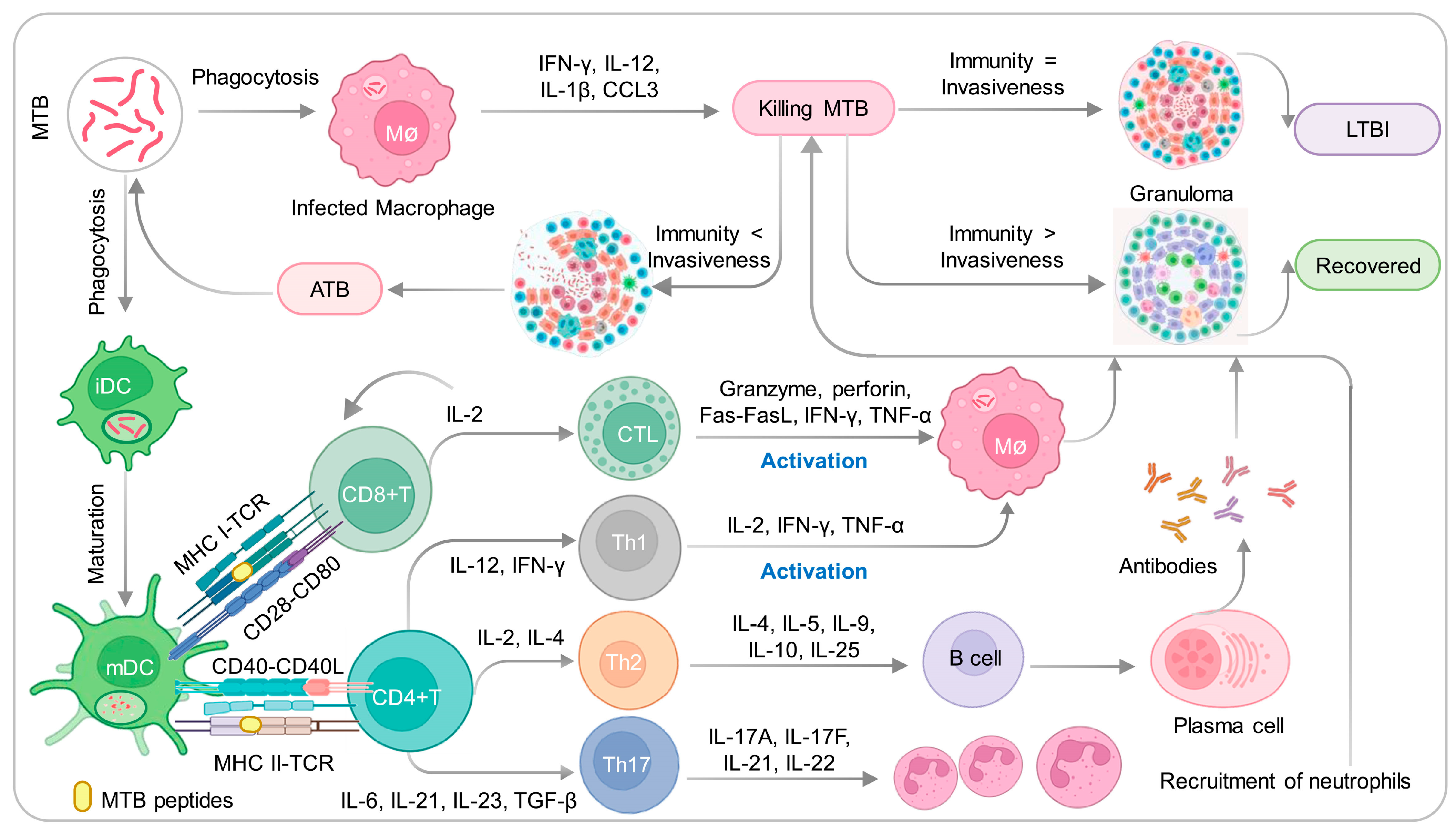
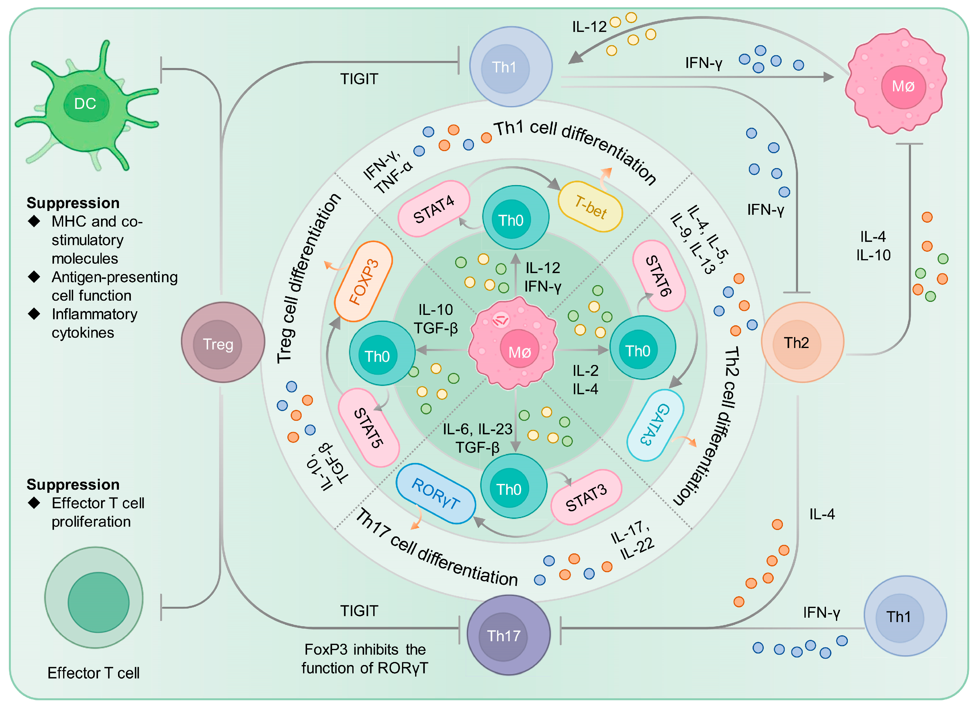
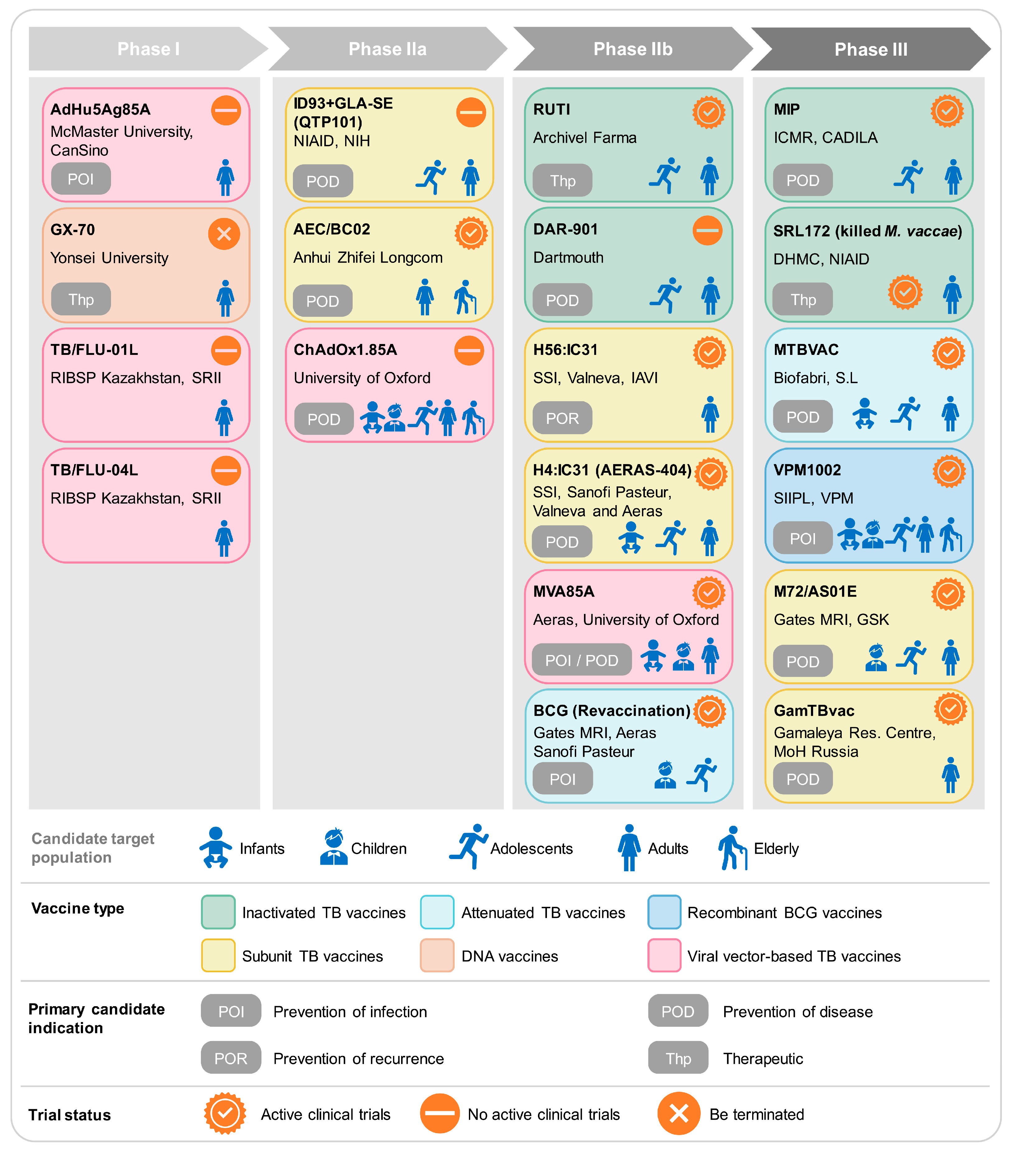
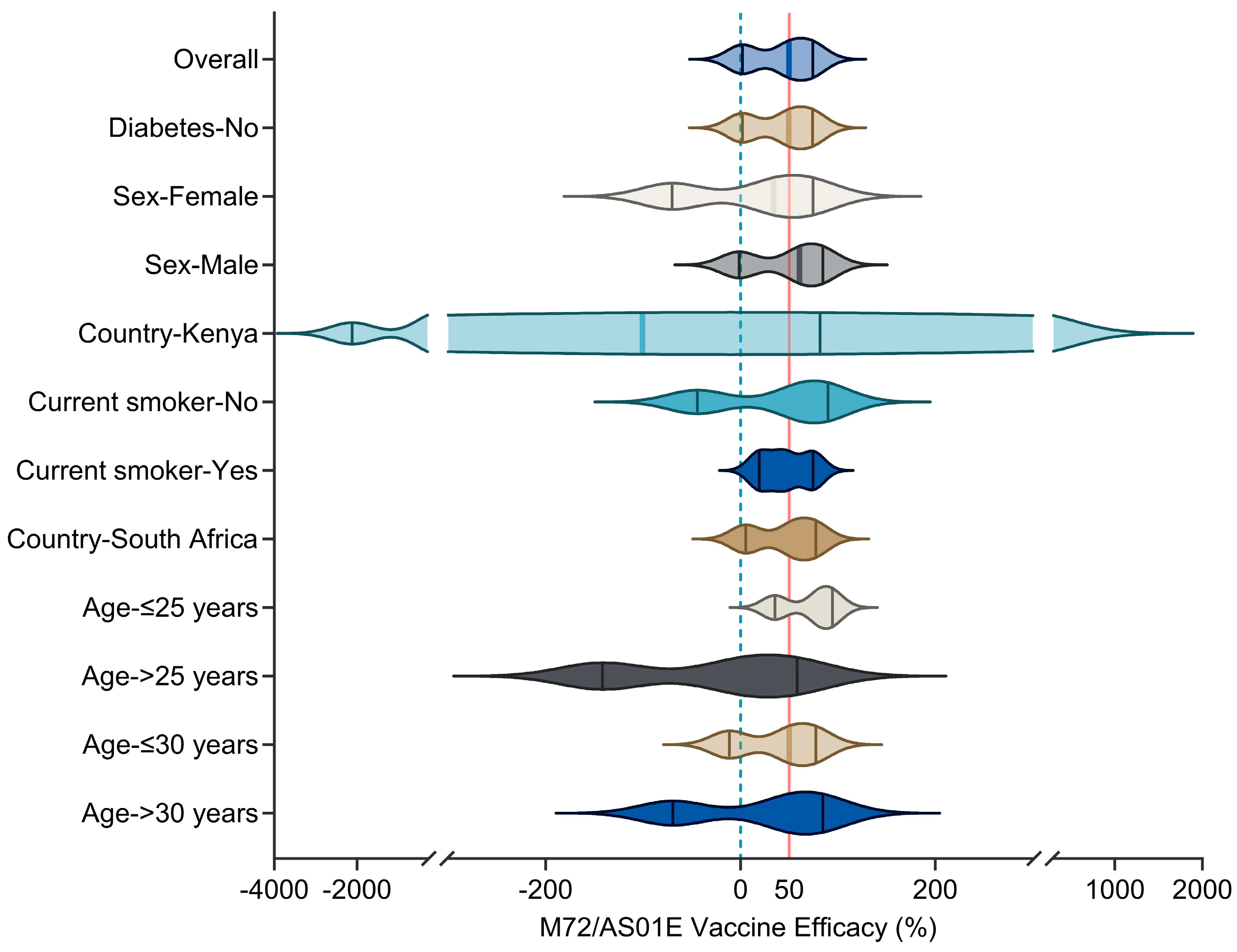
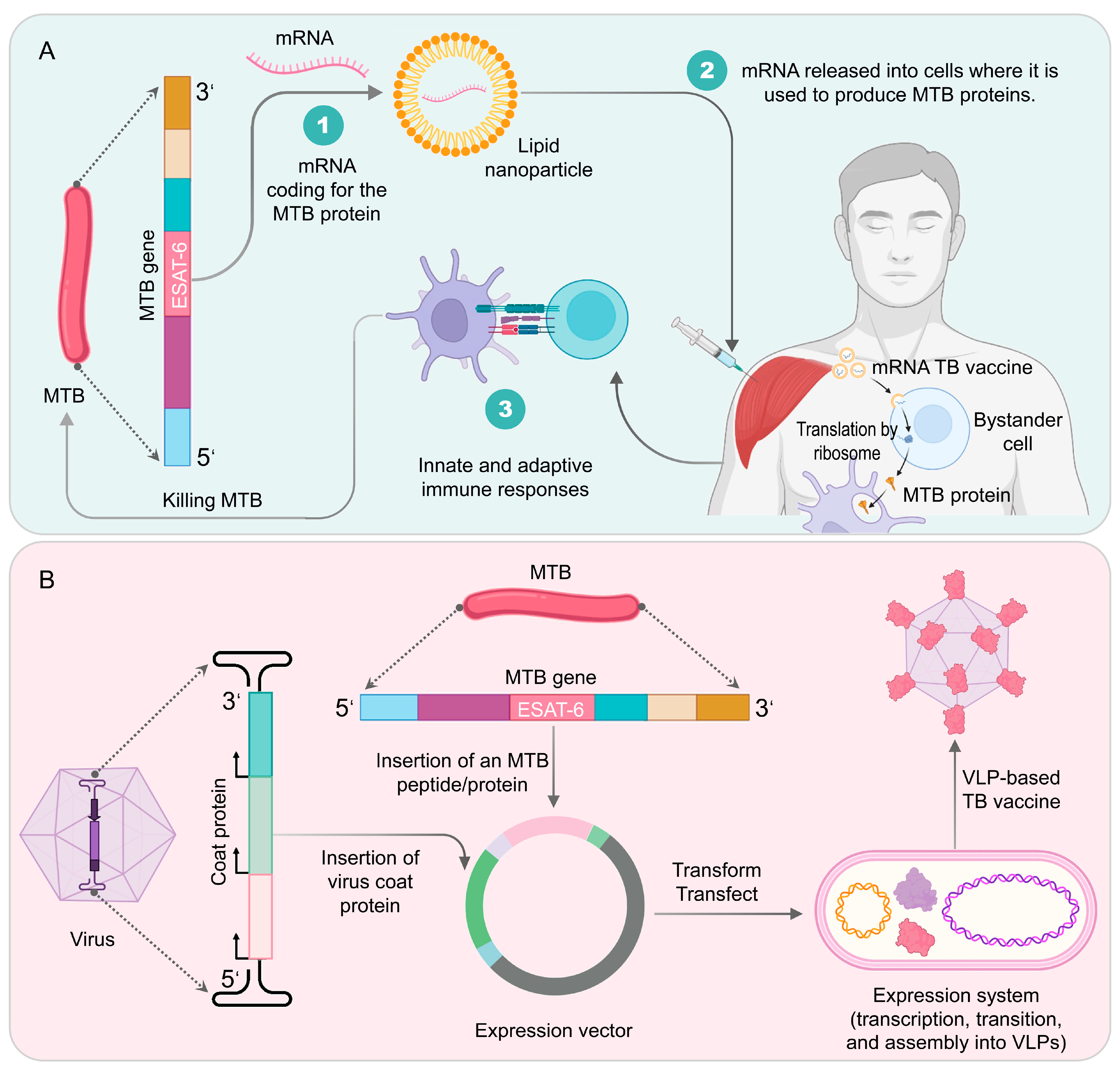
Figure 1. Innate and adaptive immune responses induced by the MTB. Upon entering the pulmonary alveoli via the respiratory tract, MTB is first recognized by resident immune cells such as alveolar macrophages, immature dendritic cells (iDCs), and natural killer (NK) cells. Following capture by iDCs, which migrate from the site of infection to the lymph nodes, the bacteria induces their differentiation into mature dendritic cells (mDCs) with enhanced antigen-presenting and MHC expression capacity. Through the assistance of CD28-CD80 and CD40-CD40L, MHC I and MHC II molecules on dendritic cells recognize and activate CD4+ and CD8+ T lymphocytes. CD4+ T lymphocytes differentiate into Th1, Th2, or Th17 subsets depending on the microenvironmental cytokines and contribute to the control of MTB infection. Th1 and Th2 immunity counterbalance each other and maintain immune homeostasis. CD8+ T lymphocytes further differentiate into cytotoxic T lymphocytes (CTLs) that produce granzyme, perforin, Fas-FasL, IFN-γ, TNF-α, and other molecules to activate macrophages and eliminate the bacteria. Depending on the interplay between innate and adaptive immune responses and the virulence of MTB, infection can lead to either recovery, latent tuberculosis infection (LTBI), or active tuberculosis (ATB).
Vaccines 11 01304 g001
Figure 2. Interplay and balance of CD4+ T cell subtypes in MTB infection. APCs, such as macrophages, present MTB antigen peptides to naive T cells (Th0 cells) through MHC II molecules, inducing the differentiation of T cells into different subtypes depending on the cytokine microenvironment. Upon stimulation with IL-12 and IFN-γ secreted by macrophages, Th0 cells activate STAT4 and T-bet, differentiate into Th1 subtype, and release cytokines such as IFN-γ and TNF-α to combat MTB infection. Similarly, macrophages can facilitate the differentiation of Th0 cells into Th2, Th17, and Treg subtypes by secreting different cytokines, including IL-2 and IL-4, IL-6, IL-23, and TGF-β, as well as IL-10 and TGF-β. The interplay among Th1, Th2, Th17, and Treg subtypes is complex and balanced, and they work together to exert immune responses and maintain the host defense against MTB infection.
Vaccines 11 01304 g002
Figure 3. TB vaccine clinical pipeline. The current pipeline delivers data pertinent to vaccine candidates undergoing clinical development, while information on candidates in preclinical development remains uncollected. Abbreviations: RIBSP, Research Institute for Biological Safety Problems; SRII, Smorodintsev Research Institute of Influenza; NIAID, National Institute of Allergy and Infectious Diseases; NIH, National Institutes of Health; SSI, Statens Serum Institut; IAVI, International AIDS Vaccine Initiative; ICMR, Indian Council of Medical Research; CADILA, Cadila Pharmaceuticals Ltd.; DHMC, Dartmouth Hitchcock Medical Center; SIIPL, Serum Institute of India Private Limited; VPM, Vakzine Projekt Management GmbH; Gates MRI, Bill & Melinda Gates Medical Research Institute; GSK, GlaxoSmithKline.
Vaccines 11 01304 g003
Figure 4. Efficacy of M72/AS01E subunit vaccine against confirmed cases of PTB in individuals without HIV infection. The analysis utilized an unadjusted Cox regression model in the efficacy cohort as per the protocol. The data used in this figure were obtained from the previous study [79]. Copyright © 2019 Massachusetts Medical Society.
Vaccines 11 01304 g004
Figure 5. Illustrates the generation of mRNA TB vaccines and VLP-based TB vaccines. In (A), the DNA encoding the target protein of MTB is obtained and transcribed into mRNA. The mRNA is then loaded with lipid nanoparticles and other carriers to create a TB mRNA vaccine for intramuscular immunization. When these mRNA vaccines are injected into the human body, ribosomes in bystander cells assist in translating the target protein. Bystander cells engulf the produced protein, activating both innate and adaptive immune responses to eliminate MTB. In (B), a coat protein from a virus is cloned into an expression vector. The expression vector is then modified to include the MTB protein, such as ESAT-6 or CFP-10. The resulting expression vector contains both the coat protein and the MTB protein. It is transformed or transfected into an expression system to produce the proteins. Finally, the MTB protein assembles into VLPs, resulting in a VLP-based TB vaccine.
Vaccines 11 01304 g005
Table 1. List of TB vaccines currently in clinical trials.
VaccineNCT
Number
PhasePopulationSample SizeArms and InterventionsPrimary Outcome MeasuresResultsRef.
M72/AS01ENCT01755598ILTBI individuals3575Experiment: 2 doses of M72/AS01E (10 µg), spaced one month apart, i.m. in the arm triangle area.
Placebo comparator: 2 doses of placebo with a 1-month interval, i.m. in the arm triangle area.
Incident rates of definite PTB disease, not associated with HIV-infection, meeting the case definitionLTBI population vaccinated with M72/AS01E can prevent latent infections from developing into TB, the vaccine efficacy in the 36th month was 49.7%[78,79]
NCT00950612IIAdolescents60Experiment: 2 doses of M72/AS01E (10 µg), with an interval of one month, i.m. in the arm triangle area.
Placebo comparator: 2 doses of placebo with a one-month interval, i.m. in the arm triangle area.
Number of subjects with solicited local symptoms and solicited general symptoms, unsolicited AEs and SAEs, different levels biochemical and hematological levelsM72/AS01E induced strong T cell and antibody responses, including NK cells and IFN- γ generation[80]
NCT04556981IIIHIV-positive patients402Experiment: 2 doses of M72AS01E (10 µg), i.m., at days 1 and 29.
Placebo comparator: control sodium chloride 0.9%, i.m., at days 1 and 29.
Number of participants with solicited AEs, unsolicited AEs, and SAEsNA
GamTBvacNCT03255278IHealthy adults60Placebo comparator: s.c. 0.5 mL placebo.
Safety and portable study group: s.c. 0.25 mL GamTBvac.
Experimental 1: s.c. 0.25 mL GamTBvac.
Experimental 2: s.c. 0.5 mL GamTBvac.
Experimental 3: s.c. 1 mL GamTBvac.
The number of AEsDifferent doses of vaccines were evaluated for immunogenicity, with half dose (0.5 mL) having the best effect[81]
NCT03878004IIHealthy adults180Experiment: s.c. 2 doses of 0.5 mL GamTBvac, with an interval of 8 weeks.
Placebo comparator: s.c. 2 doses of 0.5 mL placebo, with an interval of 8 weeks.
1. Level of IFN-γ secretion in whole blood or PBMC fraction
2. Number of participants with AEs
The vaccine is well tolerated and induces specific and persistent Th1 and Humoral immunity responses[82]
NCT04975737IIIHealthy adults7180Experiment: s.c. 2 doses of 0.5 mL GamTBvac, with an interval of 8 weeks.
Placebo comparator: s.c. 2 doses of 0.5 mL placebo, with an interval of 8 weeks.
Preventive efficacy (Ep)NA
H56:IC31NCT01967134ILTBI/healthy adults25Experiment: LTBI negative, i.m. 3 doses of 15 µg/500 nmol H56:IC31 at days 0, 56 and 112.
Experiment: LTBI positive, i.m., 3 doses of 50 µg/500 nmol H56:IC31, days 0, 56 and 112.
Number of participants with at least 1 AE until day 210H56:IC31 vaccine induces antigen-specific IgG response and CD4+ T cells expressing Th1 cytokines[83]
NCT02378207IbAdolescent84Experiment: i.m. 0.5 mL H4:IC31 or H56:IC31 at days 0 and 56.
Active comparator: i.m. 0.1 mL BCG at day 0.
Placebo comparator: i.m. 0.5 mL NaCI at days 0 and 56.
1. Number of participants with AEs
2. Percentage of participants with response rates to TB antigens as compared to baseline
Both H4: IC31 and H56: IC31 induce CD4+ T cell responses, and H4:IC31 and H56:IC31 induce serum IgG[84]
NCT01865487IIaHealthy adults/LTBI98Placebo control: i.m. 2 doses of NaCl.
Experimental groups: 5/500, 15/500, 50/500 H56ug/IC31nmol QFT-negative and/or -positive, 2 doses, days 0 and 56.
Number and percentage of unsolicited and solicited AEsH56:IC31 induces a persistent antigen-specific Th1 response without being affected by MTB infection[85]
NCT02503839I/IIATB patients51Experimental 1: 120 g of etoricoxib, po. at days 0 and 140.
Experimental 2: 140 µg H56:IC31 and no etoricoxib i.m. at days 84 and 140.
Placebo comparator: Standard TB treatment.
Experimental 3: 120 g etoricoxib po. at days 0 and 140; 140 µg H56:IC31 i.m. at days 0 and 140.
1. Participants with AEs
2. Immunogenicity of etoricoxib/H56:IC31 vaccine
H56:IC31 vaccination is safe and immunogenic in ATB patients[86]
NCT03512249IIbATB patients900Experiment: i.m. 2 doses of 0.5 mL H56:IC31 at days 0 and 56.
Placebo comparator: i.m. 2 doses of 0.5 mL NaCI at days 0 and 56.
Rate of TB disease recurrenceNA
H4:IC31NCT02074956
NCT02066428
IHealthy adults60Experimental 1–4 groups: i.m. 5 µg, 15 µg, 50 µg, or 150 µg/500 nmol, H4:IC31 at days 0 and 56.
Experimental 5: i.m. H4:IC31 (15 µg/100 nmol).
Placebo: NaCl.
Safety profile of two injections of H4:IC31 at different dose levels; evaluate the safety of one or two injections of two H4:IC31 antigen amounts administered with three different amounts of adjuvantThe evidence provided by the dose escalation test indicates that the optimal antigen adjuvant dose combination is H4, 5, 15, or 50 µg and IC31 500 nmol[87]
NCT02075203IIAdolescents989Experiment: i.d. 2 doses of H4:IC31 (15 mcgH4/500 nmol IC31) at days 0 and 56.
Active comparator: i.d. BCG at day 0.
Control group: i.d. 2 doses of 0.9% NaCl (0.9%) at day 0.
1. Safety profile of H4:IC31 and BCG revaccination
2. Number of participants testing positive for MTB at day 84
The immune efficacy of the H4: IC31 vaccine is 30.5% (p = 0.16). 44 (14.3%) H4:IC31 group experienced QFT conversion, 41 (13.1%) BCG group experienced QFT conversion, and 49 (15.8%) placebo group experienced QFT conversion[88]
ID93/GLA-SENCT01599897IHealthy adults60Experimental 1–4: i.m. 2 µg ID93 + 2 µg GLA-SE, 10 µg ID93 + 2 µg GLA-SE, 2 µg ID93 + 5 µg GLA-SE, 10 µg ID93 + 5 µg GLA-SE at days 0, 28, and 56, respectively.
Active comparator 1–2: i.m. 2 µg or 10 µg ID93 at days 0, 28, and 56.
Number of patients experiencing AEsShowing a satisfactory safety profile and eliciting a functional humoral and T-helper 1 type cellular response[89]
NCT02465216IIaHealthy adults60Experimental 1–3: i.m. 2 μg ID93 + 2 μg GLA-SE, 10 μg ID93 + 2 μg GLA-SE, 2 μg ID93 + 5 μg GLA-SE at days 0 and 56, respectively.
Experimental 4: i.m. 3 doses of 2 µg ID93+ 5 µg GLA-SE at days 0, 28, and 56.
Active comparator: i.m., NaCI at days 0, 28, and 112.
Number of AEsThe antigen-specific IgG and CD4 T cell responses induced by a dose of 2 μg ID93 + 5 μg GLA-SE were significantly higher than those induced by placebo, and lasted for 6 months[90]
NCT03806686IIaHealthy adults107Experimental 1–2: i.m. 2 μg ID93 + 5 µg GLA-SE, 5 μg ID93 +5 μg GLA-SE at days 0, 28, and 56, respectively.
Active comparator: i.m. 0.5 mL NaCI at days 0, 28, and 56.
AEsNA
AEC/BC02NCT04239313IHealthy adults30Experiment: i.m. low-dose AEC/BC02; Active comparison: i.m. low-dose adjuvant; active comparator: i.m. placebo.The number of AEs after i.m.NA
NCT05284812IILTBI individuals200Negative control group: not vaccinated. Sentinel group: 18–59-year-olds received low- or high-dose injections; ≥60 years old received low or high-dose injections, i.m. Experimental group 1: EC positive, i.m. low-dose AEC/BC02. Experimental group 2: EC positive, i.m. high-dose AEC/BC02. Experimental group 3: EC positive, i.m. high-dose AEC/BC02 into 1,3, and 6 doses; i.m. 2, 4, 5 doses of placebo. Placebo group: EC positive, i.m. placebo. Adjuvant group: high-dose adjuvant for AEC/BC02.The number of AEs after i.m. injectionNA
VPM1002NCT00749034IHealthy male80Experiment: i.d. 0.05 mL VPM1002 (5 × 10³, 5 × 10⁴, 5 × 10⁵ CFUs).
Active comparator: i.d. 0.05 mL BCG (2–8 × 10⁵ CFUs).
Physical examination, vital signs, ECG, liver sonography, chest X-ray, laboratory safety parameters, tolerability, recording of concomitant medication and AEsVPM1002 has safety and immunogenicity in response to B and T cells[91]
NCT01479972IINewborn infants 48Experiment: i.d. 0.05 mL VPM1002.
Active comparator: i.d. 0.05 mL BCG.
1. Safety
2. Immunogenicity
Inoculation with VPM1002 can induce multifunctional CD+ 4 and CD8+ T cells[92]
NCT02391415IINewborn infants 416Experiment: HIV-unexposed infants, i.d. 0.05 mL VPM1002.
Control group: HIV-unexposed infants, i.d. 0.05 mL BCG.
Experiment: HIV-unexposed infants, i.d. 0.05 mL VPM1002 (Hyg+).
Control group: HIV-exposed infants, i.d. 0.05 mL BCG.
Experiment: HIV-exposed infants, i.d. 0.05 mL VPM1002.
The difference between the VPM1002 and BCG vaccination groups in the incidence of grade 3 and 4 adverse drug reactions and IMP-related ipsilateral or generalized lymphadenopathy of 10 mm or greater (diameter)VPM1002 is safe for both HIV-exposed and -unexposed infants; both VPM1002 and BCG have immunogenicity, and from the 6th week onwards, the immune response intensity induced by BCG is greater than that of VPM1002[93]
NCT03152903IIIHealthy adults2000Experiment: i.d. 0.05 mL VPM1002.
Active comparator: i.d. 0.05 mL placebo.
Percentage of bacteriologically confirmed TB recurrence casesNA
NCT04351685IIINewborn infants 6940Experimental group: i.d. 0.05 mL VPM1002.
Active comparator: i.d. 0.05 mL BCG.
Incident cases of QFT conversionNA
MTBVACNCT02013245IHealthy adults 34Experimental 1: i.d. 0.1 mL MTBVAC (5 × 10³ CFUs).
Experimental 2: i.d. 0.1 mL MTBVAC (5 × 10⁴ CFUs).
Experimental 3: i.d. 0.1 mL MTBVAC (5 × 10⁵ CFUs).
Active comparator: i.d. 0.1 mL BCG (5 dose 5 × 10⁵ CFU).
Number of participants with AEs up to 210 days after vaccinationMTBVAC exhibits good safety in healthy adults, similar to BCG[94]
NCT02729571IbNewborn infants 54Experimental 1: i.d. 0.1 mL MTBVAC (5 × 10⁵ CFUs)/0.1 mL BCG SII (5 × 10⁵ CFUs) adults.
Experimental 2: i.d. 0.05 mL MTBVAC (2.5 × 10³ CFUs/2.5 × 104 CFUs/2.5 × 105 CFUs) infants.
Experimental 3: i.d. 0.05 mL BCG (2.5 × 10⁵ CFUs) infants.
Safety and reactogenicity in infants and adultsMTBVAC has good safety and immunogenicity, and can induce long-lasting CD4 cell responses in infants[95]
NCT02933281Ib/IIaHealthy adults144Experimental 1–4: QFT negative, i.d. MTBVAC 5 × 10³, 5 × 10⁴, 5 × 10⁵, or 5 × 10⁶ CFUs at day 0, respectively.
Experimental 5–8: QFT positive, i.d. MTBVAC 5 × 10³, 5 × 10⁴, 5 × 10⁵, or 5 × 10⁶ CFUs at day 0, respectively.
Active comparator: i.d. BCG 5 × 10⁵ CFUs at day 0.
Safety and reactogenicity of MTBVAC at escalating dose levels compared to BCG vaccine by assessing number of participants with AEs and SAEsNA
NCT03536117IIaNewborn infants99Experimental 1–3: i.d. 0.05 mL MTBVAC (2.5 × 10⁴, 2.5 × 10⁵, or 2.5 × 10⁶ CFUs), respectively.
Active comparator: i.d. 0.05 mL BCG (2.5 × 10⁵ CFUs).
1. Number of participants with treatment-related AEs as defined in protocol
2. Immunogenicity analysis in infants
NA
NCT04975178IIINewborn infants 6960Experiment: i.d. 0.05 mL MTBVAC.
Active comparator: i.d. 0.05 mL BCG.
Prevention of TB disease in healthy HIV-uninfected and HIV-exposed uninfected neonatesNA
BCG (revaccination)NCT02378207IIbAdolescents84Active comparator: BCG (2–8 × 10⁵ CFUS), i.m. as 0.1 mL in either deltoid muscle at day 0.
Placebo comparator: control sodium chloride 0.9%, i.m. as 0.5 mL in alternating deltoid muscle at days 0 and 56.
Number of participants with AEs; percentage of participants with response rates to TB antigens as compared to baselineBCG revaccination had acceptable safety and induced robust, multifunctional BCG-specific CD4+ T cells[84]
NCT02075203IIAdolescents989Active comparator: BCG SSI vaccine, 1 dose on day 0; placebo comparator: placebo, 2 doses on days 0 and 56.Safety profile of BCG revaccination; number of participants testing positive for MTB at day 84BCG revaccination was immunogenic and reduced the rate of sustained QFT conversion, with an efficacy of 45.4% (p = 0.03)[88]
NCT04152161IIbChildren and adolescents1820Experiment: a single 0.1 mL of BCG vaccine SSI, i.d. in deltoid region of the upper arm.
Placebo: a single 0.1 mL of normal saline, i.d. in deltoid region of the upper arm.
Number of participants with sustained QFT conversionNA
MIPNCT00341328IIICategory-I PTB patients300Experimental group: i.d., 6 doses MIP vaccine given 0.2 mL at baseline, then 0.1 mL every 2 weeks for 8 weeks.
Control group: i.d., 6 doses placebo given 0.2 mL at baseline, then 0.1 mL every 2 weeks for 8 weeks.
The time of sputum conversion, the cure rate; the relapse in patients of category-I pulmonary TB, and any clinical adverse reactions NA
NCT00265226IIICategory-II PTB patients1020Experimental group: intradermal administration of mycobacterium W vaccine and category II ATT as per RNTCP guidelines.
Control group: placebo administration and category II ATT drugs as per RNTCP guidelines.
1. Sputum conversion time
2. Cure rate
3. Clinical adverse reactions
At the 39th week follow-up, sputum culture conversion rate was 94.2% (309/328) in the MIP group and 89.17% (280/314) in the placebo group[96]
RUTINCT00546273IHealthy adults 24Experimental groups 1–4: RUTI (5 µg, 25 µg, 100 µg, or 200 µg) i.d., at days 0 and 28.
Placebo comparator: placebo i.d. at days 0 and 28.
1. VAS Pain Score
2. Local and systemic events occurrence, intensity, and relationship to vaccination
3. Number of clinically relevant abnormalities detected
RUTI has been proven to trigger specific reactions against MTB[97]
NCT01136161IIaLTBI individuals95Experimental groups 1–5: HIV-negative, 5 µg, 25 µg, 100 µg, 200 µg RUTI or placebo i.d. at days 28 and 56, respectively.
Experimental groups 6–10: HIV-positive, 5 µg, 25 µg, 100 µg, 200 µg RUTI or Placebo i.d. at days 28 and 56, respectively.
1. Local tolerability, focal tolerability
2. Systemic tolerability, vital signs and physical examination, ECG, and laboratory tests
The best cellular multi-antigen response was achieved when administered at 25 µg RUTI[98]
NCT04919239IIbATB patients140Experiment: 25 µg RUTI, i.d.
Placebo comparator: 25 µg placebo, i.d.
Percentage of patients with sputum culture negativeNA
NCT05455112 IIbATB patients44Experiment: 25 µg RUTI, i.d.
Placebo comparator: 25 µg NaCI, i.d.
1. Early bactericidal activity (EBA) 0–14
2. AEs and grade 3–4 AEs
NA
DAR-901NCT02063555IHealthy adults 59Experiment: 0.1 mL DAR-901 i.d., at 0, 2 and 4 months.
Active comparator: 0.1 mL NaCI i.d., at 2 and 4 months; 0.1 mL BCG i.d., at 4 months.
Placebo comparator: 0.1 mL NaCI, i.d. at 0, 2, and 4 months.
SafetyDAR-901 induces low-intensity multifunctional memory CD4+ T cell response and reaching peak in 7 days[99]
NCT02712424IIbAdolescents625Experimental group: 0.1 mL DAR-901, i.d.
Placebo comparator: 0.1 mL NaCI, i.d.
New infection with MTBThe three-dose series of DAR-901 is safe and well tolerated, but cannot prevent initial or sustained IGRA conversion[100]
MVA85ANCT00460590IHealthy adults 24Experimental groups 1–3: adults vaccinated with BCG, adults without BCG vaccination, or teenagers i.d. 5 × 10⁷ pfu MVA85A, respectively.The safety of a single injection of MVA85A in healthy subjects by collecting data on AEsMVA85A vaccine induces the production of long-lasting, multifunctional, Ag85A specific CD4+ T cells[101]
NCT01650389IIHIV-exposed infants248Experimental group: 1 × 10⁸ pfu MVA85A vaccine within 96 h of birth, i.d.
Placebo comparator: equal volume intradermal administration within 96 h of birth i.d.
SafetyMVA85A immunization in newborns exposed to HIV is safe and induces a moderate antigen-specific immune response without affecting BCG vaccination[102]
NCT00953927IIInfants2797Experiment: 1 × 10⁸ pfu MVA85A vaccine, i.d.
Placebo comparator: 1 × 10⁸ pfu candida skin test antigen, i.d.
Safety profile of MVA85A in BCG-vaccinated, HIV-negative infants32 out of 1399 MVA85A subjects (2%) achieved the primary efficacy endpoint
39 out of 1395 controls (3%) achieved the primary efficacy endpoint
[103]
ChAdOx1.85ANCT01829490IHealthy adults 42Experimental groups 1–2: 1 dose of 5 × 10⁹ or 2.5 × 10¹⁰ vp of ChAdOx1.85A, i.m., respectively.
Experimental group 3: 1 dose of 2.5 × 10¹⁰ vp of ChAdOx1.85A, followed by a boost dose of 1 × 10⁸ pfu of MVA85A, at 56 days later, i.m.
Experimental group 4: 2 doses of 2.5 × 10¹⁰ vp of ChAdOx1.85A i.m., at days 0 and 28, followed by a boost dose of 1 × 10⁸ pfu of MVA85A, at day 119, i.m.
Safety of ChAdOx1 85A vaccination with and without MVA85A boost vaccination in healthy, BCG-vaccinated adultsChAdOx1.85A induces Ag85A specific CD4+ T and CD8+ T cell responses[104]
NCT03681860IIaHealthy adults 72Experiment: i.m. 5 × 10⁹ vp ChAdOx1.85A.
Experimental 2: 3 adults i.m. 2.5 × 10¹⁰ vp ChAdOx1.85A.
Experimental 3: 3 adolescents i.m. 5 × 10⁹ vp ChAdOx1.85A.
Experimental 4: 3 adolescents i.m. 2.5 × 10¹⁰ vpChAdOx1.85A.
Placebo comparator: 30 EMaBS adolescents i.m. 2.5 × 10¹⁰ vp ChAdOx1.85A, further strengthened MVA85A, other adolescents BCG revaccinated.
Safety and immunogenicityNA[104]
AdHu5Ag85ANCT02337270IHealthy adults36Experiment: aerosol 106 Ad5Ag85A at day 0.
Experiment: aerosol 2 × 10⁶ Ad5Ag85A at day 0.
Experiment: i.m. 108 Ad5Ag85A at day 0.
Number of participants reporting AEsCompared with i.m., aerosol delivered AdHu5Ag85A vaccine has advantages in inducing respiratory epithelium immunity[105]
TB/FLU-01LNCT03017378IHealthy adults36Active comparator: TB/FLU-01L (intranasal application).
Active comparator: TB/FLU-01L (sublingual application).
AEs, solicited local and systemic reactions, unsolicited AEs, SAEs, including abnormal laboratory findingsNA
TB/FLU-04LNCT02501421IHealthy adults44Experiment: TB/FLU-04L, at days 1 and 21.
Placebo comparator: placebo, at days 1 and 21.
Immediate reactions, solicited local and systemic reactions, unsolicited events and abnormal laboratory findings, SAEsNA
Abbreviations: i.m., intramuscular injection; s.c., subcutaneous injection; i.d., intradermal injection; po., oral administration; EC: recombinant mycobacterium tuberculosis fusion protein ESAT6-CFP10; AEs: adverse events; SAEs: serious adverse events; EMaBS: Entebbe mother and baby study; NA, not available.
Disclaimer/Publisher’s Note: The statements, opinions and data contained in all publications are solely those of the individual author(s) and contributor(s) and not of MDPI and/or the editor(s). MDPI and/or the editor(s) disclaim responsibility for any injury to people or property resulting from any ideas, methods, instructions or products referred to in the content.

