
The Occurrence of Acute Disseminated Encephalomyelitis in SARS-CoV-2 Infection/Vaccination: Our Experience and a Systematic Review of the Literature
 ,
,  , ,
, ,
Abstract
:1. Introduction
1.1. General Incidence of ADEM
1.2. Etiology
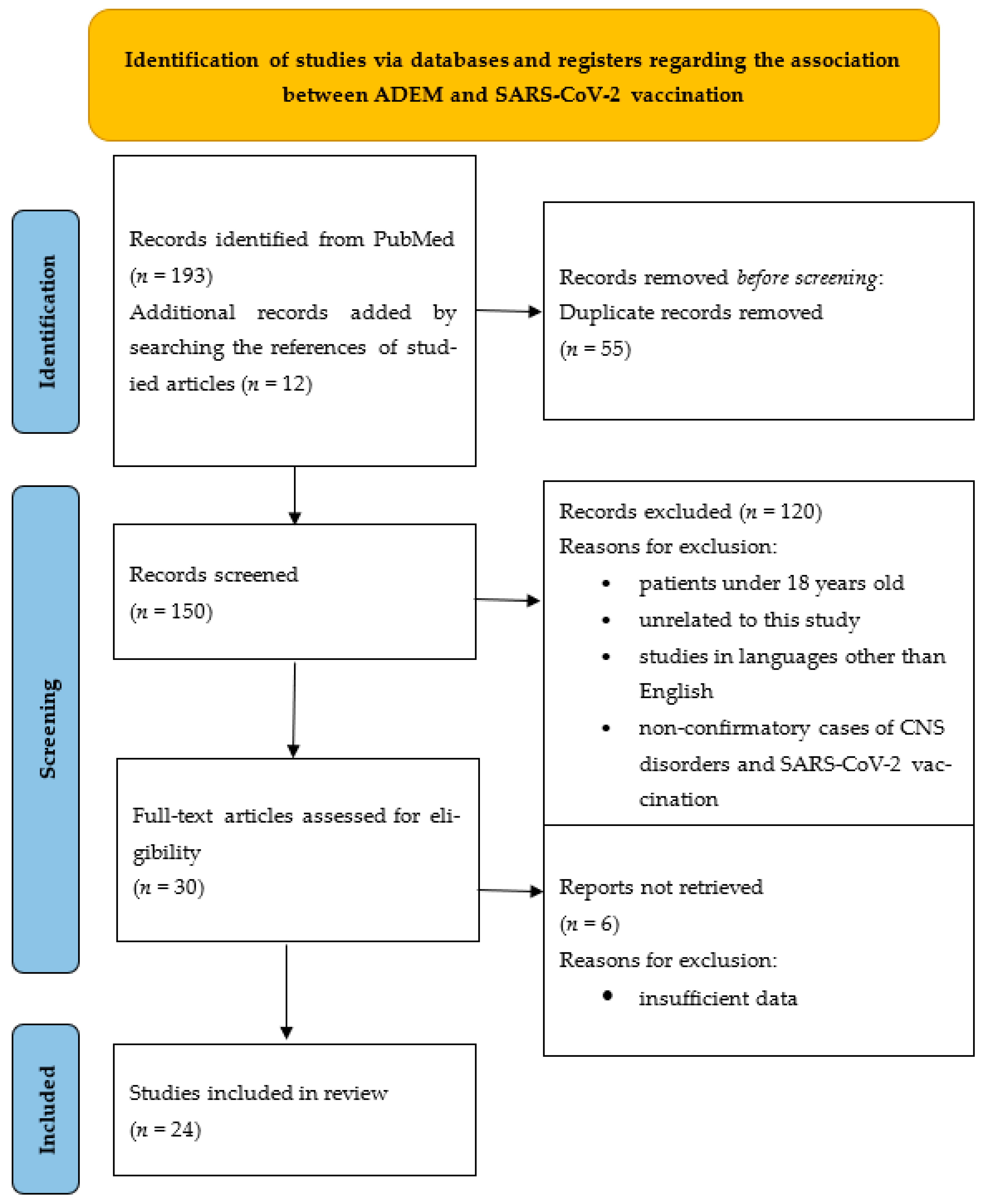
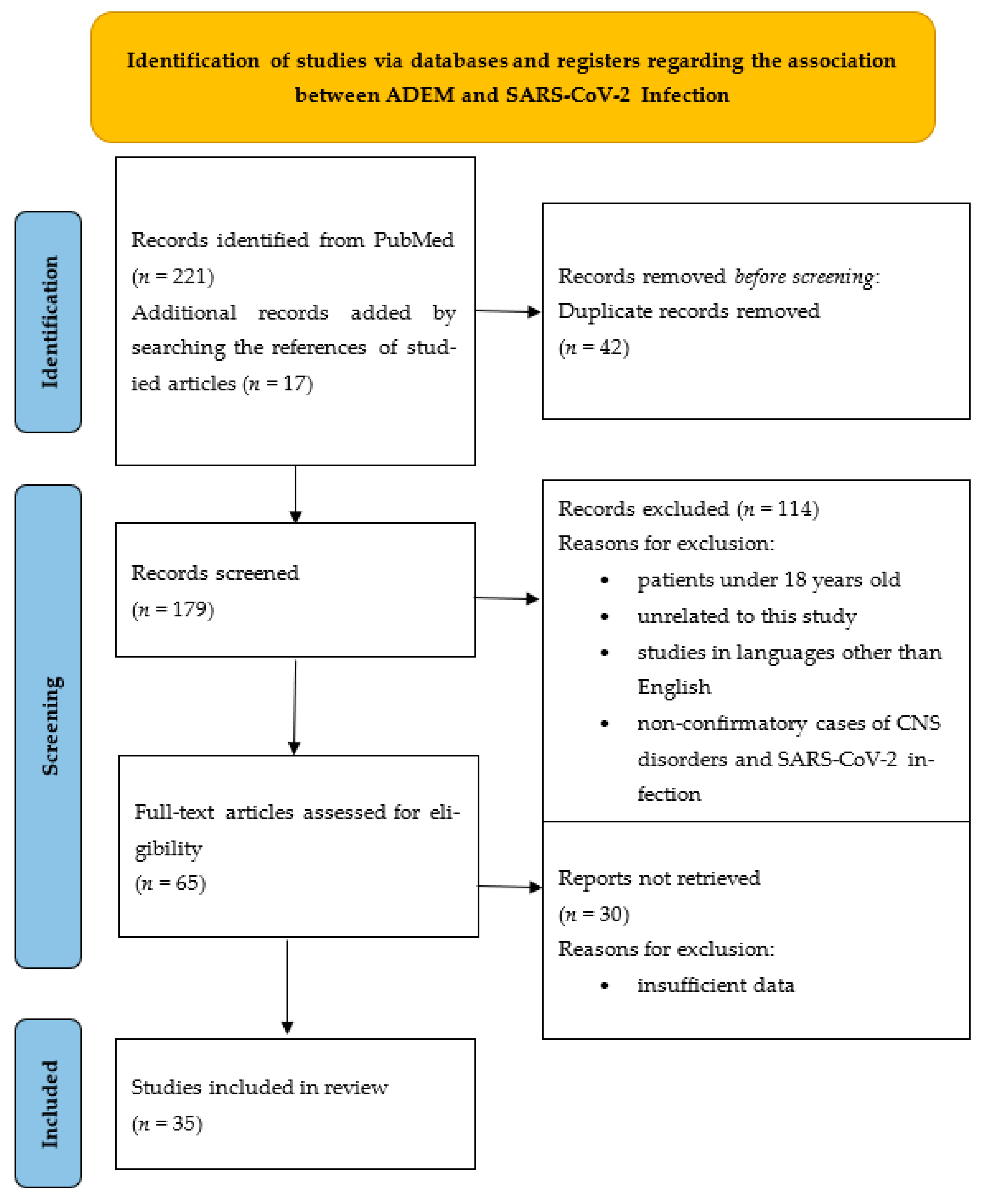
2. Materials and Methods
3. Results
3.1. Our Clinical Experience
- (1)
- The temporal association between the infection/vaccine and disease;
- (2)
- Clinical features;
- (3)
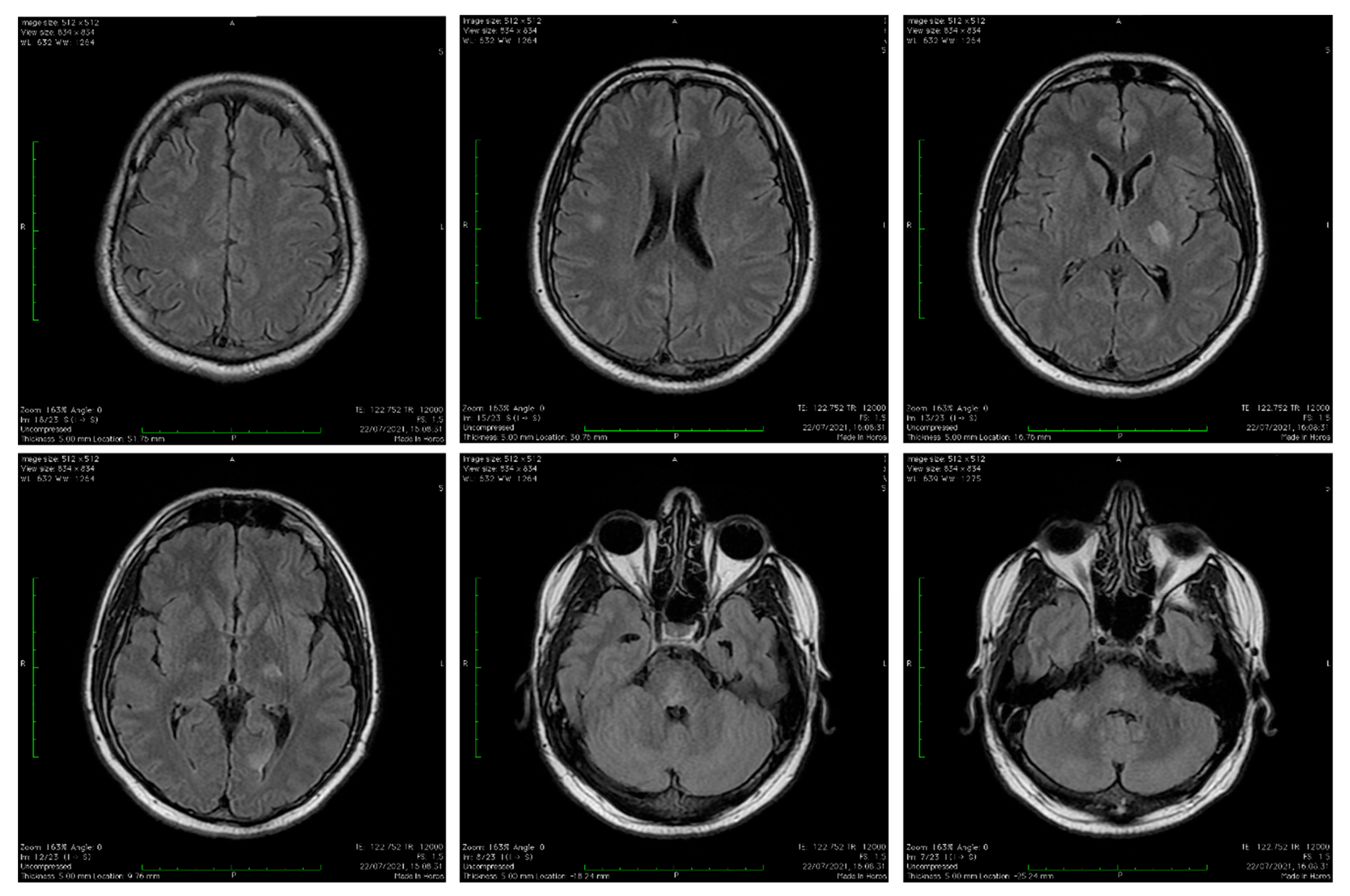
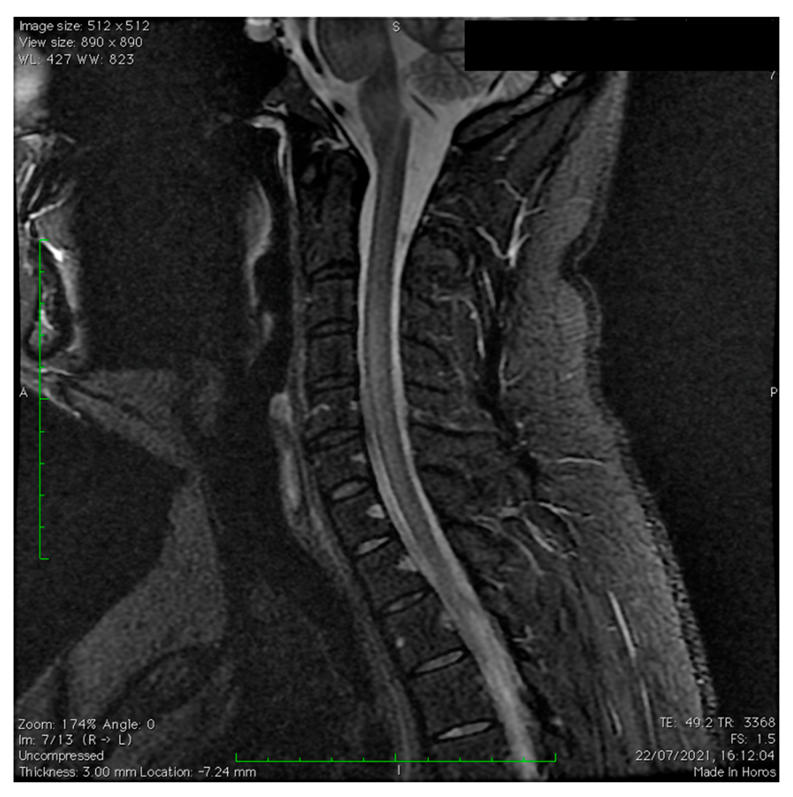
- Appearance on MRI images;
- (4)
- Exclusion of other etiologies;
- (5)
- Favorable response to corticosteroids.
3.2. Literature Review
4. Discussion
4.1. Pathophysiology of ADEM
- (a)
- The concept of molecular mimicry is based on the similarity of an amino-acid sequence (epitope) between myelin proteins of the host and invading pathogens [75,76]. The antigen-presenting cells (dendritic cells) process the pathogen, activating T cells which in turn activate B cells. Both of these cell types are able to enter into the central compartment during the process of immune surveillance and can be reactivated by local antigen-presenting cells (microglia), producing a local inflammatory immune reaction [8,75]. The injection of CD4+ T lymphocytes from immunized animals that recognize myelin-associated protein can initiate the disease in healthy animals [75,77].
- (b)
- CNS infection with a pathogen results in nervous tissue damage with the penetration of autoantigens in systemic circulation through a disrupted BBB. These autoantigens reach the lymphatic organs, where they are processed and initiate a self-reactive T-cell response with nonspecific activation of an autoreactive T-cell clone [8,75].
4.2. Pathophysiology of ADEM after SARS-CoV-2 Infection
- (1)
- Systemic circulation can contribute to the distribution of the virus in the cerebral blood flow and from here, due to sluggish blood flow in the context of inflammation, viral neuroinvasion is facilitated [82].
- (2)
- The virus crosses the BBB due to increased permeability in the context of a cytokine storm [9].
- (3)
- (4)
- The spike protein of the virus binds to cell-surface angiotensin-converting enzyme type 2 (ACE2) receptors found in various tissues and infects the endothelial cells of the BBB or the epithelial cells of the blood–CSF barrier at the level of the choroid plexus—mediating cellular entry of the virus towards the central compartment (brain and brainstem—the nucleus of the solitary tract and the paraventricular nuclei) [1,35,83].
- (5)
4.3. ADEM Diagnosis
- (1)
- Multifocal damage of CNS at first manifestation due to an inflammatory demyelinating cause.
- (2)
- Encephalopathy that cannot be explained by a rise in fever.
- (3)
- Lack of other clinical events or new lesions on MRI in the 3 months following onset.
- (4)
- Brain and/or spine MRI shows lesions in the acute phase (3 months).
- (5)
- Brain lesions on MRI are diffuse and poorly demarcated and have the following characteristics:
- (a)
- Large-size lesions of 1–2 cm that mainly affect the white matter.
- (b)
- Hypointense T1 lesions affecting white matter are rare.
- (c)
4.4. General Considerations on Postinfectious and Postvaccinal ADEM in the Context of COVID-19
- (1)
- mRNA-based vaccines in which human cells are stimulated to produce SARS-CoV-2 proteins and express the viral spike protein on their surface by means of genetically transferred information. The human body then initiates a defensive response against it.
- (2)
- Viral vector-based vaccines in which an adenovirus is used to deliver fragments of the SARS-CoV-2 genome to human cells.
- (3)
5. Conclusions
Author Contributions
Funding
Institutional Review Board Statement
Informed Consent Statement
Data Availability Statement
Acknowledgments
Conflicts of Interest
References
- Brola, W.; Wilski, M. Neurological consequences of COVID-19. Pharmacol. Rep. 2022, 74, 1208–1222. [Google Scholar] [CrossRef] [PubMed]
- Stoian, A.; Bajko, Z.; Maier, S.; Cioflinc, R.A.; Grigorescu, B.L.; Motătăianu, A.; Bărcutean, L.; Balasa, R.; Stoian, M. High-dose intravenous immunoglobulins as a therapeutic option in critical illness polyneuropathy accompanying SARS-CoV-2 infection: A case-based review of the literature (Review). Exp. Ther. Med. 2021, 22, 1182. [Google Scholar] [CrossRef] [PubMed]
- Stoian, A.; Stoian, M.; Bajko, Z.; Maier, S.; Andone, S.; Cioflinc, R.A.; Moțățăianu, A.; Barcuțean, L.; Bălașa, R. Autoimmune Encephalitis in COVID-19 Infection: Our Experience and Systematic Review of the Literature. Biomedicines 2022, 10, 774. [Google Scholar] [CrossRef] [PubMed]
- Evans, M.; Hanbury, G.; Lim, E.; Dixon, L.; Singh-Curry, V.; Nicholas, R. 118 Acute Disseminated Encephalomyelitis Associated with SARS-CoV-2 Vaccine in Multiple Sclerosis: A Case Report. J. Neurol. Neurosurg. Psychiatry 2022, 93, e2. [Google Scholar] [CrossRef]
- Kania, K.; Ambrosius, W.; Tokarz Kupczyk, E.; Kozubski, W. Acute disseminated encephalomyelitis in a patient vaccinated against SARS-CoV-2. Ann. Clin. Transl. Neurol. 2021, 8, 2000–2003. [Google Scholar] [CrossRef]
- Boesen, M.S.; Blinkenberg, M.; Koch-Henriksen, N.; Thygesen, L.C.; Uldall, P.V.; Magyari, M.; Born, A.P. Implications of the International Paediatric Multiple Sclerosis Study Group consensus criteria for paediatric acute disseminated encephalomyelitis: A nationwide validation study. Dev. Med. Child. Neurol. 2018, 60, 1123–1131. [Google Scholar] [CrossRef] [Green Version]
- Osborn, A.G.; Hedlund, G.L.; Salzman, K.L. Osborn’s Brain: Imaging, Pathology, and Anatomy; Elsevier: Amsterdam, The Netherlands, 2017. [Google Scholar]
- Menge, T.; Hemmer, B.; Nessler, S.; Wiendl, H.; Neuhaus, O.; Hartung, H.P.; Kieseier, B.C.; Stüve, O. Acute disseminated encephalomyelitis: An update. Arch. Neurol. 2005, 62, 1673–1680. [Google Scholar] [CrossRef]
- Varadan, B.; Shankar, A.; Rajakumar, A.; Subramanian, S.; Sathya, A.C.; Hakeem, A.R.; Kalyanasundaram, S. Acute hemorrhagic leukoencephalitis in a COVID-19 patient—A case report with literature review. Neuroradiology 2021, 63, 653–661. [Google Scholar] [CrossRef]
- Anilkumar, A.C.; Foris, L.A.; Tadi, P. Acute Disseminated Encephalomyelitis; StatPearls [Internet]; StatPearls Publishing: Treasure Island, FL, USA, 2022. [Google Scholar]
- Leake, J.A.; Albani, S.; Kao, A.S.; Senac, M.O.; Billman, G.F.; Nespeca, M.P.; Paulino, A.D.; Quintela, E.R.; Sawyer, M.H.; Bradley, J.S. Acute disseminated encephalomyelitis in childhood: Epidemiologic, clinical and laboratory features. Pediatr. Infect. Dis. J. 2004, 23, 756–764. [Google Scholar] [CrossRef]
- Nagaratnam, S.A.; Ferdi, A.C.; Leaney, J.; Lee, R.L.K.; Hwang, Y.T.; Heard, R. Acute disseminated encephalomyelitis with bilateral optic neuritis following ChAdOx1 COVID-19 vaccination. BMC Neurol. 2022, 22, 54. [Google Scholar] [CrossRef]
- Zelada-Ríos, L.; Pacheco-Barrios, K.; Galecio-Castillo, M.; Yamunaqué-Chunga, C.; Álvarez-Toledo, K.; Otiniano-Sifuentes, R. Acute disseminated encephalomyelitis and COVID-19: A systematic synthesis of worldwide cases. J. Neuroimmunol. 2021, 359, 577674. [Google Scholar] [CrossRef]
- Ahmad, H.R.; Timmermans, V.M.; Dakakni, T. Acute Disseminated Encephalomyelitis After SARS-CoV-2 Vaccination. Am. J. Case Rep. 2022, 23, e936574. [Google Scholar] [CrossRef]
- Tenembaum, S.; Chitnis, T.; Ness, J.; Hahn, J.S. International Pediatric MS Study Group. Acute disseminated encephalomyelitis. Neurology 2007, 68. S23–36. [Google Scholar] [CrossRef] [Green Version]
- Garg, R.K.; Paliwal, V.K. Spectrum of neurological complications following COVID-19 vaccination. Neurol. Sci. 2022, 43, 3–40. [Google Scholar] [CrossRef]
- Piyasirisilp, S.; Hemachudha, T. Neurological adverse events associated with vaccination. Curr. Opin. Neurol. 2002, 15, 333–338. [Google Scholar] [CrossRef]
- Bennetto, L.; Scolding, N. Inflammatory/post-infectious encephalomyelitis. J. Neurol. Neurosurg. Psychiatry 2004, 75 (Suppl. S1), i22–i28. [Google Scholar] [CrossRef] [Green Version]
- Vogrig, A.; Janes, F.; Gigli, G.L.; Curcio, F.; Negro, I.D.; D’Agostini, S.; Fabris, M.; Valente, M. Acute disseminated encephalomyelitis after SARS-CoV-2 vaccination. Clin. Neurol. Neurosurg. 2021, 208, 106839. [Google Scholar] [CrossRef]
- Yazdanpanah, F.; Iranpour, P.; Haseli, S.; Poursadeghfard, M.; Yarmahmoodi, F. Acute disseminated encephalomyelitis (ADEM) after SARS-CoV-2 vaccination: A case report. Radiol. Case Rep. 2022, 17, 1789–1793. [Google Scholar] [CrossRef]
- Al-Quliti, K.; Qureshi, A.; Quadri, M.; Abdulhameed, B.; Alanazi, A.; Alhujeily, R. Acute Demyelinating Encephalomyelitis Post-COVID-19 Vaccination: A Case Report and Literature Review. Diseases 2022, 10, 13. [Google Scholar] [CrossRef]
- Lee, S.; Hor, J.Y.; Koh, K.L.; Chia, Y.K. Central Nervous System Demyelination Following COVID-19 mRNA-Based Vaccination: Two Case Reports and Literature Review. J. Cent. Nerv. Syst. Dis. 2022, 14, 11795735221102748. [Google Scholar] [CrossRef]
- Poli, K.; Poli, S.; Ziemann, U. Multiple Autoimmune Syndromes Including Acute Disseminated Encephalomyelitis, Myasthenia Gravis, and Thyroiditis Following Messenger Ribonucleic Acid-Based COVID-19 Vaccination: A Case Report. Front. Neurol. 2022, 13, 913515. [Google Scholar] [CrossRef] [PubMed]
- Kumar, A.; Sabharwal, P.; Gupta, P.; Singh, V.K.; Rao, B.K. A Fatal Case of Acute Disseminated Encephalomyelitis: A Diagnosis to Ponder in Pandemic. Indian J. Crit. Care Med. 2022, 26, 518. [Google Scholar] [CrossRef] [PubMed]
- Miyamoto, K.; Koh, J.; Takahashi, M.; Niwa, M.; Ito, H. A case of anti-MOG antibody-positive ADEM following COVID-19 mRNA vaccination. Neurol. Sci. 2022, 43, 3513–3514. [Google Scholar] [CrossRef]
- Cao, L.; Ren, L. Acute disseminated encephalomyelitis after severe acute respiratory syndrome coronavirus 2 vaccination: A case report. Acta Neurol. Belg. 2022, 122, 793–795. [Google Scholar] [CrossRef] [PubMed]
- Lazaro, L.G.; Perea Cossio, J.E.; Luis, M.B.; Tamagnini, F.; Paguay Mejia, D.A.; Solarz, H.; Fernandez Liguori, N.A.; Alonso, R.N. Acute disseminated encephalomyelitis following vaccination against SARS-CoV-2: A case report. Brain Behav. Immun. Health 2022, 20, 100439. [Google Scholar] [CrossRef]
- Shimizu, M.; Ogaki, K.; Nakamura, R.; Kado, E.; Nakajima, S.; Kurita, N.; Watanabe, M.; Yamashiro, K.; Hattori, N.; Urabe, T. An 88-year-old woman with acute disseminated encephalomyelitis following messenger ribonucleic acid-based COVID-19 vaccination. eNeurologicalSci 2021, 25, 100381. [Google Scholar] [CrossRef]
- Rinaldi, V.; Bellucci, G.; Romano, A.; Bozzao, A.; Salvetti, M. ADEM after ChAdOx1 nCoV-19 vaccine: A case report. Mult. Scler. J. 2022, 28, 1151–1154. [Google Scholar] [CrossRef]
- Maramattom, B.V.; Lotlikar, R.S.; Sukumaran, S. Central nervous system adverse events after ChAdOx1 vaccination. Neurol. Sci. 2022, 43, 3503–3507. [Google Scholar] [CrossRef]
- Bastide, L.; Perrotta, G.; Lolli, V.; Mathey, C.; Vierasu, O.I.; Goldman, S.; Vandergheynst, F. Atypical acute disseminated encephalomyelitis with systemic inflammation after a first dose of AztraZaneca COVID-19 vaccine. A case report. Front. Neurol. 2022, 13, 995875. [Google Scholar] [CrossRef]
- Mousa, H.; Patel, T.H.; Meadows, I.; Ozdemir, B. Acute Disseminated Encephalomyelitis (ADEM) After Consecutive Exposures to Mycoplasma and COVID Vaccine: A Case Report. Cureus 2022, 14, e26258. [Google Scholar] [CrossRef]
- Ancau, M.; Liesche-Starnecker, F.; Niederschweiberer, J.; Krieg, S.M.; Zimmer, C.; Lingg, C.; Kumpfmüller, D.; Ikenberg, B.; Ploner, M.; Hemmer, B.; et al. Case Series: Acute Hemorrhagic Encephalomyelitis After SARS-CoV-2 Vaccination. Front. Neurol. 2022, 12, 820049. [Google Scholar] [CrossRef]
- Permezel, F.; Borojevic, B.; Lau, S.; de Boer, H.H. Acute disseminated encephalomyelitis (ADEM) following recent Oxford/AstraZeneca COVID-19 vaccination. Forensic Sci. Med. Pathol. 2022, 18, 74–79. [Google Scholar] [CrossRef]
- Stefanou, M.I.; Karachaliou, E.; Chondrogianni, M.; Moschovos, C.; Bakola, E.; Foska, A.; Melanis, K.; Andreadou, E.; Voumvourakis, K.; Papathanasiou, M.; et al. Guillain-Barré syndrome and fulminant encephalomyelitis following Ad26.COV2.S vaccination: Double jeopardy. Neurol. Res. Pract. 2022, 4, 6. [Google Scholar] [CrossRef]
- Mumoli, L.; Vescio, V.; Pirritano, D.; Russo, E.; Bosco, D. ADEM anti-MOG antibody-positive after SARS-CoV2 vaccination. Neurol. Sci. 2022, 43, 763–766. [Google Scholar] [CrossRef]
- Garg, R.K.; Malhotra, H.S.; Kumar, N.; Pandey, S.; Patil, M.R.; Uniyal, R.; Rizvi, I. Tumefactive Demyelinating Brain Lesion Developing after Administration of Adenovector-Based COVID-19 Vaccine: A Case Report. Neurol. India 2022, 70, 409–411. [Google Scholar]
- Sivji, M.; Vidya, M.V.; Pratap, T.; Jalal, M.J.A. Acute disseminated encephalomyelitis post-SARS-COV-2 vaccination with Chadox1 nCov-19 (AZD1222)—A rare case report. East. J. Med. Sci. 2022, 7, 47–50. [Google Scholar] [CrossRef]
- El Fargani, R.; El Fouar, H.; Idalene, M.; Ihbibane, F.; Tassi, N. Acute disseminated encephalomyelitis post SARS-CoV2 vaccination. Int. J. Adv. Res. 2022, 10, 715–718. [Google Scholar] [CrossRef]
- Parsons, T.; Banks, S.; Bae, C.; Gelber, J.; Alahmadi, H.; Tichauer, M. COVID-19-associated acute disseminated encephalomyelitis (ADEM). J. Neurol. 2020, 267, 2799–2802. [Google Scholar] [CrossRef]
- Novi, G.; Rossi, T.; Pedemonte, E.; Saitta, L.; Rolla, C.; Roccatagliata, L.; Inglese, M.; Farinini, D. Acute disseminated encephalomyelitis after SARS-CoV-2 infection. Neurol.-Neuroimmunol. Neuroinflamm. 2020, 7, e797. [Google Scholar] [CrossRef]
- Neppala, S.; Sundarakumar, D.K.; Caravella, J.W.; Chigurupati, H.D.; Patibandla, P. COVID-19-associated familial acute disseminated encephalomyelitis (ADEM): A case report. IDCases 2021, 26, e01264. [Google Scholar] [CrossRef]
- Berrichi, S.; Bouayed, Z.; Berrajaa, S.; Bahouh, C.; Oulalite, A.M.; Douqchi, B.; Bella, I.; Bkiyar, H.; Housni, B. Acute Disseminated Encephalomyelitis: A rare form of COVID-19’s neurotropism. Ann. Med. Surg. 2021, 71, 102940. [Google Scholar] [CrossRef] [PubMed]
- Ghosh, R.; Dubey, S.; Mandal, A.; Ray, B.K.; Benito-León, J. Complex movement disorders in SARS-CoV-2 infection induced acute disseminated encephalomyelitis. J. Neuroimmunol. 2021, 358, 577655. [Google Scholar] [CrossRef]
- Langley, L.; Zeicu, C.; Whitton, L.; Pauls, M. Acute disseminated encephalomyelitis (ADEM) associated with COVID-19. BMJ Case Rep. 2020, 13, e239597. [Google Scholar] [CrossRef] [PubMed]
- McCuddy, M.; Kelkar, P.; Zhao, Y.; Wicklund, D. Acute demyelinating encephalomyelitis (ADEM) in COVID-19 infection: A case series. MedRxiv 2020, 68, 1192–1195. [Google Scholar]
- Lopes, C.C.B.; Brucki, S.M.D.; Passos Neto, C.E.B.; Corazza, L.A.; Baima, J.P.S.; Fiorentino, M.D.; Tatsch, J.F.S.; Martin, M.D.G.M.; Lucato, L.T.; Gomes, H.R.; et al. Acute Disseminated Encephalomyelitis in COVID-19: Presentation of two cases and review of the literature. Arq. Neuropsiquiatr. 2020, 78, 805–810. [Google Scholar] [CrossRef]
- Kumar, A.; Olivera, A.; Mueller, N.; Howard, J.; Lewis, A. Delayed SARS-COV-2 leukoencephalopathy without severe hypoxia. J. Neurol. Sci. 2020, 418, 117146. [Google Scholar] [CrossRef]
- Oumerzouk, J.; Nabil, M.; Klevor, R.; Belasri, S.; Chraa, M.; Louhab, N.; Kissani, N. Clinicoradiological and prognostic features of COVID-19-associated acute disseminated encephalomyelitis. Rev. Neurol. 2022, 178, 144–150. [Google Scholar] [CrossRef]
- Abdi, S.; Ghorbani, A.; Fatehi, F. The association of SARS-CoV-2 infection and acute disseminated encephalomyelitis without prominent clinical pulmonary symptoms. J. Neurol. Sci. 2020, 416, 117001. [Google Scholar] [CrossRef]
- Esmaeili, S.; Abbasi, M.H.; Mojtahed, M.; Mirzaasgari, Z.; Emamikhah, M.; Makiani, M.J.; Nazarian, H. Acute disseminated encephalitis (ADEM) as the first presentation of COVID-19; A case report. Ann. Med. Surg. 2022, 77, 103511. [Google Scholar] [CrossRef]
- Rossi, T.; Novi, G.; D’Agostino, I.; di Cello, L.; Soldati, M.R.; Telani, S.; Ripandelli, G. Bilateral optic neuritis as the presenting sign of post SARS-CoV-2 acute disseminated encephalomyelitis. Am. J. Ophthalmol. Case Rep. 2022, 25, 101273. [Google Scholar] [CrossRef]
- Gelibter, S.; Bellavia, G.; Arbasino, C.; Arnò, N.; Glorioso, M.; Mazza, S.; Murelli, R.; Sciarretta, M.; Dallocchio, C. Encephalopathy as a prognostic factor in adults with acute disseminated encephalomyelitis following COVID-19. J. Neurol. 2022, 269, 2293–2300. [Google Scholar] [CrossRef]
- Verriello, L.; Pez, S.; Pauletto, G.; D’Agostini, S.; Nilo, A.; Gigli, G.L.; Valente, M. Neurological disorders in COVID-19: A case of Acute Disseminated Encephalomyelitis in an adult patient. Acta Bio-Med. Atenei Parm. 2022, 93, e20222140. [Google Scholar] [CrossRef]
- Shahmirzaei, S.; Moghadasi, A.N. Association of COVID-19 and acute disseminated encephalomyelitis (ADEM) in the absence of pulmonary involvement. Autoimmun. Rev. 2021, 20, 102753. [Google Scholar] [CrossRef]
- Zoghi, A.; Ramezani, M.; Roozbeh, M.; Darazam, I.A.; Sahraian, M.A. A case of possible atypical demyelinating event of the central nervous system following COVID-19. Mult. Scler. Relat. Disord. 2020, 44, 102324. [Google Scholar] [CrossRef]
- Zanin, L.; Saraceno, G.; Panciani, P.P.; Renisi, G.; Signorini, L.; Migliorati, K.; Fontanella, M.M. SARS-CoV-2 can induce brain and spine demyelinating lesions. Acta Neurochir. 2020, 162, 1491–1494. [Google Scholar] [CrossRef]
- Utukuri, P.; Bautista, A.; Lignelli, A.; Moonis, G. Possible acute disseminated encephalomyelitis related to severe acute respiratory syndrome coronavirus 2 infection. Am. J. Neuroradiol. 2020, 41, E82–E83. [Google Scholar] [CrossRef]
- Karsidag, S.; Sahin, S.; Ates, M.F.; Cinar, N.; Kendirli, S. Demyelinating disease of the central nervous system concurrent with COVID-19. Cureus 2021, 13, e17297. [Google Scholar] [CrossRef]
- El Beltagi, A.H.; Vattoth, S.; Abdelhady, M.; Ahmed, I.; Paksoy, Y.; Abou Kamar, M.; Alsoub, H.; Almaslamani, M.; Alkhal, A.L.; Own, A.; et al. Spectrum of neuroimaging findings in COVID-19. Br. J. Radiol. 2021, 94, 20200812. [Google Scholar] [CrossRef]
- Freire-Álvarez, E.; Guillén, L.; Lambert, K.; Baidez, A.; García-Quesada, M.; Andreo, M.; Alom, J.; Masiá, M.; Gutiérrez, F. COVID-19-associated encephalitis successfully treated with combination therapy. Clin. Infect. Pract. 2020, 7, 100053. [Google Scholar] [CrossRef]
- Umapathi, T.; Quek, W.M.J.; Yen, J.M.; Khin, H.S.W.; Mah, Y.Y.; Chan, C.Y.J.; Ling, L.M.; Yu, W.Y. Encephalopathy in COVID-19 patients; viral, parainfectious, or both? eNeurologicalSci 2020, 21, 100275. [Google Scholar] [CrossRef]
- Kızılırmak, R.; Özalp, M.; Osmanağaoğlu, M.A.; Boz, C. A Case Report of Acute Disseminated Encephalomyelitis in a Pregnant Woman After COVID-19 Infection. Turk. J. Neurol. 2021, 27, 49–51. [Google Scholar] [CrossRef]
- Benevides, M.L.; Elias, S.; Fagundes, D.A.; Martins, R.F.; Dutra, M.M.; Rodrigues de Oliveira Thais, M.E.; Rodrigues, G.M.; Nunes, J.C.; Martins, G.L. Acute Hemorrhagic Leukoencephalopathy Triggered by COVID-19 Infection. Neurohospitalist 2022, 12, 524–528. [Google Scholar] [CrossRef] [PubMed]
- Chalil, A.; Baker, C.S.; Johnston, R.B.; Just, C.; Debicki, D.B.; Mayich, M.S.; Bosma, K.J.; Steven, D.A. Acute hemorrhagic encephalitis related to COVID-19. Neurol. Clin. Pract. 2021, 11, e147–e151. [Google Scholar] [CrossRef] [PubMed]
- Paterson, R.W.; Brown, R.L.; Benjamin, L.; Nortley, R.; Wiethoff, S.; Bharucha, T.; Jayaseelan, D.L.; Kumar, G.; Raftopoulos, R.E.; Zambreanu, L.; et al. The emerging spectrum of COVID-19 neurology: Clinical, radiological and laboratory findings. Brain 2020, 143, 3104–3120. [Google Scholar] [CrossRef]
- Dixon, L.; Varley, J.; Gontsarova, A.; Mallon, D.; Tona, F.; Muir, D.; Luqmani, A.; Jenkins, I.H.; Nicholas, R.; Jones, B.; et al. COVID-19-related acute necrotizing encephalopathy with brain stem involvement in a patient with aplastic anemia. Neurol.-Neuroimmunol. Neuroinflamm. 2020, 7, e789. [Google Scholar] [CrossRef]
- Haqiqi, A.; Samuels, T.L.; Lamb, F.J.; Moharrum, T.; Myers, A.E. Acute haemorrhagic leukoencephalitis (Hurst disease) in severe COVID-19 infection. Brain Behav. Immun. Health 2021, 12, 100208. [Google Scholar] [CrossRef]
- Fitouchi, S.; Heger, B.; Kremer, L.; Kremer, S.; Ohlmann, P. A case of acute disseminate encephalomyelitis after SARS-CoV-2 related acute respiratory distress syndrome. J. Neuroradiol. 2021, 48, 464–465. [Google Scholar] [CrossRef]
- Handa, R.; Nanda, S.; Prasad, A.; Anand, R.; Zutshi, D.; Dass, S.K.; Bedi, P.K.; Pahuja, A.; Shah, P.K.; Sharma, B. COVID-19-associated acute haemorrhagic leukoencephalomyelitis. Neurol. Sci. 2020, 41, 3023–3026. [Google Scholar] [CrossRef]
- Yong, M.H.; Chan, Y.F.Z.; Liu, J.; Sanamandra, S.K.; Kheok, S.W.; Lim, K.C.; Sewa, D.W. A rare case of acute Hemorrhagic leukoencephalitis in a COVID-19 patient. J. Neurolo Sci. 2020, 416, 117035. [Google Scholar] [CrossRef]
- Baghal, M.; Ahmed, M.; Ibrahim, M.; Jafri, S.; Elias, S. An Unusual Case of Acute Hemorrhagic Necrotizing Encephalomyelitis in a COVID-19 Patient. Cureus 2021, 13, e15542. [Google Scholar] [CrossRef]
- Badrawi, N.; Kumar, N.; Albastaki, U. Post COVID-19 vaccination neuromyelitis optica spectrum disorder: Case report & MRI findings. Radiol. Case Rep. 2021, 16, 3864–3867. [Google Scholar] [CrossRef]
- Balasa, R.; Maier, S.; Barcutean, L.; Stoian, A.; Motataianu, A. The direct deleterious effect of Th17 cells in the nervous system compartment in multiple sclerosis and experimental autoimmune encephalomyelitis: One possible link between neuroinflammation and neurodegeneration. Rev. Rom. Med. Lab. 2020, 28, 9–17. [Google Scholar] [CrossRef] [Green Version]
- Garg, R.K. Acute disseminated encephalomyelitis. Postgrad. Med. J. 2003, 79, 11–17. [Google Scholar] [CrossRef] [Green Version]
- Caldemeyer, K.S.; Smith, R.R.; Harris, T.M.; Edwards, M.K. MRI in acute disseminated encephalomyelitis. Neuroradiology 1994, 36, 216–220. [Google Scholar] [CrossRef]
- Noorbakhsh, F.; Johnson, R.T.; Emery, D.; Power, C. Acute disseminated encephalomyelitis: Clinical and pathogenesis features. Neurol. Clin. 2008, 26, 759–780. [Google Scholar] [CrossRef]
- Román, G.C.; Gracia, F.; Torres, A.; Palacios, A.; Gracia, K.; Harris, D. Acute Transverse Myelitis (ATM): Clinical Review of 43 Patients With COVID-19-Associated ATM and 3 Post-Vaccination ATM Serious Adverse Events with the ChAdOx1 nCoV-19 Vaccine (AZD1222). Front. Immunol. 2021, 12, 653786. [Google Scholar] [CrossRef]
- Talbot, P.J.; Boucher, A.; Duquette, P.; Gruslin, E. Coronaviruses and Neuroantigens: Myelin proteins, myelin genes. In Experimental Models of Multiple Sclerosis; Lavi, E., Constantinescu, C.S., Eds.; Springer: Boston, MA, USA, 2005; pp. 781–791. [Google Scholar] [CrossRef] [Green Version]
- Boss, J.; Davis, L.E. Smallpox and smallpox vaccination: Neurological implications. Neurology 2003, 60, 1241–1245. [Google Scholar] [CrossRef]
- Stonehouse, M.; Gupte, G.; Wassmer, E.; Whitehouse, W.P. Acute disseminated encephalomyelitis: Recognition in the hands of general paediatricians. Arch. Dis. Child. 2003, 88, 122–124. [Google Scholar] [CrossRef] [Green Version]
- Gasmi, A.; Tippairote, T.; Mujawdiya, P.K.; Gasmi Benahmed, A.; Menzel, A.; Dadar, M.; Bjørklund, G. Neurological Involvements of SARS-CoV2 Infection. Mol. Neurobiol. 2021, 58, 944–949. [Google Scholar] [CrossRef]
- Siracusa, L.; Cascio, A.; Giordano, S.; Medaglia, A.A.; Restivo, G.A.; Pirrone, I.; Saia, G.F.; Collura, F.; Colomba, C. Neurological complications in pediatric patients with SARS-CoV-2 infection: A systematic review of the literature. Ital. J. Pediatr. 2021, 247, 123. [Google Scholar] [CrossRef]
- Wu, Y.; Xu, X.; Chen, Z.; Duan, J.; Hashimoto, K.; Yang, L.; Liu, C.; Yang, C. Nervous system involvement after infection with COVID-19 and other coronaviruses. Brain Behav. Immun. 2020, 87, 18–22. [Google Scholar] [CrossRef] [PubMed]
- Song, E.; Bartley, C.M.; Chow, R.D.; Ngo, T.T.; Jiang, R.; Zamecnik, C.R.; Dandekar, R.; Loudermilk, R.P.; Dai, Y.; Liu, F.; et al. Divergent and self-reactive immune responses in the CNS of COVID-19 patients with neurological symptoms. Cell Rep. Med. 2021, 2, 100288. [Google Scholar] [CrossRef] [PubMed]
- Zuhorn, F.; Graf, T.; Klingebiel, R.; Schäbitz, W.R.; Rogalewski, A. Postvaccinal Encephalitis after ChAdOx1 nCov-19. Ann. Neurol. 2021, 90, 506–511. [Google Scholar] [CrossRef] [PubMed]
- Ewer, K.J.; Barrett, J.R.; Belij-Rammerstorfer, S.; Sharpe, H.; Makinson, R.; Morter, R.; Flaxman, A.; Wright, D.; Bellamy, D.; Bittaye, M. T cell and antibody responses induced by a single dose of ChAdOx1 nCoV-19 (AZD1222) vaccine in a phase 1/2 clinical trial. Nat. Med. 2021, 27, 270–278. [Google Scholar] [CrossRef] [PubMed]
- Giannotta, G.; Giannotta, N. Vaccines and neuroinflammation. Int. J. Pub. Health Safe 2018, 3, 1000163. [Google Scholar]
- Hervé, C.; Laupèze, B.; Del Giudice, G.; Didierlaurent, A.M.; Tavares Da Silva, F. The how’s and what’s of vaccine reactogenicity. NPJ Vaccines 2019, 4, 39. [Google Scholar] [CrossRef] [Green Version]
- Vojdani, A.; Vojdani, E.; Kharrazian, D. Reaction of human monoclonal antibodies to SARS-CoV-2 proteins with tissue antigens: Implications for autoimmune diseases. Front. Immunol. 2020, 11, 617089. [Google Scholar] [CrossRef]
- Wingerchuk, D.M.; Lucchinetti, C.F. Comparative immunopathogenesis of acute disseminated encephalomyelitis, neuromyelitis optica, and multiple sclerosis. Curr. Opin. Neurol. 2007, 20, 343–350. [Google Scholar] [CrossRef]
- Wucherpfennig, K.W.; Strominger, J.L. Molecular mimicry in T cell-mediated autoimmunity: Viral peptides activate human T cell clones specific for myelin basic protein. Cell 1995, 80, 695–705. [Google Scholar] [CrossRef] [Green Version]
- Krupp, L.B.; Tardieu, M.; Amato, M.P.; Banwell, B.; Chitnis, T.; Dale, R.C.; Ghezzi, A.; Hintzen, R.; Kornberg, A.; Pohl, D.; et al. International Pediatric Multiple Sclerosis Study Group criteria for pediatric multiple sclerosis and immune-mediated central nervous system demyelinating disorders: Revisions to the 2007 definitions. Mult. Scler. 2013, 19, 1261–1267. [Google Scholar] [CrossRef]
- Yazdanpanah, F.; Hamblin, M.R.; Rezaei, N. The immune system and COVID-19: Friend or foe? Life Sci. 2020, 256, 117900. [Google Scholar] [CrossRef]
- Voysey, M.; Clemens, S.A.C.; Madhi, S.A.; Weckx, L.Y.; Folegatti, P.M.; Aley, P.K.; Angus, B.; Baillie, V.L.; Barnabas, S.L.; Bhorat, Q.E.; et al. Safety and efficacy of the ChAdOx1 nCoV-19 vaccine (AZD1222) against SARS-CoV-2: An interim analysis of four randomised controlled trials in Brazil, South Africa, and the UK. Lancet 2021, 397, 99–111. [Google Scholar] [CrossRef]
- Teijaro, J.R.; Farber, D.L. COVID-19 vaccines: Modes of immune activation and future challenges. Nat. Rev. Immunol. 2021, 21, 195–197. [Google Scholar] [CrossRef]
- Heinz, F.X.; Stiasny, K. Distinguishing features of current COVID-19 vaccines: Knowns and unknowns of antigen presentation and modes of action. NPJ Vaccines 2021, 6, 104. [Google Scholar] [CrossRef]
- Zubair, A.S.; McAlpine, L.S.; Gardin, T.; Farhadian, S.; Kuruvilla, D.E.; Spudich, S. Neuropathogenesis and neurologic manifestations of the coronaviruses in the age of coronavirus disease 2019: A review. JAMA Neurol. 2020, 77, 1018–1027. [Google Scholar] [CrossRef]
- Moutal, A.; Martin, L.F.; Boinon, L.; Gomez, K.; Ran, D.; Zhou, Y.; Stratton, H.J.; Cai, S.; Luo, S.; Gonzalez, K.B.; et al. SARS-CoV-2 spike protein co-opts VEGF-A/neuropilin-1 receptor signaling to induce analgesia. Pain 2021, 162, 243–252. [Google Scholar] [CrossRef]
- Davies, J.; Randeva, H.S.; Chatha, K.; Hall, M.; Spandidos, D.A.; Karteris, E.; Kyrou, I. Neuropilin 1 as a new potential SARS CoV 2 infection mediator implicated in the neurologic features and central nervous system involvement of COVID 19. Mol. Med. Rep. 2020, 22, 4221–4226. [Google Scholar] [CrossRef]
- Mayi, B.S.; Leibowitz, J.A.; Woods, A.T.; Ammon, K.A.; Liu, A.E.; Raja, A. The role of neuropilin-1 in COVID-19. PLoS Pathog. 2021, 17, e1009153. [Google Scholar] [CrossRef]
- Daly, J.L.; Simonetti, B.; Klein, K.; Chen, K.E.; Williamson, M.K.; Antón-Plágaro, C.; Shoemark, D.K.; Simón-Gracia, L.; Bauer, M.; Hollandi, R.; et al. Neuropilin-1 is a host factor for SARS-CoV-2 infection. Science 2020, 370, 861–865. [Google Scholar] [CrossRef]
- Goss, A.L.; Samudralwar, R.D.; Das, R.R.; Nath, A. ANA Investigates: Neurological complications of COVID-19 vaccines. Ann. Neurol. 2021, 89, 856–857. [Google Scholar] [CrossRef]
- European Medicines Agency | COVID-19 Vaccine Safety Update 2021. Available online: https://www.fda.gov/news-events/press-announcements/fda-updates-warnings-fluoroquinolone-antibiotics (accessed on 2 June 2023).
- Zhang, T.; Rodricks, M.B.; Hirsh, E. COVID-19-associated acute disseminated encephalomyelitis: A case report. medRxiv 2020. [Google Scholar] [CrossRef] [Green Version]
- Osborn, A.G.; Salzman, K.L.; Barkovich, A.J. Diagnostic Imaging: Brain; Amirsys: Salt Lake City, UT, USA, 2010. [Google Scholar]
- Wender, M. Acute disseminated encephalomyelitis (ADEM). J. Neuroimmunol. 2011, 231, 92–99. [Google Scholar] [CrossRef] [PubMed]
- Ketelslegers, I.A.; Visser, I.E.R.; Neuteboom, R.F.; Boon, M.; Catsman-Berrevoets, C.E.; Hintzen, R.Q. Disease course and outcome of acute disseminated encephalomyelitis is more severe in adults than in children. Mult. Scler. 2011, 17, 441–448. [Google Scholar] [CrossRef]
- Suppiej, A.; Cainelli, E.; Casara, G.; Cappellari, A.; Nosadini, M.; Sartori, S. Long-term neurocognitive outcome and quality of life in pediatric acute disseminated encephalomyelitis. Pediatr. Neurol. 2014, 50, 363–367. [Google Scholar] [CrossRef] [PubMed]
- Hurst, E.W. Acute hæmorrhagic leucoencephalitis: A previously undefined entity. Med. J. Austral. 1941, 2, 1–6. [Google Scholar] [CrossRef]
- Javed, A.; Khan, O. Acute disseminated encephalomyelitis. Handb. Clin. Neurol. 2014, 123, 705–717. [Google Scholar] [CrossRef]
- Grzonka, P.; Scholz, M.C.; De Marchis, G.M.; Tisljar, K.; Rüegg, S.; Marsch, S.; Fladt, J.; Sutter, R. Acute hemorrhagic leukoencephalitis: A case and systematic review of the literature. Front. Neurol. 2020, 11, 899. [Google Scholar] [CrossRef]
- Waak, M.; Malone, S.; Sinclair, K.; Phillips, G.; Bandodkar, S.; Wienholt, L.; Robertson, T.; Whitehead, B.; Trnka, P.; Kothur, K.; et al. Acute hemorrhagic leukoencephalopathy: Pathological features and cerebrospinal fluid cytokine profiles. Pediatr. Neurol. 2019, 100, 92–96. [Google Scholar] [CrossRef]
- Princiotta Cariddi, L.; Tabaee Damavandi, P.; Carimati, F.; Banfi, P.; Clemenzi, A.; Marelli, M.; Giorgianni, A.; Vinacci, G.; Mauri, M.; Versino, M. Reversible Encephalopathy Syndrome (PRES) in a COVID-19 patient. J. Neurol. 2020, 267, 3157–3160. [Google Scholar] [CrossRef]
- Dale, R.C.; de Sousa, C.; Chong, W.K.; Cox, T.C.; Harding, B.; Neville, B.G. Acute disseminated encephalomyelitis, multiphasic disseminated encephalomyelitis and multiple sclerosis in children. Brain 2000, 12, 2407–2422. [Google Scholar] [CrossRef] [Green Version]
- Maier, S.; Moțățăianu, A.; Bărcuțean, L.; Balint, A.; Huțanu, A.; Bajko, Z.; Stoian, A.; Andone, S.; Bălașa, R. Interferon β 1A, an immunomodulator in relapsing remitting multiple sclerosis patients: The effect on pro inflammatory cytokines. Farmacia 2020, 68, 65–75. [Google Scholar] [CrossRef]
- Stoian, A.; Moțățăianu, A.; Bărcuțean, L.; Maier, S.; Bajko, Z.; Voidăzan, S.; Fărcaș, A.; Bălașa, R. Understandig the mechanism of action of intravenous immunoglobulins: A ten years’ experience in treating Guillain Barrésyndrome. Farmacia 2020, 68, 426–435. [Google Scholar] [CrossRef]
- Almaghrabi, N.; Saab, A. Adult-onset acute disseminated encephalomyelitis: A case report. Radiol. Case Rep. 2021, 16, 2469–2473. [Google Scholar] [CrossRef]
- Rahmlow, M.R.; Kantarci, O. Fulminant demyelinating diseases. Neurohospitalist 2013, 3, 81–91. [Google Scholar] [CrossRef] [Green Version]
- Stoian, A.; Șerban, G.; Bajko, Z.; Andone, S.; Mosora, O.; Bălașa, A. Therapeutic plasma exchange as a first choice therapy for axonal Guillain Barré syndrome: A case based review of the literature (Review). Exp. Ther. Med. 2021, 21, 265. [Google Scholar] [CrossRef]
- Stoian, A.; Bălasa, R.; Grigorescu, B.L.; Maier, S.; Andone, S.; Cocuz, I.G.; Bajko, Z.; Filep, C.R.; Stoian, M. Guillain-Barré syndrome associated with COVID-19: A close relationship or just a coincidence? (Review). Exp. Ther. Med. 2021, 22, 916. [Google Scholar] [CrossRef]
- Stoian, A.; Moțățăianu, A.; Bajko, Z.; Balașa, A. Guillain-Barré and Acute Transverse Myelitis Overlap Syndrome Following Obstetric Surgery. J. Crit. Care Med. 2020, 6, 74–79. [Google Scholar] [CrossRef] [Green Version]


| No./Reference | Age /Sex | Vaccine | RT-PCR Test Swab | Neurologic Onset | Neurological Symptoms | Brain Computed Tomography (CT)/MRI Spine MRI | CSF | Other Studies Carried Out | Treatment | Outcome |
|---|---|---|---|---|---|---|---|---|---|---|
| 1. Vogrig, A., et al. [19] | 56/F | Pfizer (1st dose) | N | 1 week | Horizontal gaze-evoked nystagmus, weakness in left upper limb, left-sided dysmetria, left hemi-ataxic gait | Brain MRI: Hyperintense lesions on FLAIR-weighted images involving left cerebellar peduncle, no contrast enhancement. On a second MRI, new supratentorial areas of hyperintensity on FLAIR sequences were observed. | White blood cells (WBC): normal; Proteins (P): normal; Glucose (G): normal; Anti-SARS-CoV-2 antibodies (−); Oligoclonal bands (OCB) (−); Anti-aquaporin-4 antibodies (AQP4) (−); Anti-myelin oligodendrocyte glycoprotein antibodies (anti-MOG) (−); Microbiological studies (herpes simplex virus (HSV), varicella zoster virus (VZV), human herpes virus 6 (HHV6), Epstein–Barr Virus (EBV), cytomegalovirus (CMV), tick-borne encephalitis (TBE), Borrelia) (−) | Markers of systemic autoimmunity (including antinuclear antibody (ANA), extractable nuclear antigen antibodies (ENA), antineutrophil cytoplasm antibodies (ANCA), antiphospholipid antibodies, complement C3 and C4) (−); Anti-SARS-CoV-2 antibodies in serum (+) | Prednisone 75 mg q.d. with gradual tapering | Favorable: progressive improvement in gait stability |
| 2. Kania, K., et al. [5] | 19/F | Moderna | N | 2 weeks | Nuchal rigidity, bilateral Babinski signs | Brain MRI: Multiple hyperintense lesions on T2- and FLAIR-weighted images involving both brain hemispheres, pons, medulla oblongata, and cerebellum, some with contrast enhancement. Spine MRI: Hyperintense area from medulla oblongata to Th11 segment and few contrast-enhancing lesions. | WBC: 294/mm3 (lymphocytic pleocytosis); Red blood cells (RBC): 77/mm3; P: 648 mg/L; Bacterial culture (−); Fungal culture (−); (HSV, VZV, HHV6, EBV, CMV) (−); Neisseria meningitidis (−); Streptococcus pneumoniae (−); Group B streptococcus (−); Hemophilus influenzae (−); Listeria monocytogenes (−) | OCB in serum and CSF (+); AQP4 (−); Anti-MOG (−) | Methylprednisolone | Full recovery |
| 3. Yazdanpanah, F., et al. [20] | 37/M | Sinopharm | N | 1 month | Bilateral facial nerve paralysis, tetraparesis 2/5 Medical Research Council (MRC) | Brain MRI: Multiple hyperintense lesions on T2- and FLAIR-weighted images involving left cerebral peduncle, pons, and medulla, some with contrast enhancement. Spine MRI: Normal. | WBC: 2/mm3; RBC: 32/mm3; P: 56 mg/dL; G: 97 mg/dL; OCB (−) | Autoimmune disease markers, vasculitis, viral markers (−) | Methylprednisolone 7 g followed up for 2 weeks for corticosteroid tapering | Full recovery |
| 4. Al-Quliti, K., et al. [21] | 56/F | AstraZeneca | N | 10 days | Meningism, bilateral-adduction- gaze deficit, tetraparesis 4/5 MRC in upper limbs and 3/5 in lower limbs, diminished deep tendon reflexes | Brain MRI: Multiple bilateral and asymmetric hyperintense lesions on T2- and FLAIR-weighted images involving no contrast enhancement. | WBC: 2/mm3; RBC: 32/mm3; P: 1.76 g/L; G: 4.62 g/L | - | 1 g of methylprednisolone for 5 days | Full recovery |
| 5. Lee, S., et al. [22] | 56/M | Pfizer (1st dose) | N | 3 days | Confused with behavior changes | Brain MRI: Multiple hyperintense lesions on T2- and FLAIR-weighted images involving bilateral frontal, temporal, and parietal lobes; no contrast enhancement. | WBC: normal; P: normal; G: normal; Microbiological studies (HSV, VZV, HHV, EBV, CMV, etc.) (−); Antibodies against extracellular/intracellular and synaptic neuronal antigens (−); OCB (−) | AQP4 (−); Anti-MOG (−) | 1 g of MP for 3 days followed by intravenous immunoglobulins; (IVIG) for 5 days; oral prednisolone in tapering doses over 8 weeks | Favorable: substantial reduction in the signal intensities on follow-up brain MRI |
| 6. Lee, S., et al. [22] | 48/M | Pfizer (1st dose) | N | 1 day | Left-sided facial pain, numbness in left upper limb | Brain MRI: Hyperintense lesion on T2- and FLAIR-weighted images involving the right and dorsal sides of the pons with contrast enhancement. | WBC: normal; P: normal; G: normal; Bacterial culture (−); Fungal culture (−); OCB (−); IgG index: normal | AQP4 (−); Anti-MOG (−); ANA (−); ENA (−); Angiotensin-converting enzyme (ACE) (−) | 1 g of MP for 3 days followed by 1 mg/kg of oral prednisolone | Full recovery |
| 7. Poli, K., et al. [23] | 65/M | Pfizer (3rd dose) | Not performed | 3 days | Left-sided hemiparesis 4/5 MRC, contralateral dissociated sensory loss, right-sided vestibulocochlear nerve deficit | Brain MRI: Multiple hyperintense lesions on T2- and FLAIR-weighted images involving right cerebellar peduncle, pons, and medulla oblongata with contrast enhancement. | WBC: 54/mm3 (lymphocytic pleocytosis); P: normal; G: normal; Screening for bacterial, viral, and fungal neuroinfections (−) | AQP4 (−); Anti-MOG (−); Onconeural antibodies (−); Antiganglioside antibodies (−); Sarcoidosis markers (−); Markers of systemic autoimmunity (−); OCB in serum and CSF (−) | 1 g of MP for 5 days followed by 2 g/kg IVIG for 5 days followed by 7 therapeutic plasma exchange (TPE) treatments | Favorable: improvement in neurological status after plasmapheresis |
| 8. Nagaratnam, S.A., et al. [12] | 36/F | AstraZeneca (1st dose) | Not performed | 2 weeks | Bilateral visual impairment, painful eye movements | Brain MRI: Multiple hyperintense lesions on T2- and FLAIR-weighted images involving subcortical white matter, bilateral internal capsules, pons, and left middle cerebellar peduncle with contrast enhancement. Spine MRI: Normal. | WBC: 59/mm3; P: 0.4 g/L; G: 4.8 g/L; OCB (+) | AQP4 (−); Anti-MOG (−) | 1 g of MP for 3 days; because the patient’s condition worsened, she received a further course of 3 doses of 1 g of MP followed by 50 mg oral prednisolone in tapering doses | Favorable: significant improvements in visual acuity and color vision |
| 9. Kumar, A., et al. [24] | 40/F | AstraZeneca (1st dose) | N | 2 weeks | Quadriplegia, reduced sensation to touch and pain in lower limbs, exaggerated deep tendon reflexes, bilateral Babinski sign | Brain MRI: Altered signal on T2- and FLAIR-weighted images involving the visualized cervical cord–medulla region and the right temporal lobe. | WBC: normal; RBC: normal; P: normal; OCB (−); Bacterial culture (−); Fungal culture (−); PCR panels (including VZV, EBV, and CMV) (−); Venereal Disease Research Laboratory (VDRL) (−) | Serum and CSF autoimmune encephalitis panel (−); Anti-MOG (−); ENA (−); Anti-double-stranded deoxyribonucleic acid (DNA) (−) | MP 1 g/day followed by 0.4 g/kg/day IVIG | Patient passed away within a week of admission |
| 10. Miyamoto, K., et al. [25] | 54/F | Pfizer (2nd dose) | NA | 12 days | Somnolence, urinary retention, bilateral ocular abduction palsy and facial paralysis, sluggish movement, muscle stiffness | Brain MRI: Multiple hyperintense lesions on T2- and FLAIR-weighted images involving cerebral white matter, bilateral basal ganglia, and midbrain. | WBC: 23/uL; P: 31.2 mg/mL; Anti-MOG (+) | Autoimmune encephalitis panel (−); AQP4 (−); Serum anti-MOG (+) | 1 g of MP for 3 days followed by 7 plasma exchanges followed by 400 mg/kg/day IVIG for 5 days | Favorable |
| 11. Cao, L., et al. [26] | 24/F | Sinopharm | N | 2 weeks | Somnolence and memory decline, Mini-Mental State Evaluation (MMSE): 11/30 | Brain MRI: Abnormal signals in the bilateral temporal cortex. | WBC: 51 × 106/L; Antibodies to major pathogens (−); Bacterial culture (−); Fungal culture (−); SARS-CoV-2 antibodies (−); AQP4 (−); Anti-MOG (−); Anti-MBP (−); Anti-glial fibrillary acidic protein (GFAP) (−); Autoimmune encephalitis panel (−); Paraneoplastic markers (−) | Serum SARS-CoV-2 antibodies (−); OCB in serum and CSF (−); Human immunodeficiency virus (HIV) (−); Autoimmune vasculitis antibodies (−); Anticardiolipin antibodies (−); Antinuclear antibodies (−) | IVIG 20 g/day for 5 days | Favorable: MMSE 29/30 |
| 12. Lazaro, LG., et al. [27] | 26/F | Gam-COVID-Vac (1st dose) (human adenovirus viral vector) | NA | 4 weeks | Right upper limb weakness, gait ataxia, deferred memory, disorientation, headache, anosognosia, incoherent speech | Brain MRI: Multiple hyperintense lesions on T2- and FLAIR-weighted images. | WBC: 3/uL; P: 50,6 mg/dL; G: 78,3 mg/dL; CSF markers for viral and bacterial agents (−); OCB (+) | Anti-MOG (−); HIV (−); VDRL (−); Hepatitis B/C (−); Brain tissue biopsy (−) | 1 g of MP for 5 days | Full recovery |
| 13. Shimizu, M., et al. [28] | 88/F | Pfizer (2nd dose) | NA | 29 days | Impaired consciousness, gaze-evoked nystagmus | Brain MRI: Multiple hyperintense lesions on T2- and FLAIR-weighted images in the bilateral middle cerebellar peduncles. | CSF markers for viral bacterial and fungal agents (−); OCB (−) | Antinuclear antibodies (−); Autoimmune vasculitis antibodies (−); Onconeural antibodies (−); Antiganglioside antibodies (−) | 1 g of MP for 3 days | Favorable |
| 14. Rinaldi, V., et al. [29] | 45/M | AstraZeneca (1st dose) | NA | 12 days | Nystagmus on lateral gaze bilaterally and right arm pronator drift | Brain MRI: Multiple hyperintense lesions on T2-weighted images in the pons, right cerebellar peduncle, and right thalamus, some with contrast enhancement. Spine MRI: Multiple hyperintense lesions on T2-weighted images at the cervical, dorsal, and conus medullaris levels. | WBC: 44/uL; P: normal; CSF cytology (−); CSF markers for viral and bacterial agents (−); OCB (−); | Herpes viruses (−); HIV (−); Mycoplasma pneumoniae (−); Borrelia burgdorferi (−); AQP4 (−); Anti-MOG (−); Antinuclear antibodies (−); Antineutrophil cytoplasmic antibodies (−); Anticardiolipin antibodies (−) | 1 g of methylprednisolone for 5 days followed by oral prednisone tapering | Favorable |
| 15. Maramattom, BV., et al. [30] | 46/M | AstraZeneca (1st dose) | N | 5 days | Progressive paraparesis | Brain MRI: T2, FLAIR hyperintensities in bilateral middle cerebellar peduncle, pons, medulla, and left thalamocapsular region. Spine MRI: Longitudinally extensive transverse myelitis. | WBC: 63/uL; P: 52 mg/dL; G: 93 mg/dL; CSF markers for viral and bacterial agents (−) | AQP4 (−); Anti-MOG (−); ANCA (−) | IV MP and plasma exchange | Favorable |
| 16. Maramattom, BV., et al. [30] | 64/M | ChAdOX1 vaccine (2nd dose) | NA | 20 days | Ascending paresthesia in the lower limbs, hand paresthesia, epigastric band-like sensation, leg stiffness; in evolution: spastic paraparesis with paraplegia in the lower limbs | Brain MRI: Bilateral hemispheric hyperintensities of the corticospinal tract. Spine MRI: Multifocal cord hyperintensities | Normal | Lab test for autoimmune encephalitis, paraneoplastic panel: negative | IVIG for 5 days (2 g/kg), IV MP 1 g/day (3 days); after 1 month, rituximab 1 g IV | Favorable |
| 17. Bastide, L., et al. [31] | 49/F | ChAdOx1 nCoV-19 AstraZeneca (1st dose) | N | 1–2 weeks | Paresthesia in both legs, sphincter dysfunction, hypoesthesia with Th8 level, sensory ataxia | Brain MRI: Multiple hyperintense lesions on FLAIR-weighted images in the periventricular and deep white matter; no contrast enhancement. Spine MRI: Multiple hyperintense lesions on T2-weighted images at the cervical and dorsal levels with contrast enhancement. | WBC: 8/uL; P: 101 mg/dL; CSF markers for viral and bacterial agents (−) | AQP4 (−); Anti-MOG (−); ANA (−); ANCA (−); OCB (−); Serum protein electrophoresis (−); Hepatitis A virus (HAV) (−); Hepatitis B virus (HBV) (−); EBV (−); CMV (−); HIV (−); HSV (−); Syphilis (−); Borrelia b. (−); Toxoplasma (−); John Cunningham (JC) virus (−) | 1 g of methylprednisolone for 5 days followed by 5 sessions of therapeutic plasma exchange followed by rituximab 1 gr IV and another course of IV MP | Neurological improvement 8 months later |
| 18. Mousa, H, et al. [32] | 44/F | SARS-CoV-2 messenger ribonucleic acid (mRNA) vaccine (1st dose)/possible overlap with M. pneumoniae infection | Negative PCR test | 6 days | Blurred vision in the right eye, then in the left eye, numbness and tingling in lower limbs, lower back pain, urinary retention, followed by motor deficit in the lower limbs 1/5, abolished deep tendon reflexes in the lower limbs, diminished sensation at touch and pinprick with a sensory level at T3 | T2 spine MRI: A 12 mm lesion at T11-12 level, multifocal and diffuse abnormal signal intensity at C3-C4 and upper thoracic spine suggestive of an active demyelinating plaque. T2 brain MRI: Multiple supratentorial and infratentorial lesions consistent with a demyelinating disease. | WBC: 105 cells, P: 98 mg/dL, myelin basic protein: 10.2 mcg/L, IgG: 11.6 mg/dL, IgA: 1.8 mg/dL, IgM: 1.8 mg/dL, albumin: 59 mg/dL, IgM M. pneumonia Ab: 1943 mg/dL, IgG M. pneumonia Ab: 4.07 mg/dL, EBV DNA qPCR 693 IU/mL, negative oligoclonal bands | Blood lab tests: WBC: 12,100/mL, K: 3.4 mEq/L | 5 days of IV pulse therapy, 5 sessions of plasma exchange, and steroid taper | Improvement in visual and urinary symptoms but still with severe neurological deficit with scotoma in both eyes and lower limb deficit |
| 19. Ancău, M, et al. [33] | 61/M | ChAdOx1 nCoV-19 vaccine (1st dose) | N | 2 days | Headache, apathy, loss of consciousness, generalized seizures, coma | Brain CT diffuse hypodense areas: The right subcortical frontotemporal and right thalamic regions. Brain MRI: Bilateral cortical and subcortical FLAIR hyperintense lesions; hemorrhagic involvement of the basal ganglia. | WBC: 1/µL, negative oligoclonal bands, CSF/serum ratio for albumin of 22.8 × 10⁻³, negative CSF for viral and bacterial agents | AQP4, MOG, ANA, ANCA, antiphospholipid antibodies, neuronal and paraneoplastic antibodies: negative | 1 g methylprednisolone IV/day for 5 days followed by 5 sessions of therapeutic plasma exchange with concomitant methylprednisolone administration and subsequent corticosteroid tapering | A slight improvement in alertness, reduction in brain lesions; after 14 weeks of rehabilitation, the patient was still in vegetative state |
| 20. Ancău, M, et al. [33] | 25/F | ChAdOx1 nCoV-19 vaccine (1st dose) | N | 9 days | Severe cephalgia, thoracic back pain, mild weakness, numbness in lower legs, evolving to paraplegia and anesthesia below T6, abolished deep tendon reflexes, urinary retention | Spine MRI: Longitudinal edema in the thoracic spinal cord with mild contrast enhancement and focal central hemorrhages. Brain MRI: Bihemispheric white matter lesions with contrast enhancement. | Granulocytic pleocytosis: 241 WBC/µL, a highly elevated CSF/serum quotient for albumin of 164.7 × 10⁻³, negative oligoclonal bands | Negative testing for bacterial and viral infections, paraneoplastic antibodies, AQP4, MOG, ANA, ANCA, anti-double-stranded DNA antibodies | 1 g methylprednisolone IV/5 days, followed by 7 plasma exchange sessions with concomitant methylprednisolone administration with subsequent corticosteroid tapering | Improvements in cephalgia and in the sensory components but persistent paraplegia at 6-week follow-up |
| 21. Ancău, M, et al. [33] | 55/F | ChAdOx1 nCoV-19 vaccine (1st dose) | N | 9 days | Dizziness, nausea, meningism followed by severe tetraparesis and coma, increased intracranial pressure, anisocoria, nonreactive mydriasis, hydrocephaly and transtentorial herniation | Brain MRI: Hyperintense and hemorrhagic lesions with frontotemporal distribution but also in the right parietal, temporal, and right occipital lobes, and the left frontobasal region. | Mixed granulocytic and lymphocytic pleocytosis: 10 WBC/µL, negative CSF oligoclonal bands | Negative laboratory testing for infectious agents; negative testing for paraneoplastic antibodies, AQP4, MOG | 1 g methylprednisolone IV/5 days with a subsequent tapering of steroids, then a repeated high dose of steroids due to worsening condition | Death |
| 22. Permezel, F., et al. [34] | 63/M | Oxford/AstraZeneca COVID-19 vaccine (1st dose) | NA | 12 days | Fatigue, vertigo, abdominal pain (ketoacidosis + myocardial infarction) followed by declining cognition, disorientation, impaired attention; later in evolution: poorly responsive, required intubation | Noncontrast brain MRI: Numerous foci in T2 and T2 FLAIR with periventricular and juxtacortical distribution. | NA | Infective causes and malignancy were ruled out | Corticosteroids and plasmapheresis | Death |
| 23. Ahmad, HR., et al. [14] | 61/F | Pfizer-BioNTech SARS-CoV-2 | N | The symptoms began around the first dose of vaccine | Difficulties in communication due to speech changes, generalized weakness, altered mental status; later in evolution: encephalopathy and tachypnea that required intubation | Brain and cervical spine MRI (without contrast): An acute leukoencephalopathy process affecting the deep white matter extending downward to the brainstem and cerebellum. | P: 61 mg/dL, without other significant changes | K: 3.2 mmol/L, bicarbonate: 11 mmol/L, chloride: 120 mmol/L; cortisol, procalcitonin, glucose level, thyroid function tests, antinuclear antibody, infectious disease panel, paraneoplastic antibodies: normal limits; urine: positive for tetrahydrocannabinol | 1 g methylprednisolone IV/5 days in addition to IvIG 2 g/kg in 5 days | The patient regained consciousness, followed commands, and was oriented, but with generalized weakness |
| 24. Stefanou, MI., et al. [35] | 47/M | Ad26.COV2.S | N | 27 days | Acral paresthesia, flaccid paraparesis with ascending evolution, followed by T6 sensory level, severe tetraparesis | Brain and spine MRI: Neuroimaging findings suggestive of encephalomyelitis (overlapped with GBS). | P: 5.6 g/L, cells: 2/mm3 | Negative infectious, autoimmune work-up | IVIg 2 gr/kg, IV MP 5 gr | Improvement in the symptoms with mild residual paraparesis at discharge |
| 25. Mumoli, L., et al. [36] | 45/M | ChAdOx1 nCoV19 | NA; negative IgM and IgG antibodies | 7 days | Burning sensations in the back, back pain, followed by numbness and hypoesthesia in the knees, thighs, and perineum, urinary retention, then gait difficulties, febrile status, myalgia, paraparesis, sensory deficit up to D5 | First brain CT: Normal. Spinal cord MRI: A central nonexpansive short tau inversion recovery (STIR) signal lesion from D10 to conus without contrast enhancement. Brain MRI: Multiple lesions, hyperintense T2 and FLAIR with bilateral subcortical/cortical gray-white matter lesions without gadolinium enhancement. | Cells: 43 cells, mild hyperproteinorachia, negative oligoclonal bands, negative cultures | Mild leukocytosis, negative extensive serological panel for infections, autoimmune diseases except anti-MOG antibodies 1:2560 | IV MP 1 g/day, 5 days | Improvement in sensibility gait symptoms |
| 26. Garg, RK., et al. [37] | 56/F | Adenovector-based ChAdOx1 nCoV-19 (COVISHIELDTM) vaccine | NA | 2 days | Weakness of the right upper and lower limbs, brisk deep tendon reflexes on the right side, extensive right plantar response | Brain MRI: T2 and FLAIR hyperintensities in the white matter of the left parietal lobe with extension towards corpus callosum with no gadolinium enhancement. | NA | WBC: 21,400/mm3 (polymorphs 86%; lymphocyte 12%; eosinophils and monocytes 1% each), C-reactive protein: 3.0 mg/L, without other pathological changes | Oral MP 32 mg/day for 2 weeks, followed by tapered doses (8 mg/week) | Good evolution, independent in daily activities |
| 27. Sivji, M., et al. [38] | 49/F | ChAdOx1 nCoV-19 vaccine (AZD1222) (COVISHIELD) (2nd dose) | N | 3 weeks | Right lower limb paresthesia with ascending evolution, difficulties in walking and climbing stairs; in evolution: weakness in the right hand, then slurred speech, central facial weakness, sensory impairment below T12 level | Brain MRI: Hyperintense lesions in the right temporal lobe and left posterior lobe. | Normal | Normal blood tests, including autoimmune testing | 2 courses of IV MP 1 g/day (5 days) with further tapering of the steroids in the next 10 weeks | Good evolution, without motor deficit at 3-month follow-up |
| 28. El Fargani, R., et al. [39] | 34/M | Sinopharm vaccine | N | 20 days | Headache, vomiting, photophobia, acute febrile confusion; Glasgow Coma Scale (GCS): 13 points | Brain CT: Left temporal hypodensity. Brain MRI: Signal defect in the supratentorial and infratentorial white matter, basal ganglia lesions. | 37 elements/mm3, predominantly lymphocytes; negative test for infectious diseases, sterile culture, normal levels of proteins and glucose | WBC: 17,820/uL with neutrophilia (13,250/uL) | IV MP 1 g/day (5 days), then oral MP 1 g/kg/day but without improvement, followed by 5 sessions of therapeutic plasma exchange | Clinical improvement |
| Study | Age /Sex | RT-PCR Test Swab | COVID-19 Severity | Neurological Onset | Neurological Symptoms | Brain CT/MRI Spine MRI | CSF | Other Studies Carried Out | Treatment | Outcome |
|---|---|---|---|---|---|---|---|---|---|---|
| 1. Parsons, T., et al. [40] | 51/F | P | Severe—required intubation | NA | Unresponsive (GCS 3) | Brain MRI (day 24): Hyperintense lesions on FLAIR imaging in deep hemispheric and juxtacortical white matter with mild contrast enhancement (repeated during hospitalization, showing an increase in the number and distribution of FLAIR lesions). | WBC: 1/mm3; RBC: 2095/mm3; P: 62 mg/dL; G: 56 mg/dL; Bacterial culture (−); Fungal culture (−); PCR panel (including HSV, VZV, EBV, and CMV) (−); PCR for SARS-CoV-2 (−) | OCB in serum and CSF (+); ANA (−); ANCA (−); AQP4 (−); HIV (−); Syphilis (−) | MP 1 g/day, 5 days; IVIG 0.4 g/kg/day, 5 days from day 31 | Favorable: alertness improved gradually, followed simple commands on day 36; able to speak on day 39; fully oriented on day 59 |
| 2. Novi, G., et al. [41] | 64/F | N | Influenza-like syndrome | 3–4 weeks after COVID-19 symptom onset | Irritability, headache, severe vision loss, right abdominal sensory level, left lower limb hyperreflexia, Babinski sign on left side | Brain MRI: Multiple T1 post-gadolinium-enhancing lesions; bilateral optic nerve enhancement. Spine MRI: A spinal cord lesion at the T8 level. | WBC: 22/mm3 (lymphocytic pleocytosis); P: 452 mg/L; PCR for SARS-CoV-2 (+) | OCB in serum and CSF (+); AQP4 (−); Anti-MOG (−) | MP 1 g/day, 5 days, tapered with oral prednisone 75 mg/d associated with IVIG (2 g/kg in 5 days) | Favorable: progressive recovery of visual acuity |
| 3. Neppala, S., et al. [42] | 68/M | P | Severe—required intubation | NA | Unresponsive (GCS 3) | Brain MRI: Bilateral multifocal white matter FLAIR signal hyperintensities; no contrast enhancement. Spine MRI: No abnormalities in the spinal cord. | WBC: 3/mm3; RBC: 50/mm3; P: 28 mg/dL; G: 109 mg/dL; CSF cultures (−); PCR panels (including COVID-19 RNA, HSV, VZV, EBV, and CMV) (−) | OCB in serum and CSF (−); Anti-MOG (−); ANA (−); ANCA (−); HIV (−); Syphilis (−); AFB (−) | 40 mg IV methylprednisolone for a few weeks | Favorable: complete resolution of motor aphasia, and muscle strength improved to 4/5 MRC |
| 4. Neppala, S., et al. [42] | 49/M | P | Severe—required intubation | NA | Unresponsive (GCS 3) | Brain MRI: Bilateral multifocal white matter FLAIR signal hyperintensities; no contrast enhancement. Spine MRI: No abnormalities in the spinal cord. | WBC: 9/mm3; RBC: 1100/mm3; P: 91 mg/dL; G: 66 mg/dL; CSF cultures (−); PCR panels (COVID-19 RNA, HSV, VZV, EBV, and CMV) (−) | OCB in serum and CSF (−); Anti-MOG (−); ANA (−); ANCA (−); HIV (−); Syphilis (−) | 40 mg IV methylprednisolone for a few weeks | Favorable: complete resolution of motor aphasia, and muscle strength improved to 3/5 MRC |
| 5. Zelada-Riíos, L. et al. [13] | 35/M | NA | Mild: dry cough and fatigue resolved in 3 days | 7 days after COVID-19 symptom onset | Nystagmus, bilateral VI cranial nerve palsy, absent gag reflex, dysarthria, tetraparesis 4/5 MRC, hyperreflexia in lower limbs, ataxia, gait impairment | Brain MRI: Multiple disseminated T2 and FLAIR hyperintensities; minimal contrast enhancement. Spine MRI: Diffuse, confluent intramedullary lesions with faint contrast enhancement between C5 and C7. | WBC: normal; RBC: normal; P: 47 mg/dL; G: normal | Serum anti-SARS-CoV-2 IgM/IgG antibodies (+); OCB in serum and CSF (−); Anti-MOG (−) | Two cycles of MP 1 g/day for 5 days tapered with oral prednisone | Favorable: improvement in neurological symptoms after 10 days |
| 6. Berrichi, S., et al. [43] | 38/F | P | Cough, fever, oxygen saturation of 88% on ambient air | 2 weeks after COVID-19 symptom onset | Incoherent speech, aggressiveness, visual and auditory hallucination, flaccid paraparesis, umbilicus sensory level, urinary retention | Brain MRI: FLAIR nodular hyperintensities in the juxtacortical frontal and temporal white matter, left thalamus, and brainstem. Spine MRI: T2 hyperintensities with contrast enhancement along the posterior column of the cervical spinal cord. | WBC: 17/mm3; IgG index 1.2 OCB (+) | - | An intravenous injection of 400 mg of Tocilizumab, and high doses of methylprednisolone | Favorable: neurological symptoms slowly regressed |
| 7. Ghosh, R., et al. [44] | 34/F | P | Severe: fever, anorexia, weakness, headache | NA | Myoclonus, gait ataxia (neurological status worsened over the next 3 days; she became unresponsive to external stimuli) | Brain MRI: Multiple disseminated T2 and FLAIR hyperintensities in both fronto-parieto-occipital subcortical regions, bilateral thalami, red nuclei, and basis points of brainstem. Spine MRI: Normal. | WBC: 9/mm3 (all lymphocytes); P: 89 mg/dL; CSF was tested for viral, bacterial, and parasitic infections, tuberculosis, paraneoplastic, autoimmune encephalitis (−) | AQP4 (−), anti-MOG (−); paired sera for viral, bacterial, and parasitic infections, tuberculosis, paraneoplastic, autoimmune encephalitis (−) | MP 1 g/d for 5 days and IVIG (2 g/kg in 5 days) on 22nd day of admission | Favorable: movement disorders persisted after MP; myoclonus and gait ataxia improved significantly after IVIG |
| 8. Langley, L., et al. [45] | 53/M | P | Severe—required intubation | NA | No verbal response to pain, motor response was limited to right-hand twitching, no limb reflexes | Brain MRI: Multiple hyperintense lesions within the subcortical and deep white matter of the frontoparietal lobes bilaterally; no contrast enhancement. | C: normal;P: normal CSF cultures (−); PCR panel (including HSV, VZV, EBV, adenovirus, and CMV) (−) | OCB in serum and CSF (+) | MP 1 g/d for 3 days and tapered with oral prednisolone | Favorable: at discharge, muscle strength had improved to 3/5 MRC in left arm and 5/5 MRC in lower limbs |
| 9. McCuddy, M., et al. [46] | 37/F | P | Severe—required intubation | Diagnosis of ADEM on day 22 of hospitalization | Tetraparesis with paraplegia | Brain MRI: Multiple T2 hyperintense lesions involving cerebral white matter, corpus callosum, pons, and medulla; some lesions present contrast enhancement. | WBC: 2/mm3; P: 95 mg/dL; G: 85 mg/dL; Meningitis/encephalitis panel (−); OCB (−); PCR for SARS-CoV-2 (−) | NA | Decadron 20 mg iv 5 days and 10 mg 5 days | Partially favorable: 50 days after admission, presented with partial return of strength in upper extremities and regaining of some function in distal lower limbs |
| 10. McCuddy, M., et al. [46] | 56/M | P | Severe—required intubation | Diagnosis of ADEM on day 20 of hospitalization | Unresponsive | Brain MRI: Multiple T2 hyperintense lesions involving cerebral white matter and deep bilateral cerebellum. | WBC: 1/mm3; P: 55 mg/dL; G: 112 mg/dL; CSF culture (−); Lyme disease (−); Multiple sclerosis (MS) panel (−); VDRL (−); OCB (−); PCR for SARS-CoV-2 (−) | NA | Solumedrol 1 g/day, 5 days and IVIG 25 g/day, 3 days | Unfavorable: not opening eyes, unresponsive to painful stimuli, remains on ventilation |
| 11. McCuddy, M., et al. [46] | 70/F | P | Severe—required intubation | Diagnosis of ADEM on day 16 of hospitalization | Unresponsive to verbal stimuli, withdraws to pain | Brain MRI: Multiple T2 hyperintense lesions involving cerebral white matter, corpus callosum, and pons with minimum enhancement. | WBC: 0/mm3; P: 63 mg/dL; G: 87 mg/dL; Meningitis/encephalitis panel (−); CSF culture (−); Lyme disease (−); MS panel (−); VDRL (−); OCB (−); PCR for SARS-CoV-2 (−) | NA | Solumedrol 1 g/day, 5 days and IVIG 25 g/day, 3 days | Partially favorable: opened eyes spontaneously, flexion withdrawal, weaning from the ventilator |
| 12. Lopes, C.C.B., et al. [47] | 59/F | P | Severe—required intubation | NA | Patient in coma, with asymmetric flexor motor responses, hyperreflexia, and bilateral Babinski sign | Brain MRI: Hyperintensity on T2- and FLAIR-weighted images in the cerebral and cerebellar white matter and corpus callosum. | WBC: normal; RBC: normal; P: normal; G: normal; CSF culture (−); PCR for SARS-CoV-2 (−); OCB (−) | H1N1 (−) | - | Patient died of systemic complication |
| 13. Lopes, C.C.B., et al. [47] | 41/M | P | Severe—required intubation | 20 days after COVID-19 symptom onset | Poor verbal interaction, decreased spontaneous movement of the four limbs, with normal withdrawal response to pain | Brain MRI: Hyperintense lesions on FLAIR-weighted images in centrum semiovale, bilaterally, right thalamus, globus pallidus bilateral, and bilateral internal capsule. | WBC: normal; RBC: normal; P: normal; G: normal; Negative microbiologic analysis; OCB (−); PCR for SARS-CoV-2 (−) | - | NA | Favorable: after two weeks, patient had mild attentional and executive dysfunction |
| 14. Kumar, A., et al. [48] | 35/F | P N at admission | Anosmia and ageusia | 2 months | Gait instability, later became unarousable to noxious stimulation | Brain MRI: Symmetric hyperintense lesions on FLAIR-weighted images involving bilateral cerebral peduncles. On the second MRI, lesions had progressed. Spine MRI: Normal. | WBC: 1/mm3; RBC: 0/mm3; P: 22 mg/dL; G: 76 mg/dL; Meningitis/encephalitis panel (−); CSF culture (−); OCB (−) | Myelin basic protein (MBP) (+); AQP4 (−); Anti-MOG (−); ANA (−); Antimitochondrial antibodies (AMA) (−); Anti-JO-1 (−); Anti-liver kidney microsomal antibody (LKM) (−); Antiphospholipid antibodies and IgG (−); Ceruloplasmin (−); Lyme serologies (−) | MP 1 mg/kg for 5 days and IVIG (2 g/kg in 3 days); as the neurological status did not improve, she received 5 days of PLEX | Unfavorable: after 48 days of hospitalization, her condition did not improve and she was transferred to a long-term care facility |
| 15. Oumerzouk, J., et al. [49] | 58/M | P | Mild: nausea and vomiting | 3 days after COVID-19 symptom onset | Left central vestibular syndrome and left lateropulsion | Brain MRI: Hyperintense lesions on FLAIR-weighted images in right thalamus, left cerebellar, and right parietal regions. | WBC: normal; RBC: normal; P: normal; G: normal; CSF culture (−); OCB (−); PCR for SARS-CoV-2 (−) | EMG: normal | MP 1 g/d for 5 days followed by oral prednisone 1 mg/kg/day, which was then gradually tapered over 10 weeks | Favorable with rapid regression of the symptoms; a brain MRI performed 15 days after full recovery was completely normal |
| 16. Oumerzouk, J., et al. [49] | 25/M | P | Moderate/severe: fever, cough, respiratory distress | 2 weeks after COVID-19 symptom onset | Rhythmic movements of the right arm, vertigo, Romberg’s sign positive followed by a rapid deterioration in neurological status (GCS 5) | Brain MRI: Hyperintense lesions on T2- and FLAIR-weighted images in left temporal and bilateral frontoparietal lobes; multiple punctiform signal voids in T2 of the two cerebral hemispheres and vermis. | WBC: normal; RBC: normal; P: normal; G: normal | Hepatitis B and C (−); Toxoplasmosis (−); HIV (−); Syphilis (−); stereotaxic brain biopsy: demyelination and perivenular inflammation without signs of a neoplasm | Intravenous methylprednisolone 1 g | Patient died after 7 days of hospitalization |
| 17. Oumerzouk, J., et al. [49] | 54/F | P | Moderate/severe: fever, cough, headache, dyspnea | 9 days after COVID-19 symptom onset | Altered level of consciousness, cerebellar syndrome, numbness of the four limbs, spastic tetraparesis | Brain MRI: Multiple confluent hyperintense lesions on T2- and FLAIR-weighted images involving cerebral white matter, left middle cerebellar peduncles, thalamus, and lenticular nucleus. | WBC: 4/mm3; RBC: 0/mm3; P: 36 mg/dL; G: 59 mg/dL; CSF culture (−); OCB (−) | - | MP 1 g/d for 3 days followed by oral prednisone 1 mg/kg/day, for 6 weeks, which was then gradually tapered | Favorable: after 3 months, patient presented left spastic hemiparesis with a left cerebellar syndrome |
| 18. Abdi, S., et al. [50] | 58/M | P | No complaints of pulmonary symptoms | NA | Altered level of consciousness, impaired movement of the left upper limb | Brain MRI: Multiple confluent hyperintense lesions on FLAIR-weighted images involving cerebral white matter, deep gray matter, and midbrain, without prominent enhancement on T1-weighted images. | WBC: 0/mm3; P: 15 mg/dL; G: 105 mg/dL; PCR panels (including HSV, VZV, EBV, and CMV) (−); OCB (−) | Tuberculosis (−); Brucella antibodies (−); HIV (−) | Intravenous dexamethasone 8 mg/day | Favorable: improvement in mental status after two days |
| 19. Esmaeili, S., et al. [51] | 67/M | P | Severe—required intubation | 2 days after COVID-19 symptom onset | Altered level of consciousness, could not speak consistently, and could not obey simple tasks | Brain MRI: Hyperintense lesions on FLAIR-weighted images involving cerebral white matter, middle cerebellar peduncles, corpus callosum, the basal ganglia, thalami, midbrain, and pons with enhancements in the midbrain and left parietal lobe. | WBC: normal; RBC: normal; P: normal; G: normal; PCR panels (including HSV, VZV, EBV, and CMV) (−); PCR for SARS-CoV-2 (−); OCB (−) | - | MP 1 g/day for 5 days followed by IVIG (0.4 g/kg in 5 days) | The patient died after 4 weeks of hospitalization |
| 20. Rossi, T., et al. [52] | 59/F | N | Flu-like syndrome | 4 weeks after COVID-19 symptom onset | Severe visual field defect in both eyes, color vision deficiency, pain in all directions of gaze, left lower limb hyperreflexia with Babinski sign, and right-sided sensory impairment | Brain MRI: Bilateral optic nerve enhancement. Spine MRI: D7-D8 spinal cord lesion with enhancements. | WBC: 22/mm3 (lymphocytic pleocytosis); P: 452 mg/L; G: normal; PCR for SARS-CoV-2 (+) | - | MP 1 g/day for 5 days followed by IVIG (2 g/kg in 5 days) and oral prednisone 75 mg/day (that was subsequently tapered) | Favorable: ten days later, vision improved and the number of gadolinium-enhancing lesions reduced in MRI |
| 21. Gelibter, S., et al. [53] | 53/M | P | Mild respiratory symptoms and fever | 2 weeks after COVID-19 symptom onset | Bilateral blindness, altered level of consciousness, dysarthria, ophthalmoplegia, left hemiparesis, ataxia, left upper limb dystonia | Brain MRI: Hyperintense lesions on FLAIR-weighted images involving bilateral cerebral white matter, with incomplete gadolinium enhancement. Spine MRI: Dorsal enhancing lesions. | WBC: 1/mm3; P: 74 mg/dL; G: normal; PCR panels (including HSV, VZV, EBV, and CMV) (−); PCR for SARS-CoV-2 (−); AQP4 (−); Anti-MOG (−); OCB (−) | Anti-Hu, anti-Yo, anti-Ri, anti-Tr, anti-CV2, anti-Ma proteins, anti-amphiphysin, and anti-glutamic acid decarboxylase (GAD) tested negative | MP 1 g/day for 7 days, followed by intravenous tapering for a total of 10.5 g and IVIG (2 g/kg in 5 days); 2 more sessions of IVIG were performed | Mild recovery |
| 22. Verriello, L., et al. [54] | 58/M | N | Mild respiratory symptoms | 4 weeks after COVID-19 symptom onset | Ataxic gait, left-sided dysmetria, mild left hemiparesis | Brain MRI: Hyperintense lesions on T2- and FLAIR-weighted images involving deep and periventricular white matter of frontoparietal and occipital lobes, corpus callosum, cerebellum, and brainstem. | WBC: normal; P: 508 mg/L; OCB (−); CSF markers for viral and bacterial agents (−); PCR for SARS-CoV-2 (−) | Serum anti-SARS-CoV-2 IgG antibodies (+) | MP 1 g/day for 5 days tapered with oral prednisone | Mild recovery |
| 23. Shahmirzaei, S., et al. [55] | 30/M | N | No symptoms | Not clear | Gait ataxia, right-sided internuclear ophthalmoparesis, confusion | Brain MRI: Hyperintense lesions on T2- and FLAIR-weighted images suggestive of acute disseminated encephalomyelitis. | WBC: 0/mm3; RBC: 16/mm3; P: 45.7 mg/dL; G: 58 mg/dL; CSF culture (−); OCB (+) | AQP4 (−); Anti-MOG (−); IgG anti-SARS-CoV-2 antibodies in serum (+) | MP 1 g/day for 5 days followed by rituximab 1 g iv | Favorable |
| 24. Zoghi, A., et al. [56] | 21/M | N | Fever, nonproductive cough, and a sore throat | 2 weeks | Tetraparesis 4+/5 MRC in upper limbs and 2/5 in lower limbs, urinary retention, T8 sensory level | Brain MRI: Long corticospinal tract lesions in internal capsules extending to the cerebral peduncles and pons and corpus callosum hyperintensity signal abnormalities on T2- and FLAIR-weighted images. Cervical spine MRI: Longitudinally extensive transverse myelitis. | WBC: 150/mm3 (lymphocytic pleocytosis); P: 281 mg/dL; G: 34 mg/dL; Bacterial culture (−); Fungal culture (−); PCR panels (including HSV, HI, etc.); AQP4 (−); Anti-MOG (−); PCR for SARS-CoV-2 (−) | HIV (−); Hepatitis B and C (−); antiphospholipid antibodies (−); ACE (−); human leukocyte antigen (HLA) B5 and B51 (−); Serum AQP4 (−); Serum anti-MOG (−) | Daily plasma exchange for 5 days | Slow recovery |
| 25. Zanin, L., et al. [57] | 54/F | P | Anosmia and ageusia a few days prior, severe hypoxia after admission, required intubation | NA | GCS of 12 (E3 M6 V3), without focal sensorimotor deficits | Brain and spine MRI: Hyperintense lesions on T2-weighted images involving periventricular white matter, bulbo–medullary junction, cervical and dorsal spinal cord. | WBC: normal; P: normal; G: normal; PCR panels (including SARS-CoV-2, HSV, VVZ, CMV) | - | Dexamethasone 20 mg/day for 10 days followed by 10 mg/day for 10 days | Favorable: the patient was transferred to rehabilitation, without sensorimotor deficits after 12 days |
| 26. Utukuri, P.S., et al. [58] | 44/M | P | None | NA | Urinary retention, paraparesis, dysarthria, bilateral arm ataxia | Brain MRI: Nonenhancing, hyperintense lesions on T2-weighted images involving periventricular and juxtacortical white matter. Spine MRI: Nonenhancing, hyperintense lesions on T2-weighted images throughout cervical and thoracic spinal cord. | WBC: 6/mm3; P: 36 mg/dL; Bacterial culture (−); Viral PCR studies (−); PCR for SARS-CoV-2 (+); OCB (−) | - | IVMP followed by IVIG | Modest improvement |
| 27. Karsidag, S., et al. [59] | 18/F | P | Fever, diarrhea, fatigue, and hyposmia | 2 weeks after COVID-19 symptom onset | Horizontal nystagmus, truncal ataxia, and cerebellar dysmetria on both sides | Brain MRI: Hyperintense lesions on T2- and FLAIR-weighted images involving bilateral periventricular and subcortical white matter, some with contrast enhancement. | WBC: 0/mm3; P: 11 mg/dL; G: 52 mg/dL; Bacterial culture (−); Fungal culture (−); Viral PCR studies (including SARS-CoV-2) (−); OCB (−) | - | MP 1 g/day for 7 days | Started to walk independently |
| 28. Karsidag, S., et al. [59] | 42/F | Diagnosis of COVID-19 one month prior | 3 weeks after COVID-19 symptom onset | Paresthesia in the left mandibular branch of the trigeminal nerve, left hemiparesis 4/5 MRC, Babinski sign was found positive on the left foot | Brain MRI: Hyperintense lesions on T2- and FLAIR-weighted images involving bilateral periventricular white matter, some with contrast enhancement. | WBC: 0/mm3; P: 22 mg/dL; G: 48 mg/dL; Bacterial culture (−); Fungal culture (−); Viral PCR studies (including SARS-CoV-2) (−); OCB (−); Mycobacterium tuberculosis (−); Syphilis (−); AQP4 (−) | - | MP 1 g/day for 7 days | Favorable: symptoms improved | |
| 29. Karsidag, S., et al. [59] | 32/F | Diagnosis of COVID-19 four months prior | 4 months after COVID-19 symptom onset | Hypoesthesia in the mandibular branch of the trigeminal nerve and weakness in the right leg and Babinski sign on the right side after two months | Brain MRI: Hyperintense lesions on T2- and FLAIR-weighted images involving bilateral periventricular white matter, cerebellum, and left pontocerebellar junction, some with contrast enhancement. Spine MRI after 2 months: New abnormal signal intensities with contrast enhancement at the C 6–7 levels. | WBC: 0/mm3; P: 30 mg/dL; G: 80 mg/dL; Bacterial culture (−); Fungal culture (−); Viral PCR studies (−); PCR for SARS-CoV-2 (+); OCB (+); Mycobacterium tuberculosis (−); Syphilis (−); AQP4 (−); ANA (−); ENA (−) | - | 64 mg of oral methylprednisolone; after 2 months, she received MP 1 g/day for 10 days | Full recovery | |
| 30. El Beltagi, A.H., et al. [60] | 25/M | P | Flu-like symptoms | 3 weeks after COVID-19 symptom onset | Quadriparesis 4/5 MRC, ataxia | Brain MRI: Hyperintense lesions on T2- and FLAIR-weighted images involving both brain hemispheres and midbrain, some with contrast enhancement. Spine MRI: T2 hyperintensities along the cervicodorsal spinal cord. | Lymphocytic pleocytosis with elevated glucose and protein levels; Bacterial culture (−); Viral PCR studies (−); Mycobacterium tuberculosis (−); Autoimmune panel (−) | - | NA | NA |
| 31. Freire- Álvarez, E., et al. [61] | 39/M | P | Fatigue, fever, headache | 2 weeks after COVID-19 symptom onset | Minimal stiff neck, drowsiness, paraphasia | Brain MRI: Hyperintensities in the cortical and subcortical right frontal regions, right thalamus and mammillary body, bilateral temporal lobes, and cerebral peduncles. | WBC: 20/mm3; P: 198 mg/dL; G: 48 mg/dL; Bacterial culture (−); Fungal culture (−); Viral PCR studies including SARS-CoV-2 (−) | HIV (−); Treponema pallidum (−); Borrelia burgdorferi (−) | 0.4 g/kg/day IVIG for 5 days, iv. tocilizumab for 3 days | Without significant neurological improvement |
| 32. Umapathia, T., et al. [62] | 59/M | P | Asymptomatic | Unknown | Encephalopathy, quadriplegia | Brain MRI: Hyperintense lesions on T2- and FLAIR-weighted images involving periventricular and deep white matter bilaterally, cerebral peduncles, pons, middle cerebellar peduncles, and cerebellar white matter. | WBC: 6/mm3; RBC: 22/mm3; P: 0.56 g/L; G: 2.7 mmol/; Bacterial culture (−); Fungal culture (−); Viral PCR studies including SARS-CoV-2 (−); OCB (−); Autoimmune encephalitis panel (−) | Syphilis (−); Autoimmune encephalitis panel (−); AQP4 (−); Anti-MOG (−); ANA (−); Paraneoplastic panel (−) | MP 1 g/day for 7 days | Partially favorable: at 3.5 months of illness, he was able to open eyes spontaneously |
| 33. Kızılırmak, R., et al. [63] | 32/F | P | Nausea | 2 weeks after COVID-19 symptom onset | Altered level of consciousness, GCS of 9, neck stiffness, right upper extremity spasticity | Brain MRI: Hyperintense lesions on T2- and FLAIR-weighted images involving left frontal subcortical white matter and corpus callosum genus. | WBC: 10/mm3; P: 55 mg/dL; Bacterial culture (−); Viral PCR studies including SARS-CoV-2 (−); OCB (−) | - | MP 1 g/day for 5 days and 6 cycles of plasmapheresis every other day | Partial improvement in the symptoms |
| 34. Benevides, M.L., et al. [64] | 50/F | P | Myalgia, fever, vomiting, headache | 3 days after COVID-19 symptom onset | 2 episodes of grand mal seizures | Brain MRI: Hypointense lesions on the right temporal lobe, left parietal lobe, and both occipital lobes in SWI. | WBC: 8/mm3; RBC: 768/mm3; P: 150 mg/dL; G: 67 mg/dL; Bacterial culture (−); Fungal culture (−); PCR panels (including SARS-CoV-2, HSV, VVZ, CMV) | HIV (−); Hepatitis B and C (−); Syphilis (−); antinuclear antibodies (−) | MP 1 g/day for 5 days | Full recovery |
| 35. Varadan, B., et al. [9] | 46/M | P prior admission | Fever, dyspnea | 5 weeks after COVID-19 symptom onset | Altered mental status, left hemiparesis 0/5 MRC in upper limb and 3/5 MRC in lower limb | Brain MRI: Hyperintense lesions on T2- and FLAIR-weighted images involving bilateral frontal, parietal lobes, left thalamus, left cerebral peduncle, and medulla. Second MRI: Progression in number and size of the lesions with florid intralesional hemorrhage. | Lymphocytic pleocytosis with increased protein; Bacterial culture (−); Fungal culture (−) | - | MP 1 g/day for 5 days | Patient died |
| 36. Chalil, A., et al. [65] | 48/F | P | Myalgia, dry cough, dyspnea, fever | 2 weeks after COVID-19 symptom onset | Equal and nonreactive pupils, absent gag and corneal reflexes | Brain CT: Bilateral parietal and occipital intraparenchymal hemorrhage with intraventricular extension. Brain MRI: Cortical gadolinium enhancement with hyperintense T2 and FLAIR signal surrounding the hemorrhage. | WBC: 76/mm3; PCR for SARS-CoV-2 (−) | - | Briefly treated with tocilizumab | Patient is undergoing rehabilitation with severe neurologic deficits |
| 37. Paterson, R. W., et al. [66] | 52/M | P | Myalgia, cough, dyspnea, fever | Approx. 2 weeks after COVID-19 symptom onset | Impaired consciousness (responding to pain only) | Brain MRI: Bilateral white matter changes with hemorrhage. | OCB (−); Viral PCR panel (−) | - | Supportive treatment | Slow recovery |
| 38. Dixon, L., et al. [67] | 59/F | P | Fever, cough, and headache | 10 days | Impaired consciousness, at admission GCS 11 of 15, and after 12 h the patient’s GCS fell to 5 (E1,V1,M3) | Brain MRI: Extensive abnormal signal and microhemorrhage within the dorsolateral putamina, ventrolateral thalamic nuclei, subinsular regions, corpus callosum, cingulate gyri, and subcortical white matter with peripheral contrast enhancement. | WBC: 4/mm3; P: 2.3 g/L; Bacterial culture (−); Viral PCR panel (including SARS-CoV-2, HSV, VVZ, CMV, etc.) | - | High dose of dexamethasone | The patient died after 10 days of hospitalization |
| 39. Paterson, R. W., et al. [66] | 52/M | P | Cough, dyspnea, fever | NA | Headache, flaccid tetraparesis, areflexia; after 3 days, developed dysphagia and ophthalmoplegia | Brain MRI: Multifocal confluent lesions in internal and external capsules, splenium, and deep white matter which increased in size and showed multiple microhemorrhages after 5 days. Spine MRI: Gadolinium enhancement of the cervical and lumbar roots. | WBC: 0/mm3; P: increased level; PCR for SARS-CoV-2 (−) | Nerve conduction studies: supported a diagnosis of GBS | MP 1 g/day for 5 days | Neurological improvement following treatment on day 3 |
| 40. Paterson, R. W., et al. [66] | 47/F | P | Cough, fever, and shortness of breath | 1 week after COVID-19 symptom onset | Severe headache, left-sided numbness, left-sided weakness | Brain MRI: Right hemispheric edema with a leading edge on contrast imaging; small areas of T2 hyperintense changes in the left hemisphere. | - | Brain biopsy showed perivenular inflammation supporting aggressive hyperacute ADEM | MP 1 g/day for 5 days, followed by oral prednisolone 60 mg daily and 5 days of IVIG, hemicraniectomy | Slow recovery |
| 41. Haqiqi, A., et al. [68] | 56/M | P | Flu-like symptoms | 2–3 weeks after COVID-19 symptom onset | Impaired consciousness | Brain MRI on day 27: Normal. Brain MRI on day 60: Extensive abnormal signal throughout the white matter bilaterally with hemorrhage compatible with hemorrhagic leukoencephalitis. | WBC: 8/mm3; RBC: 6/mm3; P: 0.71 g/L; G: 4.3 mmol/L; Bacterial culture (−); PCR panels (including SARS-CoV-2, HSV, VVZ, CMV, etc.) | West Nile virus serum IgM (−); OCB in serum and CSF (+); CMV (−); Hepatitis A (−); Hepatitis B (−); Hepatitis C (−); HIV (−); EBV (−) | NA | Patient was discharged to a neurorehabilitation center |
| 42. Fitouchi, S et al. [69] | 54/M | P | ARDS, respiratory support, thoracic CT scan: lesions up to 50% of the lung parenchyma | 10 days after COVID-19 onset | GCS 5 | Brain MRI: Nodular FLAIR hyperintense lesions, right optic nerve, subcortical white matter, bilateral corticospinal tracts, located in parietal and occipital lobes with mild contrast enhancement; mild mass effect; normal medullar MRI. | Lumbar puncture (LP): normal cell count, normal protein, glucose, and lactate dehydrogenase (LDH) levels; negative bacterial cultures, SARS-CoV-2 (−), HSV (−), VZV (−), CMV (−), EBV (−), HHV6 (−), enterovirus (−), HIV (−), OCB in serum and CSF (+) | Anti-N-Methyl-D-Aspartate (NMDA), α-amino-3-hydroxy-5-methyl-4-isoxazolepropionic acid (AMPA), aminobutyric acid (GABA), dipeptidyl-peptidase–like protein 6 (DPPX), leucine-rich glioma inactivated 1 (Lgi1), contactin-associated protein like 2 (Caspr2), glycine, and GFAP (−) | MP 1 g/day from day 15—no response TPE from day 21 (5 sessions)—no response Rituximab 1 g (days 28 and 42)—no response | Discharged with persistent vegetative state |
| 43. Handa, R., et al. [70] | 33/M | P | Fever | 2 days after COVID-19 symptom onset | Generalized tonic–clonic seizures, GCS of 7 (E2 V1 M4), tetraparesis, areflexia | Brain MRI: Symmetrical FLAIR hyperintensities involving bilateral subcortical frontoparietal lobes, corpus callosum, medulla, and visualized cervical cord with petechial hemorrhages. | WBC: 5/mm3; P: 35 mg/dL; G: 75 mg/dL; Bacterial culture (−); HSV 1 and 2 (−); Tuberculosis (−); Cryptococcal antigen (−) | - | MP 1 g/day for 5 days | Favorable: patient became conscious and responsive |
| 44. Yong, MH, et al. [71] | 61/M | P | Fever, tachypnea; in evolution: respiratory failure | 18 days after COVID-19 onset | GCS 3 | Brain MRI: Asymmetrical, multifocal lesions in the subcortical white matter, overlying cortex; bilateral thalamus and cerebellar hemisphere involvement; mass effect of largest lesion; innumerable widespread petechial hemorrhages; incomplete ring-like enhancement of the thalamic lesions. | NA (increased intracranial pressure) | Lymphopenia (0.58 × 10⁹/L); LDH 2239 u/L, Ferritin 6575 μg/L, C-Reactive-Proteine (CRP) 228 mg/L, D-dimer > 32 mg/L, and interleukin-6 level 154 ng/mL | TPE (1 session followed by citrate toxicity) IVIg 2 g/kg in 5 days MP 1 g/day (5 days) | Improved to GCS 13 Tetraparetic, dysphasic |
| 45. Baghal, M., et al. [72] | 56/M | Recently discharged after being treated for SARS-CoV-2 infection | 1–2 weeks after COVID-19 symptom onset | Right-sided unilateral loss of vision, severe slurring of speech, loss of ability to walk without assistance | Brain MRI: Signs consistent with acute hemorrhagic necrotizing encephalitis. | WBC: 10/mm3; P: 300 mg/L; G: 5.2 mmol/L; Bacterial culture (−) | Blood culture (−) | 1 g IVIG for 3 days followed by oral prednisone 60 mg/day with gradual tapering over 6 weeks | Favorable: gradual improvements in weakness and visual impairment | |
| Variables | Postinfection n = 45 | Postvaccination n = 29 | p-Value |
|---|---|---|---|
| Male/Female Sex no. (%) | 26 (57.78%) 19 (42.22%) | 13 (44.82%) 16 (55.18%) | 0.27 |
| COVID-19 Infection Severity, no. (%) | |||
| Asymptomatic | 4 (8.89%) | - | 0.21 |
| Mild | 16 (35.56%) | - | 0.01 |
| Moderate | 11 (24.44%) | - | 0.04 |
| Severe | 14 (31.11%) | - | 0.02 |
| Neurological Symptoms, no. (%) | |||
| Cranial Nerve Involvement | 16 (35.56%) | 14 (48.27%) | 0.27 |
| Motor Deficit | 23 (51.11%) | 20 (68.96%) | 0.13 |
| Cerebellar Involvement | 10 (22.22%) | 4 (13.79%) | 0.37 |
| Babinski Sign | 5 (11.11%) | 4 (13.79%) | 0.73 |
| Coma | 12 (26.67%) | 7 (24.13%) | 0.80 |
| Sensory Deficit | 9 (20%) | 11 (37.93%) | 0.09 |
| Nuchal Rigidity | 2 (4.44%) | 4 (13.73%) | 0.17 |
| Epileptic Seizures | 2 (4.44%) | - | 0.43 |
| Imagistic and Diagnostic, no. (%) | |||
| Brain MRI | 45 (100%) | 28 (96.55%) | 0.35 |
| Spine MRI | 11 (24.44%) | 10 (34.48%) | 0.35 |
| Enhancing Lesions | 19 (42.22%) | 8 (27.58%) | 0.20 |
| AHLE | 10 (22.22%) | 3 (10.34%) | 0.20 |
| CSF-OCB | 8 (17.78%) | 4 (13.33%) | 0.65 |
| CSF-Anti-MOG | - | 1 (3.44%) | 0.35 |
| Treatment, no. (%) | |||
| Corticosteroids | 37 (82.22%) | 28 (96.55%) | 0.09 |
| IVIg | 14 (31.11%) | 10 (34.48%) | 0.61 |
| Rituximab | 2 (4.44%) | 2 (6.89%) | 0.67 |
| TPE | 5 (11.11%) | 8 (27.58%) | 0.08 |
| Corticosteroids + IVIg | 13 (28.89%) | 9 (31.03%) | 0.66 |
| Corticosteroids + Rituximab | 2 (4.44%) | 2 (6.89%) | 0.67 |
| Corticosteroids + TPE | 4 (8.89%) | 8 (27.58%) | 0.04 |
| IVIg + Rituximab | - | 2 (6.89%) | 0.18 |
| IVIg + TPE | 2 (4.44%) | 3 (10.34%) | 0.35 |
| Rituximab + TPE | 1 (2.22%) | 1 (3.44%) | 0.77 |
| Corticosteroids + IVIg + Rituximab | - | 2 (6.89%) | 0.18 |
| Corticosteroids + IVIg + TPE | 2 (4.44%) | 3 (10.34%) | 0.35 |
| IV-Ig + Rituximab + TPE | - | 1 (3.44%) | 0.35 |
| All the Treatments | - | 1 (3.44%) | 0.35 |
| Outcomes, no. (%) | |||
| Full Recovery | 3 (6.67%) | 6 (20.68%) | 0.09 |
| Minor Sequelae | 15 (33.33%) | 17 (58.62%) | 0.02 |
| Major Sequelae | 21 (46.67%) | 3 (10.34%) | 0.002 |
| Death | 5 (11.11%) | 3 (10.34%) | 0.87 |
| Full Recovery All Patients | Full Recovery Postinfection | Full Recovery Postvaccination | ||
|---|---|---|---|---|
| Corticosteroid Monotherapy | Correlation Coefficient | 0.384 | 0.274 | 0.529 |
| p-value | 0.001 | 0.08 | 0.003 | |
| IV-Ig | Correlation Coefficient | −0.277 | −0.202 | −0.371 |
| p-value | 0.02 | 0.20 | 0.04 | |
| Monotherapy | Correlation Coefficient | 0.352 | 0.244 | 0.493 |
| p-value | 0.003 | 0.11 | 0.007 | |
| Coma | Correlation Coefficient | −0.310 | −0.318 | −0.449 |
| p-value | 0.007 | 0.03 | 0.01 | |
| AHLE | Correlation Coefficient | −0.06 | 0.07 | 0.665 |
| p-value | 0.59 | 0.64 | <0.001 | |
| Poor Outcome All Patients | Poor Outcome Postinfection | Poor Outcome Postvaccination | ||
|---|---|---|---|---|
| Corticosteroids | Correlation Coefficient | −0.337 | −0.277 | 0.09 |
| p-value | 0.03 | 0.08 | 0.61 | |
| TPE | Correlation Coefficient | 0.382 | 0.314 | 0.256 |
| p-value | 0.01 | 0.04 | 0.18 | |
| Corticosteroids + TPE | Correlation Coefficient | 0.337 | 0.277 | 0.256 |
| p-value | 0.03 | 0.08 | 0.18 | |
| Coma | Correlation Coefficient | 0.501 | 0.389 | 0.449 |
| p-value | <0.001 | 0.008 | 0.01 | |
| AHLE | Correlation Coefficient | 0.314 | 0.132 | 0.665 |
| p-value | 0.006 | 0.38 | <0.001 | |
Disclaimer/Publisher’s Note: The statements, opinions and data contained in all publications are solely those of the individual author(s) and contributor(s) and not of MDPI and/or the editor(s). MDPI and/or the editor(s) disclaim responsibility for any injury to people or property resulting from any ideas, methods, instructions or products referred to in the content. |
© 2023 by the authors. Licensee MDPI, Basel, Switzerland. This article is an open access article distributed under the terms and conditions of the Creative Commons Attribution (CC BY) license (https://creativecommons.org/licenses/by/4.0/).
Share and Cite
Stoian, A.; Bajko, Z.; Stoian, M.; Cioflinc, R.A.; Niculescu, R.; Arbănași, E.M.; Russu, E.; Botoncea, M.; Bălașa, R. The Occurrence of Acute Disseminated Encephalomyelitis in SARS-CoV-2 Infection/Vaccination: Our Experience and a Systematic Review of the Literature. Vaccines 2023, 11, 1225. https://doi.org/10.3390/vaccines11071225
Stoian A, Bajko Z, Stoian M, Cioflinc RA, Niculescu R, Arbănași EM, Russu E, Botoncea M, Bălașa R. The Occurrence of Acute Disseminated Encephalomyelitis in SARS-CoV-2 Infection/Vaccination: Our Experience and a Systematic Review of the Literature. Vaccines. 2023; 11(7):1225. https://doi.org/10.3390/vaccines11071225
Chicago/Turabian StyleStoian, Adina, Zoltan Bajko, Mircea Stoian, Roxana Adriana Cioflinc, Raluca Niculescu, Emil Marian Arbănași, Eliza Russu, Marian Botoncea, and Rodica Bălașa. 2023. "The Occurrence of Acute Disseminated Encephalomyelitis in SARS-CoV-2 Infection/Vaccination: Our Experience and a Systematic Review of the Literature" Vaccines 11, no. 7: 1225. https://doi.org/10.3390/vaccines11071225