
Multisystem Inflammatory Syndrome in Children (MIS-C), Possibly Due to COVID-19 mRNA Vaccination


Abstract
:1. Introduction
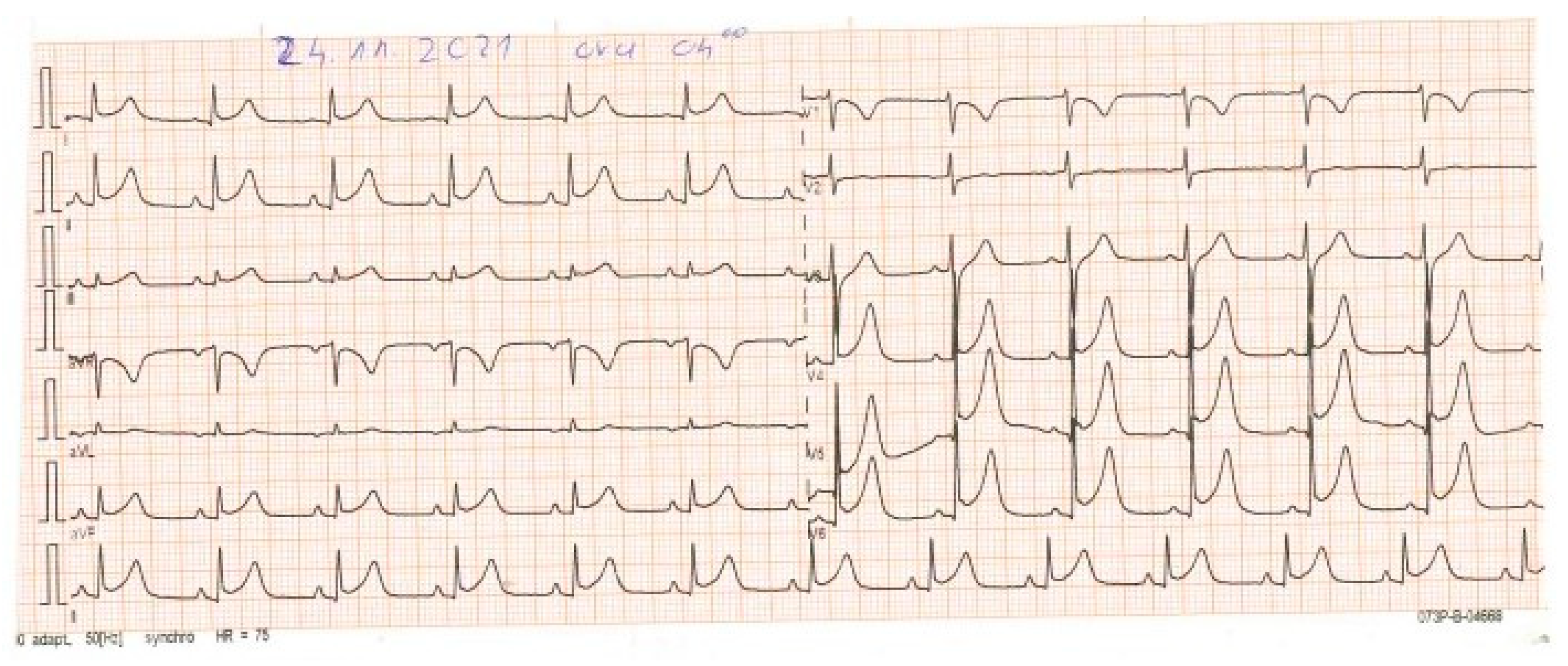
Case Report
2. Discussion
3. Conclusions
Author Contributions
Funding
Institutional Review Board Statement
Informed Consent Statement
Data Availability Statement
Conflicts of Interest
Abbreviations
| CDC | Centers for Disease Control and Prevention |
| FDA | Food and Drug Administration |
| MIS-C | multisystem inflammatory syndrome in children |
| MIS-C/A | multisystem inflammatory syndrome in children/adults |
| MIS-V | multisystem inflammatory syndrome following SARS-CoV-2 vaccination |
| SARS-CoV-2 | severe acute respiratory syndrome coronavirus 2 |
References
- Godfred-Cato, S.; Bryant, B.; Leung, J.; Oster, M.E.; Conklin, L.; Abrams, J.; Roguski, K.; Wallace, B.; Prezzato, E.; Koumans, E.H.; et al. COVID-19–Associated Multisystem Inflammatory Syndrome in Children—United States, March–July 2020. MMWR Morb. Mortal. Wkly. Rep. 2020, 69, 1074–1080. [Google Scholar] [CrossRef] [PubMed]
- Feldstein, L.R.; Rose, E.B.; Horwitz, S.M.; Collins, J.P.; Newhams, M.M.; Son, M.B.F.; Newburger, J.W.; Kleinman, L.C.; Heidemann, S.M.; Martin, A.A.; et al. Multisystem Inflammatory Syndrome in U.S. Children and Adolescents. N. Engl. J. Med. 2020, NEJMoa2021680. [Google Scholar] [CrossRef]
- Davies, P.; Evans, C.; Kanthimathinathan, H.K.; Lillie, J.; Brierley, J.; Waters, G.; Johnson, M.; Griffiths, B.; du Pré, P.; Mohammad, Z.; et al. Intensive Care Admissions of Children with Paediatric Inflammatory Multisystem Syndrome Temporally Associated with SARS-CoV-2 (PIMS-TS) in the UK: A Multicentre Observational Study. Lancet Child Adolesc. Health 2020, 4, 669–677. [Google Scholar] [CrossRef] [PubMed]
- Whittaker, E.; Bamford, A.; Kenny, J.; Kaforou, M.; Jones, C.E.; Shah, P.; Ramnarayan, P.; Fraisse, A.; Miller, O.; Davies, P.; et al. Clinical Characteristics of 58 Children with a Pediatric Inflammatory Multisystem Syndrome Temporally Associated with SARS-CoV-2. JAMA 2020, 324, 259–269. [Google Scholar] [CrossRef] [PubMed]
- Levy, M.; Recher, M.; Hubert, H.; Javouhey, E.; Fléchelles, O.; Leteurtre, S.; Angoulvant, F. Multisystem Inflammatory Syndrome in Children by COVID-19 Vaccination Status of Adolescents in France. JAMA 2022, 327, 281–283. [Google Scholar] [CrossRef] [PubMed]
- Yousaf, A.R.; Cortese, M.M.; Taylor, A.W.; Broder, K.R.; Oster, M.E.; Wong, J.M.; Guh, A.Y.; McCormick, D.W.; Kamidani, S.; Schlaudecker, E.P.; et al. Reported Cases of Multisystem Inflammatory Syndrome in Children Aged 12–20 Years in the USA Who Received a COVID-19 Vaccine, December, 2020, through August, 2021: A Surveillance Investigation. Lancet Child Adolesc. Health 2022, 6, 303–312. [Google Scholar] [CrossRef] [PubMed]
- Salzman, M.B.; Huang, C.-W.; O’Brien, C.M.; Castillo, R.D. Multisystem Inflammatory Syndrome after SARS-CoV-2 Infection and COVID-19 Vaccination. Emerg. Infect. Dis. 2021, 27, 1944–1948. [Google Scholar] [CrossRef]
- Jain, E.; Donowitz, J.R.; Aarons, E.; Marshall, B.C.; Miller, M.P. Multisystem Inflammatory Syndrome in Children after SARS-CoV-2 Vaccination. Emerg. Infect. Dis. 2022, 28, 990–993. [Google Scholar] [CrossRef]
- Santilli, V.; Manno, E.C.; Giancotta, C.; Rossetti, C.; Cotugno, N.; Amodio, D.; Rotulo, G.A.; Deodati, A.; Bianchi, R.; Lucignani, G.; et al. Two Pediatric Cases of Multisystem Inflammatory Syndrome with Overlapping Neurological Involvement Following SARS-CoV-2 Vaccination and Unknown SARS-CoV2 Infection: The Importance of Pre-Vaccination History. Vaccines 2022, 10, 1136. [Google Scholar] [CrossRef]
- Riphagen, S.; Gomez, X.; Gonzalez-Martinez, C.; Wilkinson, N.; Theocharis, P. Hyperinflammatory Shock in Children during COVID-19 Pandemic. Lancet 2020, 395, 1607–1608. [Google Scholar] [CrossRef]
- Verdoni, L.; Mazza, A.; Gervasoni, A.; Martelli, L.; Ruggeri, M.; Ciuffreda, M.; Bonanomi, E.; D’Antiga, L. An Outbreak of Severe Kawasaki-like Disease at the Italian Epicentre of the SARS-CoV-2 Epidemic: An Observational Cohort Study. Lancet 2020, 395, 1771–1778. [Google Scholar] [CrossRef] [PubMed]
- Sood, M.; Sharma, S.; Sood, I.; Sharma, K.; Kaushik, A. Emerging Evidence on Multisystem Inflammatory Syndrome in Children Associated with SARS-CoV-2 Infection: A Systematic Review with Meta-Analysis. SN Compr. Clin. Med. 2021, 3, 38–47. [Google Scholar] [CrossRef] [PubMed]
- CDC. COVID-19 Vaccination. Centers for Disease Control and Prevention. Available online: https://www.cdc.gov/coronavirus/2019-ncov/vaccines/safety/adverse-events.html (accessed on 4 March 2023).
- Rabail, R.; Ahmed, W.; Ilyas, M.; Rajoka, M.S.R.; Hassoun, A.; Khalid, A.R.; Khan, M.R.; Aadil, R.M. The Side Effects and Adverse Clinical Cases Reported after COVID-19 Immunization. Vaccines 2022, 10, 488. [Google Scholar] [CrossRef] [PubMed]
- Chin, S.E.; Bhavsar, S.M.; Corson, A.; Ghersin, Z.J.; Kim, H.S. Cardiac Complications Associated with COVID-19, MIS-C, and MRNA COVID-19 Vaccination. Pediatr. Cardiol. 2022, 43, 483–488. [Google Scholar] [CrossRef] [PubMed]
- CDC. Multisystem Inflammatory Syndrome (MIS). Centers for Disease Control and Prevention. Available online: https://www.cdc.gov/mis/mis-c/hcp_cstecdc/index.html (accessed on 4 March 2023).
- Brlić, P.K.; Pavletić, M.; Lerga, M.; Krstanović, F.; Matešić, M.P.; Miklić, K.; Malić, S.; Mikša, L.; Pajcur, M.; Peruč, D.; et al. SARS-CoV-2 Spike and Nucleocapsid Antibody Response in Vaccinated Croatian Healthcare Workers and Infected Hospitalized Patients: A Single Center Cohort Study. Viruses 2022, 14, 1966. [Google Scholar] [CrossRef]
- VAERS—FAQs. Available online: https://vaers.hhs.gov/faq.html (accessed on 4 March 2023).
- Ouldali, N.; Bagheri, H.; Salvo, F.; Antona, D.; Pariente, A.; Leblanc, C.; Tebacher, M.; Micallef, J.; Levy, C.; Cohen, R.; et al. Hyper Inflammatory Syndrome Following COVID-19 MRNA Vaccine in Children: A National Post-Authorization Pharmacovigilance Study. Lancet Reg. Health Eur. 2022, 17, 100393. [Google Scholar] [CrossRef]
- Yalçinkaya, R.; Öz, F.N.; Polat, M.; Uçan, B.; Teke, T.A.; Kaman, A.; Özdem, S.; Savaş Şen, Z.; Cinni, R.G.; Tanir, G. A Case of Multisystem Inflammatory Syndrome in a 12-Year-Old Male After COVID-19 MRNA Vaccine. Pediatr. Infect. Dis. J. 2022, 41, e87–e89. [Google Scholar] [CrossRef]
- Wangu, Z.; Swartz, H.; Doherty, M. Multisystem Inflammatory Syndrome in Children (MIS-C) Possibly Secondary to COVID-19 MRNA Vaccination. BMJ Case Rep. 2022, 15, e247176. [Google Scholar] [CrossRef]
- Liu, A.; Love, A.; Katz, S.; Patrick, A.; Parra, D.; Halasa, N.; Miller, M.R. Multisystem Inflammatory Syndrome in a Fully Vaccinated 18-Year-Old without Known SARS-CoV-2 Infection. Pediatr. Rheumatol. Online J. 2022, 20, 74. [Google Scholar] [CrossRef]
- Abdelgalil, A.A.; Saeedi, F.A. Multisystem Inflammatory Syndrome in a 12-Year-Old Boy after MRNA-SARS-CoV-2 Vaccination. Pediatr. Infect. Dis. J. 2022, 41, e93–e94. [Google Scholar] [CrossRef]
- Lee, J.S.; Cho, K.S.; Choe, Y.J. A Suspected Case of Multisystem Inflammatory Disease in Children Following COVID-19 Vaccination: A Case Report and Systematic Literature Review. Pediatr. Infect. Dis. J. 2022, 41, e456–e460. [Google Scholar] [CrossRef] [PubMed]


| Variable | Patient | |||||
|---|---|---|---|---|---|---|
| Age (years) | 16 | |||||
| Sex | F | |||||
| Body weight | 55 kg | |||||
| Height | 167 cm | |||||
| Initial manifestations | fatigue, fever, body pain, a loss of appetite, nausea, vomiting, and diarrhea | |||||
| Manifestations at admission | somnolent, pale, and dehydrated, with cyanotic lips and cold extremities, | |||||
| Blood pressure | 79/38 mmHg | |||||
| Heart rate | 100 beats/min | |||||
| Respiratory rate | 45 breaths/min | |||||
| Oxygen blood saturation (SpO2) | 93% with O2 flow of 3–5 L/min | |||||
| Laboratory findings | Day 1 | Day 2 | Day 3 | Day 8 | Day 14 | |
| ESR (5–10) | 70 | 42 | 16 | |||
| CRP (0.0–6.0 mg/L) | 234 | 186 | 143 | 70.9 | 20 | 6.4 |
| PCT (0.0–0.5 ng/mL) | 5.66 | 6.73 | 1.32 | 0.73 | ||
| Interleukin 6 (<7.0 pg/mL) | 56.68 | |||||
| Ferritin (15–150 ng/mL) | 269.1 | |||||
| Cholesterol (3.60–5.70 mmol/L) | 3.2 | 4.76 | ||||
| Triglicerides (0.45–1.81 mmol/L) | 0.75 | 2.46 | ||||
| Urea (1.70–8.30 mmol/L) | 6.23 | 6.43 | 7.27 | 8.78 | 9.88 | |
| Uric Ac (155–430 umol/L) | 327 | |||||
| Creatinine (53–115 umol/L) | 109 | 115 | 96.1 | 105.1 | 90.6 | |
| Albumin (35.0–52.0 g/L) | 33.6 | 39.0 | 33.8 | 33.6 | 32.9 | |
| Total protein (64.0–83.0 g/L) | 60.5 | 62.2 | 70.3 | 78.1 | 68.6 | 67 |
| Glycaemia (3.6–6.4 mmol/L) | 8.05 | 11.69 | 11.69 | 12.54 | 6.61 | |
| Total bilirubine (3.6–6.4 mmol/L) | 21.4 | 10.2 | ||||
| Direct bilirubine (0–5.1 umol/L) | 4.8 | 3.49 | ||||
| ALT (3–41 U/L) | 165 | 213 | 35 | 39 | 20 | 29 |
| AST (2–37 U/L) | 124 | 143 | 74 | 80 | 25 | 45 |
| ALP (43–115 U/L) | 29 | 29 | 31 | 53 | 56 | |
| GGT (3–55 U/L) | 16 | |||||
| CK (38–171 U/L) | 32 | 201/430 | 180 | 46 | 17 | 42 |
| CK-MB (5–25 U/L) | 10 | 44 | ||||
| Troponina I (0–0.04 ng/m) | 0.18 | 6.37 | ||||
| LDH (230–460 U/L) | 202 | 396 | 671 | 732 | 521 | |
| Complete blood cell count (CBC) | ||||||
| WBC (3.5–10) | 2.7 | 6.5 | 15.1 | 14.6 | 11.6 | 7.4 |
| RBC (3.8–5.8) | 4.70 | 6.07 | 5.64 | 5.95 | 5.06 | 4.58 |
| HGB (11.0–16.5) | 12.2 | 16.2 | 14.8 | 134 | 13.3 | 11.3 |
| HCT (35.0–50.0) | 44.1 | 57.7 | 53.4 | 48.3 | 47.8 | 42.4 |
| PLT (150–390) | 122 | 125 | 145 | 132 | 131 | 222 |
| LYM (17.0–48.0%) | 42.3 | 22.3 | 7.5 | 15.3 | 30.4 | 26.1 |
| GRA (43.0–76.0%) | 45.8 | 67.0 | 89.5 | 81.9 | 65.3 | 69.4 |
| Capillary blood gases and electrolytes | ||||||
| Ph (7.35–7.45) | 7.47 | 7.46 | 7.49 | 7.53 | 7.45 | 7.4 |
| pCO2 (34–46 mm Hg) | 27 | 33 | 35 | 34 | 39 | 32 |
| pO2 (80–105 mm Hg) | 51 | 51 | 57 | 68 | 54 | 74 |
| Na+ (130–145 mmol/L) | 129 | 130 | 134 | 139 | 137 | 135 |
| K+ (3.4–5.1 mmol/L) | 4.5 | 3.8 | 3.5 | 3.5 | 2.6 | 3.5 |
| Ca++ (1.17–1.24 mmol/L) | 1.15 | 1.21 | 1.17 | 1.15 | 1.19 | 1.19 |
| HCO3 (22–26 mmol/L) | 19.7 | 23.5 | 26.7 | 28.4 | 27.1 | 25.3 |
| BE (−4 to +2 mmol/L) | −4 | −0.3 | 3.4 | 5.7 | 3.1 | 2.2 |
| Coagulation tests | ||||||
| D-dimer (˂200) | 222 | 599 | 155 | 117 | 55 | 44 |
| PT (70–120%) | 73% | |||||
| INR (099–1.2) | 1.2 | |||||
| PTT (25–35″) | 25″ | |||||
| TT (12–22) | 18″ | |||||
| Protrombina | 100% | |||||
| Proakcelerina | 106% | |||||
| Prokonvertina | 72% | |||||
| F VIII | 105% | |||||
| F IX | 103% | |||||
| F X | 88% | |||||
| F XI | 90% | |||||
| F XII | 70% | |||||
| The serology for SARS-CoV-2 and other infections | ||||||
| Day 20 | Day 20 | |||||
| IgM SARS-CoV 2 (>1.1 positive) | 0.4 | 2.1 | ||||
| IgG SARS-CoV 2 (>1.1 positive) | 2.6 | 2.1 | >2500 | |||
| Anti SARS-CoV2 spike (>1.1) | >2500 | >2500 | ||||
| IgM rubeola | Negative | |||||
| IgG rubeola | Positive | |||||
| IgM HSV | Negative | |||||
| IgG HSV | Positive | |||||
| IgM toxoplasma | Negative | |||||
| IgG toxoplasma | Positive | |||||
| IgM CMG | Negative | |||||
| IgG CMV | Positive | |||||
| Bacterial culture tests | ||||||
| Throat culture | Sterile; twice | |||||
| Blood culture | Sterile; twice | |||||
| Urine culture | Sterile; twice | |||||
Disclaimer/Publisher’s Note: The statements, opinions and data contained in all publications are solely those of the individual author(s) and contributor(s) and not of MDPI and/or the editor(s). MDPI and/or the editor(s) disclaim responsibility for any injury to people or property resulting from any ideas, methods, instructions or products referred to in the content. |
© 2023 by the authors. Licensee MDPI, Basel, Switzerland. This article is an open access article distributed under the terms and conditions of the Creative Commons Attribution (CC BY) license (https://creativecommons.org/licenses/by/4.0/).
Share and Cite
Keka-Sylaj, A.; Ramosaj, A.; Baloku, A.; Zogaj, L.; Gjaka, P. Multisystem Inflammatory Syndrome in Children (MIS-C), Possibly Due to COVID-19 mRNA Vaccination. Vaccines 2023, 11, 956. https://doi.org/10.3390/vaccines11050956
Keka-Sylaj A, Ramosaj A, Baloku A, Zogaj L, Gjaka P. Multisystem Inflammatory Syndrome in Children (MIS-C), Possibly Due to COVID-19 mRNA Vaccination. Vaccines. 2023; 11(5):956. https://doi.org/10.3390/vaccines11050956
Chicago/Turabian StyleKeka-Sylaj, Alije, Atifete Ramosaj, Arbana Baloku, Leonora Zogaj, and Petrit Gjaka. 2023. "Multisystem Inflammatory Syndrome in Children (MIS-C), Possibly Due to COVID-19 mRNA Vaccination" Vaccines 11, no. 5: 956. https://doi.org/10.3390/vaccines11050956