
Sudden Onset of IgA Vasculitis Affecting Vital Organs in Adult Patients following SARS-CoV-2 Vaccines


Abstract
:1. Introduction
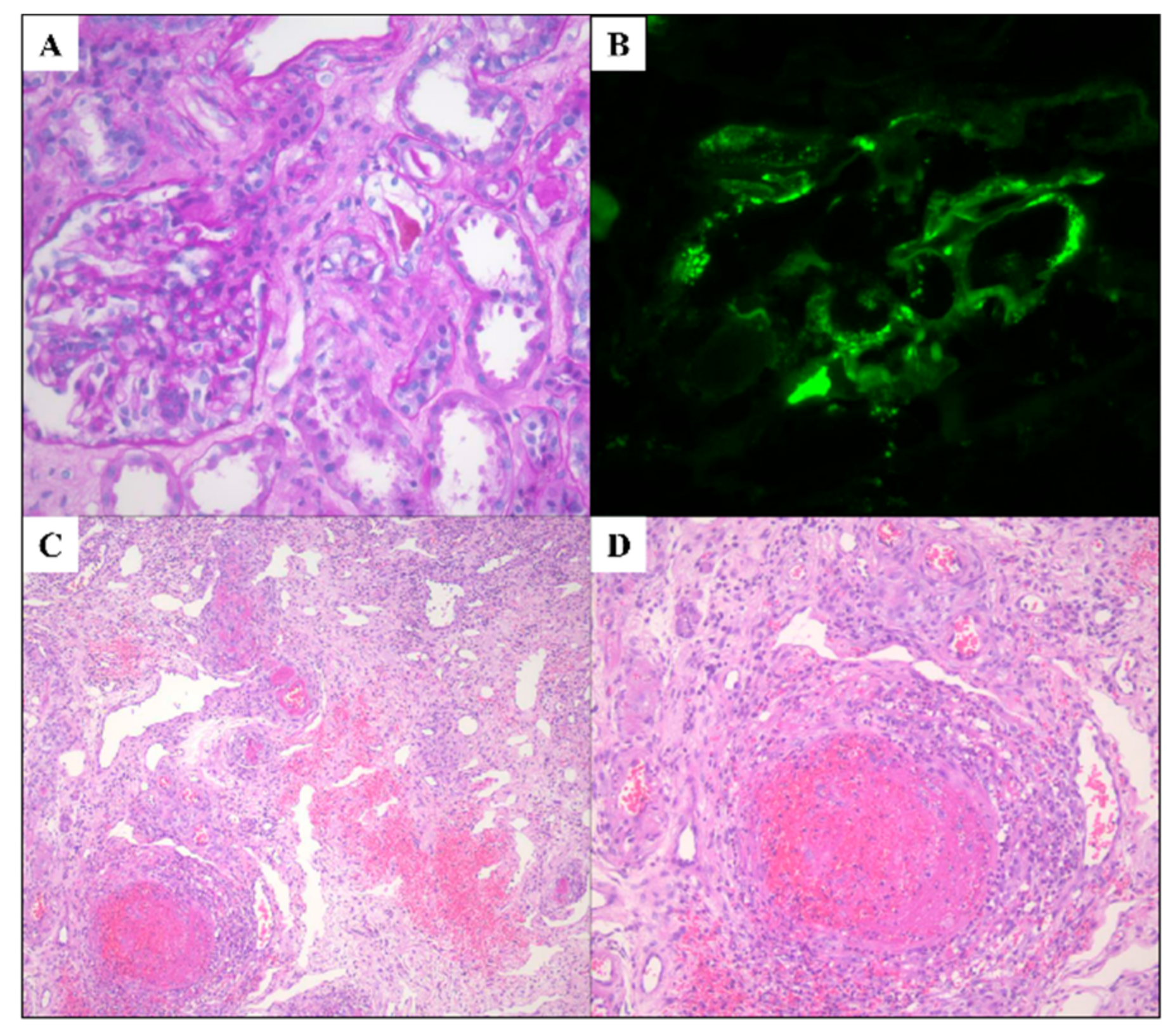
2. Case 1
3. Case 2
4. Case 3
5. Discussion

| Author, the Year of Report | Age/Gender | Underlying Disease | Type of Vaccination | New Onset or Relapse | Time to Present Symptom after Vaccination (Days) | Vaccine Dose | Proteinuria (UPCR, g/g) | Hematuria | Other Clinical Symptoms | Treatment | Outcome |
|---|---|---|---|---|---|---|---|---|---|---|---|
| Anderegg et al., 2021 [19] | 39/M | HTN | mRNA-1273 (Moderna) | N | 0 | 2 | NA | Macrohematuria | Fever, flu-like symptom | PD, CYC | Hematuria persisted |
| Abramson et al., 2021 [20] | 30/M | None | mRNA-1273 (Moderna) | N | 1 | 2 | 0.8 | >30 cells/HPF | Fever, headache | RAASi | Proteinuria reduction |
| Kudose et al., 2021 [21] | 50/F | HTN, Obesity, anti-phospholipid syndrome | mRNA-1273 (Moderna) | N | 2 | 2 | 2.0 | >50 cells/HPF | Generalized weakness, decreased appetite | NA | Hematuria resolved |
| Kudose et al., 2021 [21] | 19/M | Microhematuria | mRNA-1273 (Moderna) | N | 2 | 2 | None | Numerous red blood cells | NA | NA | Hematuria resolved |
| Park et al., 2021 [22] | 22/F | IgA vasculitis | mRNA-1273 (Moderna) | R | 2 | 2 | 0.4 | >50 cells/HPF | None | Supportive | Hematuria resolved |
| Park et al., 2021 [22] | 39/F | None | mRNA-1273 (Moderna) | NA | 2 | 2 | 0.9 | >50 cells/HPF | None | Supportive | Hematuria resolved |
| Park et al., 2021 [22] | 50/M | Chronic kidney disease | mRNA-1273 (Moderna) | NA | 1 | 2 | 3.56 | >50 cells/HPF | None | RAASi | Hematuria resolved |
| Park et al., 2021 [22] | 67/M | Chronic kidney disease | mRNA-1273 (Moderna) | NA | 1 month | 1 | 2.90 | >50 cells/HPF | Purpuric rash | PD | Hematuria resolved |
| Perrin et al., 2021 [23] | 22/M | IgA nephropathy | mRNA-1273 (Moderna) | R | 2 and 25 after first dose, 2 after second dose | 2 | 0.34 | Gross hematuria | Arthralgia | NA | Hematuria resolved |
| Grossman et al., 2021 [24] | 94/M | None | mRNA-1273 (Moderna) | N | 10 | 2 | 3+ | 3+ | Purpuric rash | PD | Hematuria resolved |
| Negrea et al., 2021 [25] | 38/F | IgA nephropathy | mRNA-1273 (Moderna) | R | Several hours after | 2 | 1.4 | Micro hematuria | Body ache, fatigue, headache | NA | NA |
| Negrea et al., 2021 [25] | 38/F | IgA nephropathy | mRNA-1273 (Moderna) | R | Several hours after | 2 | 0.4 | Micro hematuria | Body ache, fatigue, headache | NA | NA |
| Obeid et al., 2021 [7] | 78/F | IgA vasculitis | mRNA-1273 (Moderna) | R | 7 | 1 | NA | 150 × 10⁶/L | Diarrhea, Abdominal pain | MP | Improved rapidly |
| Perrin et al., 2021 [23] | 27/F | IgA nephropathy, Hemodialysis | BNT162b2 (Pfizer-BioNTech) | R | 2 | 2 | 1.9 | Gross hematuria | Abdominal pain, Urticaria | NA | Hematuria resolved |
| Perrin et al., 2021 [23] | 41/F | IgA nephropathy, kidney transplantation | BNT162b2 (Pfizer-BioNTech) | R | 2 | 1 | 0.47 | Gross hematuria | None | NA | Hematuria resolved |
| Maye et al., 2021 [26] | 23/M | NA | BNT162b2 (Pfizer-BioNTech) | R | 1 | 2 | UACR 4.9 mg/mmol | 165 cells/mm3 | Purpuric rash | PD | Hematuria resolved |
| Mohamed et al., 2021 [27] | 50/M | Seasonal allergy | BNT162b2 (Pfizer-BioNTech) | N | 14 | 1 | 1.1 | 10 cells/HPF | Purpuric rash | PD, RAASi | Decreased UPCR of 0.5 g/day |
| Sirufo et al., 2021 [11] | 76/F | None | ChAdOx1 (Oxford/AstraZeneca) | N | 7 | 1 | Neg | 72 cells/HPF | Purpuric rash | Deflazacort | Hematuria resolved |
6. Conclusions
Author Contributions
Funding
Institutional Review Board Statement
Informed Consent Statement
Conflicts of Interest
References
- Song, Y.; Huang, X.; Yu, G.; Qiao, J.; Cheng, J.; Wu, J.; Chen, J. Pathogenesis of IgA Vasculitis: An Up-To-Date Review. Front. Immunol. 2021, 12, 771619. [Google Scholar] [CrossRef] [PubMed]
- Pillebout, E.; Sunderkötter, C. IgA vasculitis. Semin. Immunopathol. 2021, 43, 729–738. [Google Scholar] [CrossRef] [PubMed]
- Piram, M.; Mahr, A. Epidemiology of immunoglobulin A vasculitis (Henoch-Schönlein): Current state of knowledge. Curr. Opin. Rheumatol. 2013, 25, 171–178. [Google Scholar] [CrossRef] [PubMed]
- Nossent, J.; Raymond, W.; Isobel Keen, H.; Preen, D.; Inderjeeth, C. Morbidity and mortality in adult-onset IgA vasculitis: A long-term population-based cohort study. Rheumatology 2021, 61, 291–298. [Google Scholar] [CrossRef]
- Gendreau, S.; Porcher, R.; Thoreau, B.; Paule, R.; Maurier, F.; Goulenok, T.; Frumholtz, L.; Bernigaud, C.; Ingen-Housz-Oro, S.; Mekinian, A.; et al. Characteristics and risk factors for poor outcome in patients with systemic vasculitis involving the gastrointestinal tract. Semin. Arthritis Rheum. 2021, 51, 436–441. [Google Scholar] [CrossRef]
- Hetland, L.E.; Susrud, K.S.; Lindahl, K.H.; Bygum, A. Henoch-Schönlein Purpura: A Literature Review. Acta Derm. Venereol. 2017, 97, 1160–1166. [Google Scholar] [CrossRef] [Green Version]
- Obeid, M.; Fenwick, C.; Pantaleo, G. Reactivation of IgA vasculitis after COVID-19 vaccination. Lancet Rheumatol. 2021, 3, e617. [Google Scholar] [CrossRef]
- Wu, H.H.L.; Kalra, P.A.; Chinnadurai, R. New-Onset and Relapsed Kidney Histopathology Following COVID-19 Vaccination: A Systematic Review. Vaccines 2021, 9, 1252. [Google Scholar] [CrossRef]
- Iwata, H.; Kamiya, K.; Kado, S.; Nakaya, T.; Kawata, H.; Komine, M.; Ohtsuki, M. Case of immunoglobulin A vasculitis following coronavirus disease 2019 vaccination. J. Dermatol. 2021, 48, e598–e599. [Google Scholar] [CrossRef]
- Badier, L.; Toledano, A.; Porel, T.; Dumond, S.; Jouglen, J.; Sailler, L.; Bagheri, H.; Moulis, G.; Lafaurie, M. IgA vasculitis in adult patient following vaccination by ChadOx1 nCoV-19. Autoimmun. Rev. 2021, 20, 102951. [Google Scholar] [CrossRef]
- Sirufo, M.M.; Raggiunti, M.; Magnanimi, L.M.; Ginaldi, L.; De Martinis, M. Henoch-Schönlein Purpura Following the First Dose of COVID-19 Viral Vector Vaccine: A Case Report. Vaccines 2021, 9, 1078. [Google Scholar] [CrossRef] [PubMed]
- Sugita, K.; Kaneko, S.; Hisada, R.; Harano, M.; Anno, E.; Hagiwara, S.; Imai, E.; Nagata, M.; Tsukamoto, Y. Development of IgA vasculitis with severe glomerulonephritis after COVID-19 vaccination: A case report and literature review. CEN Case Rep. 2022, 1–6. [Google Scholar] [CrossRef] [PubMed]
- Watanabe, Y.; Mendonça, L.; Allen, E.R.; Howe, A.; Lee, M.; Allen, J.D.; Chawla, H.; Pulido, D.; Donnellan, F.; Davies, H.; et al. Native-like SARS-CoV-2 Spike Glycoprotein Expressed by ChAdOx1 nCoV-19/AZD1222 Vaccine. ACS Cent. Sci. 2021, 7, 594–602. [Google Scholar] [CrossRef]
- Farkash, I.; Feferman, T.; Cohen-Saban, N.; Avraham, Y.; Morgenstern, D.; Mayuni, G.; Barth, N.; Lustig, Y.; Miller, L.; Shouval, D.S.; et al. Anti-SARS-CoV-2 antibodies elicited by COVID-19 mRNA vaccine exhibit a unique glycosylation pattern. Cell Rep. 2021, 37, 110114. [Google Scholar] [CrossRef] [PubMed]
- Suzuki, H.; Moldoveanu, Z.; Julian, B.A.; Wyatt, R.J.; Novak, J. Autoantibodies Specific for Galactose-Deficient IgA1 in IgA Vasculitis with Nephritis. Kidney Int. Rep. 2019, 4, 1717–1724. [Google Scholar] [CrossRef] [Green Version]
- Allez, M.; Denis, B.; Bouaziz, J.D.; Battistella, M.; Zagdanski, A.M.; Bayart, J.; Lazaridou, I.; Gatey, C.; Pillebout, E.; Chaix Baudier, M.L.; et al. COVID-19-Related IgA Vasculitis. Arthritis Rheumatol. 2020, 72, 1952–1953. [Google Scholar] [CrossRef] [PubMed]
- Jedlowski, P.M.; Jedlowski, M.F. Coronavirus disease 2019-associated immunoglobulin A vasculitis/Henoch-Schönlein purpura: A case report and review. J. Dermatol. 2022, 49, 190–196. [Google Scholar] [CrossRef]
- Farooq, H.; Aemaz Ur Rehman, M.; Asmar, A.; Asif, S.; Mushtaq, A.; Qureshi, M.A. The pathogenesis of COVID-19-induced IgA nephropathy and IgA vasculitis: A systematic review. J. Taibah Univ. Med. Sci. 2022, 17, 1–13. [Google Scholar] [CrossRef]
- Anderegg, M.A.; Liu, M.; Saganas, C.; Montani, M.; Vogt, B.; Huynh-Do, U.; Fuster, D.G. De novo vasculitis after mRNA-1273 (Moderna) vaccination. Kidney Int. 2021, 100, 474–476. [Google Scholar] [CrossRef]
- Abramson, M.; Mon-Wei Yu, S.; Campbell, K.N.; Chung, M.; Salem, F. IgA Nephropathy After SARS-CoV-2 Vaccination. Kidney Med. 2021, 3, 860–863. [Google Scholar] [CrossRef]
- Kudose, S.; Friedmann, P.; Albajrami, O.; D’Agati, V.D. Histologic correlates of gross hematuria following Moderna COVID-19 vaccine in patients with IgA nephropathy. Kidney Int. 2021, 100, 468–469. [Google Scholar] [CrossRef] [PubMed]
- Park, K.; Miyake, S.; Tai, C.; Tseng, M.; Andeen, N.K.; Kung, V.L. Letter regarding: “A Case of Gross Hematuria and IgA Nephropathy Flare-Up Following SARS-CoV-2 Vaccination”. Kidney Int. Rep. 2021, 6, 2246–2247. [Google Scholar] [CrossRef] [PubMed]
- Perrin, P.; Bassand, X.; Benotmane, I.; Bouvier, N. Gross hematuria following SARS-CoV-2 vaccination in patients with IgA nephropathy. Kidney Int. 2021, 100, 466–468. [Google Scholar] [CrossRef] [PubMed]
- Grossman, M.E.; Appel, G.; Little, A.J.; Ko, C.J. Post-COVID-19 vaccination IgA vasculitis in an adult. J. Cutan. Pathol. 2022, 49, 385–387. [Google Scholar] [CrossRef]
- Negrea, L.; Rovin, B.H. Gross hematuria following vaccination for severe acute respiratory syndrome coronavirus 2 in 2 patients with IgA nephropathy. Kidney Int. 2021, 99, 1487. [Google Scholar] [CrossRef]
- Maye, J.A.; Chong, H.P.; Rajagopal, V.; Petchey, W. Reactivation of IgA vasculitis following COVID-19 vaccination. BMJ Case Rep. 2021, 14, e247188. [Google Scholar] [CrossRef]
- Mohamed, M.M.B.; Wickman, T.J.; Fogo, A.B.; Velez, J.C.Q. De Novo Immunoglobulin A Vasculitis Following Exposure to SARS-CoV-2 Immunization. Ochsner. J. 2021, 21, 395–401. [Google Scholar] [CrossRef]
- Bellanti, J.A. COVID-19 vaccines and vaccine hesitancy: Role of the allergist/immunologist in promotion of vaccine acceptance. Allergy Asthma Proc. 2021, 42, 386–394. [Google Scholar] [CrossRef]

| Patient Characteristics | Patient 1 | Patient 2 | Patent 3 |
|---|---|---|---|
| Age | 60 | 63 | 70 |
| Sex | M | M | M |
| Type of vaccination | ChAdOx1 (Oxford/AstraZeneca) | mRNA-1273 (Moderna) | ChAdOx1 (Oxford/AstraZeneca) |
| Time to present symptoms | 10 days | 1 days | 14 days |
| Baseline Cr level (mg/dL) | 0.76 | 0.67 | 0.59 |
| Peak Cr level after COVID-19 vaccine (mg/dL) | 0.98 | 1.29 | 3.34 |
| Gross hematuria before COVID-19 vaccine | 0–2/HPF | 0–2/HPF | 0–2/HPF |
| Gross hematuria after COVID-19 vaccine | 21–50/HPF | 21–50/HPF | 21–50/HPF |
| Proteinuria before COVID-19 vaccine | Neg | + | + |
| Peak UPCR after COVID-19 vaccine (mg/g) | 3902 | 2214 | 15764 |
| UPCR 2 months after COVID-19 vaccine (mg/g) | 584 | 454 | 6618 |
| Other clinical symptoms | Purpura, Abdominal pain, Melena | Purpura, Abdominal pain, Melena, Arthralgia | Purpura, Abdominal pain, Melena, Arthralgia |
| Current treatment | PD | PD | MMF, PD |
Publisher’s Note: MDPI stays neutral with regard to jurisdictional claims in published maps and institutional affiliations. |
© 2022 by the authors. Licensee MDPI, Basel, Switzerland. This article is an open access article distributed under the terms and conditions of the Creative Commons Attribution (CC BY) license (https://creativecommons.org/licenses/by/4.0/).
Share and Cite
Choi, Y.; Lee, C.H.; Kim, K.M.; Yoo, W.-H. Sudden Onset of IgA Vasculitis Affecting Vital Organs in Adult Patients following SARS-CoV-2 Vaccines. Vaccines 2022, 10, 923. https://doi.org/10.3390/vaccines10060923
Choi Y, Lee CH, Kim KM, Yoo W-H. Sudden Onset of IgA Vasculitis Affecting Vital Organs in Adult Patients following SARS-CoV-2 Vaccines. Vaccines. 2022; 10(6):923. https://doi.org/10.3390/vaccines10060923
Chicago/Turabian StyleChoi, Yunjung, Chang Hun Lee, Kyoung Min Kim, and Wan-Hee Yoo. 2022. "Sudden Onset of IgA Vasculitis Affecting Vital Organs in Adult Patients following SARS-CoV-2 Vaccines" Vaccines 10, no. 6: 923. https://doi.org/10.3390/vaccines10060923