Article

SARS-CoV-2 Antibody and T Cell Response after a Third Vaccine Dose in Hemodialysis Patients Compared with Healthy Controls

1
Institute for Medical-Chemical Laboratory Diagnostics, Mistelbach-Gänserndorf State Clinic, 2130 Mistelbach, Austria
2
Department for Internal Medicine III–Nephrology and Diabetology, Mistelbach-Gänserndorf State Clinic, 2130 Mistelbach, Austria
3
Department for Internal Medicine, Hainburg State Clinic, 2410 Hainburg, Austria
4
Institute of Medical Statistics, Center for Medical Statistics, Informatics and Intelligent Systems, Medical University of Vienna, 1090 Vienna, Austria
*
Author to whom correspondence should be addressed.
Vaccines 2022, 10(5), 694; https://doi.org/10.3390/vaccines10050694
Submission received: 1 April 2022 / Revised: 8 April 2022 / Accepted: 11 April 2022 / Published: 28 April 2022

Abstract

:
Hemodialysis (HD) patients have an increased risk of severe SARS-CoV-2 infection. In this study, we assess the impact of a third vaccine dose (3D) on antibody levels and T cell response in HD patients and a healthy control group in a prospective cohort study consisting of 60 HD patients and 65 healthy controls. Each participant received two doses of the BNT-162b2 mRNA vaccine and an mRNA vaccine 3D. The SARS-CoV-2 antibody response was measured 6 months after the second vaccine dose and 6 to 8 weeks after the 3D. We assessed INF-γ secretion 6–8 weeks post 3D in 24 healthy controls, 17 HD patients with a normal response, and 20 low responder HD patients. The groups were compared using univariate quantile regressions and multiple analyses. After the 3D, the SARS-CoV-2-specific antibody and INF-γ titers of most HD patients were comparable to those of healthy controls. A subgroup of HD patients who had shown a diminished antibody response after the first two vaccine doses developed a significantly lower antibody and INF-γ response compared to responder HD patients and controls even after the 3D. A new strategy is needed to protect low/non-responder HD patients from severe SARS-CoV-2 infection.

1. Introduction

Severe Acute Respiratory Coronavirus-2 (SARS-CoV-2) is a coronavirus that has led to a pandemic with global consequences. Infection with this virus can cause severe respiratory failure and death [1]. Patients undergoing hemodialysis on a regular basis (HD patients) are especially prone to infection with SARS-CoV-2 [2,3], a severe course of the disease, and significantly increased mortality [4,5]. Consequently, HD patients are prioritized to receive vaccines against COVID-19. Prior studies [6,7] showed that HD patients exhibit a diminished antibody response after two vaccine doses. Importantly, a subgroup of HD patients were low/non-responders. The T cell interferon gamma (INF-γ) response in HD patients after vaccination has not been well characterized, although some studies [8] aim to improve our knowledge on this topic. In Austria, a third dose (3D) of vaccine has been recommended due to the decline of vaccine-induced antibodies and the rise of COVID-19 cases [9].
The aim of this study is to measure the impact of the third mRNA vaccine on antibody levels and INF-γ response in HD patients 6 to 8 weeks after the 3D and compare these to healthy controls who also received three vaccine shots. Using such data, we aim to assess the differences between the groups and explore whether further measures are needed to adequately protect this high-risk population.

2. Materials and Methods

We conducted a prospective cohort study to elucidate the antibody and INF-γ response to vaccination with Comirnaty (BNT-162b2, BioNTech/Pfizer, Mainz, Germany, two doses) and a booster dose of an mRNA vaccine (either Spikevax (mRNA-1273), Moderna, Cambridge, MA, USA, or BNT-162b2) administered 6 months after the second vaccine dose, in HD patients versus healthy controls vaccinated with the same regimen.

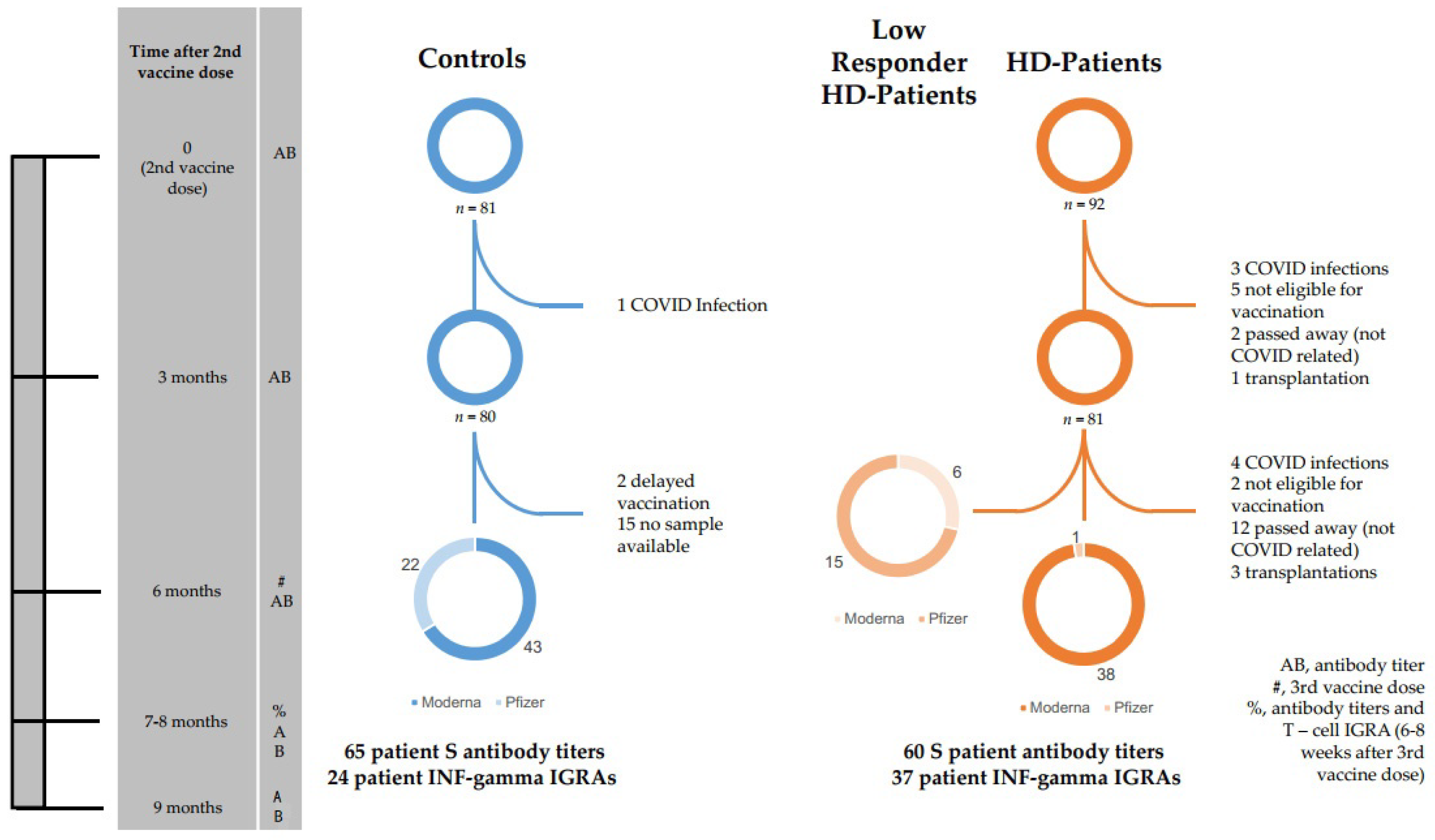
2.1. Study Population

HD patients were considered eligible if they had been on dialysis for at least 3 months and had received vaccination with Comirnaty (vaccination schedule in Section 2.1.3). The healthy control group consisted of volunteer healthcare workers who had been vaccinated using the same regimen. Participants in both groups was restricted to adults 18–99 years old. Pregnant women and individuals with known SARS-CoV-2 infection in the past (diagnosed via patient history and test for nucleocapsid (N) antibody, see Section 2.3.2) were excluded from the study. The study protocol was approved by the local ethics committee. Written informed consent was obtained.
In all, 81 dialysis patients were initially scheduled to receive the 3D. Of these, four contracted SARS-CoV-2 infection, two were not eligible for vaccination due to high CRP values, three received a transplant, and, sadly, 12 passed away (unrelated to COVID-19). Finally, 60 HD patients in the study who received their 3D were included.
Of these, 21 were identified as low/non-responders (see Section 2.1.1). In this group, two participants (11%) were women and 16 were men. The median age was 72 years (age range 49–82 years).
The other 39 HD patients were designated “responders”. In this group, 15 participants (38%) were women and 24 were men. The median age was 66 years (age range 34 to 83 years). All HD patients received their 3D six months after the second dose.
Initially, 80 volunteer healthcare workers were recruited to receive their 3D in the course of the study. Of these, two delayed their 3D and 13 opted not to test for antibodies after the 3D. Finally, 65 healthy controls were included.
This group consisted of 43 (66%) women and 22 men. The median age was 50 years (age range 29–65 years). All demographic data are summarized in Table 1.
The medical histories of dialysis patients were extracted from medical records, while the control group was assessed using a standardized questionnaire.

2.1.1. Low/Non-Responders

Twenty-one patients in the HD patient group and none in the healthy control group were identified as low-/non-responders based on their antibody titers. Titers lower than 15 BAU/ml (lower limit of detection of antibody test; non-responders) or 29 BAU/mL (low responders, cut-off based on off correlation with SARS-CoV-2 neutralization activity of patient sera [10]) four weeks after the second vaccine, as described in our previous work [7], caused patients to belong to this group.

2.1.2. Patient Selection for SARS-CoV-2 T Cell Test

The study participants were assigned an anonymization number at the beginning of the study. Some were then randomly selected to receive an INF-γ release assay (IGRA) test, due to the limited availability of test kits. From each of the three subjects’ groups (controls, HD patient responders and HD patient low/non- responders), patients were selected using a random number generator (RANDOM.ORG) set to generate integers that had a maximum value of 80, 60 and 21 (initial numbers of controls, adjusted numbers of HD patient responders and HD patient low-responders, respectively), repeated for the number of IGRA tests available. The participants whose anonymization numbers corresponded to the value generated received an IGRA test 6–8 weeks after their 3D (24 controls, 17 HD patient responders, 20 HD patient low/non-responders).

2.1.3. Vaccination Schedules

All study participants were immunized with two doses of BNT-162b2 timed three weeks between the first and second doses. All study participants received 3D five to six months after the second dose. Of these participants, 43 controls (66%) received mRNA-1273 (Moderna) and 22 controls (33%) received BNT-162b2 (Pfizer/BioNTech) as their 3D. Of the 39 responder HD patients, 38 received mRNA-1273 (Moderna) and one received BNT-162b2 (Pfizer/BioNTech). Of the 21 low-responder patients, 15 received BNT-162b2 and six received mRNA-1273.
For a graphical representation, see Figure 1.

2.2. Antibody Titers

2.2.1. Processing of Blood Samples

Blood draws were performed one week prior to and six to eight weeks after administration of the booster dose. Samples were centrifuged on a Hettich Rotanta 460r centrifuge at 3000 rpm for 10 min, aliquoted and anonymized. They were then stored at −70 °C and thawed prior to testing.

2.2.2. Serological Assessment

All samples were analyzed for nucleocapsid (N) antibodies with an Elecsys® Anti-SARS-CoV-2 test (Roche Diagnostics Austria, Vienna). A positive result in this test led to exclusion from the study due to a high probability of a past SARS-CoV-2 infection.
The antibody response elicited by vaccination was measured using an Elecsys® Anti-SARS-CoV-2 S on a Cobas e 801 platform according to specifications, diluted 100-fold. Results were recorded as ranging from 0 (≤0.40 U/mL, lower limit of detection [LOD]) to 25,000 (≥25,000 U/mL, upper LOD) and assigned to anonymized patient data, as in our previous study [7]. Results showing values greater 15BAU/mL were considered positive.
Longitudinal antibody titers were measured from samples collected 21 days, 3 months and 6 months (last collection on the day of the 3D) after the second vaccine dose. Follow-up samples were taken 6–8 weeks after 3D and 12 weeks after 3D. For the last follow-up, only 16 healthy controls were available.

2.3. SARS-CoV-2 S-Specific Interferon Gamma Release Assay (IGRA)

2.3.1. Processing of Blood Samples

In the blood draws for antibody titers 6–8 weeks after the 3D, a second sample was drawn from randomly selected patients (see Section 2.1.2) into a lithium-heparin tube. The whole blood samples were incubated in three test tubes lined with SARS-CoV-2 S antigen at 37 °C for 24 h and then centrifuged on a Hettich Rotanta 460r centrifuge for 10 min at 4000 rpm. The supernatant was then analyzed using the ELISA plates provided by the manufacturer.

2.3.2. IGRA Test

The T cell response was assessed using the WANTAI SARS-CoV-2 IGRA assay according to specifications [11]. Twenty µL of specimen diluent and 50 µL of reconstituted standard (400pg/mL in serial dilutions of 1:1, 1:2, 1:4, 1:8, 1:16 and 1:32) and the same amount of serum supernatant was used to fill the wells, and was incubated at 37 °C for 1 h. Fifty µL HRP conjugate was added, and after incubation for 1 h, the plate was washed 5 times using the washing buffer provided by the manufacturer diluted 1:20, with 1 min soak time between washes. Chromogen A and B solutions were added to the wells, and after 15 min of incubation in the dark at 37 °C, a stop solution was applied to all wells. The plate was read in a dual wavelength photometer (450 and 630 nm). The resulting INF-γ concentrations were recorded as ranging from 0 (≤3 pg/mL, LOD), or any of the conditions specified in the test manual as negative, to 400 (≥400 pg/mL, upper LOD) and assigned to anonymized patient data.

2.4. Adverse Events

Adverse events (AE) of the 3D for all groups (controls and combined responder and low/non-responder HD patients) were assessed via a standardized questionnaire.
AEs were divided into two categories: local AEs (pain at injection site, redness and/or swelling at injection site, and induration at injection site) and systemic AEs (fatigue, headache, muscle and/or joint pain, fever, gastrointestinal symptoms [diarrhea, nausea, vomiting] or other AEs).
Patients were asked to grade their AEs after 3D according to subjective severity. Grading was performed on a scale from 1 to 4. Grade 1 AE signified mild (does not interfere with activity); Grade 2 moderate (interferes with activity); Grade 3 severe (prevents daily activity); and Grade 4 (emergency department visit or hospitalization), analogous to the FDA toxicity grading scale [12].

2.5. Statistics

We investigated the influence of being a member of one of three groups (control, HD low/non-responder patients, and HD responder patients), sex and age on SARS-CoV-2 RBD-specific antibody titer 6–8 weeks after 3D. Univariate quantile (median) regressions were performed. The quantile regression was chosen due to the skewed distribution of the antibody titer (40 patients with maximum titer observation of 25,000 BAU/mL, test cut-off). Bootstrap was applied to construct standard errors and perform statistical tests for each independent factor (5000 replications). Pairwise contrasts estimating marginal means (median), standard error (SE) and 95% confidence intervals (CI) were generated for factor variables, and p-value adjustment for pairwise comparison of the group variable was performed using the Tukey method. Then, a multiple analysis was computed for all variables with a p-value less than alpha = 0.05 in the univariate analysis.
The influence of sex and age on INF-γ titers was also investigated for the three groups (controls, HD responder patients, HD low/non-responder patients). For this purpose, the same analyses as above were conducted.
Logistic regression analyses were computed to investigate the univariate influence of several risk factors on the HD patients’ response to the vaccine. Odds ratios, p-values and corresponding 95% confidence intervals were computed and the probability of being a dialysis responder was modelled.
The significance level was set to 0.05. The analyses were performed with R 4.1.1 and the R-packages) quantreg and emmeans [13] (R is part of the Free Software Foundation, Boston, MA, USA).
Boxplots and AE bar graphs were created in Excel 2019 (Microsoft, Redmond, WA, USA).

2.6. Checklist for Cohort Studies

We used the STROBE cohort checklist when writing our report [14].

3. Results

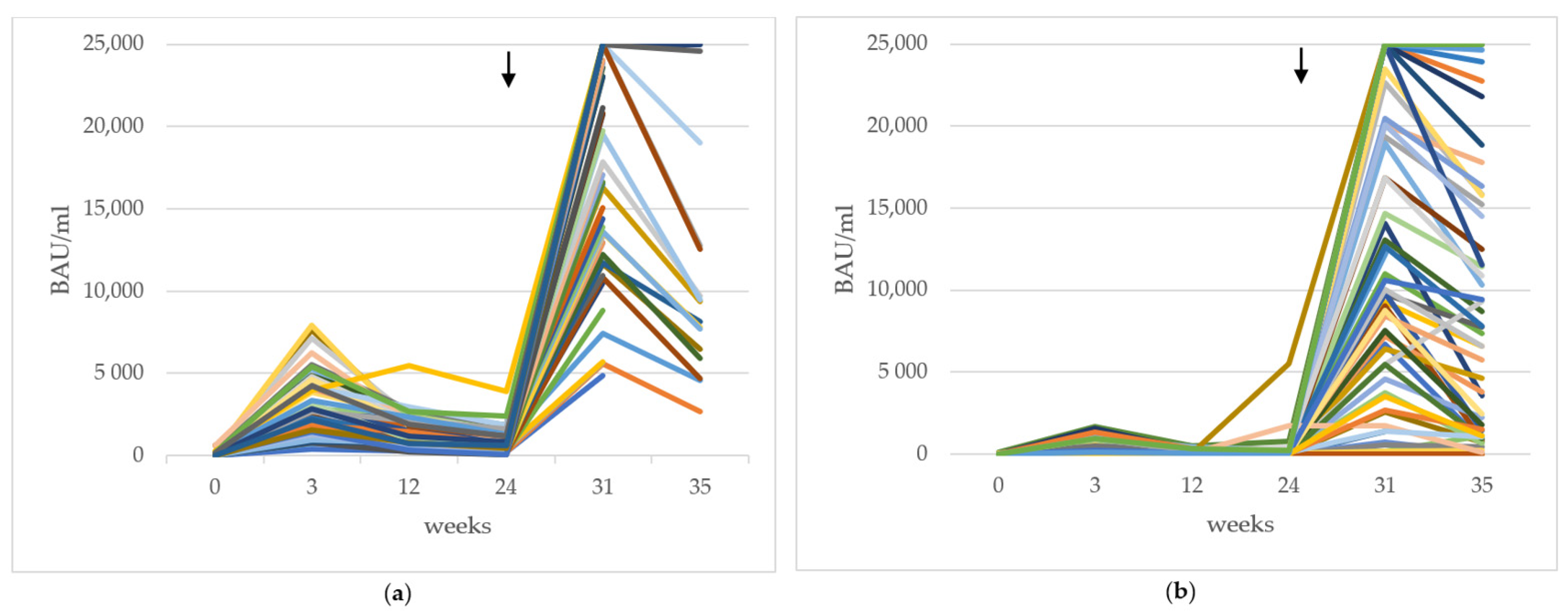
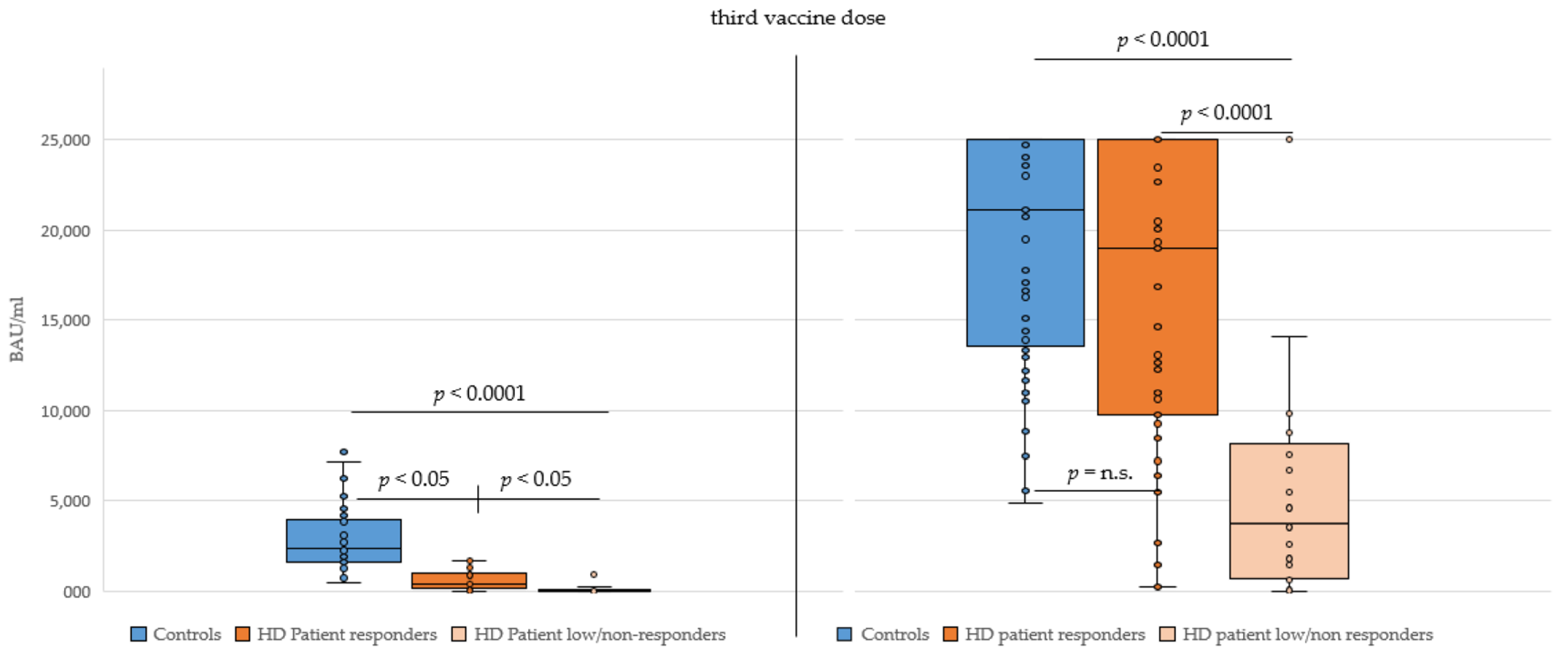
3.1. Vaccine Antibody Titer Results after Three Doses

We analyzed the SARS-CoV2 RBD specific antibody titers of 65 healthcare workers and 60 HD patients. Of the control group, 65/65 (100%) seroconverted (had a titer of >15 BAU/mL, providing a positive test result) 6–8 weeks after their 3D. Of the HD Patients, 58/60 (97%) seroconverted after the 3D (86% HD patients had had >15 BAU/mL 3 months after the second dose). A graphical representation of longitudinal antibody titers is depicted in Figure 2a for the healthy controls and in Figure 2b for the HD patients. Titers were measured in individuals at six points in time after administration of the second vaccine dose and then blotted in the graph.
Importantly, of the 60 HD patients, 21 individuals had an antibody response <29 BAU/mL 3 months after two vaccine doses (“low/non-responders”). Their antibody titers measured 6 weeks after administration of the booster shot showed a certain amount of antibody response. Three HD patients could still be classified as low/non-responders after their 3D. Of these, one suffered from plasmozytoma and another from myasthenia gravis, both conditions with consequences or therapies that could negatively influence B cell function. One low responder achieved an antibody titer of 67 BAU/mL, all 18 remaining patients achieved an antibody titer of 700 BAU/mL or greater (see Table 2), constituting a significant increase caused by the booster vaccine (p < 0.001, Mann-Whitney U).
When comparing the SARS-CoV-2 RBD-specific antibody response 6–8 weeks after the 3D in the control and HD responder patient groups, no significant difference was observed (p = 0.8), which was in contrast to after the second vaccine dose. However, significantly lower antibody titers were found for HD low/non-responder patients after the 3D compared to the control and HD responder patient groups (p < 0.0001, see Figure 3).

3.1.1. Influences of Sex and Age on Antibody Titer

Via a quantile regression test we assessed the influence of variables known to play a role in antibody response, namely, sex [15] and age [7,16] in our groups (see Section 2.5). In this study group, men had lower median SARS-CoV-2 RBD-specific titers than women, but this difference was not significant (p = 0.27). Age had a significant influence on antibody titer (p = 0.01): With increasing age, the median antibody titer values decreased. The Spearman correlation coefficient for the two variables age and titer was −0.28. Table 3 shows values and estimates of the univariate quantile regression for the influence of sex and age.
After including these variables in a multivariate analysis to adjust for sex and age differences in our groups, the differences in antibody titers 6–8 weeks after 3D between the low/non-responders and the other two groups were still significant (p < 0.0001). The control group and the responder HD patients still did not show a significant titer difference (p = 0.43), and age did not have a significant influence on the titers anymore (p = 0.33). See Table 4 for the results of the pairwise group comparison.

3.1.2. Responder vs. Low/Non-Responder HD Patients
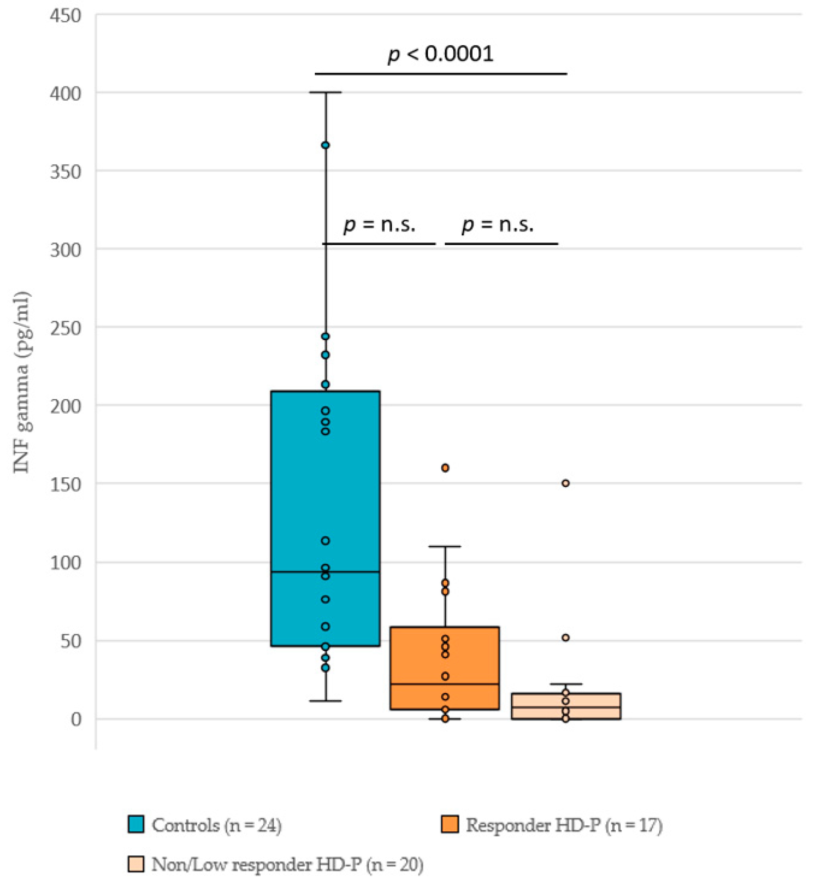
We analyzed possible influences on antibody titers that could potentially cause low or no response in patients. Table 5 shows that only sex has a significant influence on dialysis responders versus low responders (p = 0.02). In the group of 21 low responders, only 2 were female (9.5%) whereas in the responder group, 17 of 43 were female (39.5%).
All other variables are not significant. However, the sample sizes for these comparisons are small. For example, 22 (51.2%) patients received renin-angiotensin-aldosteron-system (RAAS) inhibitors in the responder group, but only six (28.6%) in the non-responder group. Only seven patients were available for an analysis of the influence of immunosuppressants (four in the low and three in the responder group).

3.2. INF-γ Titers

As a correlate of T cell activation, we compared the SARS-CoV-2-specific INF-γ response in controls (n = 24), HD responders (n = 17) and HD low/non-responders (n = 20) 6–8 weeks after the booster vaccine. In all, 96% (23/24) of the controls and 76% (28/37) of the HD patients that were tested developed an INF-γ titer greater than the cut-off (3 pg/mL). The boxplots (see Figure 4) show apparent differences in the distribution of INF-γ secretion between the three groups. The quantile regression results reveal a significant difference only for the median of the control group versus low/non-responder HD patients. However, the sample sizes per group were rather small and the chosen analysis strategy (Tukey test for pairwise comparison) further reduced the power. In the multivariate analysis, neither age nor sex had a significant influence on INF-γ titers.

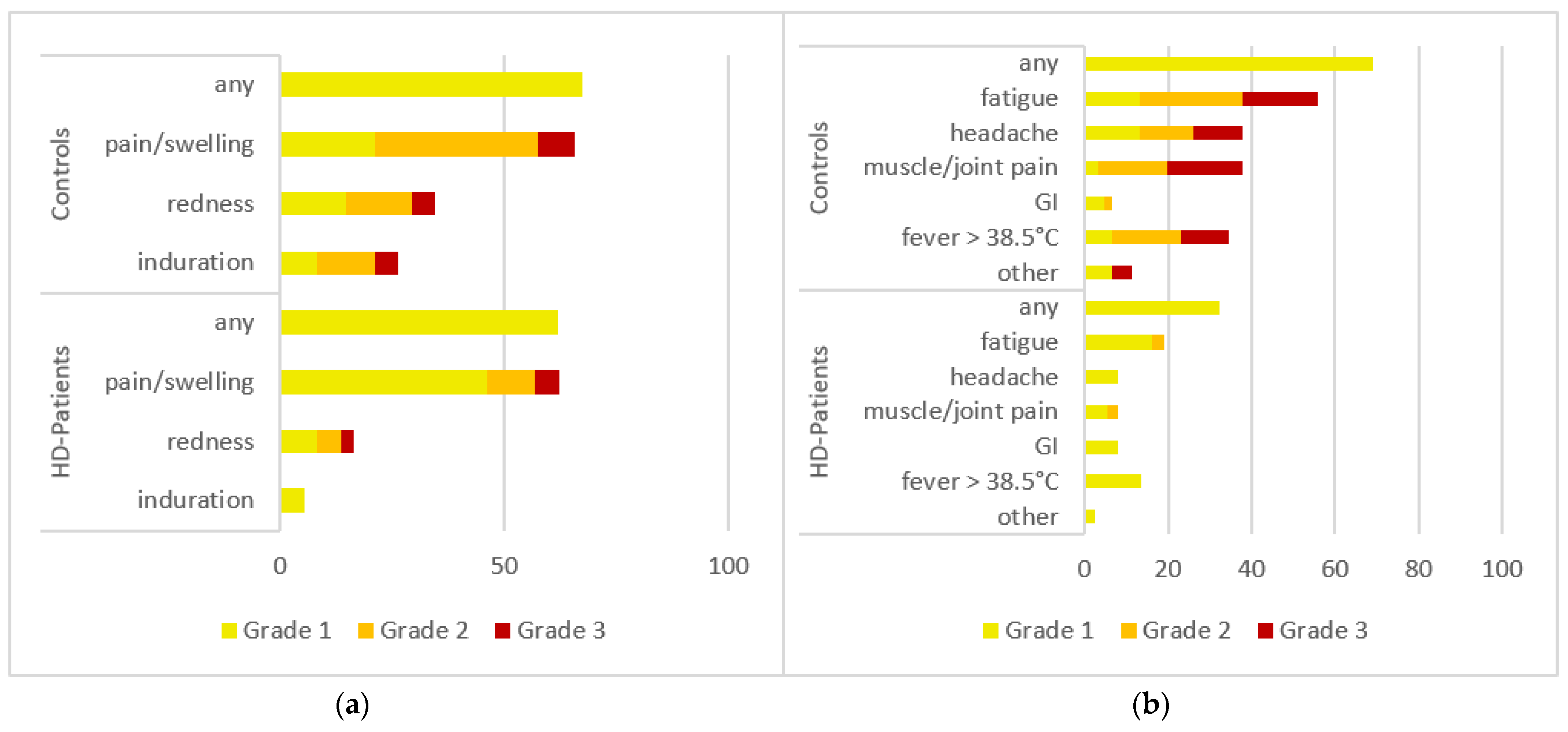
3.3. Adverse Events (AEs) after 3D

Adverse event (AE) reports were analyzed and compared descriptively between the two groups. Sixty-one control group questionnaires and 37 HD patient questionnaires were available. No Grade 4 (emergency department visit or hospitalization) AEs were reported in either group. The control group reported more local AEs after their 3D (67% vs. 62% AEs) and more systemic AEs after the 3D (69% vs. 32% AEs) compared with the HD patients (responders and low/non-responders combined) group. See Figure 5a,b for a graphical representation.

4. Discussion

We investigated the SARS-CoV-2-specific RBD titers in healthy controls and HD patients 6–8 weeks after 3D. Recently we showed a significant difference in titers between healthy controls and HD patients after two vaccine doses [7]. More HD patients seroconverted after the 3D (97% compared to 86% after the second dose), and the antibody titers did not differ significantly between controls and HD responder patients. Overall, antibody titers, which are an important factor in protection from a severe infection course, improved significantly after 3D in HD patients, even in low/non-responder HD patients. This result contradicts previous findings [8], where antibody titers in high responders did not significantly improve; however, that study’s cut-off for low/non-responders was higher than in our design.
In our study, men were shown to have a greater risk of developing low or no antibody response to the vaccine. Studies show that female patients tend to develop higher antibody titers after mRNA vaccination [15]. This fact could contribute to the difference between genders. We also analyzed other factors to elucidate low/non responder HD patient characteristics like RAAS inhibitor usage and age, shown in Table 5. Some trends (entries with a p value close to 0.05) of a factor influencing a patient to become a low/non responder can be seen, but no variable we analyzed showed statistical significance because the number of these individuals in our study is too low for reliable statistics. In the case of age, for example, studies show decreasing antibody response to vaccination with higher age [16,17,18], but in our study, no significance was found. It also seems likely that not only a single one of the factors in Table 5, but rather a combination of them would be necessary to cause a low/non-response. Thus, further studies on this subject with sufficient numbers to power statistic significance would be needed.
We measured SARS-CoV-2 antibody titers without neutralizing capacity or neutralization titers in our study. This could be viewed as a limitation; however, antibody titers are relevant, as demonstrated by recent studies focusing on the correlates of protection from severe infection. In nonhuman primates, after mRNA vaccine immunization [19] and in adoptive transfer studies [20], antibodies were identified as one such correlate. The assay we used in this study, an RBD-specific S antibody ELISA, not only correlates with neutralization tests, it was also concluded that S-specific and/or RBD-specific antibody tests can be used as a correlate of protection from severe infection [19]. This conclusion is further reinforced by data on immunization with the AstraZeneca [21] and mRNA-1273 [22] vaccines. Another study showed a better correlation of efficacy of seven different COVID-19 vaccines with binding (S) antibody titers than with neutralization assays [23]. It also provided data from human monoclonal therapy that proves the protective role of antibodies in COVID-19 [23]. The RBD antibody test used in our study also showed good correlation with the WHO International Standard and Reference Panel for anti-SARS-CoV-2 antibody [24,25], thus providing standardized results.
Cut-offs to quantify antibody titers were determined as in our previous study [7] according to correlation with a neutralization test [10]. Briefly, a serum antibody titer of 29 U/mL three weeks after the second vaccine dose was used as the cut-off to divide responder HD patients and low/non-responder HD patients. We feel that these data support the use of antibody titers as a correlate of protection and as the cut-off for our groups.
Another possible limitation of our study is the usage of different vaccines (BNT-162b2 or mRNA-1273) for the 3D. This circumstance arose due to patient preference and vaccine availability, and could potentially lead to differences in antibody titers. Studies show that while mRNA-1273 boosters induce a slightly higher antibody response [26], the effectiveness of mRNA vaccines against severe disease caused by the SARS-CoV-2 Omicron variant is comparable [27].
T cell-mediated immunity and immunological memory against SARS-CoV-2 are deemed more robust and longer lasting than antibody levels after infection [28] and more consistent against variants of concern after vaccination with mRNA vaccines [29,30,31,32,33,34]. It is known that the T cell response has clinical implications. Studies have shown a protective cross-reactivity between SARS-CoV-2 and endemic human coronaviruses for memory T cells [35]. Counts of SARS-CoV-2-specific T cells in peripheral blood predict protection in individuals with low anti-S IgG responses [36]. IGRA assays can be used as a clinically relevant marker of T cell activation [6]. Vaccines induce a highly conserved T cell immunity that is able to neutralize Omicron [37,38] and other variants, potentially even future mutations [38]. In our study, although most of the participants who were tested (96% of the control and 76% of the HD patients group) developed a SARS-CoV-2-specific INF-γ response after the 3D, INF-γ titers were significantly lower in low/non-responder HD patients. As the Omicron variant rises to become the dominant pandemic strain, this could become a potential problem. SARS-CoV-2 Omicron seems to have developed a significant level of immune evasion against antibodies elicited by other variants or vaccines, effectively reducing the neutralizing capacity of serum [39], but the virus epitopes targeted by T cells have remained relatively constant. Studies show a conserved T cell response against Omicron, whereas B cell/antibody responses against the variant and its sublineages are diminished [40,41]. Thus, T cell responses could be more important in combatting Omicron. Since low/non-responder HD patients’ INF-γ titers are lower than those of controls and responder HD patients, they may be even more susceptible to symptomatic and severe infection and/or have a higher mortality than controls and responder HD patients when confronted with this virus variant.
One limitation of our study is that our T cell test system tested INF-γ production of cells in patients’ blood. The majority of INF-γ is produced by T cells, but B cells and antigen presenting cells can also secrete INF-γ [42], and no further differentiation of the T cell response is possible with this test. Nevertheless, in concordance with the literature cited above, we feel confident that the IGRA test can be used as an accurate tool for T cell response and that our test systems are valid surrogate markers for protective immune response achieved by vaccination.
AEs were less pronounced in HD patients than in the control group; no severe AEs (myocarditis, emergency department visits) were reported. The 3D was well tolerated by both controls and HD patients, which agrees with data from recent studies [8] and the general safety profile of mRNA vaccines [43].
One further limitation is that the HD patients and controls were not matched by sex or age. This could exert an influence on the results (the healthy controls had a median age of 50, while the HD patients’ median age was 66), especially since our analysis shows an impact of age on antibody titers and an influence of sex on being a vaccine responder.
In summary, data show that HD patients like those in our study are at a high risk of infection with SARS-CoV-2 and a severe course of disease and mortality [44,45,46,47]. Most HD patients developed a robust antibody response after their 3D. Seroconversion improved from 86% to 97% in HD patients. The antibody titers induced by 3D in the HD patient responder group were not significantly lower than the antibody response in our healthy control group after the 3D. Low/non-responder HD patients developed a significantly lower antibody response after the 3D compared to controls and HD patient responders. Nonetheless, compared after two vaccine doses their antibody titers improved. The responder HD patients’ INF-γ production was equal to that of controls, but low/non-responder HD patients had significantly lower INF-γ production than the other two groups.

5. Conclusions

Taken together, these findings underline that booster vaccination is a valid strategy to enhance immune responses in most HD patients to the level of healthy controls. They also point to a vulnerable group that needs a different vaccine regimen and/or preventative measures beyond vaccination (e.g., masks, social distancing and hand hygiene, testing strategies and patient isolation). Further studies on alternative vaccination strategies (e.g., dosing and scheduling) in HD patients should be conducted to protect this group from severe SARS-CoV-2 infection.

Author Contributions

Conceptualization, B.S., H.R. and B.K.; methodology, B.S.; software, S.Z.; validation, B.S., H.R. and B.K.; formal analysis, H.R.; investigation, A.K.-V., M.G. and B.H.; resources, H.R.; data curation, B.S. and S.Z.; writing—original draft preparation, B.S.; writing—review and editing, B.K.; visualization, B.S.; supervision, B.K.; project administration, B.S. All authors have read and agreed to the published version of the manuscript.

Funding

This research received no external funding.

Institutional Review Board Statement

The study was conducted in accordance with the Declaration of Helsinki, and approved by the Ethics Committee of Lower Austria (protocol code GS1-EK-2/512-2021, date of approval March 2021).

Informed Consent Statement

Informed consent was obtained from all subjects involved in the study.

Data Availability Statement

All data can be acquired upon request from the authors.

Conflicts of Interest

The authors declare no conflict of interest.

References

  1. US National Institute of Health. Clinical Spectrum of SARS-CoV-2 Infection. Available online: https://www.covid19treatmentguidelines.nih.gov/overview/clinical-spectrum/ (accessed on 18 February 2022).
  2. Thaunat, O.; Legeai, C.; Anglicheau, D.; Couzi, L.; Blancho, G.; Hazzan, M.; Pastural, M.; Savoye, E.; Bayer, F.; Morelon, E.; et al. IMPact of the COVID-19 epidemic on the moRTAlity of kidney transplant recipients and candidates in a French Nationwide registry sTudy (IMPORTANT). Kidney Int. 2020, 98, 1568–1577. [Google Scholar] [CrossRef] [PubMed]
  3. Quintaliani, G.; Reboldi, G.; Di Napoli, A.; Nordio, M.; Limido, A.; Aucella, F.; Messa, P.; Brunori, G.; The Italian Society of Nephrology COVID-19 Research Group. Exposure to novel coronavirus in patients on renal replacement therapy during the exponential phase of COVID-19 pandemic: Survey of the Italian Society of Nephrology. J. Nephrol. 2020, 33, 725–736. [Google Scholar] [CrossRef] [PubMed]
  4. Goffin, E.; Candellier, A.; Vart, P.; Noordzij, M.; Arnol, M.; Covic, A.; Lentini, P.; Malik, S.; Reichert, L.J.; Sever, M.S.; et al. COVID-19-related mortality in kidney transplant and hemodialysis patients: A comparative, prospective registry-based study. Nephrol. Dial. Transpl. 2021, 36, 2094–2105. [Google Scholar] [CrossRef] [PubMed]
  5. Wilde, B.; Korth, J.; Jahn, M.; Kribben, A. COVID-19 vaccination in patients receiving dialysis. Nat. Rev. Nephrol. 2021, 17, 788–789. [Google Scholar] [CrossRef] [PubMed]
  6. Espi, M.; Charmetant, X.; Barba, T.; Koppe, L.; Pelletier, C.; Kalbacher, E.; Chalencon, E.; Mathias, V.; Ovize, A.; Cart-Tanneur, E.; et al. The ROMANOV study found impaired humoral and cellular immune responses to SARS-CoV-2 mRNA vaccine in virus-unexposed patients receiving maintenance hemodialysis. Kidney Int. 2021, 100, 928–936. [Google Scholar] [CrossRef]
  7. Simon, B.; Rubey, H.; Treipl, A.; Gromann, M.; Hemedi, B.; Zehetmayer, S.; Kirsch, B. Hemodialysis patients show a highly diminished antibody response after COVID-19 mRNA vaccination compared to healthy controls. Nephrol. Dial. Transpl. 2021, 36, 1709–1716. [Google Scholar] [CrossRef]
  8. Espi, M.; Charmetant, X.; Barba, T.; Mathieu, C.; Pelletier, C.; Koppe, L.; Chalencon, E.; Kalbacher, E.; Mathias, V.; Ovize, A.; et al. A prospective observational study for justification, safety, and efficacy of a third dose of mRNA vaccine in patients receiving maintenance hemodialysis. Kidney Int. 2021, 101, 390–402. [Google Scholar] [CrossRef]
  9. Fischer, K.; Holzmann, H.; Karnthaler, U.; Kiermayr, S.; Klein, J.-P.; Kohlfürst, D.; Redlberger-Fritz, M. COVID-19-Impfungen: Anwendungsempfehlungen des Nationalen Impfgremiums. Available online: https://www.sozialministerium.at/Corona-Schutzimpfung/Corona-Schutzimpfung---Fachinformationen.html (accessed on 13 December 2021).
  10. Tan, C.W.; Chia, W.N.; Qin, X.; Liu, P.; Chen, M.I.-C.; Tiu, C.; Hu, Z.; Chen, V.C.-W.; Young, B.E.; Sia, W.R.; et al. A SARS-CoV-2 surrogate virus neutralization test based on antibody-mediated blockage of ACE2–spike protein–protein interaction. Nat. Biotechnol. 2020, 38, 1073–1078. [Google Scholar] [CrossRef] [PubMed]
  11. Wantai Biopharm, Co., Ltd. WANTAI SARS-CoV-2-IGRA: Diagnostic Kit for T Cell activity to SARS-CoV-2 Product Information. Available online: https://www.szabo-scandic.com/pub/media/product_data/datasheet/WAN/WAN-DATA-WANWS-1696.pdf (accessed on 8 September 2021).
  12. U.S. Department of Health and Human Services. Toxicity Grading Scale for Healthy Adult and Adolescent Volunteers Enrolled in Preventive Vaccine Clinical Trials. Available online: https://www.fda.gov/media/73679/download (accessed on 22 December 2021).
  13. R Core Team. R: A Language and Environment for Statistical Computing. Available online: https://www.R-project.org/ (accessed on 3 February 2022).
  14. von Elm, E.; Altman, D.G.; Egger, M.; Pocock, S.J.; Gøtzsche, P.C.; Vandenbroucke, J.P. The Strengthening the Reporting of Observational Studies in Epidemiology (STROBE) statement: Guidelines for reporting observational studies. PLoS Med. 2007, 4, e296. [Google Scholar] [CrossRef] [Green Version]
  15. Demonbreun, A.R.; Sancilio, A.; Velez, M.E.; Ryan, D.T.; Pesce, L.; Saber, R.; Vaught, L.A.; Reiser, N.L.; Hsieh, R.R.; D’Aquila, R.T.; et al. COVID-19 mRNA Vaccination Generates Greater Immunoglobulin G Levels in Women Compared to Men. J. Infect. Dis. 2021, 224, 793–797. [Google Scholar] [CrossRef]
  16. Cheng, Z.J.; Xue, M.; Zheng, P.; Lyu, J.; Zhan, Z.; Hu, H.; Zhang, Y.; Zhang, X.D.; Sun, B. Factors Affecting the Antibody Immunogenicity of Vaccines against SARS-CoV-2: A Focused Review. Vaccines 2021, 9, 869. [Google Scholar] [CrossRef] [PubMed]
  17. Collier, D.A.; Ferreira, I.A.T.M.; Kotagiri, P.; Datir, R.P.; Lim, E.Y.; Touizer, E.; Meng, B.; Abdullahi, A.; Baker, S.; Dougan, G.; et al. Age-related immune response heterogeneity to SARS-CoV-2 vaccine BNT162b2. Nature 2021, 596, 417–422. [Google Scholar] [CrossRef] [PubMed]
  18. Müller, L.; Andrée, M.; Moskorz, W.; Drexler, I.; Walotka, L.; Grothmann, R.; Ptok, J.; Hillebrandt, J.; Ritchie, A.; Rabl, D.; et al. Age-dependent Immune Response to the Biontech/Pfizer BNT162b2 Coronavirus Disease 2019 Vaccination. Clin. Infect. Dis. 2021, 73, 2065–2072. [Google Scholar] [CrossRef]
  19. Corbett, K.S.; Nason, M.C.; Flach, B.; Gagne, M.; O’Connell, S.; Johnston, T.S.; Shah, S.N.; Edara, V.V.; Floyd, K.; Lai, L.; et al. Immune Correlates of Protection by mRNA-1273 Immunization against SARS-CoV-2 Infection in Nonhuman Primates. bioRxiv 2021. [Google Scholar] [CrossRef]
  20. McMahan, K.; Yu, J.; Mercado, N.B.; Loos, C.; Tostanoski, L.H.; Chandrashekar, A.; Liu, J.; Peter, L.; Atyeo, C.; Zhu, A.; et al. Correlates of protection against SARS-CoV-2 in rhesus macaques. Nature 2021, 590, 630–634. [Google Scholar] [CrossRef]
  21. Feng, S.; Phillips, D.J.; White, T.; Sayal, H.; Aley, P.K.; Bibi, S.; Dold, C.; Fuskova, M.; Gilbert, S.C.; Hirsch, I.; et al. Correlates of protection against symptomatic and asymptomatic SARS-CoV-2 infection. medRxiv 2021. [Google Scholar] [CrossRef] [PubMed]
  22. Gilbert, P.B.; Montefiori, D.C.; McDermott, A.; Fong, Y.; Benkeser, D.C.; Deng, W.; Zhou, H.; Houchens, C.R.; Martins, K.; Jayashankar, L.; et al. Immune Correlates Analysis of the mRNA-1273 COVID-19 Vaccine Efficacy Trial. medRxiv 2021. [Google Scholar] [CrossRef]
  23. Krammer, F. A correlate of protection for SARS-CoV-2 vaccines is urgently needed. Nat. Med. 2021, 27, 1147–1148. [Google Scholar] [CrossRef]
  24. Rubio-Acero, R.; Castelletti, N.; Fingerle, V.; Olbrich, L.; Bakuli, A.; Wölfel, R.; Girl, P.; Müller, K.; Jochum, S.; Strobl, M.; et al. In Search of the SARS-CoV-2 Protection Correlate: Head-to-Head Comparison of Two Quantitative S1 Assays in Pre-characterized Oligo-/Asymptomatic Patients. Infect. Dis. Ther. 2021, 10, 1505–1518. [Google Scholar] [CrossRef]
  25. WHO. WHO/BS.2020.2403 Establishment of the WHO International Standard and Reference Panel for anti-SARS-CoV-2 Antibody. Available online: https://www.who.int/publications/m/item/WHO-BS-2020.2403 (accessed on 20 April 2022).
  26. Munro, A.P.S.; Janani, L.; Cornelius, V.; Aley, P.K.; Babbage, G.; Baxter, D.; Bula, M.; Cathie, K.; Chatterjee, K.; Dodd, K.; et al. Safety and immunogenicity of seven COVID-19 vaccines as a third dose (booster) following two doses of ChAdOx1 nCov-19 or BNT162b2 in the UK (COV-BOOST): A blinded, multicentre, randomised, controlled, phase 2 trial. Lancet 2021, 398, 2258–2276. [Google Scholar] [CrossRef]
  27. Thompson, M.G.; Natarajan, K.; Irving, S.A.; Rowley, E.A.; Griggs, E.P.; Gaglani, M.; Klein, N.P.; Grannis, S.J.; DeSilva, M.B.; Stenehjem, E.; et al. Effectiveness of a Third Dose of mRNA Vaccines Against COVID-19–Associated Emergency Department and Urgent Care Encounters and Hospitalizations Among Adults During Periods of Delta and Omicron Variant Predominance—VISION Network, 10 States, August 2021–January 2022. Morb. Mortal. Wkly. Rep. 2022, 71, 139–145. [Google Scholar]
  28. Dan, J.M.; Mateus, J.; Kato, Y.; Hastie, K.M.; Yu, E.D.; Faliti, C.E.; Grifoni, A.; Ramirez, S.I.; Haupt, S.; Frazier, A.; et al. Immunological memory to SARS-CoV-2 assessed for up to 8 months after infection. Science 2021, 371, eabf4063. [Google Scholar] [CrossRef] [PubMed]
  29. Redd, A.D.; Nardin, A.; Kared, H.; Bloch, E.M.; Pekosz, A.; Laeyendecker, O.; Abel, B.; Fehlings, M.; Quinn, T.C.; Tobian, A.A.R. CD8+ T-Cell Responses in COVID-19 Convalescent Individuals Target Conserved Epitopes from Multiple Prominent SARS-CoV-2 Circulating Variants. In Open Forum Infectious Diseases; Oxford University Press on Behalf of Infectious Diseases Society of America: Oxford, UK, 2021; Volume 8. [Google Scholar] [CrossRef]
  30. Tarke, A.; Sidney, J.; Methot, N.; Yu, E.D.; Zhang, Y.; Dan, J.M.; Goodwin, B.; Rubiro, P.; Sutherland, A.; Wang, E.; et al. Impact of SARS-CoV-2 variants on the total CD4+ and CD8+ T cell reactivity in infected or vaccinated individuals. Cell Rep. Med. 2021, 2, 100355. [Google Scholar] [CrossRef] [PubMed]
  31. Geers, D.; Shamier, M.C.; Bogers, S.; den Hartog, G.; Gommers, L.; Nieuwkoop, N.N.; Schmitz, K.S.; Rijsbergen, L.C.; van Osch, J.A.T.; Dijkhuizen, E.; et al. SARS-CoV-2 variants of concern partially escape humoral but not T-cell responses in COVID-19 convalescent donors and vaccinees. Sci. Immunol. 2021, 6, eabj1750. [Google Scholar] [CrossRef]
  32. Goel, R.R.; Painter, M.M.; Apostolidis, S.A.; Mathew, D.; Meng, W.; Rosenfeld, A.M.; Lundgreen, K.A.; Reynaldi, A.; Khoury, D.S.; Pattekar, A.; et al. mRNA Vaccination Induces Durable Immune Memory to SARS-CoV-2 with Continued Evolution to Variants of Concern. bioRxiv 2021. [Google Scholar] [CrossRef]
  33. Painter, M.M.; Mathew, D.; Goel, R.R.; Apostolidis, S.A.; Pattekar, A.; Kuthuru, O.; Baxter, A.E.; Herati, R.S.; Oldridge, D.A.; Gouma, S.; et al. Rapid induction of antigen-specific CD4+ T cells is associated with coordinated humoral and cellular immunity to SARS-CoV-2 mRNA vaccination. Immunity 2021, 54, 2133–2142. [Google Scholar] [CrossRef]
  34. Liu, J.; Chandrashekar, A.; Sellers, D.; Barrett, J.; Jacob-Dolan, C.; Lifton, M.; McMahan, K.; Sciacca, M.; VanWyk, H.; Wu, C.; et al. Vaccines Elicit Highly Conserved Cellular Immunity to SARS-CoV-2 Omicron. Nature 2022, 603, 493–496. [Google Scholar] [CrossRef]
  35. Kundu, R.; Narean, J.S.; Wang, L.; Fenn, J.; Pillay, T.; Fernandez, N.D.; Conibear, E.; Koycheva, A.; Davies, M.; Tolosa-Wright, M.; et al. Cross-reactive memory T cells associate with protection against SARS-CoV-2 infection in COVID-19 contacts. Nat. Commun. 2022, 13, 80. [Google Scholar] [CrossRef]
  36. Wyllie, D.; Jones, H.E.; Mulchandani, R.; Trickey, A.; Taylor-Phillips, S.; Brooks, T.; Charlett, A.; Ades, A.E.; Moore, P.; Boyes, J.; et al. SARS-CoV-2 responsive T cell numbers and anti-Spike IgG levels are both associated with protection from COVID-19: A prospective cohort study in keyworkers. medRxiv 2021. [Google Scholar] [CrossRef]
  37. Keeton, R.; Tincho, M.B.; Ngomti, A.; Baguma, R.; Benede, N.; Suzuki, A.; Khan, K.; Cele, S.; Bernstein, M.; Karim, F.; et al. T cell responses to SARS-CoV-2 spike cross-recognize Omicron. Nature 2022, 603, 488–492. [Google Scholar] [CrossRef]
  38. Moss, P. The T cell immune response against SARS-CoV-2. Nat. Immunol. 2022, 23, 186–193. [Google Scholar] [CrossRef] [PubMed]
  39. Schmidt, F.; Muecksch, F.; Weisblum, Y.; Da Silva, J.; Bednarski, E.; Cho, A.; Wang, Z.; Gaebler, C.; Caskey, M.; Nussenzweig, M.C.; et al. Plasma neutralization properties of the SARS-CoV-2 Omicron variant. medRxiv 2021. [Google Scholar] [CrossRef]
  40. Tarke, A.; Coelho, C.H.; Zhang, Z.; Dan, J.M.; Yu, E.D.; Methot, N.; Bloom, N.I.; Goodwin, B.; Phillips, E.; Mallal, S.; et al. SARS-CoV-2 vaccination induces immunological T cell memory able to cross-recognize variants from Alpha to Omicron. Cell 2022, 185, 847–859.e11. [Google Scholar] [CrossRef] [PubMed]
  41. Iketani, S.; Liu, L.; Guo, Y.; Liu, L.; Huang, Y.; Wang, M.; Luo, Y.; Yu, J.; Yin, M.T.; Sobieszczyk, M.E.; et al. Antibody Evasion Properties of SARS-CoV-2 Omicron Sublineages. bioRxiv 2022. [Google Scholar] [CrossRef]
  42. Burke, J.D.; Young, H.A. IFN-γ: A cytokine at the right time, is in the right place. Semin. Immunol. 2019, 43, 101280. [Google Scholar] [CrossRef]
  43. Hause, A.M.; Baggs, J.; Marquez, P.; Abara, W.E.; Olubajo, B.; Myers, T.R.; Su, J.R.; Thompson, D.; Gee, J.; Shimabukuro, T.T.; et al. Safety Monitoring of COVID-19 Vaccine Booster Doses Among Persons Aged 12–17 Years—United States, December 9, 2021–February 20, 2022. Morb. Mortal. Wkly. Rep. 2022, 71, 347. [Google Scholar] [CrossRef]
  44. Windpessl, M.; Bruchfeld, A.; Anders, H.-J.; Kramer, H.; Waldman, M.; Renia, L.; Ng, L.F.P.; Xing, Z.; Kronbichler, A. COVID-19 vaccines and kidney disease. Nat. Rev. Nephrol. 2021, 17, 291–293. [Google Scholar] [CrossRef]
  45. Francis, A.; Baigent, C.; Ikizler, T.A.; Cockwell, P.; Jha, V. The urgent need to vaccinate dialysis patients against severe acute respiratory syndrome coronavirus 2: A call to action. Kidney Int. 2021, 99, 791–793. [Google Scholar] [CrossRef]
  46. Hilbrands, L.B.; Duivenvoorden, R.; Vart, P.; Franssen, C.F.M.; Hemmelder, M.H.; Jager, K.J.; Kieneker, L.M.; Noordzij, M.; Pena, M.J.; de Vries, H.; et al. COVID-19-related mortality in kidney transplant and dialysis patients: Results of the ERACODA collaboration. Nephrol. Dial. Transpl. 2020, 35, 1973–1983. [Google Scholar] [CrossRef]
  47. Jager, K.J.; Kramer, A.; Chesnaye, N.C.; Couchoud, C.; Sánchez-Álvarez, J.E.; Garneata, L.; Collart, F.; Hemmelder, M.H.; Ambühl, P.; Kerschbaum, J.; et al. Results from the ERA-EDTA Registry indicate a high mortality due to COVID-19 in dialysis patients and kidney transplant recipients across Europe. Kidney Int. 2020, 98, 1540–1548. [Google Scholar] [CrossRef]
Figure 1. Patient flowchart. This flowchart is a graphical representation of the study design. The time axis on the left shows various significant time points in the study for easy orientation. The next column lists the events that occurred at each time point. Entries in this column are explained in the box at the bottom right of the flowchart.
Vaccines 10 00694 g001
Figure 2. Antibody titres in (a) healthy controls, (b) all haemodialysis (HD) patients (responders and low/non-responders combined) over the course 35 weeks after administration of the second vaccine dose. Each colored line represents the longitudinal antibody titers of one individual. Booster vaccine doses were applied in October 2021 (week 24 in the figure, denoted with an arrow ↓). All values in binding antibody units per milliliter.
Vaccines 10 00694 g002
Figure 3. SARS-CoV-2 receptor binding domain-specific antibody titers in Controls and haemodialysis patients (HD patients, split into responder and low/non-responder groups) 6 months after the second vaccine dose (left) and 6–8 weeks after the third vaccine dose (right). Antibody titers in BAU (binding antibody units) per milliliter. A p value < 0.05 wasconsidered significant; n.s., not significant. The p value in the left figure is for controls vs. all HD patients (responders and low/non-responders combined).
Vaccines 10 00694 g003
Figure 4. Interferon gamma secretion IGRA titers in controls and HD patients (split into responder and low/non-responder groups) 6–8 weeks after the third vaccine dose. Interferon gamma titers in picograms per ml; HD-P, HD patients; p values < 0.05 were considered significant; n.s., not significant.
Vaccines 10 00694 g004
Figure 5. Adverse events (AEs) in controls (top) and HD patients (bottom). The AEs were recorded using a standardized questionnaire and graded by the patients (Grade 1: mild, does not interfere with activity; Grade 2: moderate, interferes with activity; Grade 3: severe, prevents daily activity). No Grade 4 events (emergency department visits or hospitalization) were reported. HD patients, patients on haemodialysis. All numbers represent the percentages of dialysis (n = 36) and control (n = 61) patients. (a) local AEs after the third vaccination; (b) Systemic AEs after the third vaccination.
Vaccines 10 00694 g005
Table 1. Demographics of the study population.
CharacteristicControl Group (n = 65)HD Patients, Responders (n = 39)HD Patients, Non-Responders (n = 21)
Age (year, mean, range)52 (29–65)66 (34–83)71 (49–82)
Women43 (66%)15 (38%)2 (11%)
Risk factors
Diabetes2 (3%)15 (38%)8 (38%)
COPD010 (26%)6 (29%)
Hypertension18 (28%)33 (85%)18 (86%)
Primary Kidney Disease
Diabetes-10 (26%)7 (33%)
Vascular disease-18 (46%)3 (14%)
Glomerulonephritis-4 (10%)4 (19%)
unknown-00
other-7 (18%)7 (33%)
Medication
RAAS-Inhibitors usage020 (51%)6 (29%)
Immunosuppressant usage (Steroids, CNI, MMF)2 (3%)3 (8%)4 (19%)
Vitamin D supplements usage028 (72%)18 (86%)
EPO usage035 (90%)18 (86%)
COPD, Chronic obstructive pulmonary disease. RAAS, Renin-Angiotensin-Aldosteron system. CNI, Calcineurin inhibitors. MMF, Mycophenolat-Mofetil. EPO, Erythropoetin.
Table 2. SARS-CoV-2 S antibody titers in low/non- responders on the day of and 4 weeks after the booster shot.
Patient CodeRBD Antibody Titer (BAU/mL)
Pre-BoosterPost-Booster
241<15<15
209<15<15
266<15<15
253<1567
262<15719
251<151840
250<152793
212<156657
242<153963
288<153988
227<156292
255<154043
279<153358
224<1525,000
265<1511,698
238198044
2492311,591
2762415,650
274267465
226286212
228282227
Patient codes were assigned randomly during anonymization. All titer values in BAU/mL (binding antibody units per milliliter).
Table 3. Values and estimates of the univariate quantile regression for the influence of sex and age.
VariableValueEstimateSEp Value
Sex (female)3803 34660.27
Age −3011170.01 (*)
All numbers, except for the p-value, are in binding antibody units per ml. Value and estimate, influence on titer; SE, standard error; p values < 0.05 were considered significant; (*), significant value.
Table 4. Estimated contrasts of the pairwise group comparison of SARS-CoV-2 antibody titres 6–8 weeks after 3D.
ContrastEstimateSEp-Value
Controls vs. HD responder patients215834570.8071
Controls vs. HD low/non-responder patients17,3862606<0.0001 (*)
HD low/non-responder vs. HD responder patients−15,2283173<0.0001 (*)
Contrast, the two groups that are being compared; Estimate, the estimated difference. All numbers, except for the p-value, are in binding antibody units per ml. SE, standard error; p values < 0.05 were considered significant; (*), significant value.
Table 5. Influence of variables on responder status.
Groupp ValueOR (Lower CI-Upper CI)
RAAS-inhibitors usage0.092.62 (0.88–8.53)
Immunosppressants usage0.160.32 (0.06–1.59)
Vitamin D supplements usage0.130.35 (0.07–1.24)
EPO usage0.551.62 (0.29–8.13)
Age0.110.96 (0.90–1.01)
Sex0.02 (*)6.21 (1.53–42.19)
Univariate logistic regression analysis of factors potentially influencing SARS-CoV-2 vaccine response. OR, odds ratio; CI, confidence interval; RAAS, renin-angiotensin-aldosteron system; EPO, erythropoietin; p values < 0.05 were considered significant; (*), significant value.
Publisher’s Note: MDPI stays neutral with regard to jurisdictional claims in published maps and institutional affiliations.

