
Ocular Adverse Events after Inactivated COVID-19 Vaccination in Xiamen


Abstract
:1. Introduction
2. Material and Methods
2.1. Study Design and Inclusion Criteria
2.2. Data Collection
3. Results
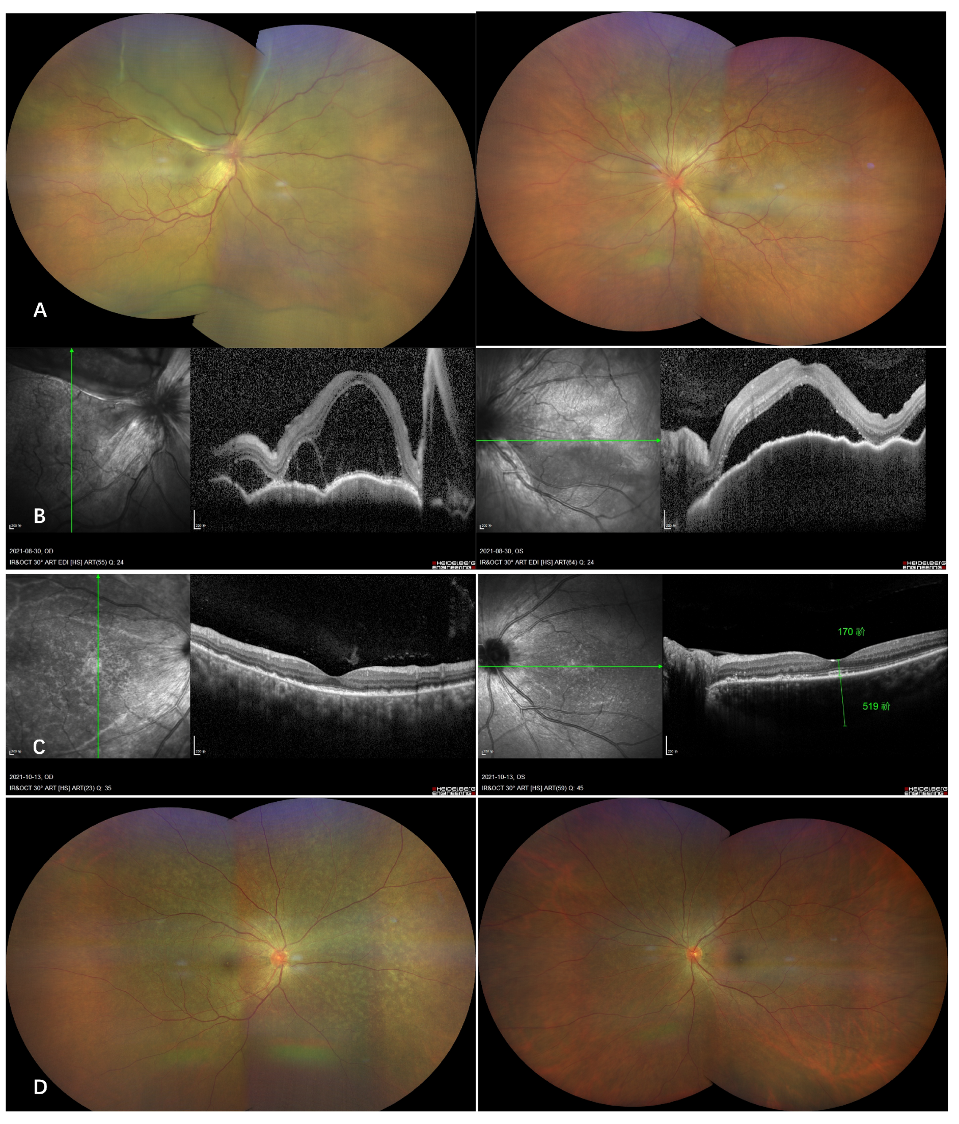
Selected Cases
- Case 1
- Case 2
- Case 3
4. Discussion
Author Contributions
Funding
Institutional Review Board Statement
Informed Consent Statement
Data Availability Statement
Conflicts of Interest
Appendix A

| Grade of Causality | Definition |
|---|---|
| Certain | Where a clinical event (including a laboratory test abnormality) occurs in a plausible time relationship to drug administration and cannot be explained by concurrent disease or other drugs or chemicals. A plausible (expected) clinical response to withdrawal of the medicine must be demonstrated and, if possible, the clinical response to restarting the medicine should also be demonstrated. |
| Probable or likely | Where a clinical event occurs with a reasonable time sequence to drug administration and is unlikely to be due to any concurrent disease or other drugs or chemicals. A plausible clinical response to withdrawal of the medicine, but not to restarting the medicine, must be demonstrated. |
| Possible | Where a clinical event occurs within a reasonable time sequence to drug administration but which could be explained by concurrent disease or other drugs or chemicals. Information on drug withdrawal may be lacking or unclear |
Appendix B
| Criteria | Yes | No | Unknown |
|---|---|---|---|
| Are there previous conclusive reports on this reaction? | +1 | 0 | 0 |
| Did the adverse reaction appear after the suspected drug was administered? | +2 | −1 | 0 |
| Did the adverse reaction improve when the drug was discontinued or a specific antagonist administered? | +1 | 0 | 0 |
| Did the adverse reaction reappear when the drug was readministered? | −2 | −1 | 0 |
| Are there alternative causes (other than the drug) that could on their own have caused the reaction? | −1 | +2 | 0 |
| Did the reaction reappear when a placebo was given? | −1 | +1 | 0 |
| Was the drug detected in the blood (or other fluids) in concentrations known to be toxic? | +1 | 0 | 0 |
| Was the reaction more severe when the dose was increased or less severe when the dose was decreased? | +1 | 0 | 0 |
| Did the patient have a similar reaction to the same or similar drugs in any previous exposure? | +1 | 0 | 0 |
| Was the adverse event confirmed by any objective evidence? | +1 | 0 | 0 |
| Total score–causal likelihood: 0–13 | 9–13 Definite 5–8 Probable 1–4 Possible 0 Doubtful | ||
References
- Geneva: World Health Organization. WHO COVID-19 Dashboard. 2020. Available online: https://covid19.who.int/ (accessed on 1 February 2022).
- Mallapaty, S. China’s COVID vaccines have been crucial—Now immunity is waning. Nature 2021, 598, 398–399. [Google Scholar] [CrossRef] [PubMed]
- Zhang, Y.; Zeng, G.; Pan, H.; Li, C.; Zhu, F. Safety, tolerability, and immunogenicity of an inactivated SARS-CoV-2 vaccine in healthy adults aged 18–59 years: A randomised, double-blind, placebo-controlled, phase 1/2 clinical trial. Lancet Infect. Dis. 2021, 21, 181–192. [Google Scholar] [CrossRef]
- Che, Y.; Liu, X.; Pu, Y.; Zhou, M.; Li, Q. Randomized, double-blinded and placebo-controlled phase II trial of an inactivated SARS-CoV-2 vaccine in healthy adults. Clin. Infect. Dis. Off. Publ. Infect. Dis. Soc. Am. 2020. [Google Scholar] [CrossRef]
- Pichi, F.; Aljneibi, S.; Neri, P.; Hay, S.; Dackiw, C.; Ghazi, N.G. Association of ocular adverse events with inactivated COVID-19 vaccination in patients in Abu Dhabi. JAMA Ophthalmol. 2021, 139, 1131–1135. [Google Scholar] [CrossRef] [PubMed]
- Ng, X.L.; Betzler, B.K.; Testi, I.; Ho, S.L.; Tien, M.; Ngo, W.K.; Zierhut, M.; Chee, S.P.; Gupta, V.; Pavesio, C.E. Ocular adverse events after COVID-19 vaccination. Ocul. Immunol. Inflamm. 2021. [Google Scholar] [CrossRef] [PubMed]
- Rabinovitch, T.; Ben-Arie-Weintrob, Y.; Hareuveni-Blum, T.; Shaer, B.; Vishnevskia-Dai, V.; Shulman, S.; Newman, H.; Biadsy, M.; Masarwa, D.; Fischer, N. Uveitis after the Bnt162b2 Mrna Vaccination Against Sars-Cov-2 Infection: A Possible Association. Retina 2021, 41, 2462–2471. [Google Scholar] [CrossRef]
- Renisi, G.; Lombardi, A.; Stanzione, M.; Invernizzi, A.; Bandera, A.; Gori, A. Anterior uveitis onset after bnt162b2 vaccination: Is this just a coincidence? Int. J. Infect. Dis. 2021, 110, 95–97. [Google Scholar] [CrossRef]
- Haseeb, A.A.; Solyman, O.; Abushanab, M.M.; Abo Obaia, A.S.; Elhusseiny, A.M. Ocular Complications following Vaccination for COVID-19: A One-Year Retrospective. Vaccines 2022, 10, 342. [Google Scholar] [CrossRef]
- Naranjo, C.A.; Busto, U.; Sellers, E.M.; Sandor, P.; Ruiz, I.; Roberts, E.; Janecek, E.; Domecq, C.; Greenblatt, D. A method for estimating the probability of adverse drug reactions. Clin. Pharmacol. Ther. 1981, 30, 239–245. [Google Scholar] [CrossRef]
- World Health Organization. Drug and Therapeutics Committees: A Practical Guide; Technical Report; World Health Organization: Geneva, Switzerland, 2003. [Google Scholar]
- Chen, X.; Wang, B.; Li, X. Acute-onset Vogt-Koyanagi-Harada-like uveitis following COVID-19 inactivated virus vaccination. Am. J. Ophthalmol. Case Rep. 2022, 26, 101404. [Google Scholar] [CrossRef]
- Jorge, L.F.; Queiroz, R.d.P.; Gasparin, F.; Vasconcelos-Santos, D.V. Presumed unilateral acute idiopathic maculopathy following H1N1 vaccination. Ocul. Immunol. Inflamm. 2020, 29, 1151–1153. [Google Scholar] [CrossRef] [PubMed]
- Jampol, L.M.; Tauscher, R.; Schwarz, H.P. COVID-19, COVID-19 vaccinations, and subsequent abnormalities in the retina: Causation or coincidence? JAMA Ophthalmol. 2021, 139, 1135–1136. [Google Scholar] [CrossRef]
- Ng, X.L.; Betzler, B.K.; Ng, S.; Chee, S.P.; Rajamani, L.; Singhal, A.; Rousselot, A.; Pavesio, C.E.; Gupta, V.; de Smet, M.D. The Eye of the Storm: COVID-19 Vaccination and the Eye. Ophthalmol. Ther. 2022, 11, 81–100. [Google Scholar] [CrossRef] [PubMed]
- Yue, L.; Xie, T.; Yang, T.; Zhou, J.; Chen, H.; Zhu, H.; Li, H.; Xiang, H.; Wang, J.; Yang, H. A third booster dose may be necessary to mitigate neutralizing antibody fading after inoculation with two doses of an inactivated SARS-CoV-2 vaccine. J. Med. Virol. 2022, 94, 35–38. [Google Scholar] [CrossRef] [PubMed]
- Watad, A.; Quaresma, M.; Brown, S.; Cohen Tervaert, J.; Rodríguez-Pint, I.; Cervera, R.; Perricone, C.; Shoenfeld, Y. Autoimmune/inflammatory syndrome induced by adjuvants (Shoenfeld’s syndrome)—An update. Lupus 2017, 26, 675–681. [Google Scholar] [CrossRef]
- Venkatesh, R.; Reddy, N.G.; Mishra, P.; Gupta, A.; Mahendradas, P.; Yadav, N.K. Unilateral Acute Idiopathic Maculopathy following Severe Acute Respiratory Syndrome Corona Virus (SARS-CoV-2) Infection. Ocul. Immunol. Inflamm. 2022. [Google Scholar] [CrossRef]
- Hasegawa, T.; Sannomiya, Y.; Toyoda, M.; Maruko, I.; Iida, T. Acute idiopathic maculopathy after COVID-19 vaccination. Am. J. Ophthalmol. Case Rep. 2022, 26, 101479. [Google Scholar] [CrossRef]
- Kim, M. Vogt-Koyanagi-Harada syndrome following influenza vaccination. Indian J. Ophthalmol. 2016, 64, 98. [Google Scholar] [CrossRef]
- Dogan, B.; Erol, M.K.; Cengiz, A. Vogt-Koyanagi-Harada disease following BCG vaccination and tuberculosis. Springerplus 2016, 5, 1–11. [Google Scholar] [CrossRef] [Green Version]
- Koong, L.R.; Chee, W.K.; Toh, Z.H.; Ng, X.L.; Agrawal, R.; Ho, S.L. Vogt-Koyanagi-harada disease associated with COVID-19 mRNA vaccine. Ocul. Immunol. Inflamm. 2021. [Google Scholar] [CrossRef]
- Saraceno, J.J.F.; Souza, G.M.; dos Santos Finamor, L.P.; Nascimento, H.M.; Belfort, R. Vogt-Koyanagi-Harada syndrome following COVID-19 and ChAdOx1 nCoV-19 (AZD1222) vaccine. Int. J. Retin. Vitr. 2021, 7, 1–7. [Google Scholar] [CrossRef] [PubMed]


| Patient No. | Age (Years) | Gender | Systemic and Ocular History | Medication | Symptoms after the First Dose of Vaccination | Vaccination Site (City) | First Dose of Vaccine Received | Second Dose of Vaccine Received |
|---|---|---|---|---|---|---|---|---|
| 1 | 33 | M | Ankylosing Spondylitis | Etanercept | No | Zhangzhou | Sinopharm * | No |
| 2 | 57 | F | No | No | No | Zhangzhou | Sinopharm * | Sinovac |
| 3 | 21 | M | No | No | fatigue, headache | Putian | Sinovac | No |
| 4 | 30 | F | No | No | No | Putian | Sinovac | Sinovac |
| 5 | 36 | F | No | No | No | Sanming | Sinovac | No |
| 6 | 28 | F | No | No | flu-like symptoms | Quanzhou | Sinopharm | No |
| 7 | 10 | F | Intermediate uveitis | Adalimumab methotrexate | No | Zhangzhou | Sinovac | No |
| Patient No. | Eyes Involved | Symptoms after 1st Vaccine | Symptoms after 2nd Vaccine | Manifestation | Time Intervals between Vaccination and Symptoms Onset (Days) | Positive Serologic Test | Treatment Received | Outcome | Causality Assessment |
|---|---|---|---|---|---|---|---|---|---|
| 1 | Unilateral | Redness, blurred vision | - | Iritis | 3 | HLA-B27+; ESR 38.0 mm/h | Periocular steroids | CR | Probable |
| 2 | Bilateral | Redness, blurred vision | Deteriorated vision | VKH | 10 | - | Oral steroids | CR | Probable |
| 3 | Bilateral | Redness, blurred vision | - | VKH-like uveitis | 1 | - | Periocular steroids | CR | Probable |
| 4 | Unilateral | Blurred vision | No symptoms | Multifocal choroiditis | 3 | - | Periocular steroids | CR | Possible |
| 5 | Unilateral | Blurred vision | - | AIM | 7 | ESR 41.8 mm/h | Oral steroids | CR | Possible |
| 6 | Bilateral | Blurred vision | - | VKH-like uveitis | 3 | - | Oral steroids | CR | Probable |
| 7 | Unilateral | Redness | - | Episcleritis | 7 | - | Topical steroid | CR | Possible |
Publisher’s Note: MDPI stays neutral with regard to jurisdictional claims in published maps and institutional affiliations. |
© 2022 by the authors. Licensee MDPI, Basel, Switzerland. This article is an open access article distributed under the terms and conditions of the Creative Commons Attribution (CC BY) license (https://creativecommons.org/licenses/by/4.0/).
Share and Cite
Chen, X.; Li, X.; Li, H.; Li, M.; Gong, S. Ocular Adverse Events after Inactivated COVID-19 Vaccination in Xiamen. Vaccines 2022, 10, 482. https://doi.org/10.3390/vaccines10030482
Chen X, Li X, Li H, Li M, Gong S. Ocular Adverse Events after Inactivated COVID-19 Vaccination in Xiamen. Vaccines. 2022; 10(3):482. https://doi.org/10.3390/vaccines10030482
Chicago/Turabian StyleChen, Xiuju, Xiaoxin Li, Haibo Li, Minghan Li, and Songjian Gong. 2022. "Ocular Adverse Events after Inactivated COVID-19 Vaccination in Xiamen" Vaccines 10, no. 3: 482. https://doi.org/10.3390/vaccines10030482