Next Article in Journal
Nucleic Acid Vaccines against SARS-CoV-2
Next Article in Special Issue
An IgY Effectively Prevents Goslings from Virulent GAstV Infection
Previous Article in Journal
Effect of Lockdowns on Hospital Staff in a COVID Center: A Retrospective Observational Study
Previous Article in Special Issue
Lumpy Skin Disease Virus with Four Knocked Out Genes Was Attenuated In Vivo and Protects Cattle from Infection
 
 
vaccines-logo
Article Menu
Article

Nanoformulations with Leishmania braziliensis Antigens Triggered Controlled Parasite Burden in Vaccinated Golden Hamster (Mesocricetus auratus) against Visceral Leishmaniasis

by
Jennifer Ottino
1,
Jaqueline Costa Leite
2,
Otoni Alves Melo-Júnior
2,
Marco Antonio Cabrera González
2,3,4,
Tatiane Furtado de Carvalho
5,
Giani Martins Garcia
3,
Maurício Azevedo Batista
2,
Patrícia Silveira
2,
Mariana Santos Cardoso
1,
Lilian Lacerda Bueno
1,
Ricardo Toshio Fujiwara
1,
Renato Lima Santos
5,
Paulo Ricardo de Oliveira Paes
5,
Denise Silveira-Lemos
2,
Olindo Assis Martins-Filho
6,
Alexsandro Sobreira Galdino
7,
Miguel Angel Chávez-Fumagalli
8,
Walderez Ornelas Dutra
2,9,
Vanessa Carla Furtado Mosqueira
3 and
Rodolfo Cordeiro Giunchetti
1,2,9,*
1
Departamento de Parasitologia, Universidade Federal de Minas Gerais (UFMG), Belo Horizonte 31270-901, Minas Gerais, Brazil
2
Departamento de Morfologia, Universidade Federal de Minas Gerais (UFMG), Belo Horizonte 31270-901, Minas Gerais, Brazil
3
Laboratório de Desenvolvimento Galênico e Nanotecnologia, Escola de Farmácia, Universidade Federal de Ouro Preto (UFOP), Ouro Preto 35400-000, Minas Gerais, Brazil
4
Instituto Nacional de Innovación Agraria, Estación Experimental Agraria Baños del Inca, Cajamarca 06000, Peru
5
Escola de Veterinária, Universidade Federal de Minas Gerais (UFMG), Belo Horizonte 31270-901, Minas Gerais, Brazil
6
Centro de Pesquisas René Rachou/Fundação Oswaldo Cruz, Belo Horizonte 30190-002, Minas Gerais, Brazil
7
Laboratório de Biotecnologia de Microrganismos, Universidade Federal de São João Del-Rei (UFSJ), Campus Centro Oeste, Divinópolis 35501-296, Minas Gerais, Brazil
8
Computational Biology and Chemistry Research Group, Vicerrectorado de Investigación, Universidad Católica de Santa María, Urb. San José S/N, Umacollo, Arequipa 04000, Peru
9
Instituto Nacional de Ciência e Tecnologia em Doenças Tropicais—INCT-DT, Belo Horizonte 31270-901, Minas Gerais, Brazil
*
Author to whom correspondence should be addressed.
Vaccines 2022, 10(11), 1848; https://doi.org/10.3390/vaccines10111848
Submission received: 13 October 2022 / Revised: 24 October 2022 / Accepted: 27 October 2022 / Published: 31 October 2022
(This article belongs to the Special Issue Animal Vaccines)

Abstract

:
Leishmaniasis is a widespread vector-borne disease in Brazil, with Leishmania (Leishmania) infantum as the primary etiological agent of visceral leishmaniasis (VL). Dogs are considered the main reservoir of this parasite, whose treatment in Brazil is restricted to the use of veterinary medicines, which do not promote a parasitological cure. Therefore, efficient vaccine development is the best approach to Canine Visceral Leishmaniasis (CVL) control. With this in mind, this study used hamsters (Mesocricetus auratus) as an experimental model in an anti-Leishmania preclinical vaccine trial to evaluate the safety, antigenicity, humoral response, and effects on tissue parasite load. Two novel formulations of nanoparticles made from poly(D, L-lactic) acid (PLA) polymer loading Leishmania braziliensis crude antigen (LB) exhibiting two different particle sizes were utilized: LBPSmG (570 nm) and LBPSmP (388 nm). The results showed that the nanoparticles were safe and harmless to hamsters and were antigenic with the induction in LBSap, LBPSmG, and LBPSmG groups of total anti-Leishmania IgG antibodies 30 days after challenge, which persists 200 days in LBSap and LBPSmP. At the same time, a less pronounced hepatosplenomegaly in LBSap, LBPSmG, and LBPSmP was found when compared to control groups, as well as a less pronounced inflammatory infiltrate and granuloma formation in the spleen. Furthermore, significant reductions of 84%, 81%, and 90% were observed in spleen parasite burden accessed by qPCR in the LBSap, LBPSmG, and LBPSmP groups, respectively. In this way, LBSap, LBPSmG, and LBPSmP formulations showed better results in vaccinated and L. infantum-challenged animals in further reducing parasitic load in the spleen and attenuating lesions in liver and splenic tissues. This results in safe, harmless nanoformulation vaccines with significant immunogenic and infection control potential. In addition, animals vaccinated with LBPSmP had an overall reduction in parasite burden in the spleen, indicating that a smaller nanoparticle could be more efficient in targeting antigen-presenting cells.

1. Introduction

Leishmaniasis is a complex of parasitic diseases transmitted during female sand flies’ blood meal by Phlebotomus genus in the Old World and Lutzomyia in the New World. Seven countries account for 90% of the cases distributed worldwide: Brazil, Ethiopia, India, Kenya, Somalia, South Sudan, and Sudan. The disease, which can be caused by about twenty different species of Leishmania parasites, has three main clinical manifestations: cutaneous, mucosal, and visceral [1]. L. infantum is the main etiological agent involved in visceral leishmaniasis (VL) in Latin America, although some studies report dogs with VL clinical symptoms that were found positive for L. amazonensis or L. braziliensis infection [2,3,4,5,6,7,8].
In Brazil and many Europe countries—primarily in the Mediterranean basin—dogs are considered the main reservoir of L. infantum, and a large percentage of them are asymptomatic, which contributes to the spread of the disease [9]. In this sense, canine visceral leishmaniasis (CVL) represents a serious public health problem, whereas control of human cases is closely linked to the breakdown of the disease cycle in dogs [10]. Control measures, such as the use of impregnated insecticide collars, raising people’s awareness, and new vaccine technologies, are rational approaches that could be implemented to control CVL.
Nanotechnology-based formulations have emerged as a safe and powerful alternative to be used in the control of various diseases, promoting more assertive drug or vaccinal antigen deliveries. This approach is able to target cells and, for vaccinal antigens, trigger the immune system, thereby improving the immunogenicity without adjuvants in the formulation [1,11,12,13]. In this sense, polymeric nanoparticles are a good alternative to be used in VL control as carriers of Leishmania antigens. Depending on the polymer, they exhibit low toxicity and good cost benefits. Furthermore, PLA-based nanocarriers are fully biodegradable and stable in biological environments, delaying antigen release before they reach the target cells [1,14].
Likewise, the choice of the appropriate experimental model is relevant in immunobiological research, with the golden hamster (Mesocricetus auratus) properly mimicking the VL course in its vertebrate hosts, such as human and dogs, unlike BALB/c mice [15,16].
The present study evaluated the association between two polymeric nanoformulations with L. braziliensis antigens as a potential immunobiological component against CVL using golden hamsters as an experimental model. Clinical, hematological, immunological, parasitological, histological and molecular parameters were evaluated to better assess the efficacy of the vaccine candidates in the animal model.

2. Materials and Methods

2.1. Ethical Statement

This project was approved by the Ethics Committee on the Use of Animals of the Federal University of Minas Gerais (CEUA-UFMG) by protocol 385/2013.

2.2. Studied Animals and Experimental Groups

Seventy-two healthy male golden hamsters (Mesocricetus auratus) between 6 and 8 weeks of age were divided into eight groups with eight animals each. The groups were established as follows: infected control group (C), saponin inoculated group (Sap), Leishmania braziliensis antigen vaccinated group (LB), L. braziliensis antigen plus saponin vaccinated group (LBSap), empty submicrometric large particle vaccinated group (PSmG), submicrometric large particle plus Leishmania braziliensis crude antigen vaccinated group (LBPSmG), empty submicrometric small particle vaccinated group (PSmP), and submicrometric small particle plus Leishmania braziliensis crude antigen vaccinated group (LBPSmP). Eight animals were used as a preimmune group.

2.3. Production of Vaccine Formulations

To prepare the Leishmania braziliensis antigen, a reference strain (MHOM/BR/75/M2904) was grown in Minimum Essential Medium (α–MEM-Gibco BRL, Waltham, MA, USA) supplemented with 10% bovine fetal serum and 2 mM penicillin under biochemical oxygen demand (BOD) at 23 °C for a maximum of 10 passages. On the fifth day, 108 stationary promastigotes were sonicated, and the antigen was aliquoted and stored at −80 °C [17]. Previous studies from our group had already demonstrated that L. braziliensis crude antigen, combined or not to saponin, are safe and innocuous in in vitro, pre-clinic and clinic models and are promising vaccines against CVL [17,18,19].
Saponin was used as the adjuvant (Sigma-Aldrich®, St. Louis, MI, USA) at a 100 μg/dose concentration diluted in phosphate saline buffer (PBS) at the inoculum time.
The nanospheres were prepared at Laboratório de Desenvolvimento Galênico e Nanotecnológico of the Escola de Farmácia from Universidade Federal de Ouro Preto by using the interfacial deposition method of a preformed polymer, as described by Fessi et al. [20].
Empty nanospheres were prepared by associating a poly(D, L-lactic) acid polymer (PLA) solution in an organic solvent with chitosan dissolved in 0.05 M acetic acid at concentrations of 50 mg/mL (large submicrometric particle) and 5 mg/mL (small submicrometric particle). Antigen-loaded nanoparticles (NP) were obtained using these same solutions by adding 1 mL of the L. braziliensis crude antigen suspension at a concentration of 1 mg/mL. The nanospheres were characterized by size, polydispersity index, and zeta potential, as described by Garcia et al. [21]. Large submicrometer particles (PSmG—empty particle) were 613 nm in sizes and small submicrometer particles (PSmP—empty particle) were 35 nm in size. The antigen encapsulated NP showed a mean size of 570 nm for large submicrometer particles (LBPSmG) and 388 nm for small submicrometer particles (LBPSmP).
Each vaccine formulation was diluted with 0.9% sterile saline buffer during its preparation, and the final volume was adjusted for 100 µL/dose.

2.4. Experimental Protocol

The animals enrolled in the study (n = 72 hamster; n = 8/group) were kept in ventilated racks with food and water ad libitum throughout the study, with intermittent light/dark cycles every 12 h, submitted to fully blinded preclinical interventions. The hamsters received a dose of the vaccine subcutaneously according to their respective groups, in intervals of 21 days up to three doses. The safety and toxicity evaluations of the nanoformulations were performed by observing the inoculum site (e.g., analysis of presence of nodules, papules, or ulcerative lesions), by evaluating animal’s behavior (hostility or apathy), and also by catching sight of signs such as local pain, alopecia, and bristling. Thirty days after beginning vaccine protocol (T30), the first blood sample of the animals was taken and, after 50 days (end of the vaccine protocol), the animals were challenged with 107 L. infantum stationary promastigotes (strain C46) resuspended in saline in a 500 µL final volume by intraperitoneal route [22]. L. infantum culture was grown under the same conditions described by Giunchetti et al. [17].
The hamsters were surveyed throughout the experimental period for the presence of lesions at the inoculum and challenge sites; clinical signs such as cachexia, alopecia, organs enlargement; behavioral changes; and weight loss. Two hundred days post challenge (T200), a new blood sample was taken, and the animals were euthanized by sodium pentobarbital overdose (150 mg/Kg), injected intraperitoneally.

2.5. Assessment of Total Anti-Leishmania IgG Antibodies

Total IgG antibodies were measured through an in house ELISA assay according the protocol described by Moreira et al. 2012 [23]. The 96-well plates were sensitized with 1 µg/well of soluble L. infantum strain C46 antigen. Unspecific ligations were avoided by adding 200 μL/well of a PBS solution containing 3% BSA. The hamster’s serum was added using a 1:100 proportion and the secondary anti-hamster IgG antibody was diluted 1:5000 times, both in PBS Tween 20 in a 0.05% concentration. The reaction was revealed with 0.03% hydrogen peroxide and 0.022 µg of ortho-phenylenediamine (OPD). An ELX800 (Biotek Instruments VT, Winooski, VT, USA) spectrophotometer was used to measure the wells’ luminescence in a 490 nm wavelength. The cut-off was established by the average of the optical density (OD) of eight non-infected hamsters (pre-immune group related to randomized samples in hamster of all groups before the immunization protocol) plus two standard deviations measured for these samples.

2.6. Histology Analysis

Fragments of the liver and spleen from 64 animals (except for the pre-immune group) were analyzed by histopathology stained with Hematoxylin and Eosin. The liver was analyzed for inflammatory lymphohistiocytic infiltrate, lymphohistiocytic and neutrophilic inflammatory infiltrate, granuloma, hydropic degeneration, and cholestasis. The spleen was evaluated for the presence of inflammatory lymphohistiocytic infiltrate, lymphohistiocytic and neutrophilic inflammatory infiltrate for red and white pulps, lymphoid hyperplasia, granuloma, amyloidosis, amastigotes, and perisplenitis.

2.7. Parasite Burden Evaluation

2.7.1. Limiting Dilution Assay

Vaccine efficacy was assessed by parasitological assay using limiting dilution in the liver and spleen samples. The organs from all the animals of each group (except for the pre-immune group) were removed in sterile condition, weighed (for total and fragment organ weight), macerated with 2 mL of α–MEM (minimum essential medium) in a tissue macerator (Pyrex®, Burlington, MA, USA), and centrifugated at 1540× g at 4 °C for 10 min. The liver and spleen homogenates were plated in duplicates in a 96-well microtitre plates in serial 5-fold successive dilutions. All the plates were maintained for 7 days in a biochemical oxygen demand incubator at 23 °C. The average parasite count in the well duplicate, where it was last visualized, was multiplied by the organ’s weight and the dilution factor corresponding to the well. The value obtained corresponded to the parasite burden in the respective organ fragment. It was then possible to estimate the parasite burden in the organs [24,25].

2.7.2. Quantitative PCR (qPCR)

The parasite gDNA extraction from the spleen was performed by using a NucleoSpin® Tissue extraction kit—Genomic DNA from tissue protocol (Macherey-Nagel, Duren, Germany) according to the manufacturer’s instructions. DNA was resuspended in 50 μL of elution buffer (kit provided) and quantified using Nanodrop N2000 equipment (Thermo Scientific, Waltham, MA, USA). The qPCR reactions were performed using 5 μL of the 2× Power Syber®-Green ABI reagent (Thermo Fisher, USA), 0.5 μL of each primer (2 μM), (kDNA.Leish.F: 5′-CGTGGGGGAGGGGCGTTCT-3′ and kDNA.Leish.R: 5′-CCGAAGCAGCCGCCCCTATT-3′) with a 135 pb amplicon size [26], and 4 μL of DNA (5 ng/μL) in a final volume of 10 μL per well. The β-actin constitutive hamster gene was selected to normalize the reaction using primers that amplify a 120 bp fragment (Forward: 5′-TCGTACGTGGGTGACGAGGC-3′; reverse: 5′-GTAGAAGGTGTGGTGCCAGA-3′). The amplification process took place following the cycles: initial denaturation at 95 °C for 10 min, followed by 40 cycles of denaturation at 95°C for 15 s, and a melt curve stage at 60 °C for 1 min.
Finally, the qPCR analysis was performed using 7500 Software v2.0.1 (Thermo Fisher, USA), and the results were plotted using the standard curve method (parasite burden was inferred through the difference between the amount of b-actin kDNA and the amount of the hamster’s sample kDNA). They were assembled from the serial dilution of DNA extracted from 108 Leishmania sp. promastigotes and 106 hamster—Mesocricetus auratus—splenocytes.

2.8. Statistical Analyses

Statistical analyses were conducted using the Prism 5.0 software package (Prism Software, Irvine, CA, USA). Data normality was assessed using the Kolmogorov–Smirnoff test. One-way analysis of variance (ANOVA) and Tukey post-test were used to investigate differences between groups. Considering the nonparametric nature of real time PCR data, the Kruskal–Wallis test was used, followed by Dunn’s test. Statistical significance was considered at a p value < 0.05.

3. Results

3.1. Immunobiological Safety and Harmless Analysis

The animals were observed after each vaccine dose for possible post-vaccination reactions, and the vaccine was shown to be safe and innocuous. Subsequent to animal challenge, the control groups (C, Sap, LB, PSmG, and PSmP) displayed more evident dermatological changes (e.g., alopecia) as compared to animals vaccinated with formulations that contained the Leishmania antigen (LBSap, LBPSmG, and LBPSmP). Notably, the controls groups (C, Sap, LB, PSmG, and PSmP) exhibited enlarged livers and spleens after euthanasia.

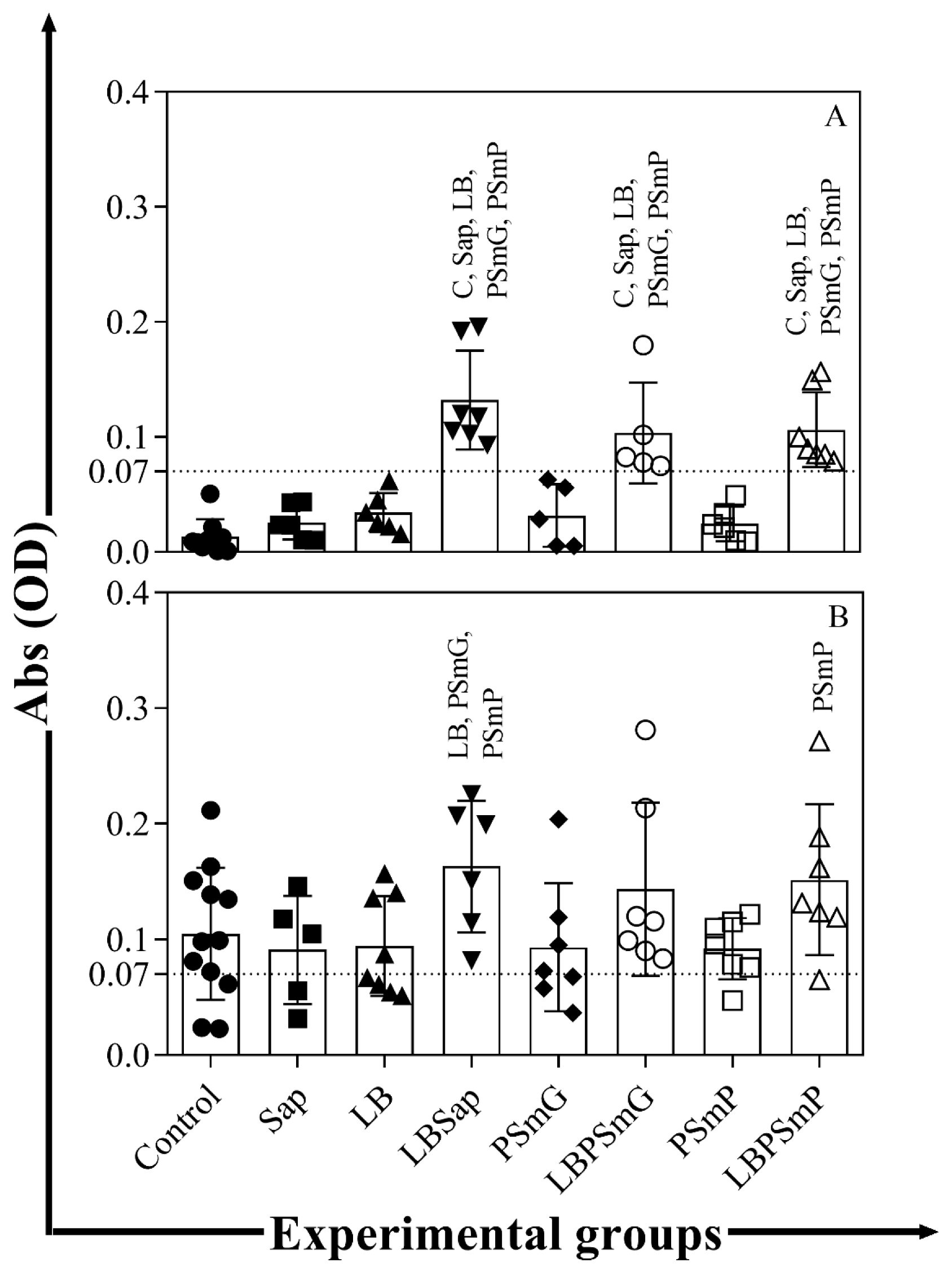
3.2. Immunogenic Potential of Nanoformulations

The LBPSmG and LBPSmG groups showed increased total anti-Leishmania l IgG levels 30 days after two vaccine boosters as compared to the other groups (C, Sap, LB, PSmG, and PSmP—p < 0.05) (Figure 1A). Furthermore, the LBSap group still presented high levels of total IgG antibodies 200 days after experimental challenge with L. infantum as compared to the LB, PSmG, and PSmP groups (p < 0.05) (Figure 1B), and the LBPSmP group showed elevated levels of IgG antibodies as compared to the PSmP group (p < 0.05) (Figure 1B).

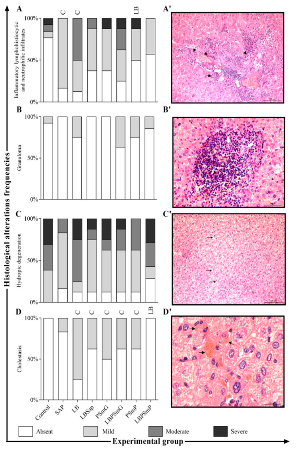
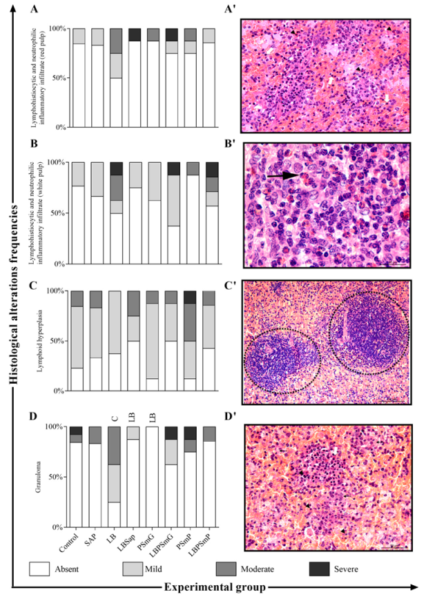
3.3. Evaluation of Changes in Parasite Target Tissues

The lesions on the liver of L. infantum-infected animals were accessed by frequency histopathological evaluation, and the LBPSmP group showed a decrease in cholestasis as compared with the LB group, as shown in the bar graphs in Figure 2A–D/ and images in Figure 2A′–D′.
The descriptive histopathological analysis of the spleen revealed a significant reduction in granuloma progression in LBSap and PSmG as compared to animals vaccinated only with the LB antigen. Additionally, perisplenitis was reduced in the Sap, LBSap, and PSmP groups compared to the C group. The frequency in which histological changes occur by group is presented in bar graphs (Figure 3A–G) and shown in representative images in Figure 3A′–G′.

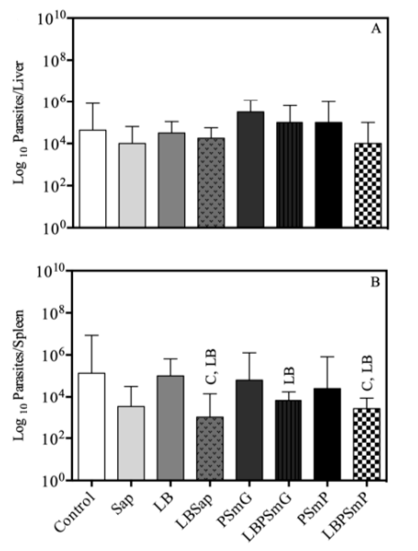
3.4. Efficacy Evaluation Based on Parasite Load

3.4.1. Limiting Dilution Assay

In addition to a tendency toward reduced parasitism in the liver (Figure 4A) in the LBSap and LBPSmP groups, the lowest parasitic load was observed in the spleen in the vaccine groups (LBSap, LBPSmG, and LBPSmP) as compared to the control groups by limiting dilution assay (Figure 4B).

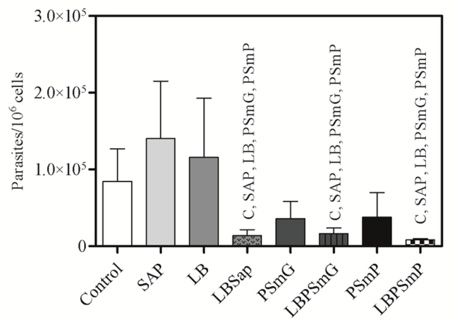
3.4.2. Quantitative Molecular Analysis (qPCR)

Data obtained in the qPCR analysis showed that the LBPSmP, LBSap, and LBPSmG groups have the lowest parasite burden/106 hamster splenocytes compared to the other groups (Figure 5). Notably, the percentage of parasite load reduction in the LBSap, LBPSmG, and LBPSmP groups were 90%, 88%, and 94%, respectively, when compared to the SAP group; 88%, 86%, and 93%, respectively, when compared to the LB group; and 84%, 81%, and 90%, respectively, when compared to the C (Control) group.

4. Discussion

The development of vaccines for pathogen control is a hallmark in modern medicine. Nanovaccines have been studied due to their efficient antigen delivery systems and ability to reach a specific target [27,28,29]. In addition, this technology has the added advantage of having no potential side effects as compared to the adjuvants used in the development of immunobiological candidates with less local and systemic side effects.
This work combines two novel nanoparticles associated with the L. braziliensis antigen as an alternative for visceral leishmaniasis control. Previous works have already demonstrated the cross protection of L. braziliensis crude antigens against L. infantum infection, as well as the genetic homology between these parasite species [17,18,19]. Giunchetti et al. (2007) [17] demonstrated a significant increase in nitric oxide (NO) levels in dogs vaccinated with LBSap in an in vitro cell proliferation assay stimulated with soluble L. chagasi antigen. This study demonstrated a protective response triggered by L. braziliensis plus saponin vaccine against visceral leishmaniasis. Furthermore, hamsters were chosen as experimental models because the evolution of their disease is similar to that in dogs and humans [30,31,32].
In this sense, clinical and laboratory studies have shown that the combination between nanoparticles and L. braziliensis antigen are safe and harmless after the three-dose vaccine protocol as no clinical, behavioral, or significant changes in weight were observed in hamsters. LBSap, an extensively studied vaccine, also presented no toxicity and deemed to be safe, as previously demonstrated. [17,18,19,33,34,35,36,37]. After L. infantum experimental challenge, and before the animals were euthanized, some dermatological disorders, such as alopecia, were observed in the C, Sap, LB, PSmG, and PSmP groups, which are considered a common clinical sign of VL [38,39].
Increased levels of total anti-Leishmania IgG antibodies were observed in this work 30 days after L. infantum challenge, which persisted 200 days after challenge in animals from the LBSap and LBPSmP groups.
These data demonstrate that the vaccines triggered a strong antigenicity profile, which was described in studies conducted in hamsters [40], mice [35], and dogs [17,33,34,37]. Nanoparticle size may also be directly related to immunogenic vaccine potential, whose physical-chemical characteristics have been extensively studied [27,41].
The LB and PSmG groups showed an increase in cholestasis and inflammatory cell infiltration intensity, indicating hepatic tissue damage. The presence of damaged hepatic ducts had already been observed in mice and preceded any other inflammatory processes in hepatic tissue, such as fibrosis and steatosis. This may lead to Küpffer cell activation with IL-6 production, along with fibrin and collagen deposition in the bile ducts, causing blockage to the liver [42]. Moreover, inflammatory cells move to the parasitized site, orchestrated by immune mechanisms, in order to enclose the infection, culminating in granuloma formation both in hamsters and dogs [38,39,43,44]. However, a reduction in these changes was clearly observed in the LBPSmP group. Hepatosplenomegaly was notably absent or less prominent in the LBSap, LBPSmG, and LBPSmP groups [15,39,45,46]. The presence of Leishmania amastigotes in parasitized tissues may induce changes in their structure and physiology [47].
In healthy individuals, the spleen is the lymphoid organ foremost related to circulating pathogen defense, and its structure may be severely affected during chronic VL disease [48]. The elimination of amastigotes in this organ is inefficient in hamsters [49,50], and some studies suggest that a disruption in spleen compartments may occur with disorders of the red and white pulp [48,51,52], which were observed in the control groups of the present study. However, the granuloma formation was slightly diminished, especially in LBSap and LBPSmP groups, and could be associated with the activation of the immune system and reduction in parasite load [44,49].
In fact, the parasite burden quantification assessed by limiting dilution and qPCR assays showed a significant reduction in Leishmania parasite burden in the LBSap, LBPSmG, and LBPSmP groups, demonstrating the efficiency in controlling parasite spread in hamsters. Similarly, in another LBSap preclinical vaccine trial using BALB/c mice, a 42% decrease in Leishmania parasites was observed in the spleen of vaccinated animals [35] as compared to the 84% reduction in hamsters in this study. Similarly, Roatt et al. [52] also reported a reduction in parasite burden in bone marrow, skin, and sand flies in a study using an L. braziliensis antigen (associated to MPL adjuvant (LBMPL)) as a vaccine in an immunotherapeutic study in naturally L. infantum-infected dogs. Importantly, the groups that presented the lowest amount of enlargement of the liver and spleen—LBSap, LBPSmG, and LBPSmP—also showed the lowest parasitic load, similar to what Lima et al. demonstrated in asymptomatic animals [53].
The LBSap, LBPSmG, and LBPSmP formulations demonstrated better results in vaccinated and L. infantum challenged animals in reducing the parasite load, making it a promising vaccine candidate. Significantly, a hallmark profile in the LBPSmP formulation includes biomarkers related to restricted parasite load and low amount of histological damage between the vaccinated groups.

5. Conclusions

The vaccine formulations proposed in this study (LBSap, LBPSmG, and LBPSmP) were innocuous, safe, and antigenic in hamsters. Moreover, they proved efficient in triggering total anti-Leishmania IgG antibodies and in attenuating tissue damage in the liver and spleen, especially LBSap and LBPSmP. Although immunophenotyping assays were not performed, the parameters evaluated in the vaccinated animals suggest the occurrence of a protective cellular profile in this pre-clinical assay. In addition, a significant parasite load reduction was observed in the spleen of hamsters vaccinated with LBPSmP indicating that a smaller nanoparticle could be more efficient in delivering the antigen to the target cells. These results should stimulate additional studies, especially regarding the nature of the immune response triggered by the nanoparticles and the mechanisms involved. The use of nanotechnology associated with Leishmania antigens could be an important strategy for visceral leishmaniasis control measures.

Author Contributions

Conceptualization, R.C.G., O.A.M.-F., R.T.F., and W.O.D.; method, J.O., J.C.L., O.A.M.-J., M.A.C.G., T.F.d.C., M.A.B., P.S., M.S.C., and G.M.G.; formal analysis, J.O., T.F.d.C., M.A.B., P.S., M.S.C., D.S.-L., A.S.G., and M.A.C.-F.; investigation, J.O., J.C.L., O.A.M.-J., M.A.C.G., and D.S.-L.; resources R.C.G., D.S.-L., O.A.M.-F., and W.O.D.; data curation, R.C.G. and J.O.; writing—original draft preparation, R.C.G., J.O., M.A.B., O.A.M.-J., and P.S.; writing—review and editing, J.O. and R.C.G.; supervision, R.C.G., P.R.d.O.P., and R.T.F.; project administration, R.C.G., L.L.B., V.C.F.M., R.T.F., R.L.S., and P.R.d.O.P.; funding acquisition, R.C.G., D.S.-L., O.A.M.-F., and W.O.D. All authors have read and agreed to the published version of the manuscript.

Funding

This research was supported by the Ministério da Saúde, Departamento de Ciência e Tecnologia (DECIT); Conselho Nacional de Desenvolvimento Científico e Tecnológico (CNPq; 440059/2021-6; 424069/2018-0); Coordenação de Aperfeiçoamento de Pessoal de Nível Superior (CAPES); Fundação de Amparo à Pesquisa do Estado de Minas Gerais (FAPEMIG; CBB-PPM-00637-17); and Instituto Nacional de Ciência e Tecnologia em Doenças Tropicais (INCT-DT).

Institutional Review Board Statement

The animal study protocol was approved by the Ethics Committee on the Use of Animals of the Federal University of Minas Gerais (CEUA-UFMG) by protocol 385/2013.

Informed Consent Statement

Not applicable.

Acknowledgments

We acknowledge the Centro de Ensaios Pré-Clínicos/ICB-UFMG for supplying the hamsters used in the preclinical experiments. The authors R.T.F., R.L.S., O.A.M.F., A.S.G., W.O.D., V.C.F.M., and R.C.G. would like to thank CNPq for their research fellowships. We thank Randall Johnson for reviewing the language of the manuscript.

Conflicts of Interest

The authors declare no conflict of interest.

References

  1. Gutiérrez, V.; Seabra, A.B.; Reguera, R.M.; Khandare, J.; Calderón, M. New Approaches from Nanomedicine for Treating Leishmaniasis. Chem. Soc. Rev. 2015, 45, 152–168. [Google Scholar] [CrossRef] [PubMed] [Green Version]
  2. Ministério da Saúde. Guia de Vigilância em Saúde-Volume 3; Ministério da Saúde: Araraquara, Brazil, 2017; Volume 3, ISBN 9788533421790. [Google Scholar]
  3. Hernández, D.; Martínez, C.; Garcia, L.; Rodríguez, N.; Convit, J. Leishmania braziliensis Causing Visceral Leishmaniasis In A Patient With Human Immunodeficiency Virus Infection, Identified With The Aid Of The Polymerase Chain Reaction. Trans. R. Soc. Trop. Med. Hyg. 1993, 87, 627–628. [Google Scholar] [CrossRef]
  4. Kaye, P.M.; Beattie, L. Lessons from Other Diseases: Granulomatous Inflammation in Leishmaniasis. Semin. Immunopathol. 2016, 38, 249–260. [Google Scholar] [CrossRef] [Green Version]
  5. Tolezano, J.E.; Uliana, S.R.B.; Taniguchi, H.H.; Araújo, M.F.L.; Barbosa, J.A.R.; Barbosa, J.E.R.; Floeter-Winter, L.M.; Shaw, J.J. The First Records of Leishmania (Leishmania) amazonensis in Dogs (Canis familiaris) Diagnosed Clinically as Having Canine Visceral Leishmaniasis from Araçatuba County, São Paulo State, Brazil. Vet. Parasitol. 2007, 149, 280–284. [Google Scholar] [CrossRef] [PubMed]
  6. Dantas-Torres, F. The Role of Dogs as Reservoirs of Leishmania Parasites, with Emphasis on Leishmania (Leishmania) infantum and Leishmania (Viannia) braziliensis. Vet. Parasitol. 2007, 149, 139–146. [Google Scholar] [CrossRef]
  7. Valdivia, H.O.; Almeida, L.V.; Roatt, B.M.; Reis-Cunha, J.L.; Pereira, A.A.S.; Gontijo, C.; Fujiwara, R.T.; Reis, A.B.; Sanders, M.J.; Cotton, J.A.; et al. Comparative Genomics of Canine-Isolated Leishmania (Leishmania) amazonensis from an Endemic Focus of Visceral Leishmaniasis in Governador Valadares, Southeastern Brazil. Sci. Rep. 2017, 7, 40804. [Google Scholar] [CrossRef] [Green Version]
  8. Estevam, L.G.T.M.; Veloso, L.B.; Silva, G.G.; Mori, C.C.; Franco, P.F.; Lima, A.C.V.M.R.; Ássimos, G.R.; Reis, I.A.; Andrade-Filho, J.D.; Araújo, M.S.S.; et al. Leishmania infantum Infection Rate in Dogs Housed in Open-Admission Shelters Is Higher than of Domiciled Dogs in an Endemic Area of Canine Visceral Leishmaniasis. Epidemiological Implications. Acta Trop. 2022, 232, 106492. [Google Scholar] [CrossRef]
  9. Marzochi, M.C.; Coutinho, S.G.; de Souza, W.J.; Toledo, L.M.D.; Grimaldi, G., Jr.; Momen, H.; Pacheco, R.d.S.; Sabroza, P.C.; de Souza, M.A.; Rangel, F.B., Jr.; et al. Canine Visceral Leishmaniasis in Rio de Janeiro, Brazil. Clinical, Parasitological, Therapeutical and Epidemiological Findings (1977–1983). Memórias do Inst. Oswaldo Cruz 1985, 80, 349–357. [Google Scholar] [CrossRef] [Green Version]
  10. Ministério da Saúde. Manual de Vigilância e Controle da Leishmaniose Visceral; Ministério da Saúde: Araraquara, Brazil, 2014; ISBN 9788533407428. [Google Scholar]
  11. Getts, D.R.; Shea, L.D.; Miller, S.D.; King, N.J.C. Harnessing Nanoparticles for Immune Modulation. Trends Immunol. 2015, 36, 419–427. [Google Scholar] [CrossRef] [Green Version]
  12. Doroud, D.; Rafati, S. Leishmaniasis: Focus on the Design of Nanoparticulate Vaccine Delivery Systems. Expert Rev. Vaccines 2012, 11, 69–86. [Google Scholar] [CrossRef]
  13. Zadeh Mehrizi, T.; Khamesipour, A.; Shafiee Ardestani, M.; Ebrahimi Shahmabadi, H.; Haji Molla Hoseini, M.; Mosaffa, N.; Ramezani, A. Comparative Analysis between Four Model Nanoformulations of Amphotericin B-Chitosan, Amphotericin B-Dendrimer, Betulinic Acid-Chitosan and Betulinic Acid-Dendrimer for Treatment of Leishmania Major: Real-Time PCR Assay Plus. Int. J. Nanomed. 2019, 14, 7593–7607. [Google Scholar] [CrossRef] [PubMed] [Green Version]
  14. Tafaghodi, M.; Khamesipour, A.; Jaafari, M.R. Immunization against Leishmaniasis by PLGA Nanospheres Encapsulated with Autoclaved Leishmania major (ALM) and CpG-ODN. Parasitol. Res. 2011, 108, 1265–1273. [Google Scholar] [CrossRef] [PubMed]
  15. Melby, P.C.; Chandrasekar, B.; Zhao, W.; Coe, J.E. The Hamster as a Model of Human Visceral Leishmaniasis: Progressive Disease and Impaired Generation of Nitric Oxide in the Face of a Prominent Th1-like Cytokine Response. J. Immunol. 2001, 166, 1912–1920. [Google Scholar] [CrossRef] [PubMed] [Green Version]
  16. Jiménez-Antón, M.D.; Grau, M.; Olías-Molero, A.I.; Alunda, J.M. Syrian Hamster as an Advanced Experimental Model for Visceral Leishmaniasis; Humana Press: New York, NY, USA, 2019; pp. 303–314. [Google Scholar]
  17. Giunchetti, R.C.; Corrêa-Oliveira, R.; Martins-Filho, O.A.; Teixeira-Carvalho, A.; Roatt, B.M.; de Oliveira Aguiar-Soares, R.D.; de Souza, J.V.; das Dores Moreira, N.; Malaquias, L.C.C.; Mota e Castro, L.L.; et al. Immunogenicity of a Killed Leishmania Vaccine with Saponin Adjuvant in Dogs. Vaccine 2007, 25, 7674–7686. [Google Scholar] [CrossRef]
  18. Vitoriano-Souza, J.; Reis, A.B.; Moreira, N.D.; Giunchetti, R.C.; Correa-Oliveira, R.; Carneiro, C.M. Kinetics of Cell Migration to the Dermis and Hypodermis in Dogs Vaccinated with Antigenic Compounds of Leishmania braziliensis plus Saponin. Vaccine 2008, 26, 3922–3931. [Google Scholar] [CrossRef]
  19. Moreira, N.d.D.; Giunchetti, R.C.; Carneiro, C.M.; Vitoriano-Souza, J.; Roatt, B.M.; Malaquias, L.C.C.; Corrêa-Oliveira, R.; Reis, A.B. Histological Study of Cell Migration in the Dermis of Hamsters after Immunisation with Two Different Vaccines against Visceral Leishmaniasis. Vet. Immunol. Immunopathol. 2009, 128, 418–424. [Google Scholar] [CrossRef] [Green Version]
  20. Fessi, H.; Puisieux, F.; Devissaguet, J.P.; Ammoury, N.; Benita, S. Nanocapsule Formation By Interfacial Polymer Deposition Following Solvent Displacement. Int. J. Pharm. 1989, 55, R1–R4. [Google Scholar] [CrossRef]
  21. Garcia, G.M.; Oliveira, L.T.; Pitta, I.D.R.; De Lima, M.D.C.A.; Vilela, J.M.C.; Andrade, M.S.; Abdalla, D.S.P.; Mosqueira, V.C.F. Improved Nonclinical Pharmacokinetics and Biodistribution of a New PPAR Pan-Agonist and COX Inhibitor in Nanocapsule Formulation. J. Control. Release 2015, 209, 207–218. [Google Scholar] [CrossRef] [Green Version]
  22. Moreira, D.; Vitoriano-souza, J.; Roatt, B.M.; Melo, P.; Vieira, D.A.; Ker, H.G.; Mirelle, J.; Cardoso, D.O.; Giunchetti, R.C.; Carneiro, M.; et al. Parasite Burden in Hamsters Infected with Two Different Strains of Leishmania (Leishmania) infantum: “Leishman Donovan Units ” versus Real-Time PCR. PLoS ONE 2012, 7, e47907. [Google Scholar] [CrossRef] [Green Version]
  23. Marques-Da-Silva, E.A.; Coelho, E.A.F.; Gomes, D.C.O.; Vilela, M.C.; Masioli, C.Z.; Tavares, C.A.P.; Fernandes, A.P.; Afonso, L.C.C.; Rezende, S.A. Intramuscular Immunization with P36(LACK) DNA Vaccine Induces IFN-γ Production but Does Not Protect BALB/c Mice against Leishmania chagasi Intravenous Challenge. Parasitol. Res. 2005, 98, 67–74. [Google Scholar] [CrossRef]
  24. Titus, R.G.; Marchand, M.; Boon, T.; Louis, J.A. A Limiting Dilution Assay for Quantifying Leishmania Major in Tissues of Infected Mice. Parasite Immunol. 1985, 7, 545–555. [Google Scholar] [CrossRef] [PubMed]
  25. Cardoso, M.S.; Bento, G.A.; de Almeida, L.V.; de Castro, J.C.; Reis-Cunha, J.L.; Barbosa, V.d.A.; de Souza, C.F.; Brazil, R.P.; Valdivia, H.O.; Bartholomeu, D.C. Detection of Multiple Circulating Leishmania Species in Lutzomyia longipalpis in the City of Governador Valadares, Southeastern Brazil. PLoS ONE 2019, 14, e0211831. [Google Scholar] [CrossRef] [PubMed] [Green Version]
  26. Vakili, B.; Nezafat, N.; Negahdaripour, M.; Ghasemi, Y. A Structural Vaccinology Approach for in Silico Designing of a Potential Self-Assembled Nanovaccine against Leishmania infantum. Exp. Parasitol. 2022, 239, 108295. [Google Scholar] [CrossRef] [PubMed]
  27. Nafari, A.; Cheraghipour, K.; Sepahvand, M.; Shahrokhi, G.; Gabal, E.; Mahmoudvand, H. Nanoparticles: New Agents toward Treatment of Leishmaniasis. Parasite Epidemiol. Control 2020, 10, e00156. [Google Scholar] [CrossRef] [PubMed]
  28. Sekhon, B.S.; Saluja, V. Nanovaccines-an Overview. Int. J. Pharm. Front. Res. 2011, 1, 101–109. [Google Scholar]
  29. Garg, R.; Dube, A. Animal Models for Vaccine Studies for Visceral Leishmaniasis. Indian J. Med. Res. 2006, 123, 439–454. [Google Scholar]
  30. Hochman, B.; Ferreira, L.M.; Bôas, F.C.V.; Mariano, M. Experimental Model in Hamster (Mesocricetus auratus) to Study Heterologous Graft of Scars and Cutaneous Diseases in Plastic Surgery. Acta Cir. Bras. 2004, 19, 69–78. [Google Scholar] [CrossRef] [Green Version]
  31. Nieto, A.; Domínguez-Bernal, G.; Orden, J.A.; De La Fuente, R.; Madrid-Elena, N.; Carrión, J. Mechanisms of Resistance and Susceptibility to Experimental Visceral Leishmaniosis: BALB/c Mouse versus Syrian Hamster Model. Vet. Res. 2011, 42, 39. [Google Scholar] [CrossRef] [Green Version]
  32. Giunchetti, R.C.; Corrêa-Oliveira, R.; Martins-Filho, O.A.; Teixeira-Carvalho, A.; Roatt, B.M.; Aguiar-Soares, R.D.d.O.; Coura-Vital, W.; Abreu, R.T.d.; Malaquias, L.C.C.; Gontijo, N.F.; et al. A Killed Leishmania Vaccine with Sand Fly Saliva Extract and Saponin Adjuvant Displays Immunogenicity in Dogs. Vaccine 2008, 26, 623–638. [Google Scholar] [CrossRef] [Green Version]
  33. Giunchetti, R.C.; Reis, A.B.; da Silveira-Lemos, D.; Martins-Filho, O.A.; Corrêa-Oliveira, R.; Bethony, J.; Vale, A.M.; da Silva Quetz, J.; Bueno, L.L.; França-Silva, J.C.; et al. Antigenicity of a Whole Parasite Vaccine as Promising Candidate against Canine Leishmaniasis. Res. Vet. Sci. 2008, 85, 106–112. [Google Scholar] [CrossRef]
  34. de Mendonça, L.Z.; Resende, L.A.; Lanna, M.F.; Aguiar-Soares, R.D.d.O.; Roatt, B.M.; Castro, R.A.d.O.E.; Batista, M.A.; Silveira-Lemos, D.; Gomes, J.d.A.S.; Fujiwara, R.T.; et al. Multicomponent LBSap Vaccine Displays Immunological and Parasitological Profiles Similar to Those of Leish-Tec® and Leishmune® Vaccines against Visceral Leishmaniasis. Parasites Vectors 2016, 9, 472. [Google Scholar] [CrossRef] [PubMed] [Green Version]
  35. Roatt, B.M.; Aguiar-soares, R.D.D.O.; Vitoriano-souza, J.; Coura, W.; Corre, R.; Martins-filho, O.A.; Leo, S.; Lana, M.D.; Gontijo, N.F.; Jose, M.; et al. Performance of LBSap Vaccine after Intradermal Challenge with L. infantum and Saliva of Lu. longipalpis: Immunogenicity and Parasitological Evaluation. PLoS ONE 2012, 7, e49780. [Google Scholar] [CrossRef] [PubMed]
  36. Aguiar-Soares, R.D.d.O.; Roatt, B.M.; Mathias, F.A.S.; Reis, L.E.S.; Cardoso, J.M.d.O.; Brito, R.C.F.d.; Ker, H.G.; Corrêa-Oliveira, R.; Giunchetti, R.C.; Reis, A.B. Phase I and II Clinical Trial Comparing the LBSap, Leishmune®, and Leish-Tec® Vaccines against Canine Visceral Leishmaniasis. Vaccines 2020, 8, 690. [Google Scholar] [CrossRef] [PubMed]
  37. Reis, A.B.; Martins-Filho, O.A.; Teixeira-Carvalho, A.; Giunchetti, R.C.; Carneiro, C.M.; Mayrink, W.; Tafuri, W.L.; Corrêa-Oliveira, R. Systemic and Compartmentalized Immune Response in Canine Visceral Leishmaniasis. Vet. Immunol. Immunopathol. 2009, 128, 87–95. [Google Scholar] [CrossRef] [Green Version]
  38. Moreira, N.d.D.; Vitoriano-Souza, J.; Roatt, B.M.; Vieira, P.M.d.A.; Coura-Vital, W.; Cardoso, J.M.d.O.; Rezende, M.T.; Ker, H.G.; Giunchetti, R.C.; Carneiro, C.M.; et al. Clinical, Hematological and Biochemical Alterations in Hamster (Mesocricetus auratus) Experimentally Infected with Leishmania infantum through Different Routes of Inoculation. Parasites Vectors 2016, 9, 181. [Google Scholar] [CrossRef] [Green Version]
  39. Costa-Pereira, C.; Moreira, M.L.; Soares, R.P.; Marteleto, B.H.; Ribeiro, V.M.; França-Dias, M.H.; Cardoso, L.M.; Viana, K.F.; Giunchetti, R.C.; Martins-Filho, O.A.; et al. One-Year Timeline Kinetics of Cytokine-Mediated Cellular Immunity in Dogs Vaccinated against Visceral Leishmaniasis. BMC Vet. Res. 2015, 11, 92. [Google Scholar] [CrossRef] [Green Version]
  40. Hojatizade, M.; Soleymani, M.; Tafaghodi, M.; Badiee, A.; Chavoshian, O.; Jaafari, M.R. Chitosan Nanoparticles Loaded with Whole and Soluble Leishmania Antigens, and Evaluation of Their Immunogenecity in a Mouse Model of Leishmaniasis. Iran. J. Immunol. IJI 2018, 15, 281–293. [Google Scholar] [CrossRef]
  41. Kolios, G.; Valatas, V.; Kouroumalis, E. Role of Kupffer Cells in the Pathogenesis of Liver Disease. World J. Gastroenterol. WJG 2006, 12, 7413–7420. [Google Scholar] [CrossRef]
  42. Melo, F.A.; Moura, E.P.; Ribeiro, R.R.; Alves, C.F.; Caliari, M.V.; Tafuri, W.L.; Calabrese, K.D.S.; Tafuri, W.L. Hepatic Extracellular Matrix Alterations in Dogs Naturally Infected with Leishmania (Leishmania) chagasi. Int. J. Exp. Pathol. 2009, 90, 538–548. [Google Scholar] [CrossRef]
  43. Vianna, V.; Takiya, C.; De Brito-Gitirana, L. Histopathologic Analysis of Hamster Hepatocytes Submitted to Experimental Infection with Leishmania donovani. Parasitol. Res. 2002, 88, 829–836. [Google Scholar] [CrossRef]
  44. Requena, J.M.; Soto, M.; Doria, M.D.; Alonso, C. Immune and Clinical Parameters Associated with Leishmania infantum Infection in the Golden Hamster Model. Vet. Immunol. Immunopathol. 2000, 76, 269–281. [Google Scholar] [CrossRef]
  45. Fiuza, J.A.; Dey, R.; Davenport, D.; Abdeladhim, M.; Meneses, C.; Oliveira, F.; Kamhawi, S.; Valenzuela, J.G.; Gannavaram, S.; Nakhasi, H.L. Intradermal Immunization of Leishmania donovani Centrin Knock-Out Parasites in Combination with Salivary Protein LJM19 from Sand Fly Vector Induces a Durable Protective Immune Response in Hamsters. PLoS Negl. Trop. Dis. 2016, 10, e0004322. [Google Scholar] [CrossRef] [PubMed]
  46. Riça-Capela, M.J.; Cortes, S.; Leandro, C.; Peleteiro, M.C.; Santos-Gomes, G.; Campino, L. Immunological and Histopathological Studies in a Rodent Model Infected with Leishmania infantum Promastigotes or Amastigotes. Parasitol. Res. 2003, 89, 163–169. [Google Scholar] [CrossRef] [PubMed]
  47. d’El-R Hermida, M.; de Melo, C.V.B.; Lima, I.d.S.; Oliveira, G.G.d.S.; Dos-Santos, W.L.C. Histological Disorganization of Spleen Compartments and Severe Visceral Leishmaniasis. Front. Cell. Infect. Microbiol. 2018, 8, 394. [Google Scholar] [CrossRef] [Green Version]
  48. Stanley, A.C.; Engwerda, C.R. Balancing Immunity and Pathology in Visceral Leishmaniasis. Immunol. Cell Biol. 2007, 85, 138–147. [Google Scholar] [CrossRef]
  49. Smelt, S.C.; Engwerda, C.R.; McCrossen, M.; Kaye, P.M. Destruction of Follicular Dendritic Cells during Chronic Visceral Leishmaniasis. J. Immunol. 1997, 158, 3813–3821. [Google Scholar]
  50. dos-Santos, W.L.; Pagliari, C.; Santos, L.G.; Almeida, V.A.; e Silva, T.L.; Coutinho, J.d.J.; Souza, T.; Duarte, M.I.; de Freitas, L.A.; Costa, C.H. A Case of Conventional Treatment Failure in Visceral Leishmaniasis: Leukocyte Distribution and Cytokine Expression in Splenic Compartments. BMC Infect. Dis. 2014, 14, 491. [Google Scholar] [CrossRef]
  51. Lima, V.M.F.d.; Fattori, K.R.; de Souza, F.; Eugênio, F.R.; Santos, P.S.P.d.; Rozza, D.B.; Machado, G.F. Apoptosis in T Lymphocytes from Spleen Tissue and Peripheral Blood of L. (L.) chagasi Naturally Infected Dogs. Vet. Parasitol. 2012, 184, 147–153. [Google Scholar] [CrossRef] [Green Version]
  52. Roatt, B.M.; Aguiar-Soares, R.D.d.O.; Reis, L.E.S.; Cardoso, J.M.d.O.; Mathias, F.A.S.; Brito, R.C.F.d.; da Silva, S.M.; Gontijo, N.D.F.; Ferreira, S.d.A.; Valenzuela, J.G.; et al. A Vaccine Therapy for Canine Visceral Leishmaniasis Promoted Significant Improvement of Clinical and Immune Status with Reduction in Parasite Burden. Front. Immunol. 2017, 8, 217. [Google Scholar] [CrossRef] [Green Version]
  53. Lima, W.G.; Oliveira, P.S.; Caliari, M.V.; Gonçalves, R.; Michalick, M.S.M.; Melo, M.N.; Tafuri, W.L.; Tafuri, W.L. Histopathological and Immunohistochemical Study of Type 3 Complement Receptors (CD11b/CD18) in Livers and Spleens of Asymptomatic and Symptomatic Dogs Naturally Infected with Leishmania (Leishmania) chagasi. Vet. Immunol. Immunopathol. 2007, 117, 129–136. [Google Scholar] [CrossRef]
Figure 1. Evaluation of anti-Leishmania infantum humoral immune response in hamsters from the different experimental groups. Serum samples were obtained from the animals 30 days after the third dose of the vaccines (A) and 200 days after the experimental challenge (B). The graphs represent the mean values and standard deviations of total IgG optical density measured by the ELISA assay for each group. Statistical differences (p < 0.05, Mann–Whitney) between groups are indicated by acronyms in the correlated group(s). LEGEND: C—Control (full circle); Sap—Saponin (full square); LB—L. braziliensis antigen (full triangle); LBSap—L. braziliensis antigen plus saponin (full inverted triangle); PSmG—Empty submicrometric large particle (full diamond); LBPSmG—Submicrometric large particle plus Leishmania braziliensis antigen (empty circle); PSmP—Empty submicrometric small particle (empty square); LBPSmP—Submicrometric small particle plus Leishmania braziliensis antigen (empty triangle).
Vaccines 10 01848 g001
Figure 2. Histological changes in the liver after experimental L. infantum challenge in slides stained by Hematoxylin and Eosin. On the left: The x axis represents the frequency of each histological change (AD) according to the immunization group (Control—C, Sap, LB, LBSap, PSmG, LBPSmG, PSmP, and LBPSmP) shown in the y axis. On the right: The photomicrographs refer to the main histological changes found in hamster livers after experimental challenge. (A′)—Inflammatory lymphohistiocytic and neutrophilic infiltrates: hepatitis characterized by inflammatory lymphohistiocytoplasmic (white arrows) and neutrophilic (head arrows) infiltrates with rare multinucleated giant cells (black arrows) (20× magnification); (B′)—Granuloma: lymphohysto-plasma-cell and neutrophilic hepatitis with some epithelioid macrophages (white arrows), characterizing hepatic micro-granulomas (40× magnification); (C′)—Hydropic degeneration: multifocal, discrete hydropic degeneration evidenced by the presence of intracytoplasmic vacuoles in hepatocytes (black arrow) (20× magnification); (D′)—Cholestasis: accumulation of intracytoplasmic pigment in hepatocytes (head arrow), compatible with bile pigment, multifocal, discrete, associated with moderate multifocal random lymphohistiocytic inflammatory infiltrate and hydropic, multifocal, discrete degeneration (100× magnification). Statistical differences (p < 0.05, Mann–Whitney) between groups are indicated by acronyms in the correlated group(s).
Vaccines 10 01848 g002
Figure 3. Histological changes in the spleen after L. infantum experimental challenge in slides stained by Hematoxylin and Eosin. On the left: The x axis represents the frequency of each histological change (AG) according to the immunization group (Control—C, Sap, LB, LBSap, PSmG, LBPSmG, PSmP, and LBPSmP) shown in the y axis. On the right: The photomicrographs refer to the main histological changes found in hamster spleens after experimental challenge. (A′)—Lymphohistiocytic and neutrophilic inflammatory infiltrate (red pulp) (head arrows): inflammatory histiocytic inflammatory with some neutrophils (white arrows), multifocal, moderate (40× magnification); (B′)—Lymphohistiocytic and neutrophilic inflammatory infiltrate (white pulp): moderate multifocal inflammatory infiltrate (black arrow) (100× magnification); (C′)—Lymphoid hyperplasia: discrete multifocal lymphoid hyperplasia associated with moderate diffuse hyperemia (20× magnification) (circled areas); (D′)—Granuloma: inflammatory histiocytic infiltrate, with some lymphocytes (head arrows) and plasmacytes (white arrow) and rare multinucleated giant cells, forming microgranulomas, multifocal to coalescent, intense, associated with the deposition of eosinophilic (black arrows), amorphous and acellular material in red pulp, compatible with amyloidosis, diffuse and moderate (40× magnification); (E′)—Amyloidosis: deposition of mild eosinophilic, amorphous and acellular material in the red pulp (black arrows), compatible with intense diffuse amyloidosis (100× magnification); (F′)—Amastigotes: microgranulomas containing intracytoplasmic Leishmania infantum amastigotes (black arrow) in histiocytes (increase of 100×); (G′)- Perisplenitis: discrete multifocal lymphohistiocytic perisplenitis (lymphocytes—head arrows; and histiocytes—white arrows) (40× magnification). Statistical differences (p < 0.05, Mann–Whitney) between groups are indicated by acronyms in the correlated group(s).
Vaccines 10 01848 g003aVaccines 10 01848 g003b
Figure 4. Parasite burden in liver (A) and spleen (B) after experimental challenge by limiting dilution assay. The x axis represents the experimental groups and the y axis represents the mean and standard deviation in log units of parasite burden in liver and spleen. Statistical differences (p < 0.05, Mann–Whitney) between groups are indicated by acronyms in the correlated group(s). LEGEND: C—Control (white bar); Sap—Saponin (light gray bar); LB—L. braziliensis antigen (medium gray bar); LBSap—L. braziliensis antigen plus saponin (dot medium gray bar); PSmG—Empty submicrometric large particle (dark gray bar); LBPSmG—Submicrometric large particle plus Leishmania braziliensis antigen (vertical line dark gray bar); PSmP—Empty submicrometric small particle (black bar); LBPSmP—Submicrometric small particle plus Leishmania braziliensis antigen (checkered bar).
Vaccines 10 01848 g004
Figure 5. Parasite load in spleen after experimental challenge obtained from qPCR reaction. The results represent the log scale of parasite burden quantification in the spleen. The x axis represents the experimental groups and the y axis represents the mean and standard deviation of parasite quantification in 106 splenocytes. Statistical differences (p < 0.05, Mann–Whitney) between groups were indicated by acronyms in the correlated group(s). LEGEND: C—Control (white bar); Sap—Saponin (light gray bar); LB—L. braziliensis antigen (medium gray bar); LBSap—L. braziliensis antigen plus saponin (dot medium gray bar); PSmG—Empty submicrometric large particle (dark gray bar); LBPSmG—Submicrometric large particle plus Leishmania braziliensis antigen (vertical line dark gray bar); PSmP—Empty submicrometric small particle (black bar); LBPSmP—Submicrometric small particle plus Leishmania braziliensis antigen (checkered bar).
Vaccines 10 01848 g005
Publisher’s Note: MDPI stays neutral with regard to jurisdictional claims in published maps and institutional affiliations.

