Article

Sex-Specific Differences and the Role of Environmental Enrichment in the Expression of Hippocampal CB1 Receptors following Chronic Unpredictable Stress

by
Evgenia Dandi
1,
Evangelia Kesidou
2,3,
Constantina Simeonidou
3,
Evangelia Spandou
3,
Nikolaos Grigoriadis
2 and
Despina A. Tata
1,*
1
Laboratory of Cognitive Neuroscience, School of Psychology, Aristotle University of Thessaloniki, 54124 Thessaloniki, Greece
2
Laboratory of Experimental Neurology and Neuroimmunology, 2nd Department of Neurology, AHEPA University Hospital, Aristotle University of Thessaloniki, 54636 Thessaloniki, Greece
3
Laboratory of Physiology, School of Medicine, Aristotle University of Thessaloniki, 54124 Thessaloniki, Greece
*
Author to whom correspondence should be addressed.
Brain Sci. 2024, 14(4), 357; https://doi.org/10.3390/brainsci14040357
Submission received: 15 March 2024 / Revised: 28 March 2024 / Accepted: 1 April 2024 / Published: 3 April 2024

Abstract

:
Stress-related mental disorders have become increasingly prevalent, thus endangering mental health worldwide. Exploring stress-associated brain alterations is vital for understanding the possible neurobiological mechanisms underlying these changes. Based on existing evidence, the brain endogenous cannabinoid system (ECS) plays a significant role in the stress response, and disruptions in its function are associated with the neurobiology of various stress-related disorders. This study primarily focuses on investigating the impact of chronic unpredictable stress (CUS) on the expression of hippocampal cannabinoid type 1 (CB1) receptors, part of the ECS, in adult male and female Wistar rats. Additionally, it explores whether environmental enrichment (EE) initiated during adolescence could mitigate the CUS-associated alterations in CB1 expression. Wistar rats, shortly after weaning, were placed in either standard housing (SH) or EE conditions for a duration of 10 weeks. On postnatal day 66, specific subgroups of SH or EE animals underwent a 4-week CUS protocol. Western blot (WB) analysis was conducted in the whole hippocampus of the left brain hemisphere to assess total CB1 protein expression, while immunohistochemistry (IHC) was performed on the right hemisphere to estimate the expression of CB1 receptors in certain hippocampal areas (i.e., CA1, CA3 and dentate gyrus-DG). The WB analysis revealed no statistically significant differences in total CB1 protein levels among the groups; however, reduced CB1 expression was found in specific hippocampal sub-regions using IHC. Specifically, CUS significantly decreased CB1 receptor expression in the CA1 and DG of both sexes, whereas in CA3 the CUS-associated decrease was limited to SH males. Interestingly, EE housing proved protective against these reductions. These findings suggest a region and sex-specific endocannabinoid response to chronic stress, emphasizing the role of positive early experiences in the protection of the adolescent brain against adverse conditions later in life.

1. Introduction

Chronic stress is implicated in the onset of various psychological disorders as well as neurodegenerative diseases caused by increased cortisol production and subsequent hypothalamic–pituitary–adrenal (HPA) axis dysregulation [1,2,3,4,5]. To better understand the effects and pathophysiology of stressful experiences, various animal paradigms have been introduced, including the Chronic Unpredictable Stress protocol, which mimics stress experienced in humans [6,7]. By studying the multifaceted stress mechanisms, scientists aim to identify potential interventions to mitigate the detrimental effects of chronic stress. The endogenous cannabinoid system (ECS) is recognized as a key component in the stress response, regulating HPA axis activity [8]. Moreover, ECS plays a significant role in the central nervous system (CNS) by modulating various brain functions, such as the reward behavior, pain perception, and learning and memory [9,10,11]. The endogenous molecules, known as endocannabinoids (eCBs), inhibit norepinephrine release [12] and their action is mediated by two types of receptors, cannabinoid type 1 and type 2 (CB1, CB2). CB2 receptors, expressed mainly in the peripheral immune system, exert an immunomodulatory effect, with CB2 agonists proposed as a potential treatment for various conditions such as chronic inflammatory pain, multiple sclerosis, and inflammatory bowel disease, among others [13,14]. In contrast, CB1 receptors, the focus of the current study, are expressed mostly in the CNS, modulating synaptic plasticity as well as behavioral, autonomic and somatic functions [15].
In fact, activation of CB1 receptors produces anxiolytic effects in various animal models of stress [16]. Studies involving CB1 knockout mice reveal aggressive responses and depressive-like behavior, indicating the involvement of endocannabinoids (eCBs) through the activation of CB1 receptors, in the regulation of emotional behavior [17]. The hippocampus, amygdala and prefrontal cortex are some of the brain regions rich in CB1 receptors, also participating in HPA axis negative feedback [18]. Under acute stress, glucocorticoids trigger the ECS, leading to the activation of CB1 receptors, downregulating HPA axis activity [19]. Conversely, chronic exogenous corticosterone (CORT) administration and/or exposure to chronic stress decreases hippocampal CB1 receptor signaling, causing dysregulation of the HPA axis activity due to increased levels of circulating CORT [20,21,22]. Despite the significance of the ECS in managing the stress response [8] and the investigation of eCBs as a potential therapeutic target for both preventing and managing stress-related mental disorders [16,23], the current understanding in this field is limited.
Existing evidence suggests that the regulation of CB1 receptors in response to CUS may be region and sex specific. In the CUS paradigm, animals are exposed daily to a variety of stressors over a specific period of time. The variety of stressors and the unpredictability in the timing and sequence of stressor administration to prevent habituation render CUS paradigm an ethologically relevant model for studying the effects of psychological stress in humans [24,25,26,27]. Research findings from studies in which CUS was implemented indicate a reduction in CB1 receptor expression in the hippocampus, while increased expression is observed in the amygdala and prefrontal cortex (PFC) [28,29]. The diminished expression of CB1 receptors in the hippocampus has been linked to anxiety and depressive-related behavior [30], while the increased activation of CB1 receptors in the amygdala and PFC prevents CORT elevations in response to stress [31]. Recent evidence-based research has underscored the significance of including both sexes for direct comparison. However, only a restricted amount of research has explored the potential positive impacts of EE against chronic stress in both sexes simultaneously [27]. Interestingly, studies encompassing both sexes have demonstrated that CUS decreases hippocampal CB1 expression in males, whereas it increases their expression in females [32,33]. In other brain areas, such as the PFC, CB1 receptor expression increases in response to CUS, but no sex differences have been identified [32]. Differences in CB1 receptor expression are not solely confined to stress-related conditions; data indicate that disparities may also be evident in naive males and females, and these differences appear to be influenced by factors such as sex hormones, brain region, and the developmental stage [34].
Pharmacological agents modulating the ECS have demonstrated efficacy in alleviating depressive and anxiety disorders as well as conditions such as post-traumatic stress disorder (PTSD, attention deficit–hyperactivity disorder (ADHD), Tourette syndrome, and psychosis [35]. Existing evidence indicates that the ECS acts as a homeostatic mechanism preventing unnecessary activation of the HPA axis and restoring its activity to baseline following stress [36]. The eCBs and their target receptors CB1 play a critical role in the brain’s adaptation to repeated stress. Consequently, the eCB signaling system is being studied as a potential therapeutic target for both preventing and treating stress-related psychopathology [16,23].
The enriched environment (EE), described as “a new way of endogenous pharmacotherapy” [37], acts protectively against the negative effects induced by stressful experiences [38,39,40] and exerts significant molecular, anatomical and functional changes in the brain [41]. In animal studies, the EE condition refers to the housing of more than two same-sex animals in large cages equipped with various objects of varying shapes and sizes along with running wheels. This environmental manipulation offers cognitive and sensory stimulation while encouraging exploration and social interaction among animals [27,42]. Numerous studies have demonstrated that EE delays the onset and progression of psychiatric disorders and neurological diseases in different animal models [43]. Early exposure to EE induces alterations in the ECS in brain regions pivotal to the stress response. In line with the above, El Rawas and colleagues found that EE led to an increase in CB1 mRNA levels in the hypothalamus and basolateral amygdala while decreasing them in basomedial amygdala. These EE-associated alterations have been implicated in the reduction in stress-related behavior [44]. In general, the increases in CB1 expression and activation in the brain as a result of EE exposure have been associated with reduced anxiety [37,45]. While the involvement of CB1 receptors in the stress response has been well documented [20,21,22] and EE has been shown to increase their expression [44], there is currently a lack of evidence concerning the beneficial effects of EE in the endogenous cannabinoid system against the negative effects caused by CUS.
To date, there have only been a limited number of studies examining the interaction between environmental enrichment (EE) and chronic unpredictable stress (CUS), with most of these investigations focusing primarily on EE–CUS interaction in male animals. The aim of the present study is to explore if housing in enriched environmental conditions during adolescence and adulthood could act protectively against the CUS-associated changes in CB1 expression in the hippocampus. It was hypothesized that animals exposed to CUS would exhibit significant decreases in the expression of CB1 receptors in the hippocampus, while EE manipulation would reverse this effect. Acknowledging the modulatory role of eCBs in the stress response and considering the importance of sex in stress-associated disorders, Wistar rats of both sexes were included in our study. To the best of our knowledge, this is the first study to examine the impact of EE, initiated in adolescence concurrently in female and male rats subjected to CUS in adulthood, on CB1 receptor expression in three distinct subregions of the hippocampus as well as in the hippocampus as a whole.

2. Materials and Methods

2.1. Animals

Wistar rats were obtained from the Veterinary Medicine School of Aristotle University of Thessaloniki. Rats originated from 12 litters (litter size: 5–10 rats) and were maintained in a controlled environment with a 12 h light/12 h dark cycle (lights on at 08:00/lights off at 20:00) at a standard temperature of 22 ± 2 °C, and they had unrestricted access to food and water. All experimental procedures were complied with the European Communities Council Directive (2010/63/EU) on the protection of animals, and the experimentation protocol was approved by the local Veterinary Medicines Directorate (#471643-1811). The sample size was calculated using the G*Power software (version 3.1.9.7) with a power of 0.8, an alpha error of 0.05 and Wilcoxon–Mann–Whitney tests.

2.2. Experimental Manipulations

On postnatal day 24 (PND24), animals were randomly housed either in standard laboratory (SH) cages (42.5 cm × 26.6 cm × 18.5 cm) or in enriched environment (EE) cages (76 cm × 45 cm × 60 cm) for a period of 10 weeks, until the termination of the experimental manipulations (PND 107). Five same-sex rats were housed in EE cages equipped with running wheels, non-chewable toys, ladders and platforms. On PND 66, a subset from SH and EE groups underwent daily exposure to a Chronic Unpredictable Stress (CUS) protocol for four weeks (PND66-95), while the other remained undisturbed (No Stress/NS) in a separate colony room. The resulting groups were as follows: Standard Housing/No Stress (SH/NS); Enriched Environment/No Stress (EE/NS); Standard housing/Chronic Unpredictable Stress (SH/CUS); Enriched Environment/Chronic Unpredictable Stress (EE/CUS). The CUS protocol included a variety of physiological, psychological, and social stressors over a four-week period. The CUS-exposed rats were subjected to two different stressors per day (light and/or dark phases), while the NS rats received 1 min handling every two days to compensate for the handling experienced by the stressed groups (for a detailed description of the protocol, please refer to [38]).

2.3. Tissue Processing

On PND 107, the animals were euthanized after being anesthetized with an intraperitoneal injection of ketamine (40 mg/kg) and xylazine (3 mg/kg). Prior to euthanasia (PND 99-106), behavioral testing was conducted to explore cognitive and emotional behavior The battery included the Barnes Maze (spatial learning and memory), the Elevated Plus Maze (anxiety), the Forced Swimming Test (depressive-like behavior) and the Open Field Test (anxiety, ambulatory activity) [38]. To assess total CB1 protein expression, Western blot (WB) analysis was conducted in the whole hippocampus of the left brain hemisphere, while immunohistochemistry (IHC) was performed on the right hemisphere to estimate the expression of CB1 receptors in certain hippocampal areas (i.e., CA1, CA3 and dentate gyrus-DG). For WB analysis, snap frozen left hippocampi were homogenized in ice-cold lysis buffer (containing 10 mM Hepes pH 7.4, 10 mM KCL, 0.1 mM EDTA, 0.1 mM EGTA, 1 mM DTT and a mixture of protease inhibitors). For IHC analysis, the right hemispheres were promptly post-fixed in 4% paraformaldehyde (3 × 24 h at 4 °C) and following preparation of tissue blocks, tissue underwent gradual hydration before being paraffin embedded. Subsequently, coronal sections (5 μm) were obtained through the dorsal hippocampus (corresponding to coronal coordinates −3.24 to −3.36 mm from bregma [46]). Figure 1 illustrates the experimental design of the study.

2.3.1. Immunohistochemical Analysis

CB1 receptor expression was estimated in the right hemisphere brain tissue of 40 animals as follows: SH/NS (n = 5 males, n = 5 females), SH/CUS (n = 5 males, n = 5 females), EE/NS (n = 5 males, n = 5 females), EE/CUS (n = 5 males, n = 5 females), using immunohistochemistry and light microscopy. In order to maintain consistent labeling conditions, tissue was collected on the same day, stored under identical conditions, and processed in parallel batches. Following deparaffinization and hydration, sections were treated with 3% hydrogen peroxide (H2O2)/methanol (10 min). Antigen retrieval was performed in citrate buffer (0.1 M, pH 6.0), followed by rinsing with phosphate-buffered saline (PBS) and incubation (1.5 h) in blocking buffer (10% fetal bovine sodium, 2% normal goat serum). Sections were exposed overnight (4 °C) to a primary antibody against CB1 receptor (Abcam, Cambridge, UK, rabbit polyclonal, 1:100), followed by incubation in a secondary antibody (Goat anti-rabbit, 1:200, 1.5 h at room temperature). Hematoxylin served as the nuclear counterstaining agent, and 3,3′ diaminobenzidine (DAB; Vector Laboratories, Newark, CA, USA) was used as the chromogen to visualize immunoreaction for CB1 receptors. Sections treated with the same procedure but in the absence of the CB1 antibody exhibited no positive immunostaining and served as negative controls.
Images of hippocampal tissue were obtained with a digital camera (Nikon DS–5M–L1) connected to a microscope (Nikon Eclipse 50i, Tokyo, Japan). The CB1 receptor immunoreactivity was assessed in the dentate gyrus (DG) molecular layer and CA3 and CA1 stratum radiatum. Two sections were selected per animal, and in each section, three IHC images per subregion (DG, CA3, CA1) were obtained. The DAB signal was blindly quantified and analyzed using the ImageJ/Fiji software (version 1.2). The mean percentage of CB1-positive tissue area per hippocampal region was calculated as an indicator of CB1 immunoreactivity (for more details, see [39]).

2.3.2. Western Blot (WB) Analysis

The CB1 protein levels in the hippocampus of 40 animals (SH/NS, n = 5 males, n = 5 females; SH/CUS, n = 5 males, n = 5 females; EE/NS, n = 5 males, n = 5 females; EE/CUS, n = 5 males, n = 5 females) were evaluated using WB. Following homogenization of snap-frozen samples in ice-cold lysis buffer, the protein concentration was estimated using a DC protein assay kit (Biorad, Hercules, CA, USA). Total protein lysate (20–30 μg) was separated using SDS-PAGE and transferred onto polyvinylidene fluoride (PVDF) membranes (Macherey-Nagel, GmbH&Co, Düsseldorf, Germany). Protein was probed with an anti-CB1 receptor antibody (Abcam, rabbit polyclonal, 1:500), followed by washes of membranes with PBS supplemented with 0.1% Tween 20 (PBST) and incubation with HRP-conjugated goat anti-rabbit IgG antibody (Sigma-Aldrich, St. Louis, MO, USA, 1:10,000). The immunoreactivity was visualized using enhanced chemiluminescence (ECL, GenScript, Piscataway, NJ, USA). Subsequently, all membranes were stripped and re-probed with an anti-actin antibody (Cell Signaling Technology, Leiden, The Netherlands) as a loading control. The immunoblot signal was normalized to actin, and densitometric analysis was blindly performed using Image J/Fiji software.

2.4. Statistical Analysis

The main effects of stress, housing and sex and their interactions on CB1 immunoreactivity and total CB1 protein were explored with 2 (stress: NS, CUS) × 2 (type of housing: SH, EE) × 2 (sex: males, females) analyses of variance (ANOVAs). The statistical program SPSS Statistics (v. 27) was used. Data satisfied the requirements for normality. To investigate statistically significant interactions between CUS and EE, simple effects tests were employed to assess the impact of one factor at each level of other factors. These analyses were conducted separately in each sex group to further examine potential statistically significant differences between males and females that might be obscured if they have been analyzed together [47]. Simple effects tests were performed in each sex group using the COMPARE subcommand in the SYNTAX editor of SPSS [48]. Data are displayed as mean values ± standard error of the mean (SEM) and are graphed according to sex. For all analyses, statistical significance was established at p < 0.05.

3. Results

3.1. Analysis of CB1 Expression in DG, CA3 and CA1 by Immunohistochemistry

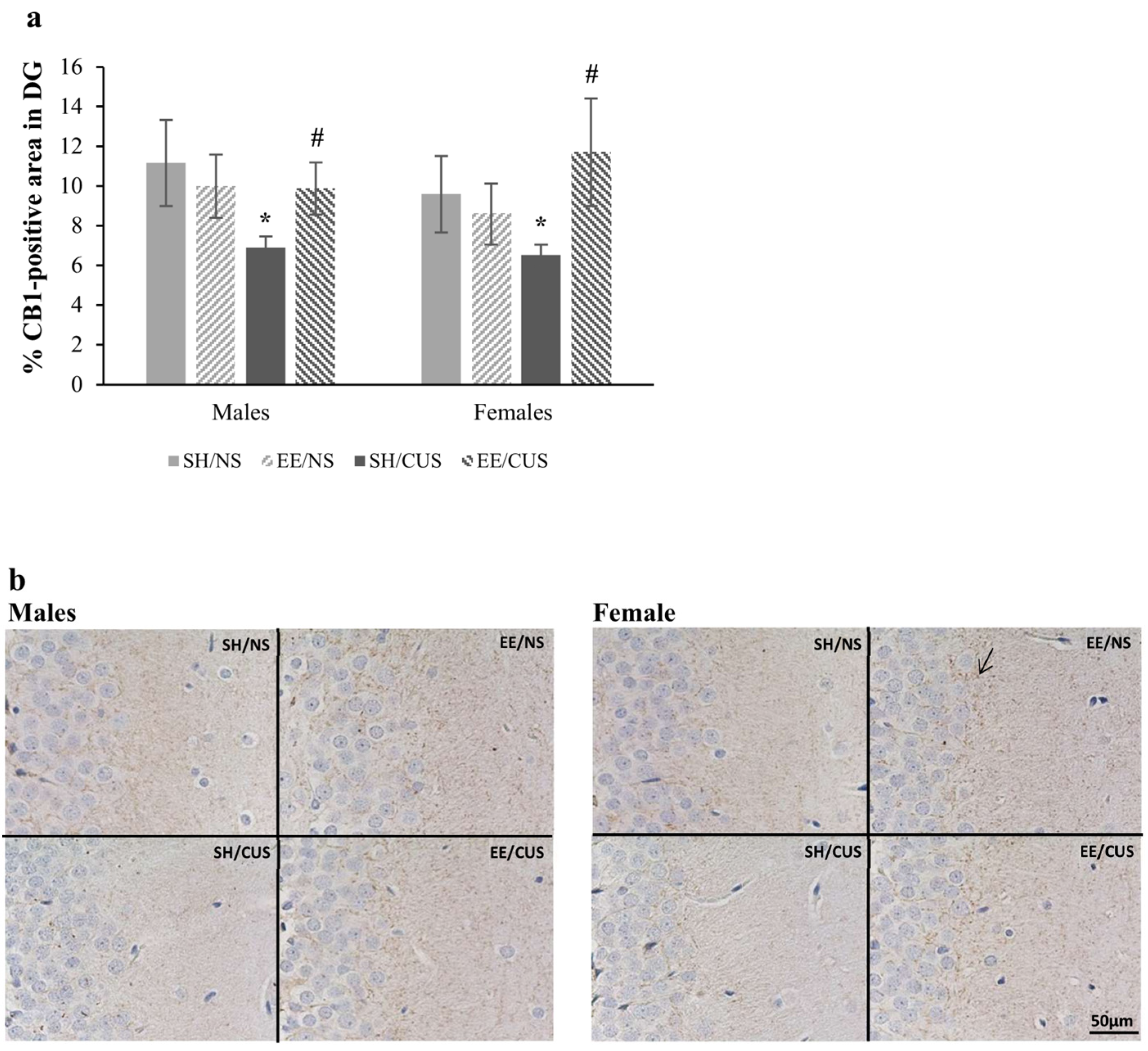
3.1.1. EE Exhibited a Protective Effect against the Stress-Induced Reductions in the Percentage of CB1-Positive Area in DG

The analysis of CB1 immunoreactivity in the DG molecular layer revealed a significant interaction between stress and housing [F(1,32) = 4.65, p = 0.039, partial η2 = 0.127]. Simple effects analysis demonstrated a significant reduction in the CB1-positive area induced by stress in SH animals [F(1,32) = 4.68, p = 0.038, partial η2 = 0.128; SH/NS = 10.4 ± 1.4 vs. CUS/SH = 6.7 ± 0.4]. Interestingly, EE reversed this stress-associated decrease [F(1,32) = 5.81, p = 0.022, partial η2 = 0.154; SH/CUS = 6.7 ± 0.4 vs. EE/CUS = 10.8 ± 1.5] (Figure 2).

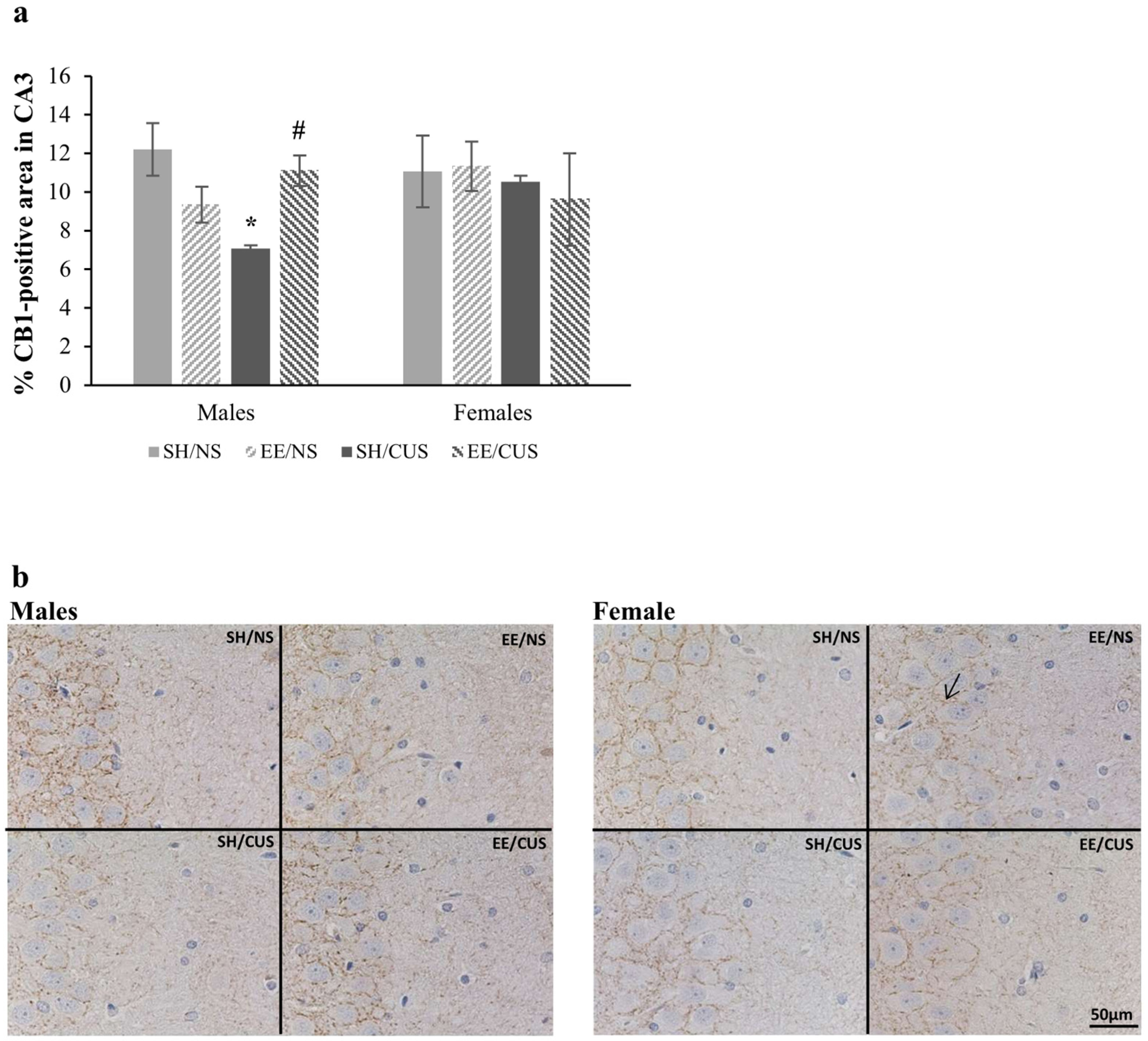
3.1.2. EE Exhibited a Protective Effect against the Stress-Induced Reductions in the Percentage of CB1-Positive Area in CA3 of Male Rats

The statistical analysis of the CB1-positive area in the CA3 stratum radiatum showed a significant interaction among stress, housing, and sex [F(1,32) = 4.58, p = 0.04, partial η2 = 0.125]. According to the analysis of simple effects, the expression of CB1 receptors was significantly reduced only in SH/CUS males [F(1,32) = 7.41, p = 0.01, partial η2 = 0.188; SH/NS = 12.2 ± 1.9 vs. SH/CUS = 7.1 ± 0.3]. Animals housed in EE conditions did not exhibit this CUS-associated decrease [F(1,32) = 4.59, p = 0.04, partial η2 = 0.125; SH/CUS = 7.1 ± 0.3 vs. EE/CUS = 11.1 ± 2.4] (Figure 3).

3.1.3. EE Exhibited a Protective Effect against the Stress-Induced Reductions in the Percentage of CB1-Positive Area in CA1

The analysis of CB1 immunoreactivity in the CA1 stratum radiatum revealed a significant interaction between stress and housing conditions [F(1,32) = 5.33, p = 0.03, partial η2 = 0.143]. Further examination through simple effects analysis demonstrated that CUS significantly decreased the expression of CB1 receptors in standardly housed rats [F(1,32) = 7.36, p = 0.011, partial η2 = 0.187; SH/NS = 10.2 ± 1 vs. SH/CUS = 6.1 ± 0.4]. EE housing reversed this outcome [F(1,32) = 5.89, p = 0.021, partial η2 = 0.155; SH/CUS = 6.1 ± 0.4 vs. EE/CUS = 9.8 ± 1.4] (Figure 4).

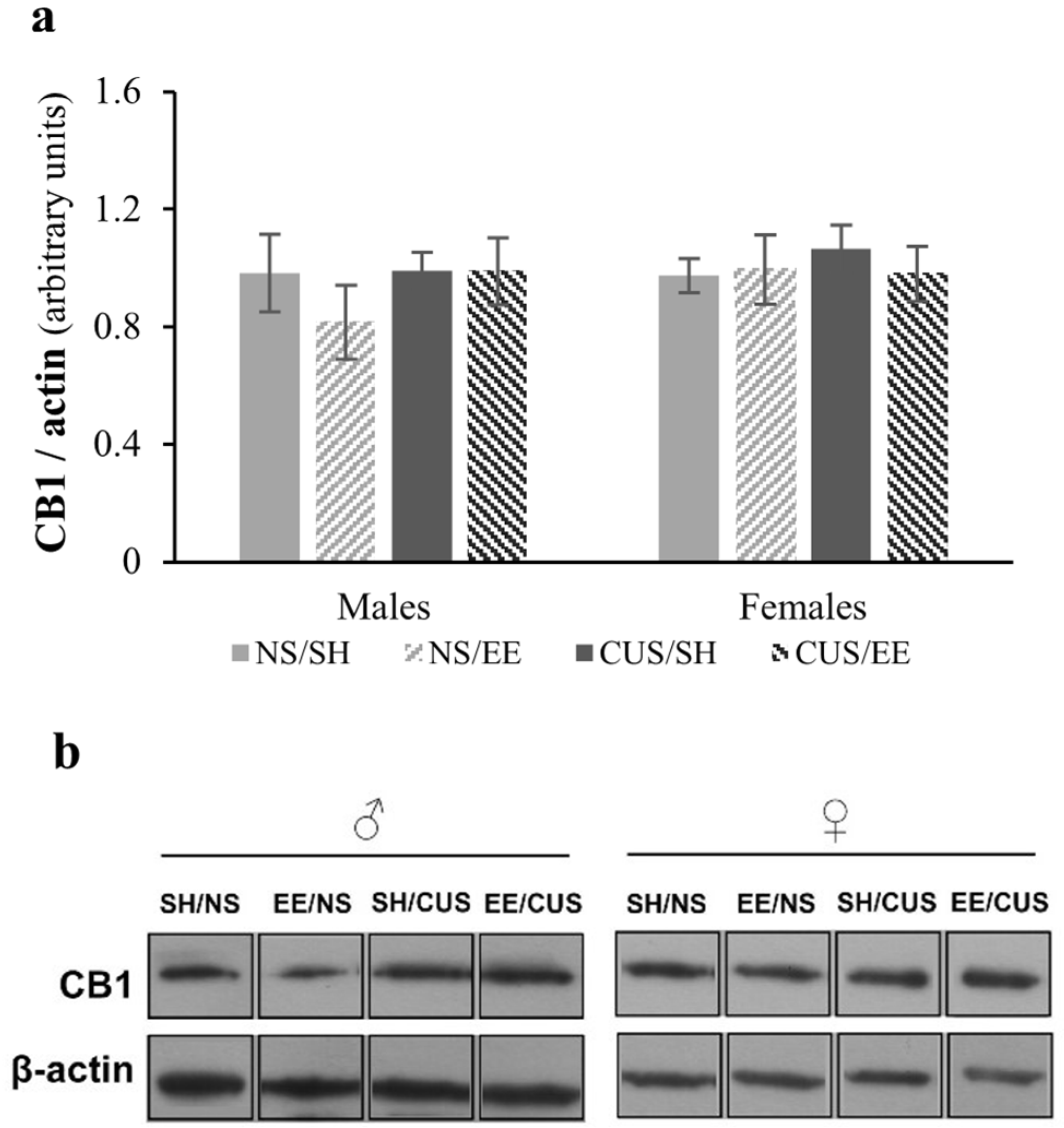
3.2. Analysis of CB1 Total Protein Expression in the Hippocampus Using Western Blotting

Statistical analysis of the CB1 total protein expression showed no significant main effect of stress [F(1,32) = 0.79, p = 0.38, partial η2 = 0.024], housing [F(1,32) = 0.64, p = 0.43, partial η2 = 0.020] or sex [F(1,32) = 0.66, p = 0.42, partial η2 = 0.020]. The factors did not interact with each other [stress × housing × sex: F(1,32) = 0.89, p = 0.35, partial η2 = 0.027] (Figure 5).

4. Discussion

Existing evidence indicates that stress-induced disruptions in the brain ECS signaling might be involved in the neurobiology of stress-related disorders such as PTSD and depression. Thus, the ECS is regarded as a potential target system for drug development in psychiatric disorders related to stress [49]. Previous studies have demonstrated that CB1 receptor inhibition increases the symptoms of depression and anxiety [50,51], even in individuals without pre-existing conditions [52]. On the contrary, the administration of CB1 receptors agonists exerts antidepressant effect [51]. The aim of the present investigation was to explore the impact of CUS and EE on the expression of CB1 receptors in the hippocampus of adult male and female Wistar rats. To date, relatively few investigations on EE–CUS interaction have been carried out, with the majority of these studies mainly employing male animals (for a review, see [27]).
Our findings suggest that the statistically significant reduction in the expression of CB1 receptors in distinct hippocampal areas following adult CUS is both sex and region specific. As WB analysis revealed, no significant difference in total CB1 protein levels was found. However, IHC analysis showed sex-related decreases in CB1 receptor expression in specific hippocampal subregions. Specifically, CUS led to a significant reduction in CB1 receptor expression in the CA1 and DG for both sexes, whereas in CA3 the decrease associated with CUS was observed only in SH males. Prior research has reported decreased levels of hippocampal CB1 receptor expression in CUS adult male rats [30]. Conversely, our findings differ from other studies that support an increase in hippocampal CB1 receptor expression in females subjected to CUS [32,33].
Determining the regional regulation of hippocampal CB1 receptor expression following stress is essential for understanding the functional connection between stress-induced CB1 receptor signaling and neuroplasticity [31]. Most of the existing studies focus on the effects of stress on the CB1 receptor in the entire hippocampus. However, our results suggest that this approach might mask some of the region-specific effects of stress. Hill and colleagues reported decreases in CB1 receptor binding in the DG, increases in the CA3 region, and no difference in CA1 in male rats after 21 days of restraint stress [31]. One potential explanation for the divergent results in relation to the present study could be attributed to the type of chronic stress employed in previous investigations. Specifically, chronic exposure to homotypic stress is less likely to increase CORT levels, and the alterations noted in CB1 receptor expression are less robust compared to those observed following CUS [21]. Additionally, sex hormones and developmental stages may contribute to these differences (for a review, see [34]). For instance, CB1 receptor expression is higher in CA3 and DG in adolescent male rats compared to adult male rats [53].
Despite the pivotal role of the ECS in the stress response [8], and the exploration of eCBs as a potential therapeutic target for both preventing and treating stress-related psychopathology [16,23], the current state of knowledge in this area is limited. To date, research has mainly focused on investigating the therapeutic role of the ECS in various conditions such as chronic pain, epilepsy, neurodegenerative diseases, and cancer, among others. In addition, the majority of studies have employed pharmacological interventions to modulate eCBs for the improvement of behavior and diseases [54]. For instance, it has been demonstrated that the activation of the ventral hippocampal CB1 receptors by cannabinoid receptor agonists can inhibit the anxiogenic-like behaviors induced by ketamine [55].
In the present study, we opted for EE as a non-pharmacological intervention to investigate its protective role against the CUS-associated changes in CB1 expression in the hippocampus. Our results indicate that EE housing, initiated in adolescence, prevented the reduction in CB1 receptor expression in all three hippocampal subregions caused by CUS. Existing evidence supports the positive impact of EE on both brain and behavior [41,56], demonstrating its efficacy in mitigating deficits associated with various brain-related disorders or exposure to adverse conditions throughout life [40,57]. While studies support that EE increases CB1 expression and activation in the brain and that these alterations are associated with reduced anxiety [37,45], there is currently a lack of evidence concerning the beneficial impact of EE on alterations in the endogenous cannabinoid system caused by CUS. To our knowledge, the current study is the first to explore the potential interaction between EE, initiated during adolescence, and CUS in adulthood, on the expression of CB1 receptors in three distinct subregions of the hippocampus in both male and female rodents.
Based on our findings, the adverse effects of CUS on cognitive function and emotional behavior [38], which were also linked to a region and sex-specific decrease in the expression of glial fibrillary acidic protein (GFAP) and synaptophysin (SYN) in the hippocampus [39], may be mediated by alterations in the CB1 receptor expression. Specifically, the learning impairments previously found exclusively in CUS males could be associated with the decreases found in SYN and CB1 receptor expression in the CA3 and CA1 hippocampal areas. Meanwhile, the decreases in SYN and CB1 receptor expression in CA1 observed in females may explain the increase in depressive behavior detected only in females [38,39]. In support of the present findings, existing evidence highlights the role of eCBs in modulating synaptic function and contributing to synaptic plasticity through retrograde signaling to CB1 and CB2 receptors [58]. Chronic exposure to repeated restraint or foot shock stress has been shown to alter CB1 receptor signaling, leading to deficits in learning and memory, as well as to dysfunctional behavioral responses to stress [59,60].

5. Conclusions

Our findings support the potential role of ECS signaling in the brain in response to chronic stress. The regional altered expression of CB1 receptors in animals exposed to CUS indicates that ECS signaling might have a region-specific regulatory role in synaptic function, while factors such as sex hormones may be associated with the sex-dependent changes. The reversal of CUS-associated reduction in the expression of CB1 receptors with EE housing emphasizes the positive role of this environmental manipulation against the detrimental effects of CUS.
Preclinical studies provide valuable insights into understanding organism function and in the development of suitable treatments, but it is crucial to interpret their findings with caution when considering clinical applications. The current study may lay the foundation for further research regarding the protective effect of enriched environmental experiences against the adverse impact of chronic stress on both behavior and the brain. The investigation of the significance of sex/gender in these effects is of great importance. The findings of the current study underscore the importance of early-life prevention, suggesting that children and adolescents raised in environments that promote cognitive flexibility and social interactions are inclined to be more resilient to stressful experiences as adults. Although the beneficial effects of EE on behavior and the brain are well-documented and existing evidence supports its beneficial role in stressful conditions, there are still important aspects that require further investigation. Future investigations should not only take into account both sexes but should also incorporate analyses of further alterations in structure and function in additional stress-associated brain regions such as the prefrontal cortex and the amygdala.

Author Contributions

Conceptualization, D.A.T., E.S. and C.S.; methodology, E.D. and N.G.; formal analysis, E.D. and E.K.; investigation, E.D. and E.K.; resources, E.S., C.S. and N.G.; data curation, D.A.T. and E.K.; writing—original draft preparation, E.D.; writing—review and editing, D.A.T., E.S. and E.D.; visualization, E.D. and E.K.; supervision, D.A.T. and E.S.; project administration, D.A.T. and E.S.; funding acquisition, D.A.T. and E.D. All authors have read and agreed to the published version of the manuscript.

Funding

This research was funded by the General Secretariat for Research and Technology (GSRT) and the Hellenic Foundation for Research and Innovation (HFRI), grant number 95144.

Institutional Review Board Statement

The animal study protocol was approved by the Institutional Review Board (or Ethics Committee) of the local Veterinary Medicines Directorate of Aristotle University (# 471643-1811, 27 July 2021). All experimental procedures were complied with the European Communities Council Directive (2010/63/EU) on the protection of animals.

Informed Consent Statement

Not applicable.

Data Availability Statement

Dataset available on request from the authors.

Conflicts of Interest

The authors declare no conflicts of interest. The funders had no role in the design of the study; in the collection, analyses, or interpretation of data; in the writing of the manuscript; or in the decision to publish the results.

References

  1. Knezevic, E.; Nenic, K.; Milanovic, V.; Knezevic, N.N. The Role of Cortisol in Chronic Stress, Neurodegenerative Diseases, and Psychological Disorders. Cells 2023, 12, 2726. [Google Scholar] [CrossRef] [PubMed]
  2. Milligan Armstrong, A.; Porter, T.; Quek, H.; White, A.; Haynes, J.; Jackaman, C.; Villemagne, V.; Munyard, K.; Laws, S.M.; Verdile, G.; et al. Chronic Stress and Alzheimer’s Disease: The Interplay between the Hypothalamic–Pituitary–Adrenal Axis, Genetics and Microglia. Biol. Rev. 2021, 96, 2209–2228. [Google Scholar] [CrossRef] [PubMed]
  3. Soares, N.M.; Pereira, G.M.; Altmann, V.; de Almeida, R.M.M.; Rieder, C.R.M. Cortisol Levels, Motor, Cognitive and Behavioral Symptoms in Parkinson’s Disease: A Systematic Review. J. Neural Transm. 2019, 126, 219–232. [Google Scholar] [CrossRef] [PubMed]
  4. Du, X.; Pang, T.Y. Is Dysregulation of the HPA-Axis a Core Pathophysiology Mediating Co-Morbid Depression in Neurodegenerative Diseases? Front. Psychiatry 2015, 6, 123351. [Google Scholar] [CrossRef] [PubMed]
  5. Pietrzak, R.H.; Laws, S.M.; Lim, Y.Y.; Bender, S.J.; Porter, T.; Doecke, J.; Ames, D.; Fowler, C.; Masters, C.L.; Milicic, L.; et al. Plasma Cortisol, Brain Amyloid-β, and Cognitive Decline in Preclinical Alzheimer’s Disease: A 6-Year Prospective Cohort Study. Biol. Psychiatry Cogn. Neurosci. Neuroimaging 2017, 2, 45–52. [Google Scholar] [CrossRef]
  6. Willner, P. Reliability of the Chronic Mild Stress Model of Depression: A User Survey. Neurobiol. Stress 2017, 6, 68–77. [Google Scholar] [CrossRef] [PubMed]
  7. Zhu, H.; Tao, Y.; Wang, T.; Zhou, J.; Yang, Y.; Cheng, L.; Zhu, H.; Zhang, W.; Huang, F.; Wu, X. Long-Term Stability and Characteristics of Behavioral, Biochemical, and Molecular Markers of Three Different Rodent Models for Depression. Brain Behav. 2019, 10, e01508. [Google Scholar] [CrossRef] [PubMed]
  8. Micale, V.; Drago, F. Endocannabinoid System, Stress and HPA Axis. Eur. J. Pharmacol. 2018, 834, 230–239. [Google Scholar] [CrossRef]
  9. Kilaru, A.; Chapma, K.D. The Endocannabinoid System. Essays Biochem. 2020, 64, 485–499. [Google Scholar]
  10. Lu, H.C.; MacKie, K. An Introduction to the Endogenous Cannabinoid System. Biol. Psychiatry 2016, 79, 516–525. [Google Scholar] [CrossRef]
  11. Woodhams, S.G.; Chapman, V.; Finn, D.P.; Hohmann, A.G.; Neugebauer, V. The Cannabinoid System and Pain. Neuropharmacology 2017, 124, 105–120. [Google Scholar] [CrossRef]
  12. Hill, M.N.; McEwen, B.S. Involvement of the Endocannabinoid System in the Neurobehavioural Effects of Stress and Glucocorticoids. Prog. Neuro-Psychopharmacol. Biol. Psychiatry 2010, 34, 791–797. [Google Scholar] [CrossRef] [PubMed]
  13. Dhopeshwarkar, A.; Mackie, K. CB2cannabinoid Receptors as a Therapeutic Target-What Does the Future Hold? Mol. Pharmacol. 2014, 86, 430–437. [Google Scholar] [CrossRef] [PubMed]
  14. Turcotte, C.; Blanchet, M.R.; Laviolette, M.; Flamand, N. The CB2 Receptor and Its Role as a Regulator of Inflammation. Cell. Mol. Life Sci. 2016, 73, 4449–4470. [Google Scholar] [CrossRef]
  15. Winters, B.L.; Vaughan, C.W. Mechanisms of Endocannabinoid Control of Synaptic Plasticity. Neuropharmacology 2021, 197, 108736. [Google Scholar] [CrossRef] [PubMed]
  16. Blessing, E.M.; Steenkamp, M.M.; Manzanares, J.; Marmar, C.R. Cannabidiol as a Potential Treatment for Anxiety Disorders. Neurotherapeutics 2015, 12, 825–836. [Google Scholar] [CrossRef] [PubMed]
  17. Viveros, M.P.; Marco, E.M.; File, S.E. Endocannabinoid System and Stress and Anxiety Responses. Pharmacol. Biochem. Behav. 2005, 81, 331–342. [Google Scholar] [CrossRef] [PubMed]
  18. Dow-Edwards, D.; Frank, A.; Wade, D.; Weedon, J.; Izenwasser, S. Sexually-Dimorphic Alterations in Cannabinoid Receptor Density Depend upon Prenatal/Early Postnatal History. Neurotoxicol. Teratol. 2016, 58, 31–39. [Google Scholar] [CrossRef] [PubMed]
  19. Mikulska, J.; Juszczyk, G.; Gawrońska-Grzywacz, M.; Herbet, M. HPA Axis in the Pathomechanism of Depression and Schizophrenia: New Therapeutic Strategies Based on Its Participation. Brain Sci. 2021, 11, 1298. [Google Scholar] [CrossRef]
  20. Akirav, I. Cannabinoids and Glucocorticoids Modulate Emotional Memory after Stress. Neurosci. Biobehav. Rev. 2013, 37, 2554–2563. [Google Scholar] [CrossRef]
  21. Hillard, C.J. Stress Regulates Endocannabinoid-CB1 Receptor Signaling. Semin. Immunol. 2014, 26, 380–388. [Google Scholar] [CrossRef] [PubMed]
  22. Tasker, J.G.; Chen, C.; Fisher, M.O.; Fu, X.; Rainville, J.R.; Weiss, G.L. Endocannabinoid Regulation of Neuroendocrine Systems. Int. Rev. Neurobiol. 2015, 125, 163–201. [Google Scholar] [CrossRef]
  23. Joshi, N.; Onaivi, E.S. Psychiatric Disorders and Cannabinoid Receptors. In Advances in Experimental Medicine and Biology; Springer: Berlin/Heidelberg, Germany, 2021; Volume 1264, pp. 131–153. [Google Scholar]
  24. Antoniuk, S.; Bijata, M.; Ponimaskin, E.; Wlodarczyk, J. Chronic Unpredictable Mild Stress for Modeling Depression in Rodents: Meta-Analysis of Model Reliability. Neurosci. Biobehav. Rev. 2019, 99, 101–116. [Google Scholar] [CrossRef] [PubMed]
  25. D’Aquila, P.S.; Brain, P.; Willner, P. Effects of Chronic Mild Stress on Performance in Behavioural Tests Relevant to Anxiety and Depression. Physiol. Behav. 1994, 56, 861–867. [Google Scholar] [CrossRef]
  26. Hill, M.N.; Hellemans, K.G.C.; Verma, P.; Gorzalka, B.B.; Weinberg, J. Neurobiology of Chronic Mild Stress: Parallels to Major Depression. Neurosci. Biobehav. Rev. 2012, 36, 2085–2117. [Google Scholar] [CrossRef]
  27. Dandi, Ε.; Spandou, E.; Dalla, C.; Tata, D. Τhe Neuroprotective Role of Environmental Enrichment against Behavioral, Morphological, Neuroendocrine and Molecular Changes Following Chronic Unpredictable Mild Stress: A Systematic Review. Eur. J. Neurosci. 2023, 58, 3003–3025. [Google Scholar] [CrossRef]
  28. McLaughlin, R.J.; Gobbi, G. Cannabinoids and Emotionality: A Neuroanatomical Perspective. Neuroscience 2012, 204, 134–144. [Google Scholar] [CrossRef]
  29. Morena, M.; Patel, S.; Bains, J.S.; Hill, M.N. Neurobiological Interactions Between Stress and the Endocannabinoid System. Neuropsychopharmacology 2016, 41, 80–102. [Google Scholar] [CrossRef]
  30. Hill, M.N.; Patel, S.; Carrier, E.J.; Rademacher, D.J.; Ormerod, B.K.; Hillard, C.J.; Gorzalka, B.B. Downregulation of Endocannabinoid Signaling in the Hippocampus Following Chronic Unpredictable Stress. Neuropsychopharmacology 2005, 30, 508–515. [Google Scholar] [CrossRef] [PubMed]
  31. Hill, M.N.; Hunter, R.G.; McEwen, B.S. Chronic Stress Differentially Regulates Cannabinoid CB1 Receptor Binding in Distinct Hippocampal Subfields. Eur. J. Pharmacol. 2009, 614, 66–69. [Google Scholar] [CrossRef] [PubMed]
  32. Marco, E.M.; Ballesta, J.A.; Irala, C.; Hernández, M.D.; Serrano, M.E.; Mela, V.; López-Gallardo, M.; Viveros, M.P. Sex-Dependent Influence of Chronic Mild Stress (CMS) on Voluntary Alcohol Consumption; Study of Neurobiological Consequences. Pharmacol. Biochem. Behav. 2017, 152, 68–80. [Google Scholar] [CrossRef] [PubMed]
  33. Reich, C.G.; Taylor, M.E.; McCarthy, M.M. Differential Effects of Chronic Unpredictable Stress on Hippocampal CB1 Receptors in Male and Female Rats. Behav. Brain Res. 2009, 203, 264–269. [Google Scholar] [CrossRef] [PubMed]
  34. Almeida, M.M.; Dias-Rocha, C.P.; Calviño, C.; Trevenzoli, I.H. Lipid Endocannabinoids in Energy Metabolism, Stress and Developmental Programming. Mol. Cell. Endocrinol. 2022, 542, 111522. [Google Scholar] [CrossRef] [PubMed]
  35. Black, N.; Stockings, E.; Campbell, G.; Tran, L.T.; Zagic, D.; Hall, W.D.; Farrell, M.; Degenhardt, L. Cannabinoids for the Treatment of Mental Disorders and Symptoms of Mental Disorders: A Systematic Review and Meta-Analysis. Lancet Psychiatry 2019, 6, 995–1010. [Google Scholar] [CrossRef] [PubMed]
  36. Surkin, P.N.; Gallino, S.L.; Luce, V.; Correa, F.; Solari, J.F.; De Laurentiis, A. Pharmacological Augmentation of Endocannabinoid Signaling Reduces the Neuroendocrine Response to Stress. Psychoneuroendocrinology 2018, 87, 131–140. [Google Scholar] [CrossRef]
  37. Jiang, S.; Zheng, C.; Wen, G.; Bu, B.; Zhao, S.; Xu, X. Down-Regulation of NR2B Receptors Contributes to the Analgesic and Antianxiety Effects of Enriched Environment Mediated by Endocannabinoid System in the Inflammatory Pain Mice. Behav. Brain Res. 2022, 435, 114062. [Google Scholar] [CrossRef] [PubMed]
  38. Dandi, E.; Spandou, E.; Tata, D.A. Investigating the Role of Environmental Enrichment Initiated in Adolescence against the Detrimental Effects of Chronic Unpredictable Stress in Adulthood: Sex-Specific Differences in Behavioral and Neuroendocrinological Findings. Behav. Process. 2022, 200, 104707. [Google Scholar] [CrossRef]
  39. Dandi, Ε.; Theotokis, P.; Petri, M.C.; Sideropoulou, V.; Spandou, E.; Tata, D.A. Environmental Enrichment Initiated in Adolescence Restores the Reduced Expression of Synaptophysin and GFAP in the Hippocampus of Chronically Stressed Rats in a Sex-Specific Manner. Dev. Psychobiol. 2023, 65, e22422. [Google Scholar] [CrossRef]
  40. Dandi, Ε.; Kalamari, A.; Touloumi, O.; Lagoudaki, R.; Nousiopoulou, E.; Simeonidou, C.; Spandou, E.; Tata, D.A. Beneficial Effects of Environmental Enrichment on Behavior, Stress Reactivity and Synaptophysin/BDNF Expression in Hippocampus Following Early Life Stress. Int. J. Dev. Neurosci. 2018, 67, 19–32. [Google Scholar] [CrossRef]
  41. Kempermann, G. Environmental Enrichment, New Neurons and the Neurobiology of Individuality. Nat. Rev. Neurosci. 2019, 20, 235–245. [Google Scholar] [CrossRef]
  42. Simpson, J.; Kelly, J.P. The Impact of Environmental Enrichment in Laboratory Rats--Behavioural and Neurochemical Aspects. Behav. Brain Res. 2011, 222, 246–264. [Google Scholar] [CrossRef] [PubMed]
  43. Vaquero-Rodríguez, A.; Ortuzar, N.; Lafuente, J.V.; Bengoetxea, H. Enriched Environment as a Nonpharmacological Neuroprotective Strategy. Exp. Biol. Med. 2023, 248, 553–560. [Google Scholar] [CrossRef] [PubMed]
  44. El Rawas, R.; Thiriet, N.; Nader, J.; Lardeux, V.; Jaber, M.; Solinas, M. Early Exposure to Environmental Enrichment Alters the Expression of Genes of the Endocannabinoid System. Brain Res. 2011, 1390, 80–89. [Google Scholar] [CrossRef] [PubMed]
  45. Banaei-Boroujeni, G.; Rezayof, A.; Alijanpour, S.; Nazari-Serenjeh, F. Targeting Mediodorsal Thalamic CB1 Receptors to Inhibit Dextromethorphan-Induced Anxiety/Exploratory-Related Behaviors in Rats: The Post-Weaning Effect of Exercise and Enriched Environment on Adulthood Anxiety. J. Psychiatr. Res. 2023, 157, 212–222. [Google Scholar] [CrossRef] [PubMed]
  46. Paxinos, G.; Watson, C. The Rat Brain in Stereotaxic Coordinates, 6th ed.; Elevier: Boston, MA, USA, 2007. [Google Scholar]
  47. Tannenbaum, C.; Ellis, R.P.; Eyssel, F.; Zou, J.; Schiebinger, L. Sex and Gender Analysis Improves Science and Engineering. Nature 2019, 575, 137–146. [Google Scholar] [CrossRef] [PubMed]
  48. Howell, G.T.; Lacroix, G.L. Decomposing Interactions Using GLM in Combination with the COMPARE, LMATRIX and MMATRIX Subcommands in SPSS. Tutor. Quant. Methods Psychol. 2012, 8, 1–22. [Google Scholar] [CrossRef]
  49. Coelho, A.A.; Lima-Bastos, S.; Gobira, P.H.; Lisboa, S.F. Endocannabinoid Signaling and Epigenetics Modifications in the Neurobiology of Stress-Related Disorders. Neuronal Signal. 2023, 7, NS20220034. [Google Scholar] [CrossRef] [PubMed]
  50. Peng, Z.W.; Zhou, C.H.; Xue, S.S.; Yu, H.; Shi, Q.Q.; Xue, F.; Chen, Y.H.; Tan, Q.R.; Wang, H.N. High-Frequency Repetitive Transcranial Magnetic Stimulation Regulates Neural Oscillations of the Hippocampus and Prefrontal Cortex in Mice by Modulating Endocannabinoid Signalling. J. Affect. Disord. 2023, 331, 217–228. [Google Scholar] [CrossRef] [PubMed]
  51. Stachowicz, K. Deciphering the Mechanisms of Reciprocal Regulation or Interdependence at the Cannabinoid CB1 Receptors and Cyclooxygenase-2 Level: Effects on Mood, Cognitive Implications, and Synaptic Signaling. Neurosci. Biobehav. Rev. 2023, 155, 105439. [Google Scholar] [CrossRef]
  52. Christensen, R.; Kristensen, P.K.; Bartels, E.M.; Bliddal, H.; Astrup, A. Efficacy and Safety of the Weight-Loss Drug Rimonabant: A Meta-Analysis of Randomised Trials. Lancet 2007, 370, 1706–1713. [Google Scholar] [CrossRef]
  53. Romero-Torres, B.M.; Alvarado-Ramírez, Y.A.; Duran-Alonzo, S.R.; Ruiz-Contreras, A.E.; Herrera-Solis, A.; Amancio-Belmont, O.; Prospéro-García, O.E.; Méndez-Díaz, M. A Potential Role of Hippocampus on Impulsivity and Alcohol Consumption Though CB1R. Pharmacol. Biochem. Behav. 2023, 225, 173558. [Google Scholar] [CrossRef]
  54. Prospéro-García, O.; Ruiz Contreras, A.E.; Ortega Gómez, A.; Herrera-Solís, A.; Méndez-Díaz, M. Endocannabinoids as Therapeutic Targets. Arch. Med. Res. 2019, 50, 518–526. [Google Scholar] [CrossRef] [PubMed]
  55. Alijanpour, S.; Rezayof, A. Activation of Ventral Hippocampal CB1 Receptors Inhibits Ketamine-Induced Anxiogenic-like Behavior: Alteration of BDNF/c-Fos Levels in the Mouse Hippocampus. Brain Res. 2023, 1810, 148378. [Google Scholar] [CrossRef]
  56. Gabriel, P.; Mastracchio, T.A.; Bordner, K.; Jeffrey, R. Impact of Enriched Environment during Adolescence on Adult Social Behavior, Hippocampal Synaptic Density and Dopamine D2 Receptor Expression in Rats. Physiol. Behav. 2020, 226, 113133. [Google Scholar] [CrossRef]
  57. Joushi, S.; Esmaeilpour, K.; Masoumi-Ardakani, Y.; Esmaeili-Mahani, S.; Sheibani, V. Effects of Short Environmental Enrichment on Early-Life Adversity Induced Cognitive Alternations in Adolescent Rats. J. Neurosci. Res. 2021, 99, 3373–3391. [Google Scholar] [CrossRef] [PubMed]
  58. Lookfong, N.A.; Raup-Konsavage, W.M.; Silberman, Y. Potential Utility of Cannabidiol in Stress-Related Disorders. Cannabis Cannabinoid Res. 2023, 8, 230–240. [Google Scholar] [CrossRef] [PubMed]
  59. Heng, L.; Beverley, J.A.; Steiner, H.; Tseng, K.Y. Differential Developmental Trajectories for CB1 Cannabinoid Receptor Expression in Limbic/Associative and Sensorimotor Cortical Areas. Synapse 2011, 65, 278–286. [Google Scholar] [CrossRef]
  60. Spiacci, G.B.L.; Antero, L.S.; Reis, D.G.; Lisboa, S.F.; Resstel, L.B. Dorsal Hippocampus Cannabinoid Type 1 Receptors Modulate the Expression of Contextual Fear Conditioning in Rats: Involvement of Local Glutamatergic/Nitrergic and GABAergic Neurotransmissions. Eur. Neuropsychopharmacol. 2016, 26, 1579–1589. [Google Scholar] [CrossRef]
Figure 1. Schematic representation of the timeline and experimental conditions of the study. EE = environmental enrichment; CUS = chronic unpredictable stress; IHC = immunohistochemistry; WB = Western blotting.
Brainsci 14 00357 g001
Figure 2. Expression of CB1 receptors in DG molecular layer. (a) CUS significantly reduced the CB1 receptor immunoreactivity for SH rats (SH/NS vs. SH/CUS, * p < 0.05). Stressed rats that were exposed to EE did not exhibit this decrease (SH/CUS vs. EE/CUS, # p < 0.05). (b) Representative photomicrographs from the DG exposed to CB1 antibody (DAB visualization). The arrow indicates positive immunostaining for CB1 receptors. Total magnification 400×, scale bar = 50 μm; n = 5 per group. SH = standard housing; EE = environmental enrichment; NS = no stress; CUS = chronic unpredictable stress.
Brainsci 14 00357 g002
Figure 3. Expression of CB1 receptors in the CA3 stratum radiatum. (a) CUS significantly decreased the CB1 receptor immunoreactivity in SH male rats only (SH/NS vs. SH/CUS, * p < 0.05). The EE condition reversed this reduction (SH/CUS vs. EE/CUS, # p < 0.05). (b) Representative photomicrographs from the CA3 exposed to CB1 antibody (DAB visualization). The arrow indicates positive immunostaining for CB1 receptors. Total magnification 400×, scale bar = 50 μm; n = 5 per group. SH = standard housing; EE = environmental enrichment; NS = no stress; CUS = chronic unpredictable stress.
Brainsci 14 00357 g003
Figure 4. Expression of CB1 receptors in the CA1 stratum radiatum. (a) CUS significantly decreased the CB1 receptor immunoreactivity in SH rats (SH/NS vs. SH/CUS, * p < 0.05). The EE condition reversed this effect (SH/CUS vs. EE/CUS, # p < 0.05). (b) Representative photomicrographs from the CA1 exposed to CB1 antibody (DAB visualization). The arrow indicates positive immunostaining for CB1 receptors. Total magnification 400×, scale bar = 50 μm; n = 5 per group. SH = standard housing; EE = environmental enrichment; NS = no stress; CUS = chronic unpredictable stress.
Brainsci 14 00357 g004
Figure 5. Total CB1 hippocampal protein expression. (a) No significant differences were found among groups (p > 0.05). (b) Representative Western blots are shown below the graph, representing the CB1 band. The immunoblot signal of CB1 was normalized to actin. n = 5 per group. SH = standard housing; EE = environmental enrichment; NS = no stress; CUS = chronic unpredictable stress.
Brainsci 14 00357 g005
Disclaimer/Publisher’s Note: The statements, opinions and data contained in all publications are solely those of the individual author(s) and contributor(s) and not of MDPI and/or the editor(s). MDPI and/or the editor(s) disclaim responsibility for any injury to people or property resulting from any ideas, methods, instructions or products referred to in the content.

