Systematic Review

Alzheimer’s Disease: Insights from Large-Scale Brain Dynamics Models

1
College of Computer Science and Technology, Taiyuan University of Technology, Taiyuan 030024, China
2
Teranslational Medicine Research Center, Shanxi Medical University, Taiyuan 030001, China
*
Author to whom correspondence should be addressed.
Brain Sci. 2023, 13(8), 1133; https://doi.org/10.3390/brainsci13081133
Submission received: 27 June 2023 / Revised: 21 July 2023 / Accepted: 25 July 2023 / Published: 28 July 2023
(This article belongs to the Special Issue Deep Research of EEG/fMRI Application in Cognition and Consciousness)

Abstract

:
Alzheimer’s disease (AD) is a degenerative brain disease, and the condition is difficult to assess. In the past, numerous brain dynamics models have made remarkable contributions to neuroscience and the brain from the microcosmic to the macroscopic scale. Recently, large-scale brain dynamics models have been developed based on dual-driven multimodal neuroimaging data and neurodynamics theory. These models bridge the gap between anatomical structure and functional dynamics and have played an important role in assisting the understanding of the brain mechanism. Large-scale brain dynamics have been widely used to explain how macroscale neuroimaging biomarkers emerge from potential neuronal population level disturbances associated with AD. In this review, we describe this emerging approach to studying AD that utilizes a biophysically large-scale brain dynamics model. In particular, we focus on the application of the model to AD and discuss important directions for the future development and analysis of AD models. This will facilitate the development of virtual brain models in the field of AD diagnosis and treatment and add new opportunities for advancing clinical neuroscience.

1. Introduction

Alzheimer’s disease (AD) is a brain disorder characterized by the accumulation of amyloid-based neuritic plaques and the abnormalities of neurodegeneration [1]. In particular, there is a progressive loss of neurons or their processes (axons and dendrites), and the corresponding neuronal function is impaired [2]. Similarly, AD is affected by age, biological sex, and other related factors. Progressive cognitive and behavioral impairment will be presented at different stages of AD (early, moderate, and severe phases) [3,4].
In recent years, substantial advancements have been made in the diagnosis, treatment, and prevention of AD via systematic implementation based on multimodal neuroimaging modalities, cognitive assessment tests, and clinical characteristics [5]. At the microscale level, many different alterations may eventually lead to synaptic decay [6,7]. For example, microcircuit dysfunction in the brain of AD patients is associated with the synaptic inhibition of excitatory neurons [8]. It has also been shown that changes in the morphology and density of dendritic spines have been postulated as the main reasons for the synaptic and neuronal loss observed in AD [9]. Additionally, hyperphosphorylated τ deposition and amyloid-β (Aβ) both impair neuronal functions and ultimately lead to axonal degeneration [4]. At a vastly different level of analysis, the macroscopic level, non-invasive neuroimaging has been used as a vector for diagnostic biomarkers of AD due to its excellent speed, considerable spatial resolution, and ability to record neuronal activity [10,11,12], and it also plays a crucial role in the clinical identification of MCI and AD [13,14]. It is understood that deficiencies caused by AD will affect cognitive functions in large-scale brain networks, but how it will do so is not yet clear. Even more important for AD, traditional non-invasive methods capturing brain activity for early changes have not yet yielded specific, generally accepted features of underlying neuronal hyperactivity [15,16,17], and newer approaches based on source localization or non-invasive brain stimulation still require further validation [10,11,18].
Large-scale brain dynamics models based on nonlinear dynamical systems allow for integrating experimental data from multiple modalities into a common framework [19] and help us to better understand the neurophysiological changes in AD. According to existing research findings, models can reveal the structural and functional relationships of the brain [20,21,22], understand the working mechanisms of the brain [23,24], explore the pathogenic mechanisms of brain diseases [25,26,27], and explain processes such as the development or evolution of the brain neural system [28,29]. Given that AD is a complex multifactorial disease characterized by neuronal degeneration and neuronal synaptic disorders in cortical and subcortical areas, as well as abnormalities of white and gray matter, it is better suited to be studied with large-scale brain dynamics models. Using neurodynamic model simulations, researchers have demonstrated that the disconnection of macroscopic anatomical structures in the brains of AD patients leads to abnormal changes in metastability, and this revealed an important relationship between metastability, cognition, and the efficiency of anatomical structures [30,31]. In addition, other researchers have used models that can analyze the characteristics of the evolutionary process to identify changes in alpha rhythms [32,33] and excitatory neurons [6,34] in the brains of AD patients and areas of abnormality [31,35,36] between AD patients and controls.
In this review, we systematically examined 24 identified studies on the application of large-scale brain dynamics models in AD from 2010 to 2023. We aim to review the large-scale brain dynamics models that are available for AD research and summarize the findings from studies using these models. In addition, we also highlight opportunities that will require the further development of models about AD (Table 1). In particular, we emphasize the importance of incorporating more information in the model as well as in the modeling process and in the selection of longitudinal data. This also presents a key challenge to adequately constrain neuroimaging data and construct models with these relevant properties. In summary, large-scale brain dynamics models can provide new opportunities for advancing clinical neuroscience.

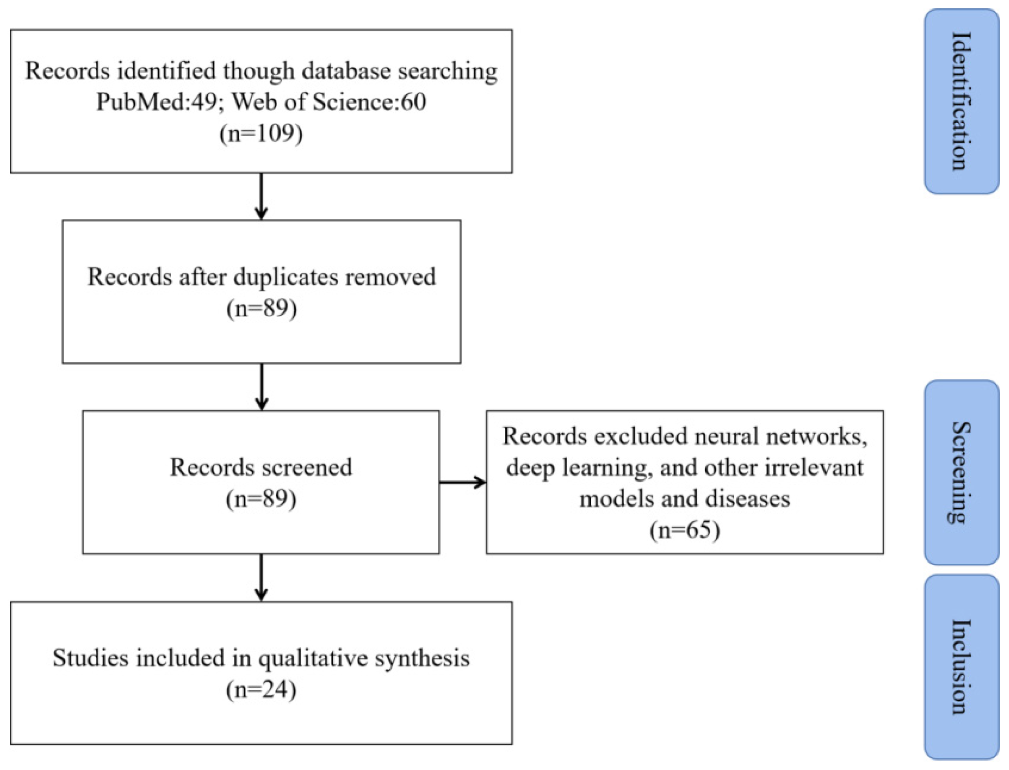
2. Materials and Methods

In this paper, we investigate the application of large-scale brain dynamics models in AD. Preferred Reporting Items for Systematic Reviews and Meta-Analyses (PRISMA) [49] was used to identify studies and narrow the collection for this review. This study was registered at PROSPERO. We performed a search on PubMed and Web of Science using the following group of keywords: “Alzheimer’s Disease” AND (“Neural dynamics” OR “Brain dynamics” OR “neuroanatomy” OR “dynamical systems” OR “Nonlinear dynamics”) AND (“Computational model” OR “Whole brain computational model” OR “computational modeling” OR “mathematical model” OR “Neural activity”) AND (“large-scale” OR “large scale” OR “macroscale”).
References from 2010 to 2023 were used for further analysis. As shown in Figure 1, after excluding unqualified studies, this review narrowed the original count of 109 studies to the final count of 24 studies.

3. Large-Scale Brain Dynamics Models

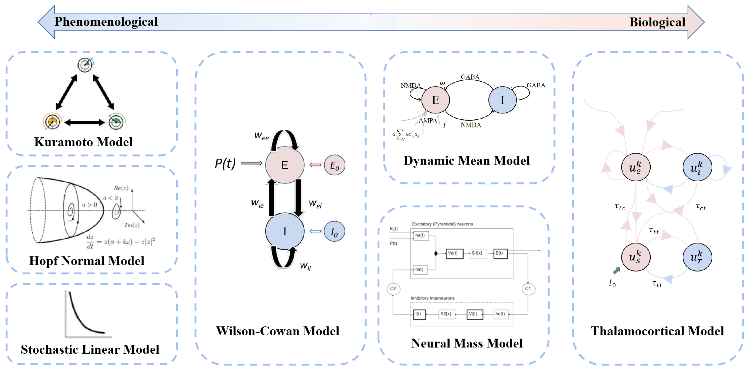
3.1. Neural Dynamics Models

In the field of neuroscience, theory and computational models are playing increasingly important roles in the challenges of understanding complex biological dynamic systems. The neurodynamic models constructed based on real collected imaging data can not only discover some phenomenon that conforms to statistical significance but also adequately explain mechanistically and conduct systematic studies on the development, evolution, or dynamics of the brain nervous system [19]. Currently, models play an important role in the “observation-experiment-theory” phase of comprehensive exploration, especially on the basis of the pioneering work of Hodgkin and Huxley [50]. As the model evolved, the neural mass model was formed in order to bridge the gap between the activity of individual neurons and the circuits [51,52]. This was later followed by the construction of the continuum neural field model [53], which can be extended in space, and the whole-brain network model that can be applied clinically [52,54].
Neural dynamics models simulate brain dynamics using biologically inspired mathematical equations that model regional activity and are coupled through the observed brain structure [21]. These equations can be built from knowledge concerning the biophysical mechanisms underlying different forms of brain activity or as phenomenological models chosen by the kind of dynamics they produce [55]. Thus, the models are also classified into two main categories: biologically informed models and phenomenologically informed models [27]. Depending on the amount of biological information covered in the models, the models are represented on the biological information axis in the position of the category to which it roughly belongs (Figure 2). In the context of phenomenologically informed models, we tend to consider more the phase transition for global connectivity and ignore the details of the model [26,43]. For example, the Kuramoto model, used to study network synchrony, considers time-delayed interactions between nodes [56,57]; the Hopf normal model has the advantage of allowing transitions between asynchronous noise activity and oscillations [35,58]; and the stochastic linear model is a linear stochastic system of coupled Ornstein–Uhlenbeck processes [59]. In the context of biologically informed models, we could treat the brain as a large set of single-cell neurons that cover the entire brain and are wired using white matter tract information from diffusion tensor imaging (DTI) [27]. For instance, a number of researchers have used the neural mass model, which describes a local population of interacting neurons, such as pyramidal and inhibitory cells [19]. This model is constructed in two ways: one uses the same conductance-based model as a single-neuron model to model the mean ensemble activity; the other uses careful empirical observations to understand and model the response of the system to inputs, as is the case with the Jansen–Rit model [19]. An additional class of models, dynamic mean-field models, has been used, which includes some of the more refined local models with an ability to model the balance between excitation and inhibition. These models are based on firing rates and synaptic gating activity [40,60]. There is also the Wilson–Cowan model [51], which uses a phase-plane approach and numerical solutions to describe the response of neuronal populations to stimulation, and the thalamocortical model [61], which describes the interaction of cortical and thalamic populations using a delayed feedback loop. A detailed review of the different models can be found in [52,62].

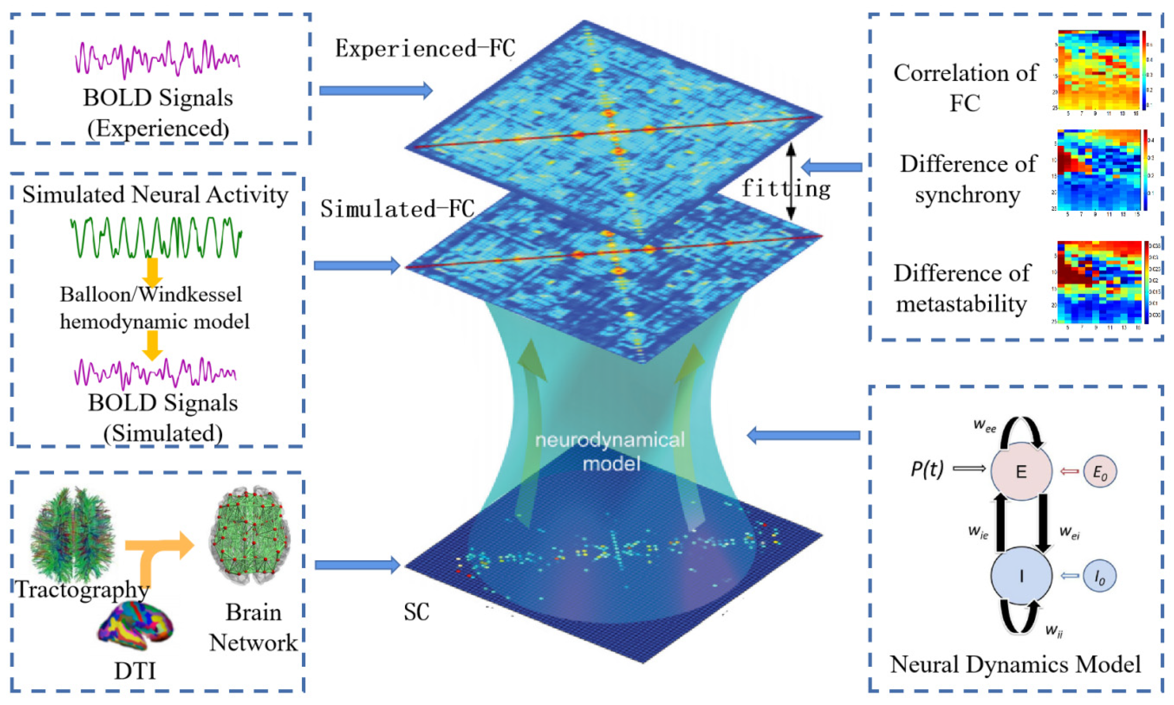
3.2. Large-Scale Dynamics Model Building

The study of model building can be divided into three parts: microscopic, mesoscopic, and macroscopic. The interrelationships between network structure and dynamics are largely elusive at the micro- and mesoscopic level [63]. From a purely neuroscientific perspective, modeling studies are important to provide fundamental insight into the driving factors of connectivity fluctuations, which will potentially bridge the gap between microscopic and macroscopic activity and connectivity [27]. The model architecture requires two key elements: (1) a structural connectivity matrix defining the coupling between areas, which can be derived from empirical data; (2) equations governing the neural dynamics of each local node, which can be constrained by known neurophysiology [64,65]. The specific modeling process is shown in Figure 3.
Structural connectivity, as an essential part of the modeling process, specifies fiber density across various white matter tracts [52]. Together with the brain parcellation that determines the number of regions and the spatial resolution where brain dynamics take place [55], large networks are constructed. In models, elements of the structural connectivity matrix set the strength of the coupling between nodes [65]. Also, in previous studies, the choice of parcellation was found to be important. For example, subcortical nodes are also crucial where the main pathology may involve subcortical structures [52] such as the hippocampus [66], the thalamus [41], and so on.
Functional connectivity is defined as the temporal dependence of the neuronal activity patterns of anatomically separated brain regions and is typically measured as the co-variation between brain signals originating at different locations [62]. This has been particularly useful for the optimization and validation of models via the comparison with the FC matrix obtained using model simulations [56,62,67].
Given network dynamics evident spontaneous emergence and their spatial similarity with the underlying structural connectivity, whole-brain network models are useful tools for investigating the biophysical mechanisms underlying brain activity [62]. By considering the dynamics emerging spontaneously from the interplay between brain areas when these are embedded in the neuroanatomical network, one can analyze the observed phenomena in the light of empirical data and formulate scenarios for the physiological origin of brain activity [62]. And a number of theoretical works have used reduced whole-brain network models to investigate the activity emerging from the interplay of brain areas in the neuroanatomical network [62,67,68]. The activity of each brain area is represented by one or more mathematical expressions representing the spontaneous behavior of isolated brain areas with an additive term representing the input received by anatomically connected areas [62].
Another point of the modeling process that needs attention is model fitting. The model parameters can be calibrated in such a way that the model dynamics closely replicate the dynamics of the brain networks extracted from the empirical data [20]. Most previous studies have used static correlations to estimate the model fit. However, as the research advanced, the use of dynamical metrics, such as functional dynamic connectivity [62], synchrony, and metastability [67], was proposed to further constrain the model fitting in order to preserve the dynamic time characteristics present in the signals at different functional states. These make use of exploring the parameter space to find the optimal parameter set by minimizing the error function or the maximizing correlation between empirical and simulated matrixes, but currently there are also iterative-fitting strategies used to update the model parameters [26,69].
Intuitively, the potential utility of brain network dynamics models is large, and it is not only influenced by biological mechanisms but also closely related to whole-brain neuroimaging data. From the summary of existing studies, it is clear that from the perspective of acquisition, the results of brain network dynamics model simulation can solve the problems of difficult data acquisition to a certain extent, such as the small number of patients with brain diseases and the inability of patients to remain quiet during acquisition. The model can also simulate data at different resolutions, allowing for a more detailed study of the content in question. In addition, the simulation results can avoid the influence of noise during the acquisition process, thus obtaining data that more closely resemble the real brain. More importantly, depending on the changes in the real data set, some of the study findings are not reproducible, but the results simulated using the model avoid such effects. From the perspective of applied research, brain network dynamics models are more able to reflect the principles of the biophysical mechanisms of the brain because they can reflect the relationship between brain structure and function. While human brains cannot be used for damage or stimulation studies due to ethical concerns, researchers can construct a personalized brain network dynamics model by changing the collected structural data so that they can be studied in greater depth. The researcher can construct a personalized brain network dynamics model by altering the collected structural data to study the data at a deeper level. The brain network dynamics model can predict the subsequent state from the existing state of the brain, thus providing a beneficial tool for the evolutionary process of brain aging and disease prevention and treatment. At the same time, this provides new opportunities to advance clinical neuroscience.

4. Applications of Large-Scale Brain Dynamics Models to Alzheimer’s Disease

Here, we focus on applications to AD (Table 1). As a neurodegenerative disease, AD is a disease that integrates the pathogenesis of excitatory synaptic deficits, slowed oscillatory rhythms, and regional changes. Most of the current modeling studies have been analyzed from these three aspects.

4.1. Slowing of Alpha Rhythm: A Biomarker for AD

Each oscillatory frequency of brain activity conveys specific physiological information on brain functional states during sleep and wake periods. The alpha rhythm represents memory and attentional processes, which functionally inhibit task-irrelevant brain regions and transmit information to task-relevant regions [70]. Low-frequency oscillations are associated with functional inhibition, so the slowing of the alpha rhythm is thought to better characterize AD [41]. Initially, Pons et al. found a slowing of the alpha rhythm and an increase in functional connectivity in older MCI patients based on a neural mass model [71]. Subsequently, a more detailed study by van Nifterick et al. found that a relatively higher theta power and a lower beta power followed by a decrease in alpha power at later stages could serve as a mechanism for well-established AD hallmarks [44]. The same conclusion was demonstrated by Ruiz-Gomez et al. using the Kuramoto model [33]. Meanwhile, current researchers have improved the thalamo-cortico-thalamic (TCT) model and suggest that this model is relevant to AD and that it is a good candidate for understanding the thalamo-cortico-thalamic neuronal mechanisms associated with alpha rhythm slowing observed in AD [32]. Based on these studies, a point was proposed: the decrease in synaptic strength has a dominant effect on the slowing of the alpha rhythm [32,34,41]. This established the basis for the following studies.

4.2. Altering Neuronal Excitability: A Therapeutic Strategy for AD

The impairment of cognitive function in Alzheimer’s disease is clearly correlated to synapse loss [6]. Using a spiking neural network model, Bachmann et al. found that the loss of excitatory synapses drives the network into a less sensitive regime, which may also account for the cognitive deficits of AD [6]. To study the potential etiology of AD and simulate the Abeta-induced disruption of interneuronal inhibitory function and consecutive hyperexcitability, Stefanovski et al. proposed a mathematical model that adds a description of molecular changes to alter population activity, where individual Abeta burden modulates regional excitation–inhibition balance, leading to local hyperexcitability with a high Abeta load in personalized virtual brains [34]. This also builds a bridge between molecular pathways and brain network modeling.
Similarly, de Haan et al. used a large-scale neural mass network to simulate the temporal evolution of activity-dependent network degeneration, and the results showed that functional network damage can be opposed by the targeted adjustment of neuronal excitability levels [39]. Van Nifterick et al. combined model and human data and found that neuronal hyperactivity may be due to hyperexcitation and/or disinhibition in early AD [44]. Therefore, we can consider increasing the excitability of excitatory neurons, followed by increasing the inhibition of inhibitory neurons as current effective therapeutic strategies.

4.3. Abnormal Regions of the Brain: Potential Therapeutic Target Areas

AD is characterized by the degradation of the connectome and of network organization by these long-range connections, with both structural and functional connectivity being affected [40]. It has been proposed that damage to the connectivity of an AD computer model focuses on nodes with high topological centrality, integration, and efficiency scores and builds connections between structure and cognition via metastability [31]. This also provides a unique perspective for AD research.
In the existing study, the relation between functional connectivity and complexity was explored using the Kuramoto mean-field model in the MCI-AD condition, and certain variations were found in the anterior and central regions in the results, specifically including the frontal and temporal lobes [36]. And in Zimmermann et al.’s study, the researchers attempted to identify biophysical model neural parameters that were associated with cognition along the spectrum from healthy controls to mild cognitive impairment (MCI) to AD, modeled the limbic subnetwork as well as the entire brain, and found that neurodegeneration and functional changes occurred first in the limbic and temporal regions of the brain and, later, in the motor and sensory areas [40].
Demirtas et al. proposed a whole-brain computational model to explore the mechanisms behind changes in whole-brain and regional connectivity and, among the results, found that regions of FC impairments in AD overlap with the default mode network (DMN) and that decreased effective connectivity (EC) strength primarily affects the left temporal lobe, instead of prompting widespread decreases in FC strength [35]. It was also found that preclinical Alzheimer’s disease (PAD) subjects exhibited decreased EC strength in several regions including the left hippocampus, while no significant EC differences were found in the MCI group [35]. This has been interpreted according to previous studies as a compensation mechanism for maintaining cognitive function under pathology-related insults in the preclinical stage of AD [72,73]. It is worth noting that Bachmann also proposes a circuit compensation mechanism for AD treatment, which restores the mean network activity by increasing the weight of the remaining connections [6]. Now, the limbic system and basal ganglia structures constitute the primary target locations for stimulation in AD [74].

5. Future Directions

Large-scale brain dynamics models of AD have been presented as useful tools to fill the gap between theoretical models and biological evidence. They link microscopic excitatory–inhibitory neurons, mesoscopic fiber tract connections, and macroscopic brain function and cognition, providing new ideas for exploring AD pathogenesis. In addition, the model-based optimization of neuromodulation schemes can be performed without ethical constraints. By giving stimulation to the AD-simulated brain and adjusting it according to the post-stimulation neural activity, it is possible to predict whether the stimulation protocol can normalize the abnormal network dynamics characteristics of AD and give an interpretable neuromodulation strategy. However, the challenges associated with the integration of findings from different levels of analysis (micro-, meso-, and macroscale) are delaying the development of models of the disease [18]. Thus, we have suggested some areas that could be further optimized.
At present, most researchers analyzing the mechanisms of brain activity from a microscopic and mesoscopic perspective have found that these influencing factors play an important role. However, large-scale brain dynamics models only focus on valid information about whole brains or the relationships between local regions, ignoring factors at a more detailed scale. In this sense, the model should be considered to include more information on biological details. In particular, in AD, degenerative changes in several neurobiological processes spanning molecular to macroscopic scales are involved, including proteinopathies, modified gene expression, synaptic alterations, vascular dysregulation, hypometabolism, and structural atrophy [75,76]. Identifying important pathways between truly microscopic-scale variables and observable macroscopic neuroimaging in Alzheimer’s disease would both advance the understanding of the underlying biology and improve the selection of therapeutic targets tailored to an individual’s particular disease subtype or presentation [76]. Therefore, we should incorporate more biological features into the model for the more accurate simulation of brain dynamics.
The investigation of the strength and directionality of interactions in the resting human brain could help to better understand the cognitive deficits associated with network disruption in certain neurological disorders and provide new biomarkers of these disorders [77]. Directed FC between the hippocampus and other brain regions had significant differences among NC, MCI, and AD subjects, and these differences were significantly correlated with cognitive and clinical measurements [78]. EC has also been proposed and has been demonstrated to reveal the direction of information flow between different brain regions, providing new insights into the underlying neuronal alterations in MCI and AD [79]. However, at present, we have not considered the directionality of the connection network when building the model, and this is one of the key elements to be considered for optimization.
We also found that only a few features of brain nodes have been focused on in some studies, and the attributes are more singular. For example, Tijms et al. used cortical thickness as a nodal attribute and constructed a gray matter brain network by calculating Pearson similarity. They found that MCI patients had lower values of standardized clustering coefficients and small-world attributes, which would become even lower as the disease progressed [80]. In contrast, the brain attribute network [81] contains nodal attributes and connectivity information, which can describe the structure and function of the brain from multiple perspectives and better characterize the subtle changes in the brains of patients with different stages of AD. If we construct a model using the brain attribute network, this will provide more advantages in studying the pathogenesis and progression of AD.
In addition, to date, studies of brain networks in AD have been mainly cross-sectional. In contrast, using longitudinal data from the whole brain, information from two points in time can be brought together simultaneously to generate different idiosyncratic networks for AD, resulting in more accurate inferences [82]. Moreover, the models can be used to analyze the characteristics of the disease evolution process and understand the progression of AD using longitudinal data, which is also important for developing treatment plans and predicting the trend of disease progression. Therefore, subsequent studies may also consider using longitudinal data for analysis.

6. Conclusions

In this review, we have shown that large-scale brain dynamics models can accurately explore and characterize brain mechanisms in AD patients. We provide a brief overview of the neural dynamics model and its large-scale modeling process. The application of models to AD concerning alpha rhythms, excitatory neurons, and abnormal regions is also summarized and discussed. Like other fields, the building of large-scale brain dynamics models for AD needs further development to realize its greater potential, and we provide methodological and technical recommendations in terms of the models themselves, the modeling process, and data selection. In summary, biophysically based whole-brain dynamics models are well positioned to impact the treatment field of AD. This also sets the stage for ushering in personalized medicine.

Author Contributions

Conceptualization, L.Y. and B.W.; methodology, L.Y. and B.W.; software, J.L. and J.S.; validation, J.L. and J.X.; formal analysis, L.Y., J.X. and D.L.; investigation, L.Y.; resources, B.W.; data curation, B.W.; writing—original draft preparation, L.Y.; writing—review and editing, L.Y. and J.L.; visualization, L.Y.; supervision, T.Y. and B.W.; project administration, B.W.; funding acquisition, B.W. All authors have read and agreed to the published version of the manuscript.

Funding

This research was funded by the National Natural Science Foundation of China, grant number 62176177; the National Natural Science Foundation of Shanxi Province, grant number 20210302123112; and the Shanxi Provincial Returnee Research Support Project, grant number 2021-039.

Conflicts of Interest

The authors declare no conflict of interest. The funders had no role in the design of the study; in the collection, analyses, or interpretation of data; in the writing of the manuscript; or in the decision to publish the results.

References

  1. Ackleh, A.S.; Elaydi, S.; Livadiotis, G.; Veprauskas, A. A continuous-time mathematical model and discrete approximations for the aggregation of beta-Amyloid. J. Biol. Dyn. 2021, 15, 109–136. [Google Scholar] [CrossRef] [PubMed]
  2. Jack, C.J.; Knopman, D.S.; Jagust, W.J.; Petersen, R.C.; Weiner, M.W.; Aisen, P.S.; Shaw, L.M.; Vemuri, P.; Wiste, H.J.; Weigand, S.D.; et al. Tracking pathophysiological processes in Alzheimer’s disease: An updated hypothetical model of dynamic biomarkers. Lancet. Neurol. 2013, 12, 207–216. [Google Scholar] [CrossRef] [Green Version]
  3. D’Amelio, M.; Rossini, P.M. Brain excitability and connectivity of neuronal assemblies in Alzheimer’s disease: From animal models to human findings. Prog. Neurobiol. 2012, 99, 42–60. [Google Scholar] [CrossRef] [PubMed]
  4. Pons, V.; Rivest, S. Targeting Systemic Innate Immune Cells as a Therapeutic Avenue for Alzheimer Disease. Pharm. Rev. 2022, 74, 1–17. [Google Scholar] [CrossRef] [PubMed]
  5. Hampel, H.; Toschi, N.; Babiloni, C.; Baldacci, F.; Black, K.L.; Bokde, A.; Bun, R.S.; Cacciola, F.; Cavedo, E.; Chiesa, P.A.; et al. Revolution of Alzheimer Precision Neurology. Passageway of Systems Biology and Neurophysiology. J. Alzheimers Dis. 2018, 64, S47–S105. [Google Scholar] [CrossRef]
  6. Bachmann, C.; Tetzlaff, T.; Duarte, R.; Morrison, A. Firing rate homeostasis counteracts changes in stability of recurrent neural networks caused by synapse loss in Alzheimer’s disease. PLoS Comput. Biol. 2020, 16, e1007790. [Google Scholar] [CrossRef]
  7. Tampellini, D. Synaptic activity and Alzheimer’s disease: A critical update. Front. Neurosci. 2015, 9, 423. [Google Scholar] [CrossRef] [Green Version]
  8. Melgosa-Ecenarro, L.; Doostdar, N.; Radulescu, C.I.; Jackson, J.S.; Barnes, S.J. Pinpointing the locus of GABAergic vulnerability in Alzheimer’s disease. Semin. Cell Dev. Biol. 2023, 139, 35–54. [Google Scholar] [CrossRef]
  9. Serrano, M.E.; Kim, E.; Petrinovic, M.M.; Turkheimer, F.; Cash, D. Imaging Synaptic Density: The Next Holy Grail of Neuroscience? Front. Neurosci. 2022, 16, 796129. [Google Scholar] [CrossRef]
  10. Mandal, P.K.; Banerjee, A.; Tripathi, M.; Sharma, A. A Comprehensive Review of Magnetoencephalography (MEG) Studies for Brain Functionality in Healthy Aging and Alzheimer’s Disease (AD). Front. Comput. Neurosci. 2018, 12, 60. [Google Scholar] [CrossRef]
  11. Cassani, R.; Estarellas, M.; San-Martin, R.; Fraga, F.J.; Falk, T.H. Systematic Review on Resting-State EEG for Alzheimer’s Disease Diagnosis and Progression Assessment. Dis. Markers 2018, 2018, 5174815. [Google Scholar] [CrossRef] [Green Version]
  12. Wei, H.; Kong, M.; Zhang, C.; Guan, L.; Ba, M. The structural MRI markers and cognitive decline in prodromal Alzheimer’s disease: A 2-year longitudinal study. Quant Imaging Med. Surg. 2018, 8, 1004–1019. [Google Scholar] [CrossRef] [PubMed]
  13. Chandra, A.; Dervenoulas, G.; Politis, M. Magnetic resonance imaging in Alzheimer’s disease and mild cognitive impairment. J. Neurol. 2019, 266, 1293–1302. [Google Scholar] [CrossRef] [PubMed] [Green Version]
  14. Talwar, P.; Kushwaha, S.; Chaturvedi, M.; Mahajan, V. Systematic Review of Different Neuroimaging Correlates in Mild Cognitive Impairment and Alzheimer’s Disease. Clin. Neuroradiol. 2021, 31, 953–967. [Google Scholar] [CrossRef] [PubMed]
  15. Maestu, F.; Cuesta, P.; Hasan, O.; Fernandez, A.; Funke, M.; Schulz, P.E. The Importance of the Validation of M/EEG With Current Biomarkers in Alzheimer’s Disease. Front. Hum. Neurosci. 2019, 13, 17. [Google Scholar] [CrossRef] [PubMed] [Green Version]
  16. Horvath, A.; Szucs, A.; Hidasi, Z.; Csukly, G.; Barcs, G.; Kamondi, A. Prevalence, Semiology, and Risk Factors of Epilepsy in Alzheimer’s Disease: An Ambulatory EEG Study. J. Alzheimers Dis. 2018, 63, 1045–1054. [Google Scholar] [CrossRef]
  17. Li, H.J.; Hou, X.H.; Liu, H.H.; Yue, C.L.; He, Y.; Zuo, X.N. Toward systems neuroscience in mild cognitive impairment and Alzheimer’s disease: A meta-analysis of 75 fMRI studies. Hum. Brain Mapp. 2015, 36, 1217–1232. [Google Scholar] [CrossRef]
  18. Maestu, F.; de Haan, W.; Busche, M.A.; DeFelipe, J. Neuronal excitation/inhibition imbalance: Core element of a translational perspective on Alzheimer pathophysiology. Ageing Res. Rev. 2021, 69, 101372. [Google Scholar] [CrossRef]
  19. Breakspear, M. Dynamic models of large-scale brain activity. Nat. Neurosci. 2017, 20, 340–352. [Google Scholar] [CrossRef]
  20. Popovych, O.V.; Manos, T.; Hoffstaedter, F.; Eickhoff, S.B. What Can Computational Models Contribute to Neuroimaging Data Analytics? Front. Syst. Neurosci. 2018, 12, 68. [Google Scholar] [CrossRef] [Green Version]
  21. Bansal, K.; Nakuci, J.; Muldoon, S.F. Personalized brain network models for assessing structure-function relationships. Curr. Opin. Neurobiol. 2018, 52, 42–47. [Google Scholar] [CrossRef] [PubMed]
  22. Honey, C.J.; Sporns, O.; Cammoun, L.; Gigandet, X.; Thiran, J.P.; Meuli, R.; Hagmann, P. Predicting human resting-state functional connectivity from structural connectivity. Proc. Natl. Acad. Sci. USA 2009, 106, 2035–2040. [Google Scholar] [CrossRef] [PubMed]
  23. Deco, G.; Tononi, G.; Boly, M.; Kringelbach, M.L. Rethinking segregation and integration: Contributions of whole-brain modelling. Nat. Rev. Neurosci. 2015, 16, 430–439. [Google Scholar] [PubMed]
  24. Cabral, J.; Vidaurre, D.; Marques, P.; Magalhaes, R.; Silva, M.P.; Miguel, S.J.; Deco, G.; Sousa, N.; Kringelbach, M.L. Cognitive performance in healthy older adults relates to spontaneous switching between states of functional connectivity during rest. Sci. Rep. 2017, 7, 5135. [Google Scholar] [PubMed] [Green Version]
  25. Lee, H.; Golkowski, D.; Jordan, D.; Berger, S.; Ilg, R.; Lee, J.; Mashour, G.A.; Lee, U. Relationship of critical dynamics, functional connectivity, and states of consciousness in large-scale human brain networks. Neuroimage 2019, 188, 228–238. [Google Scholar] [CrossRef]
  26. Deco, G.; Kringelbach, M.L. Great expectations: Using whole-brain computational connectomics for understanding neuropsychiatric disorders. Neuron 2014, 84, 892–905. [Google Scholar] [CrossRef] [Green Version]
  27. O’Neill, G.C.; Tewarie, P.; Vidaurre, D.; Liuzzi, L.; Woolrich, M.W.; Brookes, M.J. Dynamics of large-scale electrophysiological networks: A technical review. Neuroimage 2018, 180, 559–576. [Google Scholar] [CrossRef] [Green Version]
  28. Nakagawa, T.T.; Jirsa, V.K.; Spiegler, A.; McIntosh, A.R.; Deco, G. Bottom up modeling of the connectome: Linking structure and function in the resting brain and their changes in aging. Neuroimage 2013, 80, 318–329. [Google Scholar] [CrossRef] [PubMed] [Green Version]
  29. Naik, S.; Banerjee, A.; Bapi, R.S.; Deco, G.; Roy, D. Metastability in Senescence. Trends Cogn. Sci. 2017, 21, 509–521. [Google Scholar]
  30. Sase, T.; Kitajo, K. The metastable brain associated with autistic-like traits of typically developing individuals. PLoS Comput. Biol. 2021, 17, e1008929. [Google Scholar]
  31. Alderson, T.H.; Bokde, A.; Kelso, J.; Maguire, L.; Coyle, D. Metastable neural dynamics in Alzheimer’s disease are disrupted by lesions to the structural connectome. Neuroimage 2018, 183, 438–455. [Google Scholar] [CrossRef] [Green Version]
  32. Li, X.; Yang, X.; Sun, Z. Alpha rhythm slowing in a modified thalamo-cortico-thalamic model related with Alzheimer’s disease. PLoS ONE 2020, 15, e229950. [Google Scholar] [CrossRef] [PubMed]
  33. Ruiz-Gomez, S.J.; Hornero, R.; Poza, J.; Maturana-Candelas, A.; Pinto, N.; Gomez, C. Computational modeling of the effects of EEG volume conduction on functional connectivity metrics. Application to Alzheimer’s disease continuum. J. Neural. Eng. 2019, 16, 66019. [Google Scholar] [CrossRef] [PubMed]
  34. Stefanovski, L.; Triebkorn, P.; Spiegler, A.; Diaz-Cortes, M.A.; Solodkin, A.; Jirsa, V.; McIntosh, A.R.; Ritter, P. Linking Molecular Pathways and Large-Scale Computational Modeling to Assess Candidate Disease Mechanisms and Pharmacodynamics in Alzheimer’s Disease. Front. Comput. Neurosci. 2019, 13, 54. [Google Scholar] [CrossRef] [PubMed] [Green Version]
  35. Demirtas, M.; Falcon, C.; Tucholka, A.; Gispert, J.D.; Molinuevo, J.L.; Deco, G. A whole-brain computational modeling approach to explain the alterations in resting-state functional connectivity during progression of Alzheimer’s disease. Neuroimage Clin. 2017, 16, 343–354. [Google Scholar] [CrossRef]
  36. Das, S.; Puthankattil, S.D. Functional Connectivity and Complexity in the Phenomenological Model of Mild Cognitive-Impaired Alzheimer’s Disease. Front. Comput. Neurosci. 2022, 16, 877912. [Google Scholar] [CrossRef]
  37. de Haan, W.; Mott, K.; van Straaten, E.C.; Scheltens, P.; Stam, C.J. Activity dependent degeneration explains hub vulnerability in Alzheimer’s disease. PLoS Comput. Biol. 2012, 8, e1002582. [Google Scholar] [CrossRef] [Green Version]
  38. Ghorbanian, P.; Ramakrishnan, S.; Ashrafiuon, H. Stochastic coupled oscillator model of EEG for Alzheimer’s disease. In Proceedings of the 2014 36th Annual International Conference of the IEEE Engineering in Medicine and Biology Society, Chicago, IL, USA, 26–30 August 2014; pp. 706–709. [Google Scholar]
  39. de Haan, W.; van Straaten, E.; Gouw, A.A.; Stam, C.J. Altering neuronal excitability to preserve network connectivity in a computational model of Alzheimer’s disease. PLoS Comput. Biol. 2017, 13, e1005707. [Google Scholar] [CrossRef] [Green Version]
  40. Zimmermann, J.; Perry, A.; Breakspear, M.; Schirner, M.; Sachdev, P.; Wen, W.; Kochan, N.A.; Mapstone, M.; Ritter, P.; McIntosh, A.R.; et al. Differentiation of Alzheimer’s disease based on local and global parameters in personalized Virtual Brain models. Neuroimage Clin. 2018, 19, 240–251. [Google Scholar] [CrossRef]
  41. Cakir, Y. Hybrid modeling of alpha rhythm and the amplitude of low-frequency fluctuations abnormalities in the thalamocortical region and basal ganglia in Alzheimer’s disease. Eur. J. Neurosci. 2020, 52, 2944–2961. [Google Scholar] [CrossRef]
  42. Arbabyazd, L.; Shen, K.; Wang, Z.; Hofmann-Apitius, M.; Ritter, P.; McIntosh, A.R.; Battaglia, D.; Jirsa, V. Virtual Connectomic Datasets in Alzheimer’s Disease and Aging Using Whole-Brain Network Dynamics Modelling. Eneuro 2021, 8. [Google Scholar] [CrossRef]
  43. Triebkorn, P.; Stefanovski, L.; Dhindsa, K.; Diaz-Cortes, M.A.; Bey, P.; Bulau, K.; Pai, R.; Spiegler, A.; Solodkin, A.; Jirsa, V.; et al. Brain simulation augments machine-learning-based classification of dementia. Alzheimers Dement 2022, 8, e12303. [Google Scholar] [CrossRef] [PubMed]
  44. van Nifterick, A.M.; Gouw, A.A.; van Kesteren, R.E.; Scheltens, P.; Stam, C.J.; de Haan, W. A multiscale brain network model links Alzheimer’s disease-mediated neuronal hyperactivity to large-scale oscillatory slowing. Alzheimers Res. 2022, 14, 101. [Google Scholar] [CrossRef] [PubMed]
  45. Patow, G.; Stefanovski, L.; Ritter, P.; Deco, G.; Kobeleva, X. Whole-brain modeling of the differential influences of Amyloid-Beta and Tau in Alzheimer’s Disease. bioRxiv 2023, XXI, 1–5. [Google Scholar]
  46. Salimi-Nezhad, N.; Missault, S.; Notario-Reinoso, A.; Hassani, A.; Amiri, M.; Keliris, G.A. The impact of selective and non-selective medial septum stimulation on hippocampal neuronal oscillations: A study based on modeling and experiments. Neurobiol. Dis. 2023, 180, 106052. [Google Scholar] [CrossRef]
  47. Sanz, P.Y.; Fittipaldi, S.; Gonzalez, C.C.; Moguilner, S.; Cruzat, J.; Fraile-Vazquez, M.E.; Herzog, R.; Kringelbach, M.L.; Deco, G.; Prado, P.; et al. Model-based whole-brain perturbational landscape of neurodegenerative diseases. Elife 2023, 12. [Google Scholar]
  48. Alexandersen, C.G.; de Haan, W.; Bick, C.; Goriely, A. A multi-scale model explains oscillatory slowing and neuronal hyperactivity in Alzheimer’s disease. J. R. Soc. Interface 2023, 20, 20220607. [Google Scholar] [CrossRef]
  49. Liberati, A.; Altman, D.G.; Tetzlaff, J.; Mulrow, C.; Gotzsche, P.C.; Ioannidis, J.P.; Clarke, M.; Devereaux, P.J.; Kleijnen, J.; Moher, D. The PRISMA statement for reporting systematic reviews and meta-analyses of studies that evaluate healthcare interventions: Explanation and elaboration. BMJ 2009, 339, b2700. [Google Scholar] [CrossRef] [Green Version]
  50. HODGKINA, L.; HUXLEYA, F. A quantitative description of membrane current and its application to conduction and excitation in nerve. J. Physiol. 1952, 117, 500–544. [Google Scholar] [CrossRef] [PubMed]
  51. Wilson, H.R.; Cowan, J.D. Excitatory and inhibitory interactions in localized populations of model neurons. Biophys. J. 1972, 12, 1–24. [Google Scholar] [CrossRef] [Green Version]
  52. Pathak, A.; Roy, D.; Banerjee, A. Whole-Brain Network Models: From Physics to Bedside. Front. Comput. Neurosci. 2022, 16, 866517. [Google Scholar] [CrossRef] [PubMed]
  53. Amari, S. Dynamics of pattern formation in lateral-inhibition type neural fields. Biol. Cybern. 1977, 27, 77–87. [Google Scholar] [CrossRef] [PubMed]
  54. Deco, G.; Jirsa, V.; McIntosh, A.R.; Sporns, O.; Kotter, R. Key role of coupling, delay, and noise in resting brain fluctuations. Proc Natl. Acad. Sci. USA 2009, 106, 10302–10307. [Google Scholar] [CrossRef] [PubMed]
  55. Cofre, R.; Herzog, R.; Mediano, P.; Piccinini, J.; Rosas, F.E.; Sanz, P.Y.; Tagliazucchi, E. Whole-Brain Models to Explore Altered States of Consciousness from the Bottom Up. Brain Sci. 2020, 10, 626. [Google Scholar] [CrossRef]
  56. Cabral, J.; Hugues, E.; Sporns, O.; Deco, G. Role of local network oscillations in resting-state functional connectivity. Neuroimage 2011, 57, 130–139. [Google Scholar] [CrossRef] [PubMed]
  57. Breakspear, M.; Heitmann, S.; Daffertshofer, A. Generative models of cortical oscillations: Neurobiological implications of the kuramoto model. Front. Hum. Neurosci. 2010, 4, 190. [Google Scholar] [CrossRef] [Green Version]
  58. Lord, L.D.; Stevner, A.B.; Deco, G.; Kringelbach, M.L. Understanding principles of integration and segregation using whole-brain computational connectomics: Implications for neuropsychiatric disorders. Philos. Trans. A Math. Phys. Eng. Sci. 2017, 375. [Google Scholar]
  59. Saggio, M.L.; Ritter, P.; Jirsa, V.K. Analytical Operations Relate Structural and Functional Connectivity in the Brain. PLoS ONE 2016, 11, e157292. [Google Scholar]
  60. Deco, G.; Ponce-Alvarez, A.; Hagmann, P.; Romani, G.L.; Mantini, D.; Corbetta, M. How local excitation-inhibition ratio impacts the whole brain dynamics. J. Neurosci. 2014, 34, 7886–7898. [Google Scholar] [CrossRef] [Green Version]
  61. Griffiths, J.D.; McIntosh, A.R.; Lefebvre, J. A Connectome-Based, Corticothalamic Model of State- and Stimulation-Dependent Modulation of Rhythmic Neural Activity and Connectivity. Front. Comput. Neurosci. 2020, 14, 575143. [Google Scholar] [CrossRef]
  62. Cabral, J.; Kringelbach, M.L.; Deco, G. Functional connectivity dynamically evolves on multiple time-scales over a static structural connectome: Models and mechanisms. Neuroimage 2017, 160, 84–96. [Google Scholar] [CrossRef]
  63. Venkadesh, S.; Van Horn, J.D. Integrative Models of Brain Structure and Dynamics: Concepts, Challenges, and Methods. Front. Neurosci. 2021, 15, 752332. [Google Scholar] [CrossRef] [PubMed]
  64. Deco, G.; Jirsa, V.K.; McIntosh, A.R. Emerging concepts for the dynamical organization of resting-state activity in the brain. Nat. Rev. Neurosci. 2011, 12, 43–56. [Google Scholar] [CrossRef] [PubMed]
  65. Murray, J.D.; Demirtas, M.; Anticevic, A. Biophysical Modeling of Large-Scale Brain Dynamics and Applications for Computational Psychiatry. Biol. Psychiatry Cogn. Neurosci. Neuroimaging 2018, 3, 777–787. [Google Scholar] [PubMed]
  66. Deco, G.; McIntosh, A.R.; Shen, K.; Hutchison, R.M.; Menon, R.S.; Everling, S.; Hagmann, P.; Jirsa, V.K. Identification of optimal structural connectivity using functional connectivity and neural modeling. J. Neurosci. 2014, 34, 7910–7916. [Google Scholar] [PubMed] [Green Version]
  67. Vasa, F.; Shanahan, M.; Hellyer, P.J.; Scott, G.; Cabral, J.; Leech, R. Effects of lesions on synchrony and metastability in cortical networks. Neuroimage 2015, 118, 456–467. [Google Scholar] [CrossRef] [Green Version]
  68. Kringelbach, M.L.; Cruzat, J.; Cabral, J.; Knudsen, G.M.; Carhart-Harris, R.; Whybrow, P.C.; Logothetis, N.K.; Deco, G. Dynamic coupling of whole-brain neuronal and neurotransmitter systems. Proc. Natl. Acad. Sci. USA 2020, 117, 9566–9576. [Google Scholar] [CrossRef] [Green Version]
  69. Zhang, G.; Cui, Y.; Zhang, Y.; Cao, H.; Zhou, G.; Shu, H.; Yao, D.; Xia, Y.; Chen, K.; Guo, D. Computational exploration of dynamic mechanisms of steady state visual evoked potentials at the whole brain level. Neuroimage 2021, 237, 118166. [Google Scholar] [CrossRef]
  70. Jafari, Z.; Kolb, B.E.; Mohajerani, M.H. Neural oscillations and brain stimulation in Alzheimer’s disease. Prog. Neurobiol. 2020, 194, 101878. [Google Scholar] [CrossRef]
  71. Pons, A.J.; Cantero, J.L.; Atienza, M.; Garcia-Ojalvo, J. Relating structural and functional anomalous connectivity in the aging brain via neural mass modeling. Neuroimage 2010, 52, 848–861. [Google Scholar] [CrossRef]
  72. Filippi, M.; Agosta, F. Structural and functional network connectivity breakdown in Alzheimer’s disease studied with magnetic resonance imaging techniques. J. Alzheimers Dis. 2011, 24, 455–474. [Google Scholar] [PubMed]
  73. Dickerson, B.C.; Salat, D.H.; Bates, J.F.; Atiya, M.; Killiany, R.J.; Greve, D.N.; Dale, A.M.; Stern, C.E.; Blacker, D.; Albert, M.S.; et al. Medial temporal lobe function and structure in mild cognitive impairment. Ann. Neurol. 2004, 56, 27–35. [Google Scholar] [PubMed] [Green Version]
  74. Sanchez-Rodriguez, L.M.; Iturria-Medina, Y.; Baines, E.A.; Mallo, S.C.; Dousty, M.; Sotero, R.C. Design of optimal nonlinear network controllers for Alzheimer’s disease. PLoS Comput. Biol. 2018, 14, e1006136. [Google Scholar]
  75. Francis, P.T.; Ramirez, M.J.; Lai, M.K. Neurochemical basis for symptomatic treatment of Alzheimer’s disease. Neuropharmacology 2010, 59, 221–229. [Google Scholar] [PubMed]
  76. Khan, A.F.; Adewale, Q.; Baumeister, T.R.; Carbonell, F.; Zilles, K.; Palomero-Gallagher, N.; Iturria-Medina, Y. Personalized brain models identify neurotransmitter receptor changes in Alzheimer’s disease. Brain 2022, 145, 1785–1804. [Google Scholar] [CrossRef] [PubMed]
  77. Coito, A.; Michel, C.M.; Vulliemoz, S.; Plomp, G. Directed functional connections underlying spontaneous brain activity. Hum. Brain Mapp. 2019, 40, 879–888. [Google Scholar] [CrossRef] [PubMed] [Green Version]
  78. Xue, J.; Guo, H.; Gao, Y.; Wang, X.; Cui, H.; Chen, Z.; Wang, B.; Xiang, J. Altered Directed Functional Connectivity of the Hippocampus in Mild Cognitive Impairment and Alzheimer’s Disease: A Resting-State fMRI Study. Front. Aging Neurosci. 2019, 11, 326. [Google Scholar] [PubMed] [Green Version]
  79. Khazaee, A.; Ebrahimzadeh, A.; Babajani-Feremi, A. Classification of patients with MCI and AD from healthy controls using directed graph measures of resting-state fMRI. Behav. Brain Res. 2017, 322, 339–350. [Google Scholar] [CrossRef] [Green Version]
  80. Tijms, B.M.; Ten, K.M.; Gouw, A.A.; Borta, A.; Verfaillie, S.; Teunissen, C.E.; Scheltens, P.; Barkhof, F.; van der Flier, W.M. Gray matter networks and clinical progression in subjects with predementia Alzheimer’s disease. Neurobiol. Aging 2018, 61, 75–81. [Google Scholar] [CrossRef]
  81. Xu, D.; Cheng, W.; Luo, D.; Gu, Y.; Liu, X.; Ni, J.; Zong, B.; Chen, H.; Zhang, X. Adaptive Neural Network for Node Classification in Dynamic Networks. In Proceedings of the 2019 IEEE International Conference on Data Mining (ICDM), Beijing, China, 8–11 November 2019. [Google Scholar]
  82. Kundu, S.; Lukemire, J.; Wang, Y.; Guo, Y. A Novel Joint Brain Network Analysis Using Longitudinal Alzheimer’s Disease Data. Sci. Rep. 2019, 9, 19589. [Google Scholar]
Figure 1. Selection diagram, including three stages: identification, screening, and inclusion. This process led from 109 initial studies to 24 final studies.
Brainsci 13 01133 g001
Figure 2. Classification of neurodynamic models on the biological information axis.
Brainsci 13 01133 g002
Figure 3. Modeling process of large-scale brain dynamics models. The multimodal neuroimages are preprocessed to extract the empirical SC and FC. Then, a large-scale brain dynamics model with SC coupling constraints is used to simulate neuronal activity in the brain, converting the simulated neuronal activity into BOLD signals and extracting the FC. Finally, the empirical and simulated FC, synchrony, and metastability are fitted to find and determine the optimal parameters of the model to simulate the closest functional activity of the real brain.
Brainsci 13 01133 g003
Table 1. List of studies employing large-scale brain dynamics models to understand AD.
ReferencesDynamics ModelSubjectImaging ModalityMeans of Simulating ADMain Conclusion
(de Haan et al. 2012) [37]Neural Mass Model NC\MCI\ADEEGLowering synaptic strengthExcessive neuronal activity and hub vulnerability.
(Ghorbanian et al. 2014) [38]Coupled Duffing–van der Pol Oscillator ModelNC\ADEEGSimulation based on empirical dataα dominant for CTL subject and θ dominant for AD patients.
(de Haan et al. 2017) [39]Neural Mass ModelNC\ADEEGChanging neuronal excitability levels due to varied threshold potential (Vd) settingsAD-like network degeneration can be countered by global stimulation of excitatory neurons.
(Demirtas et al. 2017) [35]Hopf Normal ModelNC\PAD\MCI\ADfMRIManipulating the bifurcation parameterSynchronization ↓, FC strength ↓, and the significant EC differences in AD were located in left temporal lobe.
(Zimmermann et al. 2018) [40]Reduced Wong–Wang
Model
NC\MCI\ADfMRISimulation based on empirical dataThe model parameters correlated with cognition and the predictive capacity ↑.
(Alderson et al. 2018) [31]Kuramoto ModelNC\MCI\ADfMRILesioned the structural connections of healthy subjectsMetastability ↓; damage was centered around highly connected nodes; abnormal network topology; a link between metastable neural dynamics, cognition, and the structural integrity of the human brain.
(Stefanovski et al. 2019) [34]Jansen–Rit ModelNC\MCI\ADfMRIBased on individual PET-derived distributions of AbetaSpatial heterogeneous Abeta distribution, impaired inhibitory function, and neural frequencies ↓.
(Ruiz-Gomez et al. 2019) [33]Kuramoto ModelNC\MCI\ADEEGSimulation based on empirical dataΘ band ↑ and α band ↓.
(Cakir 2020) [41]Izhikevich Model and Neural Mass ModelNC\ADfMRISimulation based on empirical dataThe alpha rhythms ↓ in the thalamic, and fALFF of slow-4 band ↓ in the striatum.
(Li et al. 2020) [32]Thalamo-cortico-thalamic (TCT) Circuitry ModelSimulated ADEEGDecreasing synaptic connectivity parametersSynapse loss and alpha rhythm ↓.
(Bachmann et al. 2020) [6]Leaky Integrate-and-Fire (LIF) ModelSimulated ADEEGAdjusting the weight of excitatory synapsesThe loss of excitatory synapses on excitatory neurons.
(Arbabyazd et al. 2021) [42]Stochastic Linear Model (SLM) and Mean-Field Model (MFM)NC\MCI\ADfMRISimulation based on empirical dataRealistic data can be generated by whole-brain modeling.
(Triebkorn et al. 2022) [43]Jansen–Rit Neural Mass ModelNC\MCI\ADfMRIAdjusting the global scaling factor G and linked local Aβ concentrations Local hyperexcitation caused by Aβ can classify AD.
(van Nifterick et al. 2022) [44]Neural Mass ModelSCD\MCI\ADMEGAdjusting single relevant parametersOscillatory ↓; hyperexcitation.
(Das and Puthankattil 2022) [36]Kuramoto ModelNC\MCI-ADEEGEdges originating from one specific region of the cortex are set to the lowest valueFunctional connectivity ↓ and complexity ↓ in anterior and central regions.
(Patow et al. 2023) [45]Balanced Excitation–Inhibition ModelNC\MCI\ADfMRIAdjusting the inhibitory bias and scaling parametersThe neuronal activity of Aβ over tau in MCI, while tau dominates over Aβ in AD.
(Salimi-Nezhad et al. 2023) [46]Pinsky–Rinzel Neuron ModelHealthy ratsLFPEliminated 75% of MS cholinergic neuronsSelectively stimulating the remaining healthy cholinergic neurons was sufficient for network recovery.
(Sanz et al. 2023) [47]Hopf Normal ModelNC\AD\bvFTDfMRISimulation based on empirical dataKey nodes to transition from AD towards the healthy state included the hippocampus as well as temporo-posterior regions.
(Alexandersen et al. 2023) [48]Neural Mass ModelNCE/MEGAdjusting the Aβ and τP damage variablesExcitatory neuronal activity↓; oscillatory↓ independently of structural changes due to axonal damage.
Disclaimer/Publisher’s Note: The statements, opinions and data contained in all publications are solely those of the individual author(s) and contributor(s) and not of MDPI and/or the editor(s). MDPI and/or the editor(s) disclaim responsibility for any injury to people or property resulting from any ideas, methods, instructions or products referred to in the content.

