
Evaluation of High Intracranial Plaque Prevalence in Type 2 Diabetes Using Vessel Wall Imaging on 7 T Magnetic Resonance Imaging
 , and
, and
Abstract
:1. Introduction
2. Materials and Methods
2.1. Study Subjects
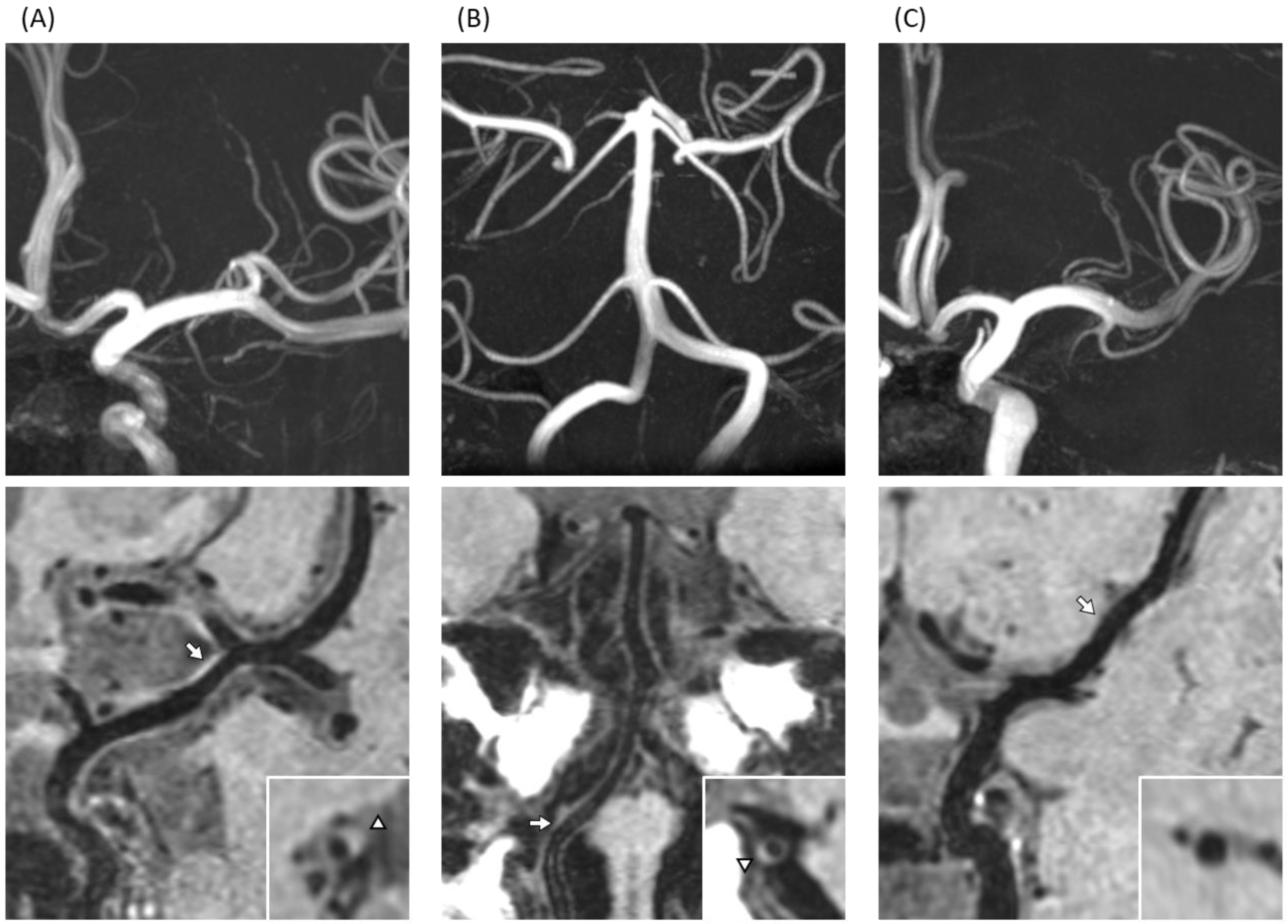
2.2. MR Protocols
2.3. Data Analysis
2.4. Statistical Analysis
3. Results
4. Discussion
5. Conclusions
Author Contributions
Funding
Institutional Review Board Statement
Informed Consent Statement
Data Availability Statement
Acknowledgments
Conflicts of Interest
References
- Sarwar, N.; Gao, P.; Seshasai, S.R.; Gobin, R.; Kaptoge, S.; Di Angelantonio, E.; Ingelsson, E.; Lawlor, D.A.; Selvin, E.; Stampfer, M.; et al. Diabetes mellitus, fasting blood glucose concentration, and risk of vascular disease: A collaborative meta-analysis of 102 prospective studies. Lancet 2010, 375, 2215–2222. [Google Scholar] [PubMed] [Green Version]
- Feigin, V.L.; Nguyen, G.; Cercy, K.; Johnson, C.O.; Alam, T.; Parmar, P.G.; Abajobir, A.A.; Abate, K.H.; Abd-Allah, F.; Abejie, A.N.; et al. Global, Regional, and Country-Specific Lifetime Risks of Stroke, 1990 and 2016. N. Engl. J. Med. 2018, 379, 2429–2437. [Google Scholar] [PubMed]
- Doi, Y.; Ninomiya, T.; Hata, J.; Fukuhara, M.; Yonemoto, K.; Iwase, M.; Iida, M.; Kiyohara, Y. Impact of glucose tolerance status on development of ischemic stroke and coronary heart disease in a general Japanese population: The Hisayama study. Stroke 2010, 41, 203–209. [Google Scholar] [CrossRef] [PubMed] [Green Version]
- Cui, R.; Iso, H.; Yamagishi, K.; Saito, I.; Kokubo, Y.; Inoue, M.; Tsugane, S. Diabetes mellitus and risk of stroke and its subtypes among Japanese: The Japan public health center study. Stroke 2011, 42, 2611–2614. [Google Scholar] [CrossRef] [PubMed] [Green Version]
- Brundel, M.; Reijmer, Y.D.; van Veluw, S.J.; Kuijf, H.J.; Luijten, P.R.; Kappelle, L.J.; Biessels, G.J. Cerebral microvascular lesions on high-resolution 7-Tesla MRI in patients with type 2 diabetes. Diabetes 2014, 63, 3523–3529. [Google Scholar] [CrossRef] [Green Version]
- Natori, T.; Sasaki, M.; Miyoshi, M.; Ito, K.; Ohba, H.; Miyazawa, H.; Narumi, S.; Kabasawa, H.; Harada, T.; Terayama, Y. Intracranial Plaque Characterization in Patients with Acute Ischemic Stroke Using Pre- and Post-Contrast Three-Dimensional Magnetic Resonance Vessel Wall Imaging. J. Stroke Cerebrovasc. Dis. 2016, 25, 1425–1430. [Google Scholar] [CrossRef]
- Narumi, S.; Sasaki, M.; Ohba, H.; Ogasawara, K.; Kobayashi, M.; Hitomi, J.; Mori, K.; Ohura, K.; Yamaguchi, M.; Kudo, K.; et al. Prediction of carotid plaque characteristics using non-gated MR imaging: Correlation with endarterectomy specimens. AJNR Am. J. Neuroradiol. 2013, 34, 191–197. [Google Scholar] [CrossRef] [Green Version]
- Natori, T.; Sasaki, M.; Miyoshi, M.; Ohba, H.; Katsura, N.; Yamaguchi, M.; Narumi, S.; Kabasawa, H.; Kudo, K.; Ito, K.; et al. Evaluating middle cerebral artery atherosclerotic lesions in acute ischemic stroke using magnetic resonance T1-weighted 3-dimensional vessel wall imaging. J. Stroke Cerebrovasc. Dis. 2014, 23, 706–711. [Google Scholar] [CrossRef]
- Song, J.W.; Moon, B.F.; Burke, M.P.; Kamesh Iyer, S.K.; Elliott, M.A.; Shou, H.; Messé, S.R.; Kasner, S.E.; Loevner, L.A.; Schnall, M.D.; et al. MR Intracranial Vessel Wall Imaging: A Systematic Review. J. Neuroimaging 2020, 30, 428–442. [Google Scholar] [CrossRef]
- Jiao, S.; Huang, J.; Chen, Y.; Song, Y.; Gong, T.; Lu, J.; Guo, T.; Zhang, J.; Zhang, C.; Chen, M. Impacts of Glycemic Control on Intracranial Plaque in Patients with Type 2 Diabetes Mellitus: A Vessel Wall MRI Study. AJNR Am. J. Neuroradiol. 2021, 42, 75–81. [Google Scholar] [CrossRef]
- Li, X.; Sun, B.; Wang, L.; Zhang, J.; Zhao, Z.; Wu, H.; Liu, X.; Zhou, Y.; Mossa-Basha, M.; Tirschwell, D.L.; et al. Association of Type 2 Diabetes Mellitus and Glycemic Control with Intracranial Plaque Characteristics in Patients with Acute Ischemic Stroke. J. Magn. Reson. Imaging 2021, 54, 655–666. [Google Scholar] [CrossRef] [PubMed]
- Sato, Y.; Ogasawara, K.; Yoshida, K.; Sasaki, M. Preoperative visualization of the marginal tentorial artery as an unusual collateral pathway in a patient with symptomatic bilateral vertebral artery occlusion undergoing arterial bypass surgery: A 7.0-T magnetic resonance imaging study. Surg. Neurol. Int. 2014, 5, 157. [Google Scholar]
- Harteveld, A.A.; van der Kolk, A.G.; Zwanenburg, J.J.; Luijten, P.R.; Hendrikse, J. 7-T MRI in Cerebrovascular Diseases: Challenges to Overcome and Initial Results. Top. Magn. Reson. Imaging 2016, 25, 89–100. [Google Scholar] [CrossRef] [PubMed]
- Yashiro, S.; Kameda, H.; Chida, A.; Todate, Y.; Hasegawa, Y.; Nagasawa, K.; Uwano, I.; Sasaki, M.; Ogasawara, K.; Ishigaki, Y. Evaluation of Lenticulostriate Arteries Changes by 7 T Magnetic Resonance Angiography in Type 2 Diabetes. J. Atheroscler. Thromb. 2018, 25, 1067–1075. [Google Scholar] [CrossRef] [Green Version]
- Harteveld, A.A.; van der Kolk, A.G.; van der Worp, H.B.; Dieleman, N.; Zwanenburg, J.J.M.; Luijten, P.R.; Hendrikse, J. Detecting Intracranial Vessel Wall Lesions with 7T-Magnetic Resonance Imaging: Patients with Posterior Circulation Ischemia Versus Healthy Controls. Stroke 2017, 48, 2601–2604. [Google Scholar] [CrossRef] [PubMed]
- van der Kolk, A.G.; Zwanenburg, J.J.; Brundel, M.; Biessels, G.J.; Visser, F.; Luijten, P.R.; Hendrikse, J. Distribution and natural course of intracranial vessel wall lesions in patients with ischemic stroke or TIA at 7.0 Tesla MRI. Eur. Radiol. 2015, 25, 1692–1700. [Google Scholar] [CrossRef]
- Zwartbol, M.H.T.; van der Kolk, A.G.; Ghaznawi, R.; van der Graaf, Y.; Hendrikse, J.; Geerlings, M.I. Intracranial Vessel Wall Lesions on 7T MRI (Magnetic Resonance Imaging). Stroke 2019, 50, 88–94. [Google Scholar] [CrossRef] [PubMed]
- Araki, E.; Goto, A.; Kondo, T.; Noda, M.; Noto, H.; Origasa, H.; Osawa, H.; Taguchi, A.; Tanizawa, Y.; Tobe, K.; et al. Japanese Clinical Practice Guideline for Diabetes 2019. Diabetol. Int. 2020, 11, 165–223. [Google Scholar] [CrossRef] [PubMed]
- Qiao, Y.; Steinman, D.A.; Qin, Q.; Etesami, M.; Schär, M.; Astor, B.C.; Wasserman, B.A. Intracranial arterial wall imaging using three-dimensional high isotropic resolution black blood MRI at 3.0 Tesla. J. Magn. Reson. Imaging 2011, 34, 22–30. [Google Scholar] [CrossRef]
- Alexander, A.L.; Buswell, H.R.; Sun, Y.; Chapman, B.E.; Tsuruda, J.S.; Parker, D.L. Intracranial black-blood MR angiography with high-resolution 3D fast spin echo. Magn. Reson. Med. 1998, 40, 298–310. [Google Scholar] [CrossRef]
- Jara, H.; Yu, B.C.; Caruthers, S.D.; Melhem, E.R.; Yucel, E.K. Voxel sensitivity function description of flow-induced signal loss in MR imaging: Implications for black-blood MR angiography with turbo spin-echo sequences. Magn. Reson. Med. 1999, 41, 575–590. [Google Scholar] [CrossRef]
- Miyazawa, H.; Natori, T.; Kameda, H.; Sasaki, M.; Ohba, H.; Narumi, S.; Ito, K.; Sato, M.; Suzuki, T.; Tsuda, K.; et al. Detecting lenticulostriate artery lesions in patients with acute ischemic stroke using high-resolution MRA at 7 T. Int. J. Stroke 2019, 14, 290–297. [Google Scholar] [CrossRef]
- Uwano, I.; Kameda, H.; Harada, T.; Kobayashi, M.; Yanagihara, W.; Setta, K.; Ogasawara, K.; Yoshioka, K.; Yamashita, F.; Mori, F.; et al. Detection of impaired cerebrovascular reactivity in patients with chronic cerebral ischemia using whole-brain 7T MRA. J. Stroke Cerebrovasc. Dis. 2020, 29, 105081. [Google Scholar] [CrossRef] [PubMed]
- Uwano, I.; Kudo, K.; Yamashita, F.; Goodwin, J.; Higuchi, S.; Ito, K.; Harada, T.; Ogawa, A.; Sasaki, M. Intensity inhomogeneity correction for magnetic resonance imaging of human brain at 7T. Med. Phys. 2014, 41, 022302. [Google Scholar] [CrossRef] [PubMed]
- Narumi, S.; Sasaki, M.; Ohba, H.; Ogasawara, K.; Kobayashi, M.; Natori, T.; Hitomi, J.; Itagaki, H.; Takahashi, T.; Terayama, Y. Predicting carotid plaque characteristics using quantitative color-coded T1-weighted MR plaque imaging: Correlation with carotid endarterectomy specimens. AJNR Am. J. Neuroradiol. 2014, 35, 766–771. [Google Scholar] [CrossRef] [PubMed] [Green Version]
- Todate, Y.; Uwano, I.; Yashiro, S.; Chida, A.; Hasegawa, Y.; Oda, T.; Nagasawa, K.; Honma, H.; Sasaki, M.; Ishigaki, Y. High Prevalence of Cerebral Small Vessel Disease on 7T Magnetic Resonance Imaging in Familial Hypercholesterolemia. J. Atheroscler. Thromb. 2019, 26, 1045–1053. [Google Scholar] [CrossRef] [PubMed] [Green Version]
- Kim, Y.S.; Lim, S.H.; Oh, K.W.; Kim, J.Y.; Koh, S.H.; Kim, J.; Heo, S.H.; Chang, D.I.; Lee, Y.J.; Kim, H.Y. The advantage of high-resolution MRI in evaluating basilar plaques: A comparison study with MRA. Atherosclerosis 2012, 224, 411–416. [Google Scholar] [CrossRef]
- Dieleman, N.; van der Kolk, A.G.; Zwanenburg, J.J.; Harteveld, A.A.; Biessels, G.J.; Luijten, P.R.; Hendrikse, J. Imaging intracranial vessel wall pathology with magnetic resonance imaging: Current prospects and future directions. Circulation 2014, 130, 192–201. [Google Scholar] [CrossRef] [Green Version]
- Turan, T.N.; Bonilha, L.; Morgan, P.S.; Adams, R.J.; Chimowitz, M.I. Intraplaque hemorrhage in symptomatic intracranial atherosclerotic disease. J. Neuroimaging 2011, 21, e159–e161. [Google Scholar] [CrossRef]
- Guggenberger, K.; Krafft, A.J.; Ludwig, U.; Raithel, E.; Forman, C.; Meckel, S.; Hennig, J.; Bley, T.A.; Vogel, P. Intracranial vessel wall imaging framework—Data acquisition, processing, and visualization. Magn. Reson. Imaging 2021, 83, 114–124. [Google Scholar] [CrossRef]
- .Zhu, C.; Haraldsson, H.; Tian, B.; Meisel, K.; Ko, N.; Lawton, M.; Grinstead, J.; Ahn, S.; Laub, G.; Hess, C.; et al. High resolution imaging of the intracranial vessel wall at 3 and 7 T using 3D fast spin echo MRI. Magn. Reson. Mater. Phy. 2016, 29, 559–570. [Google Scholar] [CrossRef] [PubMed]
- Harteveld, A.A.; van der Kolk, A.G.; van der Worp, H.B.; Dieleman, N.; Siero, J.C.W.; Kuijf, H.J.; Frijns, C.J.M.; Luijten, P.R.; Zwanenburg, J.J.M.; Hendrikse, J. High-resolution intracranial vessel wall MRI in an elderly asymptomatic population: Comparison of 3T and 7T. Eur. Radiol. 2017, 27, 1585–1595. [Google Scholar] [CrossRef] [PubMed] [Green Version]
- Choi, N.; Lee, J.Y.; Sunwoo, J.S.; Roh, H.; Ahn, M.Y.; Park, S.T.; Lee, K.B. Recently Uncontrolled Glycemia in Diabetic Patients is Associated with the Severity of Intracranial Atherosclerosis. J. Stroke Cerebrovasc. Dis. 2017, 26, 2615–2621. [Google Scholar] [CrossRef] [PubMed]
- Laiteerapong, N.; Ham, S.A.; Gao, Y.; Moffet, H.H.; Liu, J.Y.; Huang, E.S.; Karter, A.J. The Legacy Effect in Type 2 Diabetes: Impact of Early Glycemic Control on Future Complications (The Diabetes & Aging Study). Diabetes Care 2019, 42, 416–426. [Google Scholar] [PubMed] [Green Version]
- Hangai, M.; Takebe, N.; Honma, H.; Sasaki, A.; Chida, A.; Nakano, R.; Togashi, H.; Nakagawa, R.; Oda, T.; Matsui, M.; et al. Association of Advanced Glycation End Products with Coronary Artery Calcification in Japanese Subjects with Type 2 Diabetes as Assessed by Skin Autofluorescence. J. Atheroscler. Thromb. 2016, 23, 1178–1187. [Google Scholar] [CrossRef] [Green Version]
- Katakami, N. Mechanism of Development of Atherosclerosis and Cardiovascular Disease in Diabetes Mellitus. J. Atheroscler. Thromb. 2018, 25, 27–39. [Google Scholar] [CrossRef] [PubMed]


| T2D | Control | p-Value | |
|---|---|---|---|
| (n = 48) | (n = 35) | ||
| Age (years) | 53.2 ± 6.3 | 50.7 ± 5.3 | 0.067 |
| Male (%) | 64.6 | 54.3 | 0.344 |
| Body weight (kg) | 71.7 ± 11.9 | 61.6 ± 9.4 | <0.01 |
| Body mass index (kg/m2) | 26.1 ± 4.2 | 22.4 ± 2.3 | <0.01 |
| Systolic blood pressure (mmHg) | 128.8 ± 14.4 | 115.9 ± 14.0 | <0.01 |
| Diastolic blood pressure (mmHg) | 80.4 ± 10.8 | 73.5 ± 9.5 | <0.01 |
| Former or current smoking (%) | 50.0 | 14.3 | <0.01 |
| HbA1c (%) | 9.5 ± 3.0 | 5.5 ± 0.3 | <0.01 |
| AST (IU/mL) | 27.5 ± 24.2 | 21.4 ± 5.0 | 0.96 |
| ALT (IU/mL) | 33.3 ± 27.4 | 20.3 ± 9.8 | <0.01 |
| γ-GTP (IU/mL) | 62.8 ± 105.4 | 41.9 ± 47.7 | 0.278 |
| TC (mg/dL) | 191.4 ± 49.0 | 197.9 ± 35.0 | 0.554 |
| TG (mg/dL) | 149.8 ± 77.0 | 81.7 ± 35.4 | <0.01 |
| LDL-C (mg/dL) | 114.3 ± 39.1 | 108.3 ± 23.6 | 0.42 |
| HDL-C (mg/dL) | 51.6 ± 13.8 | 71.7 ± 15.3 | <0.01 |
| Hypertension, n (%) | 21 (43.8) | 0 | |
| RAS inhibitor, n | 15 | ||
| Calcium channel blocker, n | 9 | ||
| Dyslipidemia, n (%) | 32 (66.7) | 0 | |
| Statin, n | 14 | ||
| Fibrate, n | 6 |
| T2D | Control | p-Value | |
|---|---|---|---|
| Lacunar infarction | 8 (16.7%) | 0 (0%) | 0.011 |
| CMBs | 2 (4.2%) | 2 (5.7%) | 0.565 |
| PVH | 2 (4.2%) | 0 (0%) | 0.311 |
| DWMH | 8 (16.6%) | 2 (5.7%) | 0.119 |
| Brain atrophy | 2 (4.2%) | 0 (0%) | 0.331 |
| T2D | Control | p-Value | |
|---|---|---|---|
| Anterior and posterior circulation | 2.23 ± 0.23 | 0.94 ± 0.21 | <0.01 |
| Anterior circulation | 1.52 ± 0.18 | 0.51 ± 0.12 | <0.01 |
| Posterior circulation | 0.71 ± 0.12 | 0.43 ± 0.13 | 0.133 |
| Type 2 Diabetes | p-Value | ||
|---|---|---|---|
| (+) n = 38 | (−) n = 10 | ||
| Age (years) | 53.6 | 51.3 | 0.310 |
| Male (%) | 29 (76.3) | 2 (20) | <0.01 |
| sBP (mmHg) | 129.6 | 125.6 | 0.441 |
| dBP (mmHg) | 81.8 | 74.9 | 0.060 |
| HbA1c (%) | 9.3 | 10.1 | 0.572 |
| AST (IU/mL) | 29.4 | 20.1 | 0.284 |
| ALT (IU/mL) | 35.5 | 25.2 | 0.297 |
| γ-GTP (IU/mL) | 71.2 | 30.8 | 0.285 |
| TC (mg/dL) | 187.5 | 206.3 | 0.285 |
| TG (mg/dL) | 155.4 | 128.6 | 0.333 |
| LDL-C (mg/dL) | 112.6 | 120.5 | 0.577 |
| HDL-C (mg/dL) | 49.6 | 59.4 | 0.430 |
| eGFR (mL/min/1.73m2) | 74.4 | 78.8 | 0.389 |
| Former or current smoking (%) | 21(55.2) | 3(30.0) | 0.155 |
| Hypertension, n (%) | 17 (42.1) | 4 (40.0) | 0.542 |
| RAS inhibitor, n (%) | 12 (31.6) | 3 (30.0) | 0.602 |
| Calcium channel blocker, n (%) | 9 (23.7) | 0 | 0.094 |
| Dyslipidemia, n (%) | 26 (63.2) | 6 (60.0) | 0.594 |
| Statin, n (%) | 11 (28.9) | 3 (30.0) | 0.612 |
| Fibrate, n (%) | 4 (10.5) | 2 (20.0) | 0.355 |
| Diabetic neuropathy (%) | 22 (57.8) | 5 (50.0) | 0.654 |
| Diabetic retinopathy (%) | 16 (42.1) | 5 (50.0) | 0.654 |
| Diabetic nephropathy (%) | 13 (34.2) | 4 (40.0) | 0.733 |
| Lacunar infarction (%) | 8 (21.1) | 0 (0) | 0.112 |
| CMBs (%) | 2 (5.3) | 0 (0) | 0.459 |
| PVH (%) | 1 (2.6) | 1 (10.0) | 0.299 |
| DWMH (%) | 6 (15.8) | 2 (20.0) | 0.751 |
| Brain atrophy (%) | 2 (5.3) | 0 (0) | 0.459 |
Disclaimer/Publisher’s Note: The statements, opinions and data contained in all publications are solely those of the individual author(s) and contributor(s) and not of MDPI and/or the editor(s). MDPI and/or the editor(s) disclaim responsibility for any injury to people or property resulting from any ideas, methods, instructions or products referred to in the content. |
© 2023 by the authors. Licensee MDPI, Basel, Switzerland. This article is an open access article distributed under the terms and conditions of the Creative Commons Attribution (CC BY) license (https://creativecommons.org/licenses/by/4.0/).
Share and Cite
Shozushima, M.; Mori, F.; Yashiro, S.; Todate, Y.; Oda, T.; Nagasawa, K.; Hasegawa, Y.; Takebe, N.; Sasaki, M.; Ishigaki, Y. Evaluation of High Intracranial Plaque Prevalence in Type 2 Diabetes Using Vessel Wall Imaging on 7 T Magnetic Resonance Imaging. Brain Sci. 2023, 13, 217. https://doi.org/10.3390/brainsci13020217
Shozushima M, Mori F, Yashiro S, Todate Y, Oda T, Nagasawa K, Hasegawa Y, Takebe N, Sasaki M, Ishigaki Y. Evaluation of High Intracranial Plaque Prevalence in Type 2 Diabetes Using Vessel Wall Imaging on 7 T Magnetic Resonance Imaging. Brain Sciences. 2023; 13(2):217. https://doi.org/10.3390/brainsci13020217
Chicago/Turabian StyleShozushima, Masaharu, Futoshi Mori, Satoshi Yashiro, Yusuke Todate, Tomoyasu Oda, Kan Nagasawa, Yutaka Hasegawa, Noriko Takebe, Makoto Sasaki, and Yasushi Ishigaki. 2023. "Evaluation of High Intracranial Plaque Prevalence in Type 2 Diabetes Using Vessel Wall Imaging on 7 T Magnetic Resonance Imaging" Brain Sciences 13, no. 2: 217. https://doi.org/10.3390/brainsci13020217