
Stress and Right Prefrontal Transcranial Direct Current Stimulation (tDCS) Interactive Effects on Visual Working Memory and Learning

Abstract
:1. Introduction
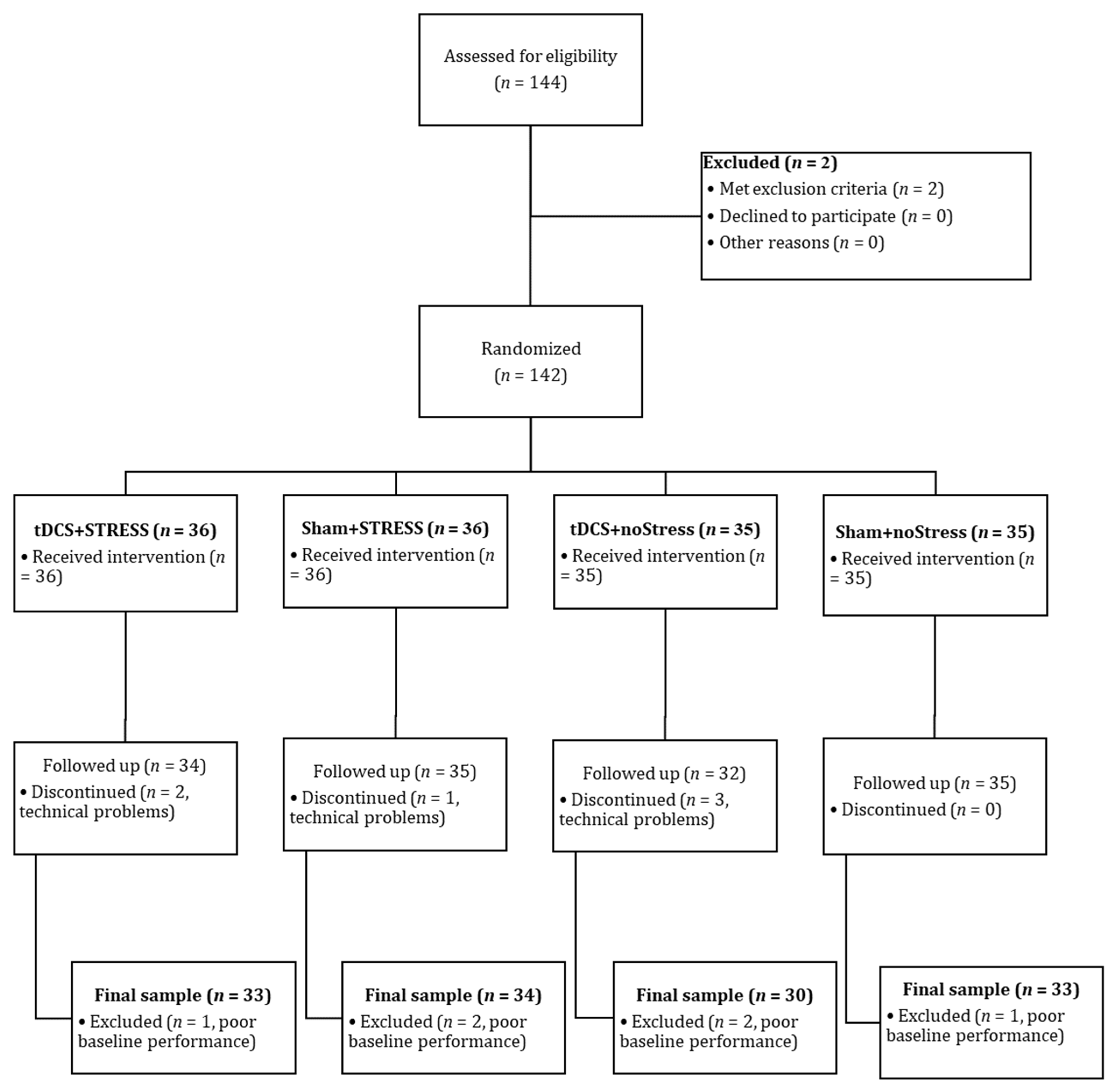
2. Method
2.1. Participants
2.2. Tools
2.2.1. Cogstate Battery
2.2.2. Stimulation Parameters
2.3. Procedure
3. Results
4. Discussion
Author Contributions
Funding
Institutional Review Board Statement
Informed Consent Statement
Data Availability Statement
Acknowledgments
Conflicts of Interest
References
- Buchanan, D.M.; Bogdanowicz, T.; Khanna, N.; Lockman-Dufour, G.; Robaey, P.; D’Angiulli, A. Systematic Review on the Safety and Tolerability of Transcranial Direct Current Stimulation in Children and Adolescents. Brain Sci. 2021, 11, 212. [Google Scholar] [CrossRef]
- Ciullo, V.; Spalletta, G.; Caltagirone, C.; Banaj, N.; Vecchio, D.; Piras, F.; Piras, F. Transcranial Direct Current Stimulation and Cognition in Neuropsychiatric Disorders: Systematic Review of the Evidence and Future Directions. Neuroscientist 2020, 27, 1073858420936167. [Google Scholar] [CrossRef]
- Barbati, S.A.; Podda, M.V.; Grassi, C. Tuning brain networks: The emerging role of transcranial direct current stimulation on structural plasticity. Front. Cell Neurosci. 2022, 16, 945777. [Google Scholar] [CrossRef]
- Farhat, L.C.; Carvalho, A.F.; Solmi, M.; Brunoni, A.R. Evidence-based Umbrella Review of Cognitive Effects of Prefrontal tDCS. Soc. Cogn. Affect. Neurosci. 2022, 17, 43–60. [Google Scholar] [CrossRef]
- Gallop, L.; Westwood, S.J.; Lewis, Y.; Campbell, I.C.; Schmidt, U. Effects of transcranial direct current stimulation in children and young people with psychiatric disorders: A systematic review. Eur. Child. Adolesc. Psychiatry 2023. [Google Scholar] [CrossRef]
- Li, Q.; Fu, Y.; Liu, C.; Meng, Z. Transcranial Direct Current Stimulation of the Dorsolateral Prefrontal Cortex for Treatment of Neuropsychiatric Disorders. Front. Behav. Neurosci. 2022, 16, 893955. [Google Scholar] [CrossRef] [PubMed]
- Brunoni, A.R.; Vanderhasselt, M.A. Working memory improvement with non-invasive brain stimulation of the dorsolateral prefrontal cortex: A systematic review and meta-analysis. Brain Cogn. 2014, 86, 1–9. [Google Scholar] [CrossRef] [PubMed]
- Herrera-Melendez, A.L.; Bajbouj, M.; Aust, S. Application of Transcranial Direct Current Stimulation in Psychiatry. Neuropsychobiology 2020, 79, 372–383. [Google Scholar] [CrossRef] [PubMed]
- Mancuso, L.E.; Ilieva, I.P.; Hamilton, R.H.; Farah, M.J. Does Transcranial Direct Current Stimulation Improve Healthy Working Memory?: A Meta-analytic Review. J. Cogn. Neurosci. 2016, 28, 1063–1089. [Google Scholar] [CrossRef] [PubMed]
- Bogdanov, M.; Schwabe, L. Transcranial Stimulation of the Dorsolateral Prefrontal Cortex Prevents Stress-Induced Working Memory Deficits. J. Neurosci. 2016, 36, 1429–1437. [Google Scholar] [CrossRef]
- Antal, A.; Fischer, T.; Saiote, C.; Miller, R.; Chaieb, L.; Wang, D.J.; Plessow, F.; Paulus, W.; Kirschbaum, C. Transcranial electrical stimulation modifies the neuronal response to psychosocial stress exposure. Hum. Brain Mapp. 2014, 35, 3750–3759. [Google Scholar] [CrossRef]
- Friehs, M.A.; Frings, C. Evidence Against Combined Effects of Stress and Brain Stimulation on Working Memory. Open Psychol. 2020, 2, 40–56. [Google Scholar] [CrossRef]
- Ankri, Y.L.E.; Braw, Y.; Luboshits, G.; Meiron, O. The effects of stress and transcranial direct current stimulation (tDCS) on working memory: A randomized controlled trial. Cogn. Affect. Behav. Neurosci. 2020, 20, 103–114. [Google Scholar] [CrossRef]
- Moscatelli, F.; Monda, V.; Limone, P.; Marsala, G.; Mancini, N.; Monda, M.; Messina, A.; De Maria, A.; Scarinci, A.; Monda, A.; et al. Acute non invasive brain stimulation improves performances in volleyball players. Physiol. Behav. 2023, 271, 114356. [Google Scholar] [CrossRef]
- Meiron, O.; Hermesh, H.; Katz, N.; Weizman, A. Executive attention deficits in schizophrenia: Putative mandatory and differential cognitive pathology domains in medicated schizophrenia patients. Psychiatry Res. 2013, 209, 1–8. [Google Scholar] [CrossRef]
- Meiron, O.; Rosset, E.; Braw, Y. Putative Neurocognitive Marker for Early Non-Salient Accelerated Cognitive Decline in Older Adults (Ch. 4). In Horizons in Neuroscience Research; Costa, A., Villalba, E., Eds.; Nova Science Publishers: Hauppauge, NY, USA, 2016; Volume 26. [Google Scholar]
- Berryhill, M.E.; Peterson, D.J.; Jones, K.T.; Stephens, J.A. Hits and misses: Leveraging tDCS to advance cognitive research. Front. Psychol. 2014, 5, 800. [Google Scholar] [CrossRef]
- Tuerk, C.; Saha, T.; Bouchard, M.F.; Booij, L. Computerized Cognitive Test Batteries for Children and Adolescents-A Scoping Review of Tools For Lab- and Web-Based Settings From 2000 to 2021. Arch. Clin. Neuropsychol. 2023, acad039. [Google Scholar] [CrossRef]
- Weiner, M.W.; Nosheny, R.; Camacho, M.; Truran-Sacrey, D.; Mackin, R.S.; Flenniken, D.; Ulbricht, A.; Insel, P.; Finley, S.; Fockler, J.; et al. The Brain Health Registry: An internet-based platform for recruitment, assessment, and longitudinal monitoring of participants for neuroscience studies. Alzheimers Dement. 2018, 14, 1063–1076. [Google Scholar] [CrossRef]
- Lupu, T.; Braw, Y.; Sacher, Y.; Ratmansky, M. Cogstate Brief Battery: Cognition and the feigning of cognitive impairment in chronic pain. Appl. Neuropsychol. Adult 2021, 29, 1332–1343. [Google Scholar] [CrossRef]
- Nixon, G.; Sarant, J.; Tomlin, D.; Dowell, R. Hearing Aid Uptake, Benefit, and Use: The Impact of Hearing, Cognition, and Personal Factors. J. Speech Lang. Hear. Res. 2021, 64, 651–663. [Google Scholar] [CrossRef]
- Sicard, V.; Stephenson, D.D.; Hergert, D.C.; Dodd, A.B.; Robertson-Benta, C.R.; Pabbathi Reddy, S.; Yeates, K.O.; Cromer, J.A.; Meier, T.B.; Campbell, R.A.; et al. Investigating the diagnostic accuracy of a paper-and-pencil and a computerized cognitive test battery for pediatric mild traumatic brain injury. Neuropsychology 2022, 36, 565–577. [Google Scholar] [CrossRef] [PubMed]
- Cromer, J.A.; Schembri, A.J.; Harel, B.T.; Maruff, P. The nature and rate of cognitive maturation from late childhood to adulthood. Front. Psychol. 2015, 6, 704. [Google Scholar] [CrossRef] [PubMed]
- Meiron, O.; Lavidor, M. Prefrontal oscillatory stimulation modulates access to cognitive control references in retrospective metacognitive commentary. Clin. Neurophysiol. 2014, 125, 77–82. [Google Scholar] [CrossRef] [PubMed]
- Roser, M.E.; Fiser, J.; Aslin, R.N.; Gazzaniga, M.S. Right hemisphere dominance in visual statistical learning. J. Cogn. Neurosci. 2011, 23, 1088–1099. [Google Scholar] [CrossRef]
- Meiron, O.; Lavidor, M. Bilateral Transcranial Alternating Current Stimulation (tACS) of the Dorsolateral Prefrontal Cortex Enhances Verbal Working Memory and Promotes Episodic Memory After-effects (Ch. 11). In Working Memory: Developmental Differences, Component Processes and Improvement Mechanisms; St Clair-Thompson, H., Ed.; Nova Science Publishers: New York, NY, USA, 2013; pp. 157–174. [Google Scholar]
- Medina, J.; Cason, S. No evidential value in samples of transcranial direct current stimulation (tDCS) studies of cognition and working memory in healthy populations. Cortex 2017, 94, 131–141. [Google Scholar] [CrossRef]
- Faul, F.; Erdfelder, E.; Lang, A.G.; Buchner, A. G*Power 3: A flexible statistical power analysis program for the social, behavioral, and biomedical sciences. Behav. Res. Methods 2007, 39, 175–191. [Google Scholar] [CrossRef]
- Faul, F.; Erdfelder, E.; Buchner, A.; Lang, A.G. Statistical power analyses using G*Power 3.1: Tests for correlation and regression analyses. Behav. Res. Methods 2009, 41, 1149–1160. [Google Scholar] [CrossRef]
- Meiron, O.; Lavidor, M. Unilateral prefrontal direct current stimulation effects are modulated by working memory load and gender. Brain Stimul. 2013, 6, 440–447. [Google Scholar] [CrossRef]
- Mehta, K.; Mohebbi, M.; Pasco, J.A.; Williams, L.J.; Walder, K.; Ng, B.L.; Gupta, V.B. Genetic polymorphism in BIN1 rather than APOE is associated with poor recognition memory among men without dementia. Sci. Rep. 2022, 12, 17802. [Google Scholar] [CrossRef]
- Mustafa, A.I.; Woods, S.P.; Loft, S.; Morgan, E.E. Lower prospective memory is associated with higher neurocognitive dispersion in two samples of people with HIV: A conceptual replication study. J. Int. Neuropsychol. Soc. 2023, 29, 677–685. [Google Scholar] [CrossRef]
- Stricker, N.H.; Lundt, E.S.; Albertson, S.M.; Machulda, M.M.; Pudumjee, S.B.; Kremers, W.K.; Jack, C.R.; Knopman, D.S.; Petersen, R.C.; Mielke, M.M. Diagnostic and Prognostic Accuracy of the Cogstate Brief Battery and Auditory Verbal Learning Test in Preclinical Alzheimer’s Disease and Incident Mild Cognitive Impairment: Implications for Defining Subtle Objective Cognitive Impairment. J. Alzheimers Dis. 2020, 76, 261–274. [Google Scholar] [CrossRef]
- Fitzgerald, P.B.; Maller, J.J.; Hoy, K.E.; Thomson, R.; Daskalakis, Z.J. Exploring the optimal site for the localization of dorsolateral prefrontal cortex in brain stimulation experiments. Brain Stimul. 2009, 2, 234–237. [Google Scholar] [CrossRef]
- Spielberger, C.D.; Gorsuch, R.L.; Lushene, R.E. Manual for the State-Trait Anxiety Inventory; Consulting Psychologists Press: Palo Alto, CA, USA, 1970. [Google Scholar]
- Rossi, V.; Pourtois, G. Transient state-dependent fluctuations in anxiety measured using STAI, POMS, PANAS or VAS: A comparative review. Anxiety Stress Coping 2012, 25, 603–645. [Google Scholar] [CrossRef] [PubMed]
- Gaab, J.; Rohleder, N.; Nater, U.M.; Ehlert, U. Psychological determinants of the cortisol stress response: The role of anticipatory cognitive appraisal. Psychoneuroendocrinology 2005, 30, 599–610. [Google Scholar] [CrossRef]
- Labuschagne, I.; Grace, C.; Rendell, P.; Terrett, G.; Heinrichs, M. An introductory guide to conducting the Trier Social Stress Test. Neurosci. Biobehav. Rev. 2019, 107, 686–695. [Google Scholar] [CrossRef] [PubMed]
- Kirschbaum, C.; Pirke, K.M.; Hellhammer, D.H. The ‘Trier Social Stress Test’—A tool for investigating psychobiological stress responses in a laboratory setting. Neuropsychobiology 1993, 28, 76–81. [Google Scholar] [CrossRef] [PubMed]
- Wiemers, U.S.; Schoofs, D.; Wolf, O.T. A friendly version of the trier social stress test does not activate the HPA axis in healthy men and women. Stress 2013, 16, 254–260. [Google Scholar] [CrossRef]
- Cohen, J. A power primer. Psychol. Bull. 1992, 112, 155–159. [Google Scholar] [CrossRef]
- Robison, M.K.; Brewer, G.A. Individual differences in working memory capacity and the regulation of arousal. Atten. Percept. Psychophys. 2020, 82, 3273–3290. [Google Scholar] [CrossRef]
- Arnsten, A.F. Stress weakens prefrontal networks: Molecular insults to higher cognition. Nat. Neurosci. 2015, 18, 1376–1385. [Google Scholar] [CrossRef]
- Datta, D.; Arnsten, A.F.T. Loss of Prefrontal Cortical Higher Cognition with Uncontrollable Stress: Molecular Mechanisms, Changes with Age, and Relevance to Treatment. Brain Sci. 2019, 9, 113. [Google Scholar] [CrossRef] [PubMed]
- Lamichhane, B.; Westbrook, A.; Cole, M.W.; Braver, T.S. Exploring brain-behavior relationships in the N-back task. Neuroimage 2020, 212, 116683. [Google Scholar] [CrossRef] [PubMed]
- Ghosal, S.; Hare, B.; Duman, R.S. Prefrontal Cortex GABAergic Deficits and Circuit Dysfunction in the Pathophysiology and Treatment of Chronic Stress and Depression. Curr. Opin. Behav. Sci. 2017, 14, 1–8. [Google Scholar] [CrossRef] [PubMed]
- Kalin, N.H. Prefrontal Cortical and Limbic Circuit Alterations in Psychopathology. Am. J. Psychiatry 2019, 176, 971–973. [Google Scholar] [CrossRef]
- Duque, A.; Cano-Lopez, I.; Puig-Perez, S. Effects of psychological stress and cortisol on decision making and modulating factors: A systematic review. Eur. J. Neurosci. 2022, 56, 3889–3920. [Google Scholar] [CrossRef] [PubMed]
- Lin, L.; Leung, A.W.S.; Wu, J.; Zhang, L. Individual differences under acute stress: Higher cortisol responders performs better on N-back task in young men. Int. J. Psychophysiol. 2020, 150, 20–28. [Google Scholar] [CrossRef] [PubMed]
- Schoofs, D.; Preuss, D.; Wolf, O.T. Psychosocial stress induces working memory impairments in an n-back paradigm. Psychoneuroendocrinology 2008, 33, 643–653. [Google Scholar] [CrossRef]
- O’Keeffe, K.; Hodder, S.; Lloyd, A. A comparison of methods used for inducing mental fatigue in performance research: Individualised, dual-task and short duration cognitive tests are most effective. Ergonomics 2020, 63, 1–12. [Google Scholar] [CrossRef]
- Allen, A.P.; Kennedy, P.J.; Dockray, S.; Cryan, J.F.; Dinan, T.G.; Clarke, G. The Trier Social Stress Test: Principles and practice. Neurobiol. Stress 2017, 6, 113–126. [Google Scholar] [CrossRef]
- Wang, J.J.; Korczykowski, M.; Rao, H.Y.; Fan, Y.; Pluta, J.; Gur, R.C.; McEwen, B.S.; John, A. Detre, Gender difference in neural response to psychological stress. Soc. Cogn. Affect. Neurosci. 2007, 2, 227–239. [Google Scholar] [CrossRef]
- McEwen, B.S.; Gianaros, P.J. Central role of the brain in stress and adaptation: Links to socioeconomic status, health, and disease. Ann. N. Y. Acad. Sci. 2010, 1186, 190–222. [Google Scholar] [CrossRef] [PubMed]


| Variables | STRESS | NoStress | |||
|---|---|---|---|---|---|
| tDCS | Sham | tDCS | Sham | Test of Difference † | |
| Age | 22.7 ± 1.7 | 22.3 ± 1.5 | 22.9 ± 2.1 | 22.4 ± 1.6 | F(3,126) = 0.85, p = 0.467 |
| Smoking | 9, 28.1% | 11, 32.4% | 10, 33.3% | 5, 15.2% | χ2 = 3.46, p = 0.325 |
| Use of hormonal contraceptives | 6, 18.2% | 12, 35.3% | 8, 27.6% | 10, 30.3% | χ2 = 2.57, p = 0.463 |
| STAI-T score | 34.9 ± 8.9 | 34.6 ± 8.1 | 35.6 ± 9.8 | 34.4 ± 7.6 | F(3,126) = 0.11, p = 0.955 |
| STAI-S score | 32.5 ± 7.0 | 33.2 ± 6.7 | 31.3 ± 6.6 | 31.7 ± 6.8 | F(3,126) = 0.49, p = 0.683 |
| VAS score | 19.7 ± 25.6 | 19.5 ± 15.2 | 19.1 ± 26.3 | 15.2 ± 23.1 | F(3,125) = 0.29, p = 0.835 |
| Cogstate 2-back accuracy ‡ | 123.9 ± 13.2 | 123.1 ± 14.5 | 126.1 ± 12.1 | 123.9 ± 13.2 | F(3,126) = 0.49, p = 0.685 |
| Cogstate OCL accuracy ‡ | 99.6 ± 7.5 | 101.8 ± 11.8 | 101.7 ± 8.5 | 103.5 ± 9.0 | F(3,126) = 0.96, p = 0.413 |
| Variable | Group | STRESS | NoStress | |||
|---|---|---|---|---|---|---|
| Δ (M ± SD) | Δ (M ± SD) | Stimulation | Stress | Stimulation × Stress | ||
| STAI-S total score (no.) | tDCS | 4.5 ± 8.9 | 0.4 ± 6.00 | F(1,126) = 0.55, p = 0.457 | F(1,126) = 11.32, p = 0.001 | F(1,126) = 0.10, p = 0.750 |
| Sham | 3.9 ± 8.8 | −1.0 ± 6.2 | ||||
| VAS total score (no.) | tDCS | 6.8 ± 29.8 | −2.9 ± 25.7 | F(1,124) = 0.36, p = 0.548 | F(1,124) = 7.06, p = 0.009 | F(1,124) = 0.39, p = 0.529 |
| Sham | 12.6 ± 28.7 | −3.00 ± 22.6 | ||||
| 2-back accuracy † | tDCS | 7.3 ± 16.9 | 1.9 ± 12.7 | F(1,126) = 0.66, p = 0.417 | F(1,126) = 3.47, p = 0.065 | F(1,126) = 0.014, p = 0.906 |
| Sham | 9.2 ± 17.3 | 4.4 ± 13.8 | ||||
| OCL accuracy † | tDCS | 0.2 ± 10.7 | 0.9 ± 9.5 | F(1,126) = 2.38, p = 0.126 | F(1,126) = 2.59, p = 0.110 | F(1,126) = 4.02, p = 0.047 |
| Sham | 6.4 ± 9.1 | 0.1 ± 10.9 | ||||
Disclaimer/Publisher’s Note: The statements, opinions and data contained in all publications are solely those of the individual author(s) and contributor(s) and not of MDPI and/or the editor(s). MDPI and/or the editor(s) disclaim responsibility for any injury to people or property resulting from any ideas, methods, instructions or products referred to in the content. |
© 2023 by the authors. Licensee MDPI, Basel, Switzerland. This article is an open access article distributed under the terms and conditions of the Creative Commons Attribution (CC BY) license (https://creativecommons.org/licenses/by/4.0/).
Share and Cite
Ankri, Y.L.E.; Braw, Y.C.; Meiron, O. Stress and Right Prefrontal Transcranial Direct Current Stimulation (tDCS) Interactive Effects on Visual Working Memory and Learning. Brain Sci. 2023, 13, 1642. https://doi.org/10.3390/brainsci13121642
Ankri YLE, Braw YC, Meiron O. Stress and Right Prefrontal Transcranial Direct Current Stimulation (tDCS) Interactive Effects on Visual Working Memory and Learning. Brain Sciences. 2023; 13(12):1642. https://doi.org/10.3390/brainsci13121642
Chicago/Turabian StyleAnkri, Yael L. E., Yoram C. Braw, and Oded Meiron. 2023. "Stress and Right Prefrontal Transcranial Direct Current Stimulation (tDCS) Interactive Effects on Visual Working Memory and Learning" Brain Sciences 13, no. 12: 1642. https://doi.org/10.3390/brainsci13121642