
Effect of Auditory Maturation on the Encoding of a Speech Syllable in the First Days of Life

 ,
,  ,
,  ,
,  and
and
Abstract
:1. Introduction
2. Materials and Methods
2.1. Ethics
2.2. Participants
2.3. Research Procedure
2.4. Data Analysis
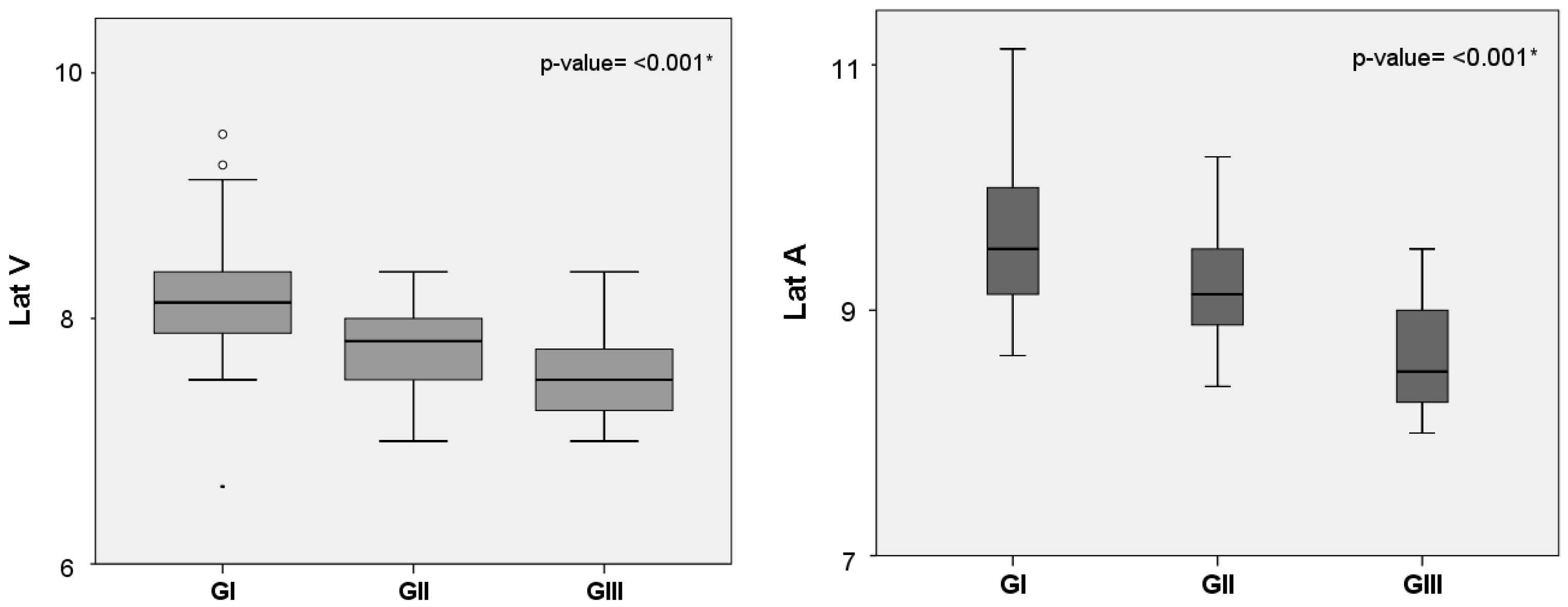
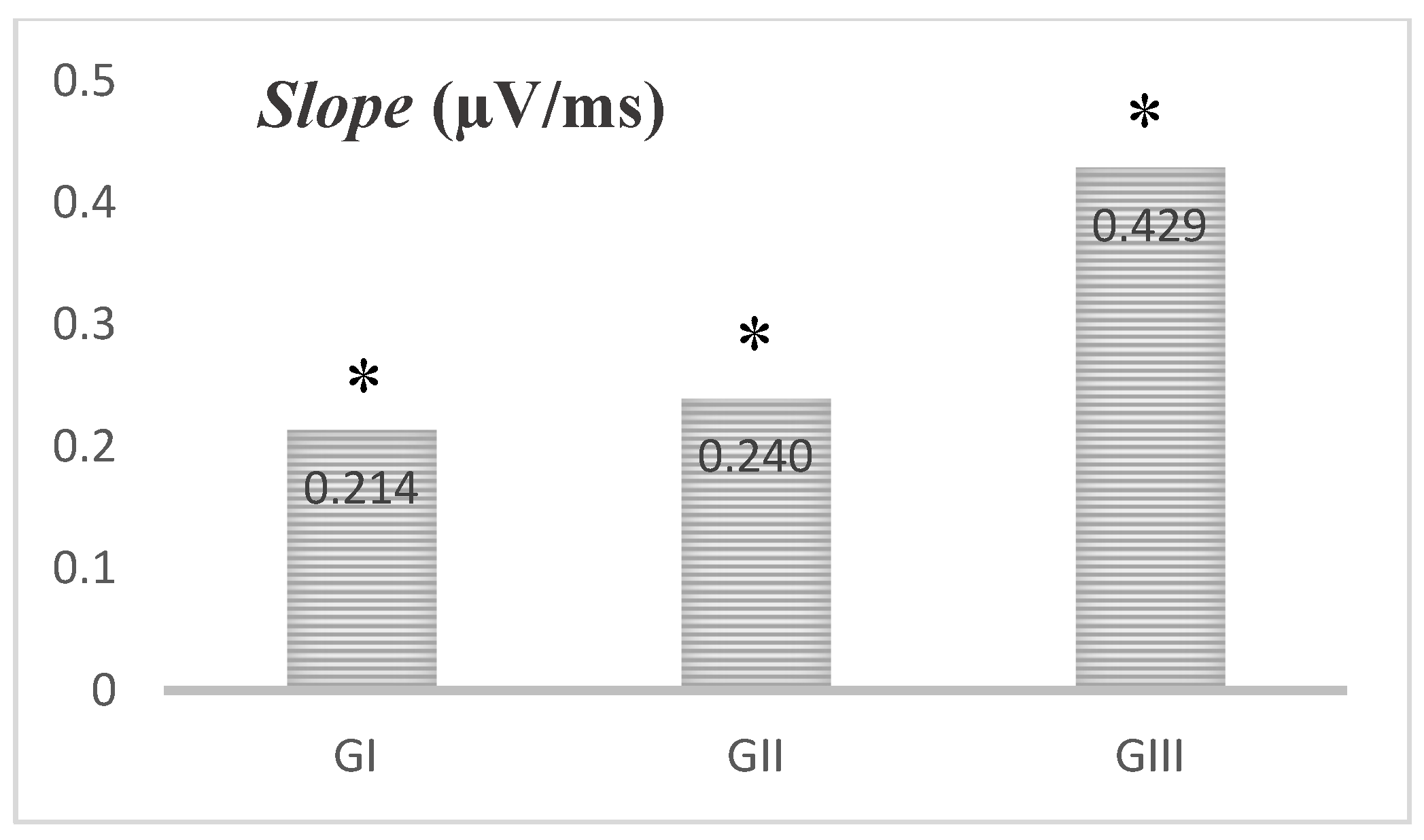
3. Results
4. Discussion
5. Conclusions
6. Patents
Author Contributions
Funding
Institutional Review Board Statement
Informed Consent Statement
Conflicts of Interest
References
- Skoe, E.; Krizman, J.; Anderson, S.; Kraus, N. Stability and plasticity of auditory brainstem function across the lifespan. Cereb. Cortex 2015, 25, 1415–1426. [Google Scholar] [CrossRef]
- Jeng, F.C. Infant and Childhood Development: Intersections between Development and Language Experience. In The Frequency-following Response: A Window into Human Communication; Kraus, N., Samira, A., White-Schwoch, T., Fay, R.R., Popper, A.R., Eds.; Springer International Publishing: Berlin, Germnany, 2017; pp. 17–43. [Google Scholar]
- Holt, L.L.; Lotto, A.J. Speech perception as categorization. Atten. Percept. Psychophys. 2010, 72, 1218–1227. [Google Scholar] [CrossRef] [PubMed]
- Zhao, T.C.; Kuhl, P.K. Linguistic effect on speech perception observed at the brainstem. Proc. Natl. Acad. Sci. USA 2018, 115, 8716–8721. [Google Scholar] [CrossRef] [PubMed] [Green Version]
- Perszyk, D.R.; Waxman, S.R. Infants’ advances in speech perception shape their earliest links between language and cognition. Sci. Rep. 2019, 9, 3293. [Google Scholar] [CrossRef]
- Skoe, E.; Kraus, N. Auditory brainstem response to complex sounds: A tutorial. Ear Hear. 2010, 31, 302–324. [Google Scholar] [CrossRef] [PubMed] [Green Version]
- Kraus, N.; Samira, A.; White-Schwoch, T. The Frequency-Following Response: A Window into Human Communication. In The Frequency-Following Response: A Window into Human Communication; Kraus, N., Samira, A., White-Schwoch, T., Fay, R.R., Popper, A.R., Eds.; Springer International Publishing: Berlin, Germany, 2017; pp. 1–15. [Google Scholar]
- Sanfins, M.D.; Garcia, M.V.; Biaggio, E.P.V.; Skarzynski, P.H. The Frequency Following Response: Evaluations in Different Age Groups; IntechOpen: London, UK, 2019. [Google Scholar] [CrossRef] [Green Version]
- Anderson, S.; Parbery-Clark, A.; White-Schwoch, T.; Kraus, N. Development of subcortical speech representation in human infants. J. Acoust. Soc. Am. 2015, 137, 3346–3355. [Google Scholar] [CrossRef] [Green Version]
- Jeng, F.C.; Schnabel, E.A.; Dickman, B.M.; Hu, J.; Li, X.; Lin, C.D.; Chung, H.K. Early maturation of frequency-following responses to voice pitch in infants with normal hearing. Percept. Mot. Skills 2010, 111, 765–784. [Google Scholar] [CrossRef]
- Jeng, F.C.; Hu, J.; Dickman, B.; Montgomery-Reagan, K.; Tong, M.; Wu, G.; Lin, C.D. Cross-linguistic comparison of frequency-following responses to voice pitch in American and Chinese neonates and adults. Ear Hear. 2011, 32, 699–707. [Google Scholar] [CrossRef] [PubMed]
- Van Dyke, K.B.; Lieberman, R.; Presacco, A.; Anderson, S. Development of phase locking and frequency representation in the infant frequency-following response. J. Speech Lang. Hear. Res. 2017, 60, 2740–2751. [Google Scholar] [CrossRef] [Green Version]
- Sanfins, M.D.; Borges, L.R.; Ubiali, T.; Colella-Santos, M.F. Speech auditory brainstem response (speech ABR) in the differential diagnosis of scholastic difficulties. Braz. J. Otorhinolaryngol. 2017, 83, 112–116. [Google Scholar] [CrossRef] [Green Version]
- Long, P.; Wan, G.; Roberts, M.T.; Corfas, G. Myelin development, plasticity, and pathology in the auditory system. Dev. Neurobiol. 2018, 78, 80–92. [Google Scholar] [CrossRef] [PubMed] [Green Version]
- American Academy of Pediatrics. Joint Committee on Infant Hearing. Position statement: Principles and guidelines for early hearing detection and intervention programs. Pediatrics 2007, 120, 898–921. [Google Scholar] [CrossRef]
- Costa, T.V.S.; Aurélio, F.S.; Silva, V.B.; Rodrigues, L.B. Standardization of the auditory brainstem response in newborns. Rev. CEFAC 2013, 15, 1482–1491. [Google Scholar] [CrossRef] [Green Version]
- Sanfins, M.; Colella-Santos, M. A review of the clinical applicability of speech-evoked auditory brainstem responses. J. Hear Sci. 2016, 6, 9–16. [Google Scholar] [CrossRef]
- Dehaene-Lambertz, G.; Dehaene, S.; Hertz-Pannier, L. Functional neuroimaging of speech perception in infants. Science 2002, 298, 2013–2015. [Google Scholar] [CrossRef] [PubMed] [Green Version]
- Jeng, F.C.; Peris, K.S.; Hu, J. Evaluation of an automated procedure for detecting frequency-following responses in American and Chinese neonates. Percept. Mot. Skills 2013, 116, 456–465. [Google Scholar] [CrossRef]
- Pinto, E.S.M.; Martinelli, M.C. Brainstem auditory evoked potentials with speech stimulus in neonates. Braz. J. Otorhinolaryngol. 2018, 86, 191–200. [Google Scholar] [CrossRef]
- Ribas-Prats, T.; Almeida, L.; Costa-Faidella, J.; Plana, M.; Corral, M.J.; Gómez-Roig, M.D.; Escera, C. The frequency-following response (FFR) to speech stimuli: A normative dataset in healthy newborns. Hear Res. 2019, 371, 28–39. [Google Scholar] [CrossRef]
- Moon, C.; Lagercrantz, H.; Kuhl, P.K. Language experienced in utero affects vowel perception after birth: A two-country study. Acta Paediatr. 2013, 102, 156–160. [Google Scholar] [CrossRef] [Green Version]
- May, L.; Byers-Heinlein, K.; Gervain, J.; Werker, J.F. Language and the newborn brain: Does prenatal language experience shape the neonate neural response to speech? Front. Psychol. 2011, 21, 222. [Google Scholar] [CrossRef] [Green Version]
- Leite, R.A.; Magliaro, F.C.L.; Raimundo, J.C.; Gândara, M.; Garbi, S.; Bento, R.F.; Matas, C.G. Effect of hearing aids use on speech stimulus decoding through speech-evoked ABR. Braz. J. Otorhinolaryngol. 2018, 84, 66–73. [Google Scholar] [CrossRef]
- Toga, A.W.; Thompson, P.M.; Sowell, E.R. Mapping brain maturation. Trends Neurosci. 2006, 29, 148–159. [Google Scholar] [CrossRef] [Green Version]
- Coffey, E.B.; Herholz, S.C.; Chepesiuk, A.M.; Baillet, S.; Zatorre, R.J. Cortical contributions to the auditory frequency-following response revealed by MEG. Nat. Commun. 2016, 24, 11070. [Google Scholar] [CrossRef] [PubMed]
- Sano, M.; Kaga, K.; Kuan, C.C.; Ino, K.; Mima, K. Early myelination patterns in the brainstem auditory nuclei and pathway: MRI evaluation study. Int. J. Pediatr. Otorhinolaryngol. 2007, 71, 1105–1115. [Google Scholar] [CrossRef]
- Bick, J.; Nelson, C.A. Early experience and brain development. Wiley Interdiscip. Rev. Cogn. Sci. 2017, 8, 1–2. [Google Scholar] [CrossRef] [PubMed]
- Kuhl, P.K. Early language acquisition: Cracking the speech code. Nat. Rev. Neurosci. 2004, 5, 831–843. [Google Scholar] [CrossRef]
- Russo, N.; Nicol, T.; Musacchia, G.; Kraus, N. Brainstem responses to speech syllables. Clin. Neurophysiol. 2004, 115, 2021–2030. [Google Scholar] [CrossRef] [PubMed] [Green Version]
- Sano, M.; Kuan, C.C.; Kaga, K.; Itoh, K.; Ino, K.; Mima, K. Early myelination patterns in the central auditory pathway of the higher brain: MRI evaluation study. Int. J. Pediatr. Otorhinolaryngol. 2008, 72, 1479–1486. [Google Scholar] [CrossRef]


| Age (Days) | V | A | C | D | E | F | O | |
|---|---|---|---|---|---|---|---|---|
| Mean | 3 to 15 | 0.14 | −0.16 | −0.09 | −0.12 | −0.19 | −0.14 | −0.16 |
| amplitude | 16 to 30 | 0.16 | −0.15 | −0.08 | −0.10 | −0.16 | −0.13 | −0.15 |
| (µV) | 31 to 45 | 0.22 | −0.23 | −0.09 | −0.13 | −0.19 | −0.19 | −0.20 |
| p-value | 0.001 * | 0.071 | 0.085 | 0.363 | 0.276 | 0.092 | 0.060 | |
Publisher’s Note: MDPI stays neutral with regard to jurisdictional claims in published maps and institutional affiliations. |
© 2021 by the authors. Licensee MDPI, Basel, Switzerland. This article is an open access article distributed under the terms and conditions of the Creative Commons Attribution (CC BY) license (https://creativecommons.org/licenses/by/4.0/).
Share and Cite
Ferreira, L.; Skarzynski, P.H.; Skarzynska, M.B.; Sanfins, M.D.; Biaggio, E.P.V. Effect of Auditory Maturation on the Encoding of a Speech Syllable in the First Days of Life. Brain Sci. 2021, 11, 844. https://doi.org/10.3390/brainsci11070844
Ferreira L, Skarzynski PH, Skarzynska MB, Sanfins MD, Biaggio EPV. Effect of Auditory Maturation on the Encoding of a Speech Syllable in the First Days of Life. Brain Sciences. 2021; 11(7):844. https://doi.org/10.3390/brainsci11070844
Chicago/Turabian StyleFerreira, Laís, Piotr Henryk Skarzynski, Magdalena Beata Skarzynska, Milaine Dominici Sanfins, and Eliara Pinto Vieira Biaggio. 2021. "Effect of Auditory Maturation on the Encoding of a Speech Syllable in the First Days of Life" Brain Sciences 11, no. 7: 844. https://doi.org/10.3390/brainsci11070844