Article

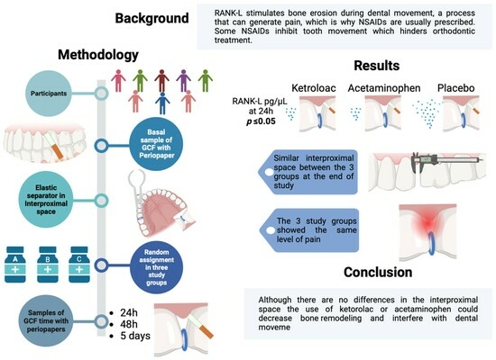
Comparison of the Effects of Ketorolac and Acetaminophen on RANK-L Levels in the Gingival Crevicular Fluid during Orthodontic Tooth Movement: A Pilot Study

by
Ruth Rodríguez-Montaño
1,2,
Yesenia Isahy Ponce-Gómez
3,
Sarah Monserrat Lomelí-Martínez
3,4,
Sonia Sifuentes-Franco
5,
Alondra del Carmen Ruiz-Gutiérrez
3,
Rubén Alberto Bayardo-González
6,
Vianeth María del Carmen Martínez-Rodríguez
3,
José Luis Meléndez-Ruíz
7 and
Juan Ramón Gómez-Sandoval
2,3,6,*
1
Department of Health and Illness as an Individual and Collective Process, University Center of Tlajomulco, University of Guadalajara (CUTLAJO-UdeG), Tlajomulco, Santa Fé Highway Km. 3.5 No.595 Lomas de Tejeda, Tlajomulco de Zuniga 45641, Jalisco, Mexico
2
Research Institute of Dentistry, Department of Integrated Dentistry Clinics, University of Guadalajara (CUCS-UdeG), 950 Sierra Mojada, Guadalajara 44340, Jalisco, Mexico
3
Periodontics Program, Department of Integrated Dentistry Clinics, University of Guadalajara (CUCS-UdeG), 950 Sierra Mojada, Guadalajara 44340, Jalisco, Mexico
4
Department of Medical and Life Sciences, University of Guadalajara (CUCiénega-UdeG), 1115 Ave. Universidad, Ocotlán 47810, Jalisco, Mexico
5
Department of Health Sciences, Centro Universitario de los Valles, University of Guadalajara (CUValles-UdeG), Highway to Guadalajara Km. 45.5, Ameca 46600, Jalisco, Mexico
6
Department of Integrated Dentistry Clinics, University of Guadalajara (CUCS-UdeG), 950 Sierra Mojada, Guadalajara 44340, Jalisco, Mexico
7
Ortodontics Program, Department of Integrated Dentistry Clinics, University of Guadalajara (CUCS-UdeG), 950 Sierra Mojada, Guadalajara 44340, Jalisco, Mexico
*
Author to whom correspondence should be addressed.
Appl. Sci. 2024, 14(4), 1464; https://doi.org/10.3390/app14041464
Submission received: 3 January 2024 / Revised: 4 February 2024 / Accepted: 8 February 2024 / Published: 10 February 2024

Abstract

:
Background: Patients usually present pain due to the release of different inflammatory mediators such as prostaglandin E2 and RANK-L. Analgesics such as acetaminophen and ketorolac can inhibit RANK-L expression and this can affect orthodontic treatment by decreasing bone remodeling and slowing orthodontic dental movement. Several studies have reported a decrease in dental movement after administering some non-steroidal anti-inflammatory drugs. Proposal: The objective was to evaluate the RANK-L levels and a possible modulation by administering acetaminophen and ketorolac in patients starting orthodontic treatment. Methodology: A double-blind, randomized clinical trial was carried out with 24 subjects divided into three study groups: calcined magnesia as a placebo, acetaminophen, and ketorolac. Gingival crevicular fluid was obtained at four time points: before pharmacological intervention, at 24 h, at 48 h, and on the 5th day. RANK-L concentrations were evaluated through ELISA analysis. Also, interproximal space generated by the elastic separator at the end of the study was recorded in the different study groups using the visual analog scale. Results: An increase in RANK-L at 24 h was observed in the placebo group compared to the ketorolac and acetaminophen groups. However, no significant differences were observed in the interproximal space at day 5 in the three study groups. Conclusion: Patients who do not take analgesics at the start of orthodontic treatment have higher levels of RANK-L. Therefore, the use of ketorolac or acetaminophen could decrease bone remodeling and interfere with orthodontic dental movement.

Graphical Abstract

1. Introduction

The World Health Organization (WHO) considers malocclusion one of the most important oral health problems. Malocclusion has repercussions on the quality of life associated with oral health. It ranks as the third most widespread dental issue in oral health; first is caries and second is periodontitis [1]. Malocclusion is a developmental condition marked by irregular associations between the alignment of teeth and the structure of dental arches [2]. Severe malocclusion can lead to both physiological and psychological effects, including impairments in chewing function, speech articulation, and aesthetic appearance. This can have a lasting negative impact on self-esteem and social interactions over time [3].
The origin of malocclusion is complex and influenced by a combination of genetic and environmental factors. Among these, the skeletal pattern, determined by genetic factors, is considered the primary contributor to the development of malocclusion [4]. However, the importance of certain environmental factors has been emphasized, including atypical swallowing, mouth breathing, and deleterious oral habits (such as thumb sucking, nail biting, or prolonged use of a baby bottle), which can disrupt the natural balance and development of the dental matrix, leading to malocclusion [4,5]. The treatment for malocclusion is orthodontics, which seeks to achieve aesthetic and functional improvement through mechanical therapy that moves the teeth to a more ideal position [5]. The displacement of teeth during orthodontic treatment is triggered by mechanical stimuli and is facilitated by changes in the periodontal ligament (PDL) and alveolar bone. Periodontal tissues surrounding the tooth root, such as the PDL, alveolar bone, and gums, undergo impact due to orthodontic force resulting from mechanical stress. The periodontal ligament (PDL) is a versatile fibrous tissue responsible for connecting the cement covering the tooth root to the alveolar bone, functioning as the primary, and a highly sensitive, receptor. The PDL detects mechanical stimuli induced by the application of orthodontic force [6,7,8,9].
In a more detailed context, when force is exerted on a tooth, osteoclastic activity is triggered on the pressure side. Simultaneously, the multiplication and differentiation of periodontal ligament (PDL) fibroblasts and mesenchymal stem cells (MSCs) lead to increased bone production by osteoblasts on the tension side. Furthermore, mechanical force promptly initiates various cell signaling pathways in osteoblasts, including calcium (Ca2+), nitric oxide (NO), interleukin-1 (IL-1), and adenosine triphosphate (ATP). The process of orthodontic tooth movement (OTM) results in the release of mediators such as NO and IL. Fluid shear stress can activate the Ca2+ signaling system, stimulating ATP release, prostaglandin E2 (PGE2) secretion, and osteoblast development. On the compression side, localized hypoxia and a decrease in blood flow in the PDL occur simultaneously. Hypoxia-inducible factor 1 (HIF-1), a transcription factor, activates the expression of Vascular Endothelial Growth Factor (VEGF) and receptor activator of nuclear factor-kappa ligand (RANK-L) in PDL fibroblasts and osteoblasts, triggering osteoclast differentiation and inducing bone resorption in the compression areas. The relationship between bone resorption and formation becomes evident during the remodeling of the alveolar bone surrounding the tooth’s root during orthodontic movement [10,11].
The receptor activator of nuclear factor-kappa B (RANK), RANK ligand (RANK-L), and osteoprotegerin (OPG) systems play a pivotal role in orchestrating bone remodeling. This intricate process entails the involvement of the tumor necrosis factor (TNF)-related ligand RANK-L and its two receptors, RANK and OPG [12,13]. More specifically, RANK-L controls the development and activation of osteoclasts. When RANK-L binds to the RANK receptor on osteoclast lineage cells, osteoclast precursors express RANK, a receptor for RANK ligand (RANK-L). Through cell–cell interactions, these precursors recognize RANK-L expressed by osteoblasts and, in the presence of macrophage colony-stimulating factor (M-CSF), undergo differentiation into osteoclasts. This binding facilitates the rapid differentiation of hematopoietic osteoclast precursors into mature osteoclasts. Mature osteoclasts, in turn, express both RANK and RANK-L, promoting their survival and stimulating bone-resorbing activity. On the other hand, osteoprotegerin (OPG), a soluble RANK-L decoy receptor predominantly produced by osteoblasts, acts to prevent osteoclast formation and bone resorption by inhibiting the interaction between RANK and RANK-L [12,13]. OPG exerts biological effects on bone cells, including the inhibition of osteoclastic differentiation, the suppression of activation of matrix osteoclasts, and the induction of apoptosis. The process of bone remodeling during orthodontic tooth movement is regulated by maintaining a balance between the binding of RANK to RANK-L and the production of OPG [12,13,14]. Mature osteoclasts release exosomes expressing RANK on their surface, establishing a binding interaction with RANK-L in osteoblasts and triggering reverse signaling. This, in turn, activates the PI3K–Akt pathway, increasing osteoblast activity. At the same time, OPG produced by osteoblasts acts as a regulator, preventing bone resorption by binding to RANK-L expressed by osteoblasts. This interaction maintains the balance between bone resorption and formation in the dynamic process of bone remodeling [15].
Some researchers have established that between 80 and 95% of patients may present some grade of pain during orthodontic procedures. The experience of pain significantly contributes to interruptions or premature discontinuations in treatment [16]. This sensory and emotional experience, often underestimated by orthodontists, not only makes orthodontic treatment difficult but it also has detrimental effects on physical and mental health [17].
In orthodontic treatment, both modeling and bone remodeling are determinants for the rate of OTM. Bone modeling during OTM is an inflammatory process, and the rate-limiting factor of dental movement is bone resorption at the bone–periodontal ligament interface [18]. Both the vascular changes generated by OTM and the release of inflammatory mediators, such as histamine, prostaglandins, leukotrienes, and cytokines, cause pain during treatment [19]. Several methods have been proposed to control pain caused by orthodontic treatment, such as administration of analgesics, vibratory stimulation, local anesthesia, chewing a “bite wafer” or gum, and, more recently, the use of low-level lasers for oral therapy [17,20]. Treatment with nonsteroidal anti-inflammatory analgesics (NSAIDs), such as ketorolac, ibuprofen, naproxen, and COX3 inhibitors such as acetaminophen, are the most commonly used analgesics to control pain caused by orthodontic dental movement [17,21].
Regarding drugs to inhibit or relieve pain during orthodontic treatment, such as acetaminophen and ketorolac, it is known that they can inhibit the expression of RANK-L, which can affect OTM and thus inhibit bone remodeling [22]. Currently, several studies have evaluated the effect of different analgesics such as ibuprofen, acetaminophen, and celecoxib on OTM [22,23,24]. However, the effect of ketorolac on RANK-L has not been evaluated, so further clinical studies are essential to determine the effect of this drug on bone resorption and, consequently, on OTM, so that the orthodontist can choose and recommend the best analgesic. Therefore, the present study aimed to compare the effects of ketorolac and acetaminophen on RANK-L during OTM.

2. Materials and Methodology

A double-blind clinical trial with randomized allocation and a placebo control group was performed. This study was approved by the Research Ethics Committee of the Center for Health Sciences of the University of Guadalajara. It was also registered in Clinical Trials under accession number, CI04820. Twenty-four patients who started orthodontic treatment at the Specialty of Orthodontics of the Integral Dental Clinics of the University Center of Health Sciences were included.

2.1. Selection Criteria

Inclusion criteria
Patients between 18 and 27 years old, who would initiate their orthodontic treatment exclusively at the University of Guadalajara’s Orthodontic Specialty.
Patients with oral health and who were systematically healthy.
Patients with the same molar class on both the right and left sides and require elastic separators in at least one quadrant before starting their treatment.
Patients also willing to participate in the study and committed to attending their scheduled sampling appointments for sample collection.
No-inclusion criteria
Patients who had previously received orthodontic treatment.
Patients who were allergic to ketorolac or acetaminophen.
Patients who were under drug treatment and/or used contraceptives.
Pregnant patients or breastfeeding women, as well as those who consumed alcohol or tobacco.
Exclusion criteria
Patients who chose to withdraw their consent.
Patients who did not attend their appointments for sample collection.
Patients who became ill during the study days, and required medication.
Patients who mentioned not having taken their medication according to the instructions.

2.2. Study Groups

The individuals included in the research project underwent the data collection questionnaire. Once they signed the consent form, they were included in the study.
The groups were formed by simple random assignment, where each participant was assigned according to the double-blind code with random numbers obtained through a random number generator (Excel v 19.0 Microsoft Office).
Group 1: Control placebo (calcined magnesia) 500 mg capsules (1.5 g daily), one capsule every 8 h for 5 days.
Group 2: Ketorolac 10 mg capsules, one capsule every 8 h (30 mg daily) for 5 days.
Group 3: Acetaminophen 500 mg capsules, one capsule every 8 h (1.5 g daily) for 5 days. The placebo and drugs of the three study groups were homologated in capsules of the same size, weight, and labeling, only identified with the letters A, B, and C for blinding.

2.3. Determination of Clinical and Demographic Variables

Gender, age, and molar class were identified through medical records and oral clinical examination to evaluate the molar class.
Each patient was provided with a booklet containing comprehensive instructions for administering the treatment, accompanied by a calendar outlining the experiment’s schedule. Patients documented the symptoms they experienced each day of the study, as well as their pain levels, through the visual analog scale (VAS), which was depicted in the same booklet with a scale ranging from 0 to 10 with an explanation that the value closest to 0 indicates the absence of pain, while a value closer to 10 indicates the presence of severe pain. Likewise, they were instructed to assign the value they deemed representative of the degree of pain they were experiencing. At the same time, a follow-up diary was provided to identify adverse effects produced by the administered drugs such as headaches, nausea, vomiting, constipation, or diarrhea.
Regarding the interproximal space (IP), it was measured using graduated acetate strips. These strips were placed in the interproximal space created by the ligature, and the number of strips that fit into the interproximal space was recorded. This measurement was carried out five days after the study, following the removal of the elastic band.
For the assessment of molar class, the anteroposterior relationship between the upper and lower permanent first molars, both on the right and left sides, was examined. Tracing paper was utilized to identify areas of occlusion with increased pressure. A Class I molar relationship is designated when the mesiobuccal cusp of the upper first molar occludes with the buccal groove of the lower first molar. In cases where the mesiobuccal cusp of the upper first molar occludes anterior to the buccal groove of the lower first molar, it is classified as a Class II molar relationship. A Class III molar relationship is identified when the mesiobuccal cusp of the upper first molar occludes more posteriorly than the buccal groove of the lower first molar.

2.4. Sample Collection

The GCF sample was taken using Periopaper (Oraflow, New York, NY, USA). Three strips were taken per site at the right upper first molar mesial zone for each sampling time (basal, 24 h, 48 h, and 5 days).
The participants were requested not to brush their teeth before sample collection to prevent bleeding and sample contamination. Excess saliva, food, or dental plaque was removed using sterile gauze. The area was isolated with cotton rolls to avoid saliva contamination and the GCF was collected by inserting the Periopaper 1 mm into the gingival crevicular sulcus for 30 s.
A basal GCF sample was taken before placement of the elastic separator (ortho technology), and then, at 24 h (T1), 48 h (T2), and 5 days (T3), GCF samples were taken again. The GCF samples were stored in Eppendorf tubes at −80 °C.
To determine the amount of GCF obtained, a standard curve was created in duplicate where the weight in mg of the Periopaper strips was recorded by placing different amounts of distilled water. It is worth mentioning that Periopaper strips can absorb up to 2 µL.
The standard curve consisted of measurements in mg of wet Periopaper strips with 0.5 µL, 1 µL, 1.5 µL, and 2 µL of distilled water and a dry Periopaper that corresponds to a value of 0 µL of distilled water.
Subsequently, the r value was analyzed to confirm the quality of the curve, ensuring it was greater than r = 0.98. Subsequently, the GCF sample collected in each Periopaper strip was weighed with an analytical balance. Then, the Periopapers’ weight was correlated with a standard curve to find the µL of GCF collected at every sample time. It is important to know that the standard curve was created every day when a GCF sample was taken.
Since three Periopaper strips were obtained in each sampling, to determine the amount of GCF obtained, the amount of gingival crevicular fluid collected from the three Periopapers strip was recorded.

2.5. Sample Preparation

The Periopaper strips were subjected to elution to extract the adsorbed GCF. The three Periopaper strips used in each sample collection were placed in an Eppendorf tube with 300 µL of PBS buffer added, and gently shaken for 15 min at 190 rpm.
Following the agitation, the Periopaper strips were secured using the Eppendorf tube cap to keep them separated from the PBS. Subsequently, they were centrifuged for 10 min at 12 rpm. The centrifugal force extracted the remaining GCF from the Periopaper strips, mixing it with the rest of the PBS at the bottom of the Eppendorf tube, leaving them dry.
The strips were carefully removed from the Eppendorf tubes to avoid re-soaking them with GCF diluted in PBS. Finally, the supernatant was stored at −80 °C until the ELISA technique was performed.

2.6. Enzyme-Linked Immunosorbent Assay ELISA

Once all samples were obtained, RANK-L levels were determined with the “Human sRANK Ligand Standard ABTS” enzyme-linked immunosorbent assay ELISA Development Kit from PreproTech. The absorbance was read using a “Powerman” spectrophotometer at 450 nm. Quantification of RANK-L in the samples was achieved by comparison with a standard curve generated from known amounts of the RANK-L standard provided by the ELISA kit.
Procedure: The capture antibody was placed in the 96 wells of the plate and allowed to incubate overnight. Next, the experiment proceeded according to the instructions. To each well, 100 µL of the LCG sample and the standard curve provided by the kit were added, and it was left to incubate for two hours.
After incubation, washes were performed, and the detection antibody was added and incubated. Subsequently, HRP-conjugated antibody was added, followed by the substrate to generate color. Readings were taken on the spectrophotometer every 5 min for 20 min until absorbance values for the highest (1.200) and lowest (0.200) concentrations of the standard curve were obtained, as indicated by the kit.

2.7. Statistical Analysis

Once the concentrations of RANK-L obtained through ELISA were known, the quantification was performed to determine how many pg/mL of RANK-L was present in 1 μL of GCF, taking into account the dilution in PBS carried out to recover the GCF from the Periopaper strips.
Normality was assessed through the Shapiro–Wilk test and, subsequently, for the identification of differences in RANK-L concentrations in the three study groups at each time point of sampling, the Mann–Whitney U test was performed.
An X2 test was performed for gender and molar class variables. Likewise, a Spearman test was performed to identify correlations between clinical characteristics and RANK-L levels. A p ≤ 0.05 was considered as statistical difference. The statistical analysis was carried out with the software SPSS v. 25.

3. Results

3.1. Clinical and Sociodemographic Data

In sociodemographic data, we can observe that women predominated in the control and ketorolac groups; however, there were no significant differences. In terms of age, the three study groups remained uniform.
Although there were no significant differences in the distribution of molar class in the three study groups, we can observe that Class I molars predominated over Class II molars.
Regarding pain, we observed that the group with the most pain was the ketorolac group and the group with the least pain was the acetaminophen group. Finally, we did not observe a significant difference between the interproximal space obtained in each study group (Table 1).

3.2. RANK-L Levels at the Four Sampling Times

Only in the control group was there a trend of increasing RANK-L levels on the fifth day of the study. However, no significant intra-group differences were observed among the four sampling times (Figure 1).

3.3. RANK-L Levels between Study Groups

The basal levels of RANK-L in the three study groups did not show a significant difference, indicating that participants did not exhibit any alterations that would modify RANK-L levels. This analysis allows for a more equitable comparison of RANK-L levels expressed in the LCG samples taken after the placement of the elastic separator.
On the other hand, 24 h after the placement of the elastic separator, a trend toward increased RANK-L concentrations was observed in the control group versus the acetaminophen and ketorolac groups; this suggests that the administration of these NSAIDs does indeed decrease the expression of RANK (Table 2).
It is worth mentioning that on the fifth day, the RANK-L levels in the ketorolac groups remained lower than in the control and acetaminophen groups; however, there was no significant difference (Figure 2).

3.4. Correlation of the Different Study Variables

A Spearman correlation was performed between RANK-L concentrations, age, interproximal space, and pain (with the VAS scale) at the different times of sampling and a positive correlation was observed between RANK-L at 24 h (T1) and 48 h (T2) (r = 0.467, p = 0.025), as well as a positive correlation between RANK-L at 48 h (T2) and 5 days (T3) (r = 0.600, p = 0.002). In contrast, a negative correlation was identified between the interproximal space and the VAS pain scale (r = −0.477 p= 0.019) (Figure 3).

4. Discussion

Malocclusion and dentofacial deformity are moderate distortions of development. Several morphological and functional factors can lead to malocclusion, which must be considered when planning orthodontic treatment. The orthodontic approach should also encompass a wide range of psychosocial and bioethical considerations to uphold dental and alveolar bone health, establish a favorable maxillomandibular bone relationship, prevent temporomandibular joint issues, and ensure the harmonious and visually pleasing appearance of both the teeth and face. Furthermore, maintaining the dentition within the expected limits of physiological relapse is crucial [25].
When it is necessary to place orthodontic metal bands on the molars during orthodontic treatment, the first step in these cases is the placement of an elastic separator to create enough space to be able to adapt the orthodontic metal bands. This procedure causes different grades of pain in patients; moreover, it can be compared to the force applied during orthodontic treatment where compression of the periodontal ligament occurs [17,26]. The separator during the first hours after placement prevents vascular circulation and thus cell differentiation begins [27].
OTM is considered to involve an inflammatory process [28]. RANK-L is a proinflammatory molecule produced mainly by osteoblasts and T lymphocytes. Its main function is to regulate preosteoclast fusion, which leads to osteoclastogenesis [29]. Some authors have shown that methods to control pain caused by orthodontic treatment, including mechanical vibration, low-level laser therapy, and chewing gum, have no clinically significant analgesic effects [20,30].
The use of pain-relieving medications, including both prescription and over-the-counter formulations, is widespread. During orthodontic treatment, substances such as acetaminophen, ibuprofen, piroxicam, and ketorolac, among others, are often consumed for a few days to alleviate the discomfort associated with specific procedures, such as the placement of separators, arch changes, and appliance activation [17,28,31,32,33,34,35,36,37]. Further clinical studies are needed to associate the effect of drugs on dental movement and bone resorption [28,38].
The “classic” NSAIDs, including derivatives of propionic acid and acetic acid (including ketorolac), inhibit both COX-1 and COX-2 [39,40]. Prostaglandins synthesized by both enzymatic isoforms not only contribute to various physiological processes, such as the regulation of vascular tone and platelet function, but also play a role in pathological processes such as inflammation [35,36,37,38,39,40,41]. As a result, in this study, the hypothesis is raised that ketorolac could potentially exert an influence on the production of RANK-L to modify the course of dental movement. However, little is known about its effect in terms of OTM and bone resorption at the molecular level.
Acetaminophen is known to not interfere with OTM [42]. It is worth mentioning that some authors no longer consider acetaminophen an NSAID. Since its analgesic action has been demonstrated to not be related to the inhibition of prostaglandin synthesis in the periphery, as this is very weak [28,43,44,45,46], Chandrasekharan et al. in 2002 identified a third catalytically functional COX enzyme, which was named COX-3. This variant was less sensitive to NSAIDs and selective COX-2 inhibitors but sensitive to acetaminophen. Acetaminophen showed selectivity for inhibiting COX-3 over COX-1 and COX-2, suggesting competitive blocking at the active site [47].
This analgesic is considered to act in the central nervous system and not through cell membranes [48], so the inhibition of prostaglandins is minimal. Therefore, it is believed that the use of acetaminophen has no effect on dental movement speed [49].
Some information is available on molecular RANK-L expression following analgesic administration in humans. Among the limited studies, Shetty et al. compared the effects of ibuprofen and acetaminophen on PGE2 levels in gingival crevicular fluid (GCF) during orthodontic movement. The conclusion suggested that acetaminophen has minimal impact on PGE2 levels, establishing it as a safe and preferred drug for pain relief in patients undergoing orthodontic treatment [28]. According to the results of the present study, a significant increase in RANK-L concentration was observed in the control group, suggesting that ketorolac and acetaminophen decrease RANK-L concentrations.
Pain control during the first treatment sessions can increase patient motivation and cooperation [50]. Recent research mentions that there is a rapid release of biochemical markers that cause pain in the first 2 h after the placement of an elastic separator or orthodontic appliance, reaching their maximum expression at 24 h [28,51]. These results support the findings of the present study since there was a higher release of RANK-L in the control group at 24 h. Furthermore, in 2006, Nishijima et al. investigated the dynamics of RANK-L and OPG levels in GCF during canine retraction without administering drugs and in an acetaminophen group. They included both in vivo and in vitro analysis using ELISA to quantify the cytokines. The findings suggest that changes in RANK-L and OPG are involved in bone resorption in response to compression force, which may have implications for the regulation of tooth movement in orthodontic treatments [27]. These results coincide with the present study: RANK-L expression was increased at 24 h in the control and acetaminophen groups.
As mentioned above, the age range of the patients included in this study was 18 to 27 years; these are considered young patients. In 2006, Kawasaki et al. conducted a comparative study on the levels of RANK-L and OPG in the GCF during OTM in juvenile and adult patients. The study involved 15 juveniles and 15 adults, with the collection of gingival crevicular fluid (GCF) samples at different time points after the application of a retraction force. Findings indicated that tooth movement was greater in juveniles than in adults after 168 h, similar to the present study. Moreover, there was an increase in RANK-L levels and a decrease in OPG levels in the GCF of both groups. The RANKL/OPG ratio was lower in adult patients compared to juveniles, suggesting that the age-related decrease in tooth movement might be linked to a reduction in the RANK-L/OPG ratio in the GCF during the initial stages of OTM [52].
The results of the present work indicate that dental movement measured through the space generated between the premolar and the upper molar in the different groups was not significant. On the contrary, Tripathi et al. compared separation and associated pain. This study concludes that the larger the interdental space, the greater the pain at 24 h [53]. This differs from the present study, in which we obtained a negative correlation between space and pain, indicating that the larger the interproximal space, the less pain the patient experiences. In addition to this, Davidovich et al. showed that the gap is created after 24 h of stimulation and that it only needs to be 0.16 mm, as this is the standard orthodontic band size [53]. However, the average spacing obtained in the present study was 0.28 mm. It is known that only 0.025 mm is required for band placement [54].
There is no correlation between the amount of RANK-L and the space obtained, which differs from what was expected. Nishijima and Kawasaki suggest that changes in RANK-L are involved in bone resorption in response to compression force, which may have implications for the regulation of tooth movement in orthodontic treatments [27,52]. In the present study, pain begins 4 h after spacer placement, gradually increases, peaks the day after spacer placement, and then subsides [55]. The grades of pain experienced in orthodontic treatment depend on the type of dental movement involved and, particularly, on the pain threshold of each individual [49]. In the present study, there was no significant difference in pain during chewing, which contradicts the literature, revealing that pain is usually aggravated during chewing, forcing the patient to use analgesics [56].
Regarding the adverse effects of the administered drugs, there were no significant differences in the manifestation of heartburn, headaches, and pain when chewing. Acetaminophen can be considered the treatment of choice because it does not cause gastrointestinal ulcers and does not affect the amount of dental movement [57]. This is consistent with our results, as no patients reported heartburn while using this medication. We attributed this finding to the lack of statistically significant differences in the number of patients evaluated, and the level, duration, and direction of the applied force, which may not have been optimal. Despite the cautions taken in sample collection, processing, and storage, the possibility of sample degradation cannot be ruled out.
When prescribing analgesics for pain relief in patients after the activation of orthodontic appliances, we must take into account the side effects and the influence they may have on dental movement.
The implications of this study are significant for orthodontic practice, as they suggest that pain management with certain analgesics can influence bone remodeling, a critical factor for effective tooth movement during orthodontic treatment. This finding is pivotal as it may lead to a re-evaluation of pain management strategies in orthodontics to avoid compromising treatment efficacy. However, the limitations, including the small sample size and the nature of it being a pilot study, indicate that the results are preliminary. Future research should take several directions. Firstly, there is a highlighted need for more extensive studies to confirm and expand upon these findings, focusing on how different doses and types of analgesics specifically impact bone remodeling and dental movement; this would provide clearer guidance for pain management in orthodontic patients.
Additionally, further research into the influence of analgesics on dental movement and bone resorption at the molecular level is suggested; such research is crucial for a better understanding of pain management in orthodontics and its impact on the effectiveness of orthodontic treatment.

5. Conclusions

In the present study, we observed a significant difference in RANK-L levels at 24 h, suggesting that the administration of ketorolac may influence dental movement. Similarly, the amount of interproximal space formed is directly proportional to the perceived pain. However, it is important to note that this study is preliminary, and further research with a larger sample size is needed to recommend to orthodontists the optimal analgesic that provides pain control without affecting the orthodontic dental movement time.

Author Contributions

Conceptualization, J.L.M.-R. and J.R.G.-S.; methodology, S.M.L.-M., S.S.-F., R.R.-M., V.M.d.C.M.-R., Y.I.P.-G. and J.R.G.-S.; software, R.A.B.-G.; formal analysis, J.R.G.-S.; investigation, R.R.-M., Y.I.P.-G., A.d.C.R.-G. and J.L.M.-R.; writing—original draft, R.R.-M., S.M.L.-M., V.M.d.C.M.-R. and J.R.G.-S.; writing—review and editing, J.R.G.-S. and R.R.-M.; supervision, J.R.G.-S.; project administration, R.A.B.-G. and J.L.M.-R. All authors have read and agreed to the published version of the manuscript.

Funding

This research did not receive any external funding.

Institutional Review Board Statement

The study adhered to the principles of the Declaration of Helsinki and received approval from the Ethics and Research Committees of Guadalajara University. Additionally, it complied with the Regulations of the General Health Law, obtaining the approval number (CI04820) August 2020, for studies involving human subjects.

Informed Consent Statement

All participants involved in the study provided informed consent.

Data Availability Statement

The data supporting the conclusions of this study are accessible from the corresponding author upon reasonable request. The data are not publicly available due to privacy.

Acknowledgments

This work was supported by the University of Guadalajara’s Program for the Improvement of Production Conditions of SNII Members (PROSNI). Special thanks to orthodontists María Concepción Mendoza Palomino and Iveth Rabago Loza.

Conflicts of Interest

The authors declare no conflicts of interest.

References

  1. Dos Santos, R.R.; Nayme, J.G.; Garbin, A.J.; Saliba, N.; Garbin, C.A.; Moimaz, S.A. Prevalence of malocclusion and related oral habits in 5-to 6-year-old children. Oral. Health Prev. Dent. 2012, 10, 311–318. [Google Scholar]
  2. Lombardo, G.; Vena, F.; Negri, P.; Pagano, S.; Barilotti, C.; Paglia, L.; Colombo, S.; Orso, M.; Cianetti, S. Worldwide prevalence of malocclusion in the different stages of dentition: A systematic review and meta-analysis. Eur. J. Paediatr. Dent. 2020, 21, 115–122. [Google Scholar] [CrossRef]
  3. Amr-Rey, O.; Sanchez-Delgado, P.; Salvador-Palmer, R.; Cibrian, R.; Paredes-Gallardo, V. Association between malocclusion and articulation of phonemes in early childhood. Angle Orthod. 2022, 92, 505–511. [Google Scholar] [CrossRef]
  4. Santana, L.G.; Flores-Mir, C.; Iglesias-Linares, A.; Pithon, M.M.; Marques, L.S. Influence of heritability on occlusal traits: A systematic review of studies in twins. Prog. Orthod. 2020, 21, 29. [Google Scholar] [CrossRef]
  5. Thomas, M. Orthodontics in the “Art” of Aesthetics. Int. J. Orthod. 2015, 26, 23–28. [Google Scholar]
  6. Davidovitch, Z. Tooth movement. Crit. Rev. Oral. Biol. Med. 1991, 24, 411–450. [Google Scholar] [CrossRef]
  7. Lekic, P.; McCulloch, C.A. Periodontal ligament cell population: The central role of fibroblasts in creating a unique tissue. Anat. Rec. 1996, 2, 327–341. [Google Scholar] [CrossRef]
  8. Terai, K.; Takano-Yamamoto, T.; Ohba, Y. Role of osteopontin in bone remodeling caused by mechanical stress. J. Bone Miner. Res. 1999, 6, 839–849. [Google Scholar] [CrossRef]
  9. Pavlin, D.; Gluhak-Heinrich, J. Effect of mechanical loading on periodontal cells. Crit. Rev. Oral. Biol. Med. 2001, 5, 414–424. [Google Scholar] [CrossRef]
  10. Wise, G.E.; King, G.J. Mechanisms of tooth eruption and orthodontic tooth movement. J. Dent. Res. 2008, 5, 414–434. [Google Scholar] [CrossRef]
  11. Yan, Y.; Sun, H.; Gong, Y. Mechanical strain promotes osteoblastic differentiation through integrin-β1-mediated β-catenin signaling. Int. J. Mol. Med. 2016, 2, 594–600. [Google Scholar] [CrossRef]
  12. Davidovitch, Z.; Nicolay, O.F.; Ngan, P.W.; Shanfeld, J.L. Neurotransmitters, cytokines, and the control of alveolar bone remodeling in orthodontics. Dent. Clin. N. Am. 1988, 32, 411–435. [Google Scholar] [CrossRef]
  13. Krishnan, V.; Davidovitch, Z. Cellular, molecular, and tissue-level reactions to orthodontic force. Am. J. Orthod. Dentofac. Orthop. 2006, 129, 469.e1–469.e32. [Google Scholar] [CrossRef]
  14. Alhashimi, N.; Frithiof, L.; Brudvik, P.; Bakhiet, M. Orthodontic tooth movement and de novo synthesis of proinflammatory cytokines. Am. J. Orthod. Dentofac. Orthop. 2001, 119, 307–312. [Google Scholar] [CrossRef]
  15. Udagawa, N.; Koide, M.; Nakamura, M.; Nakamichi, Y.; Yamashita, T.; Uehara, S.; Kobayashi, Y.; Furuya, Y.; Yasuda, H.; Fukuda, C.; et al. Osteoclast differentiation by RANKL and OPG signaling pathways. J. Bone Miner. Metab. 2021, 39, 19–26. [Google Scholar] [CrossRef]
  16. Monk, A.B.; Harrison, J.E.; Worthington, H.V.; Teague, A.; Ertan Erdinç, A.M.; Dinçer, B.; Zhang, F.; Li, F.; Yang, H.; Jin, Y.; et al. Perception of Pain during Orthodontic Treatment with Fixed Appliances. Cochrane Database Syst. Rev. 2022, 26, 79–85. [Google Scholar]
  17. Monk, A.B.; Harrison, J.E.; Worthington, H.V.; Teague, A. Pharmacological interventions for pain relief during orthodontic treatment. Cochrane Database Syst. Rev. 2017, 11, CD003976. [Google Scholar] [CrossRef]
  18. Huang, H.; Williams, R.C.; Kyrkanides, S. Accelerated orthodontic tooth movement: Molecular mechanisms. Am. J. Orthod. Dentofacial Orthop. 2014, 146, 620–632. [Google Scholar] [CrossRef]
  19. Wang, Y.H.; Angkasekwinai, P.; Lu, N.; Voo, K.S.; Arima, K.; Hanabuchi, S.; Hippe, A.; Corrigan, C.J.; Dong, C.; Homey, B.; et al. IL-25 augments type 2 immune responses by enhancing the expansion and functions of TSLP-DC-activated Th2 memory cells. J. Exp. Med. 2007, 204, 1837–1847. [Google Scholar] [CrossRef]
  20. Çelebi, F. Mechanical Vibration and Chewing Gum Methods in Orthodontic Pain Relief. Turk. J. Orthod. 2022, 35, 133–138. [Google Scholar] [CrossRef]
  21. Cheng, C.; Xie, T.; Wang, J. The efficacy of analgesics in controlling orthodontic pain: A systematic review and meta-analysis. BMC Oral. Health 2020, 20, 259. [Google Scholar] [CrossRef]
  22. Meier, M.; Bauer, K.; Proff, P.; Fanghänel, J. Meloxicam medication reduces orthodontically induced dental root resorption and tooth movement velocity: A combined in vivo and in vitro study of dental-periodontal cells and tissue. Cell Tissue Res. 2017, 368, 61–78. [Google Scholar] [CrossRef]
  23. Kaku, M.; Yamamoto, T.; Yashima, Y.; Izumino, J.; Kagawa, H.; Ikeda, K.; Tanimoto, K. Acetaminophen reduces apical root resorption during orthodontic tooth movement in rats. Arch. Oral. Biol. 2019, 102, 83–92. [Google Scholar] [CrossRef]
  24. Alshammari, A.K.; Huggare, J. Pain relief after orthodontic archwire installation-a comparison between intervention with paracetamol and chewing gum: A randomized controlled trial. Eur. J. Orthod. 2019, 41, 478–485. [Google Scholar] [CrossRef]
  25. Proffit, W.R.; Fields, H.W.; Larson, B.; Sarver, D.M. Ortodoncia Contemporánea; Elsevier Health Sciences: Amsterdam, The Netherlands, 2019; ISBN 849113591X. [Google Scholar]
  26. Roy, J.; Dempster, L.J. Dental anxiety associated with orthodontic care: Prevalence and contributing factors. Semin. Orthod. 2018, 24, 233–241. [Google Scholar] [CrossRef]
  27. Nishijima, Y.; Yamaguchi, M.; Kojima, T.; Aihara, N.; Nakajima, R.; Kasai, K. Levels of RANKL and OPG in gingival crevicular fluid during orthodontic tooth movement and effect of compression force on releases from periodontal ligament cells in vitro. Orthod. Craniofac Res. 2006, 9, 63–70. [Google Scholar] [CrossRef]
  28. Shetty, N.; Patil, A.K.; Ganeshkar, S.V.; Hegde, S. Comparison of the effects of ibuprofen and acetaminophen on PGE2 levels in the GCF during orthodontic tooth movement: A human study. Prog. Orthod. 2013, 14, 6. [Google Scholar] [CrossRef]
  29. Amanda, J.; Widayati, R.; Soedarsono, N.; Purwanegara, M.K. RANKL Concentrations in Early Orthodontic Treatment Using Passive Self-Ligating and Preadjusted Edgewise Appliance Bracket Systems. J. Phys. Conf. Ser. 2018, 1073, 042002. [Google Scholar] [CrossRef]
  30. Celebi, F.; Bicakci, A.A.; Kelesoglu, U. Effectiveness of low-level laser therapy and chewing gum in reducing orthodontic pain: A randomized controlled trial. Korean J. Orthod. 2021, 51, 313–320. [Google Scholar] [CrossRef]
  31. Sudhakar, V.; Vinodhini, T.S.; Mohan, A.M.; Srinivasan, B.; Rajkumar, B.K. The efficacy of different pre- and post-operative analgesics in the management of pain after orthodontic separator placement: A randomized clinical trial. J. Pharm. Bioallied. Sci. 2014, 6 (Suppl. 1), S80–S84. [Google Scholar] [CrossRef]
  32. Chow, J.; Cioffi, I. Pain and orthodontic patient compliance: A clinical perspective. Semin. Orthod. 2018, 24, 242–247. [Google Scholar] [CrossRef]
  33. Kaur, H.; Bansal, N.; Abraham, R. A randomized, single-blind, placebo-controlled trial to evaluate the effectiveness of verbal behavior modification and acetaminophen on orthodontic pain. Angle Orthod. 2019, 89, 617–623. [Google Scholar] [CrossRef] [PubMed]
  34. Cossellu, G.; Lanteri, V.; Lione, R.; Ugolini, A.; Gaffuri, F.; Cozza, P.; Farronato, M. Efficacy of ketoprofen lysine salt and paracetamol/acetaminophen to reduce pain during rapid maxillary expansion: A randomized controlled clinical trial. Int. J. Paediatr. Dent. 2019, 29, 58–65. [Google Scholar] [CrossRef] [PubMed]
  35. Salmassian, R.; Oesterle, L.J.; Shellhart, W.C.; Newman, S.M. Comparison of the efficacy of ibuprofen and acetaminophen in controlling pain after orthodontic tooth movement. Am. J. Orthod. Dentofac. Orthop. 2009, 135, 516–521. [Google Scholar] [CrossRef]
  36. Owayda, A.M.; Hajeer, M.Y.; Murad, R.M.T.; Al-Sabbagh, R. The efficacy of low-level laser therapy versus paracetamol-caffeine in controlling orthodontic separation pain and changes in the oral-health-related quality of life in Class I malocclusions: A 3-arm, randomized, placebo-controlled clinical trial. J. World Fed. Orthod. 2022, 11, 75–82. [Google Scholar] [CrossRef]
  37. Yassaei, S.; Vahidi, A.; Farahat, F. Comparison of the efficacy of calcium versus acetaminophen on reduction of orthodontic pain. Indian J. Dent. Res. 2012, 23, 608–612. [Google Scholar] [CrossRef]
  38. Tunçer, Z.; Polat-Ozsoy, O.; Demirbilek, M.; Bostanoglu, E. Effects of various analgesics on the level of prostaglandin E2 during orthodontic tooth movement. Eur. J. Orthod. 2014, 36, 268–274. [Google Scholar] [CrossRef] [PubMed]
  39. Parente, L.; Perretti, M. Advances in the pathophysiology of constitutive and inducible cyclooxygenases: Two enzymes in the spotlight. Biochem. Pharmacol. 2003, 65, 153–159. [Google Scholar] [CrossRef]
  40. Aranda, J.V.; Salomone, F.; Valencia, G.B.; Beharry, K.D. Non-steroidal anti-inflammatory drugs in newborns and infants. Pediatr. Clin. N. Am. 2017, 64, 1327–1340. [Google Scholar] [CrossRef]
  41. Henneman, S.; Von den Hoff, J.W.; Maltha, J.C. Mechanobiology of tooth movement. Eur. J. Orthod. 2008, 30, 299–306. [Google Scholar] [CrossRef]
  42. Hammad, S.M.; El-Hawary, Y.M.; El-Hawary, A.K. The use of different analgesics in orthodontic tooth movements. Angle Orthod. 2012, 82, 820–826. [Google Scholar] [CrossRef] [PubMed]
  43. Muth-Selbach, U.S.; Tegeder, I.; Brune, K.; Geisslinger, G. Acetaminophen inhibits spinal prostaglandin E2 release after peripheral noxious stimulation. Anesthesiology 1999, 91, 231–239. [Google Scholar] [CrossRef] [PubMed]
  44. Ayoub, S.S.; Colville-Nash, P.R.; Willoughby, D.A.; Botting, R.M. The involvement of a cyclooxygenase 1 gene-derived protein in the antinociceptive action of paracetamol in mice. Eur. J. Pharmacol. 2006, 538, 57–65. [Google Scholar] [CrossRef] [PubMed]
  45. Mitchell, J.A.; Akarasereenont, P.; Thiemermann, C.; Flower, R.J.; Vane, J.R. Selectivity of non-steroidal anti-inflammatory drugs as inhibitors of constitutive and inducible cyclooxygenase. Proc. Natl. Acad. Sci. USA 1993, 90, 11693–11697. [Google Scholar] [CrossRef]
  46. Botting, R.M. Mechanism of Action of Acetaminophen: Is There a Cyclooxygenase 3? Clin. Infect. Dis. 2000, 31, S202–S210. [Google Scholar] [CrossRef]
  47. Chandrasekharan, N.V.; Dai, H.; Roos, K.L.; Evanson, N.K.; Tomsik, J.; Elton, T.S.; Simmons, D.L. COX-3, a cyclooxygenase-1 variant inhibited by acetaminophen and other analgesic/antipyretic drugs: Cloning, structure, and expression. Proc. Natl. Acad. Sci. USA 2002, 99, 13926–13931. [Google Scholar] [CrossRef]
  48. Karthi, M.; Anbuslevan, G.J.; Senthilkumar, K.P.; Tamizharsi, S.; Raja, S.; Prabhakar, K. NSAIDs in orthodontic tooth movement. J. Pharm. Bioallied Sci. 2012, 4 (Suppl. S2), S304–S306. [Google Scholar] [CrossRef] [PubMed]
  49. Monk, A. Pharmacological Interventions for Pain Relief during Orthodontic Treatment. Ph.D. Thesis, University of Liverpool, Liverpool, UK, 2016. [Google Scholar]
  50. Hosseinzadeh Nik, T.; Shahsavari, N.; Ghadirian, H.; Ostad, S.N. Acetaminophen Versus Liquefied Ibuprofen for Control of Pain During Separation in Orthodontic Patients: A Randomized Triple Blinded Clinical Trial. Acta Med. Iran. 2016, 54, 418–421. [Google Scholar]
  51. Shen, H.; Shao, S.; Zhang, J.; Wang, Z.; Lv, D.; Chen, W.; Svensson, P.; Wang, K. Fixed orthodontic appliances cause pain and disturbance in somatosensory function. Eur. J. Oral Sci. 2016, 124, 26–32. [Google Scholar] [CrossRef]
  52. Kawasaki, K.; Takahashi, T.; Yamaguchi, M.; Kasai, K. Effects of aging on RANKL and OPG levels in gingival crevicular fluid during orthodontic tooth movement. Orthod. Craniofac Res. 2006, 9, 137–142. [Google Scholar] [CrossRef]
  53. Tripathi, T.; Singh, N.; Rai, P.; Khanna, N. Separation and pain perception of Elastomeric, Kesling and Kansal separators. Dental Press J. Orthod. 2019, 24, 42–48. [Google Scholar] [CrossRef]
  54. Davidovitch, M.; Papanicolaou, S.; Vardimon, A.D.; Brosh, T. Duration of elastomeric separation and effect on interproximal contact point characteristics. Am. J. Orthod. Dentofac. Orthop. 2008, 133, 414–422. [Google Scholar] [CrossRef] [PubMed]
  55. Asiry, M.A.; Albarakati, S.F.; Al-Marwan, M.S.; Al-Shammari, R.R. Perception of pain and discomfort from elastomeric separators in Saudi adolescents. Saudi Med. J. 2014, 35, 504–507. [Google Scholar] [PubMed]
  56. Al-Balbeesi, H.O.; Bin Huraib, S.M.; AlNahas, N.W.; AlKawari, H.M.; Abu-Amara, A.B.; Vellappally, S.; Anil, S. Pain and distress induced by elastomeric and spring separators in patients undergoing orthodontic treatment. J. Int. Soc. Prev. Community Dent. 2016, 6, 549–553. [Google Scholar] [CrossRef] [PubMed]
  57. Zarif Najafi, H.; Oshagh, M.; Salehi, P.; Babanouri, N.; Torkan, S. Comparison of the effects of preemptive acetaminophen, ibuprofen, and meloxicam on pain after separator placement: A randomized clinical trial. Prog. Orthod. 2015, 16, 34. [Google Scholar] [CrossRef] [PubMed]
Figure 1. RANK-L levels during the 5 days of the study. The data are graphically depicted through lines and vertices, illustrating the RANK-L values for each patient across the four sampling time points for each sampling time of the three study groups. A Kruskal–Wallis test was performed to identify any difference between RANK-L levels throughout the study. GCF: gingival crevicular fluid. T0: basal; T1: 24 h; T2: 48 h; and T3: 5 days.
Applsci 14 01464 g001
Figure 2. RANK-L levels in the three study groups per sampling time. Data are shown as median and interquartile range. A Kruskal–Wallis test was performed, followed by the post hoc Mann–Whitney U test, to identify differences between study groups for each sampling time. A p-value ≤ 0.05 is considered significant. GCF: gingival crevicular fluid. T0: basal; T1: 24 h; T2: 48 h; and T3: 5 days.
Applsci 14 01464 g002
Figure 3. Correlation of RANK-L with clinical characteristics. Spearman’s rank correlation test was performed to find out the association between RANKL levels and clinical characteristics. Visual analog scale of the interproximal space (IS). * p ≤ 0.050.
Applsci 14 01464 g003
Table 1. Clinic parameters and sociodemographic data.
Control
(n = 8)
Ketorolac
(n = 8)
Acetaminophen
(n = 8)
p
Male2 (25)1 (12.5)5 (62.5)0.087
Female6 (75)7 (87.5)3 (37.5)
Age (years)21.75 ± 1.5521.13 ± 1.6120.88 ± 1.350.884
Molar Class I6 (75)7 (87.5)7 (87.5)0.741
Molar Class II2 (25)1 (12.5)1 (12.5)
Pain2 (25)3 (37.5)0 (0)0.304
No pain6 (76)5 (62.5)8 (100)
VAS0.42 ± 0.421.62 ± 0.840 ± 00.634
Chewing discomfort0 (0)1 (12.5)1 (12.5)1.000
No chewing discomfort8 (100)7 (87.5)7 (87.5)
Flu1 (12.5)0 (0)0 (0)
No flu7 (87.5)8 (100)8 (100)1.000
Headache1 (12.5)1 (12.5)0 (0)
No headache7 (87.5)7 (87.5)8 (100)1.000
Heartburn1 (12.5)1 (12.5)0 (0)
No heartburn7 (87.5)7 (87.5)8 (100)1.000
Interproximal space (mm)0.28 (0.27)0.26 (0.09)0.28 (0.09)0.632
Data are shown as frequencies (percentage) by gender and molar class, pain, chewing discomfort, flu, headache, and heartburn. As means ± standard deviation for age, visual analog scale (VAS), and interproximal space. An X2 test was performed for gender and molar class variables. A Mann–Whitney U test was performed for the variables of age, VAS scale, and interproximal space. Values of p ≤ 0.05 were considered statistically significant.
Table 2. RANKL levels between study groups and sample times.
Sample TimeControl
pg/µL
Ketorolac
pg/µL
Acetaminophen
pg/µL
p
T0M (IQR)
X ¯ ± SD
0.032 (0.049)
0.225 ± 0.527
0.031 (0.008)
0.113 ± 0.236
0.032 (0.006)
0.039 ± 0.006
0.665
T1M (IQR)
X ¯ ± SD
0.038 (0.028)
0.146 ± 0.278
0.029 (0.024)
0.036 ± 0.021
0.030 (0.018)
0.047 ± 0.052
0.050 *
T2M (IQR)
X ¯ ± SD
0.033 (0.009)
0.350 ± 0.849
0.032 (0.008)
0.046 ± 0.039
0.033 (0.028)
0.042 ± 0.018
0.935
T3M (IQR)
X ¯ ± SD
0.035 (0.159)
0.111 ± 0.118
0.030 (0.012)
0.188 ± 0.446
0.035 (0.039)
0.041 ± 0.023
0.228
Data are represented by median (M), interquartile range (IQR), and mean ± standard deviation (SD). The Kruskal–Wallis test was performed to identify differences between the study groups at each time point when the sample was taken. GCF: gingival crevicular fluid. T0: basal; T1: 24 h; T2: 48 h; and T3: 5 days. * Values of p ≤ 0.05 were considered statistically significant.
Disclaimer/Publisher’s Note: The statements, opinions and data contained in all publications are solely those of the individual author(s) and contributor(s) and not of MDPI and/or the editor(s). MDPI and/or the editor(s) disclaim responsibility for any injury to people or property resulting from any ideas, methods, instructions or products referred to in the content.

