1. Introduction
The development of civilization, alongside sedentary lifestyles, poor nutrition, living life at a constant “run” and the accompanying stress, have definitely had a negative impact on the health of society. Due to changing lifestyles and the ensuing deterioration of health, we are becoming more vulnerable to damage to bones, muscles, ligaments and joints, and thereby, increasingly subjected to degenerative changes and diseases. Musculoskeletal diseases represent the leading cause of disability around the globe and incur economic burdens in the form of substantial costs for societal healthcare and for social support for individuals. Moreover, society can be described as aging (an increase in the proportion of older people), with the pace of aging much more rapid than it was in the past [
1]. The increase in the number of elderly people in society results from two basic phenomena: the decrease in the number of births and the increase in life expectancy. Numerous diseases of the musculoskeletal system accompany this phenomenon, and injuries related to various sports, traffic accidents and overweight are becoming serious social, economic and medical problems [
2]. Orthopedic trauma associated with road traffic accidents, sports injuries, collision or sudden falls, is one of the major causes of mortality and hospitalization, which results in a waste of human and financial resources.
In some ways, through the development of knowledge, humans have always attempted to search for new solutions, which is why the development of medicine as a science, in conjunction with the development of biological sciences and clinical methods, allows us to find new biomaterials to be used as implants and allows us to improve the existing properties of the materials used [
3]. The proposed solutions are aimed at improving the living comfort of society. The growing awareness of the consumer also affects the need to improve products, because when it comes to health, the latest scientific achievements are often preferred [
4]. Due to the extension of life expectancy, the demand for implants used in orthopedics is growing year by year. The global medical implant market with a value of about USD 95 billion in the year 2022 is projected to reach about USD 145 billion by the year 2030 [
5]. The global market size of orthopedic implants is estimated to reach USD 79.5 billion, which is almost twice the market size in 2019 [
6].
Implants are designed to support damaged tissue or replace diseased or damaged parts of the musculoskeletal system where the loads are very heavy and the load on the joints/muscles is variable [
7]. This means that the material must have high strength properties and high resistance to various types of corrosion. At the same time, an important factor influencing the properties of the material must be its biotolerance [
8]. The contact of a material with the desired biotolerance with a living cell does not cause any acute or chronic reactions or inflammations of the surrounding tissue [
9].
In general, titanium and its alloys are widely used for orthopedic, dental and other biomedical applications due to their low specific weight, high mechanical strength, reduced value of elastic modulus (lower than conventional orthopedic implant materials, such as stainless steel and Co–Cr alloys), good corrosion resistance in most corrosive environments (including tissue environments) and non-toxic behavior. The successful course of implant placement in a living organism is determined by “a direct structural and functional connection between the bone and the surface of the loaded implant”. In spite of these positive aspects, the metallic Ti surfaces are bioinert, rendering the surfaces less responsive to cell attachment and proliferation and effectuating the surfaces to be more prone to bacterial infections and subsequent biofilm formations. Such surfaces can result in poor osseointegration, which will subsequently lead to a retarded integration of bone cells with the implant material and the generation of reactive oxygen species at the implant–bone interface. Titanium, although characterized by high biocompatibility, can cause various defensive reactions, including the rejection of the implant, because the body’s sensitivity to elements is not the same for all individuals. The occurrence of an allergic reaction may be associated with the material used, the surgical technique or often with the condition surrounding the tissue implant. In order to circumvent these inherent bioinert surface-related limitations associated with biometallic surfaces and to improve the performance of Ti implants for successful clinical applications, there are many different processes and technologies for the surface treatment of titanium [
10]. The surface modification of titanium and its alloys consist of modifying or altering the top layer of the metal in order to change the morphology, topography and chemistry of the surface [
11].
The surface modification of titanium and its alloys consists of modifying the interface between the biomaterial surface and physiological surroundings to regulate biological responses, to improve wear–corrosion resistance (tribocorrosion behavior) and to provide excellent surface mechanical properties (increased surface hardness, compressive residual stress, scratch resistance, etc.). These surface modification techniques can be broadly classified into two categories—physical (plasma spray technology, plasma immersion ion implantation, physical vapor deposition, etc.) and chemical (chemical vapor deposition, sol gel, micro-arc oxidation, etc.). The physical type of surface modification focuses toward engineering the metallic surfaces by exposing the surfaces towards highly energetic charges, whereas chemical techniques encompass methods which chemically modify the surfaces through exposure to chemically active solutions. It is difficult to strictly separate these techniques in this manner, as most of these methods can often result in changes in the morphology, topography and chemistry of the generated surface. There are many known techniques for modifying surfaces by applying coatings to the metal, including those outlined in [
12]. Moreover, the blood compatibility of obtained materials has been studied. Techniques include:
- -
Glow techniques—these belong to the processes of thermo-chemical treatment in a gas atmosphere using the glow discharge phenomenon;
- -
Vacuum deposition techniques with chemical vapor deposition chemical methods—in these methods, as a result of chemical reactions of two or more gaseous substrates, reactions occur through the use of various forms of energy (light, plasma, heat, etc.) on the surface of the substrate or in its vicinity and are created on the surface of the substrates, creating utility products, such as coatings;
- -
The techniques of vacuum deposition with PVD physical methods—these methods are some of the techniques that increase the operational properties of usable elements by producing an anti-wear coating on their surface.
The above techniques require specialized equipment or special conditions. The simplest method for the deposition of substances on the substrate surface is the vacuum deposition process. The variety of processes and techniques for refining the surface of titanium is large. The method of modifying the surface of titanium plates proposed in this paper is cheap and fast. Therefore, it could be an alternative to the methods currently known. In addition, the varied shapes of the implants are not a problem for the method proposed in this paper, while for others, they represent a limitation. In addition, as titanium naturally does not have antibacterial, antiviral or antioxidant properties, it can be given such properties through the method of applying coatings to its surface by modifying it with appropriate substances. Such substances may be polyphenolic acids, as proposed in this paper.
Polyphenolic acids are naturally occurring organic compounds composed of numerous phenolic rings in their structures, with prevalent carboxylic and hydroxyl groups. They are characterized by antibacterial and antioxidant properties. Moreover, they are biocompatible. Therefore, the modification of the titanium surface through the deposition of this type of active substance may lead to the enrichment of its features with antibacterial, antiviral and antioxidant properties, as well as the improvement of the biocompatibility of titanium plates [
13]. Tannic acid is a type of natural tannin—a water-soluble polyphenol compound, which can be extracted from grapes, tea extract, red wine, etc. The presence of pyrogallol and catechol groups imparts antibacterial, antiviral and antioxidant properties to the tannic acid, enabling it to be an efficient surface modifier for titanium-based metallic biomaterials. The presence of tannic acid on the titanium surface can improve the osseointegration properties by reducing the reactive oxygen species generation and renders it capable of adhering to various metallic surfaces via metal ligand co-ordination. Owing to its improved molecular weight, as compared to gallic acid, tannic acid can effectively act as a bio-polymer cross-linker as well as an active additive metal for coating applications.
The aim of the study is to deposit tannic acid on the titanium surface using the vacuum deposition method. The method has not yet been used in the context of tannic acid-based coating. The morphology of obtained coatings was studied by means of a scanning electron microscope. The surface free energy of the developed surfaces was assessed by means of static contact angle studies, and total tannic acid release was also determined. Moreover, the blood compatibility of obtained materials was studied to consider materials that are safe for medical application.
2. Materials and Methods
2.1. Sample Preparation
The commercially pure grade II titanium (cp Ti) plates 1 cm × 1 cm were delivered in cooperation with Vellore Institute of Technology (India), procured from Anand Metals; tannic acid was purchased from the Sigma-Aldrich company; and bicine buffer was purchased from the Roth company. Initially, the Ti surface was polished with the aid of silicon carbide polishing sheets up to a polishing grade of a 3000 grit size, and the subsequent final polishing to obtain mirror finish was conducted by using colloidal silica solution. The polished titanium plates were cleaned by ultrasonic cleaning for 15 min and dried at room temperature. Subsequently, tannic acid solutions were prepared in bicine buffer of two different concentrations (5% and 10%). The cleaned titanium plates were placed in the prepared solutions of tannic acid and left in a desiccator under a vacuum atmosphere for a time period of 24 h in order to deposit the tannic acid on the surface of the titanium plates. After 24 h, the titanium plates were removed from the acid solutions and allowed to dry at room temperature for 3 days. Obtained titanium plates coated by tannic acid were characterized.
2.2. Scanning Electron Microscope (SEM)
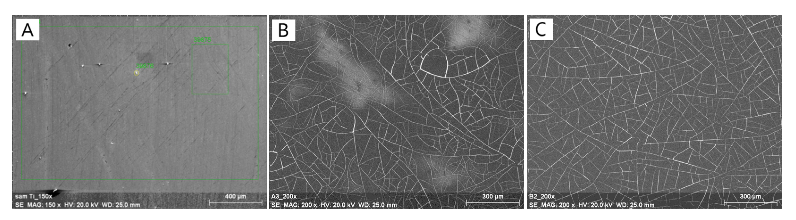
The surface morphology of the tested samples was examined using a scanning electron microscope (SEM; LEO Electron Microscopy Ltd., Cambridge, UK), operated at an accelerating voltage of 20 kV, and the samples were sputter-coated with gold as the tannic acid surfaces are non-conductive. The resolution of the images was 300 µm, and for the sample without the applied layer of tannic acid (polished Ti samples as the control), images were taken at a resolution of 400 µm.
2.3. Surface Free Energy
The contact angle studies were conducted using two different liquids (glycerin and diiodomethane), which were measured for 15 s after the droplet placement on the sample surface under ambient temperature conditions by using a goniometer (DSA 10 Control Unit, Krüss, Germany) equipped with a drop shape analysis system. Surface free energy (IFT (s)), as well as its polar (IFT (s,P)) and dispersive (IFT (s,D)) components, were then calculated using the Owens–Wendt method (also known as the Kaelble–Owens–Wendt method). This model, which considers the polar and dispersive components of surface energy, considers the molecular interactions in the surface layer as equal to the geometric mean of intermolecular interactions within each substance. Based on the Owens–Wendt model, the surface free energy is calculated based on the following equation [
14].
where
represents the total surface free energy of the surface and
and
denotes the dispersion and polar constituents of the surface free energy, respectively. All the samples were thoroughly cleaned prior to the contact angle measurements to remove the presence of surface contaminants if any were present.
2.4. Total Tannic Acid Release
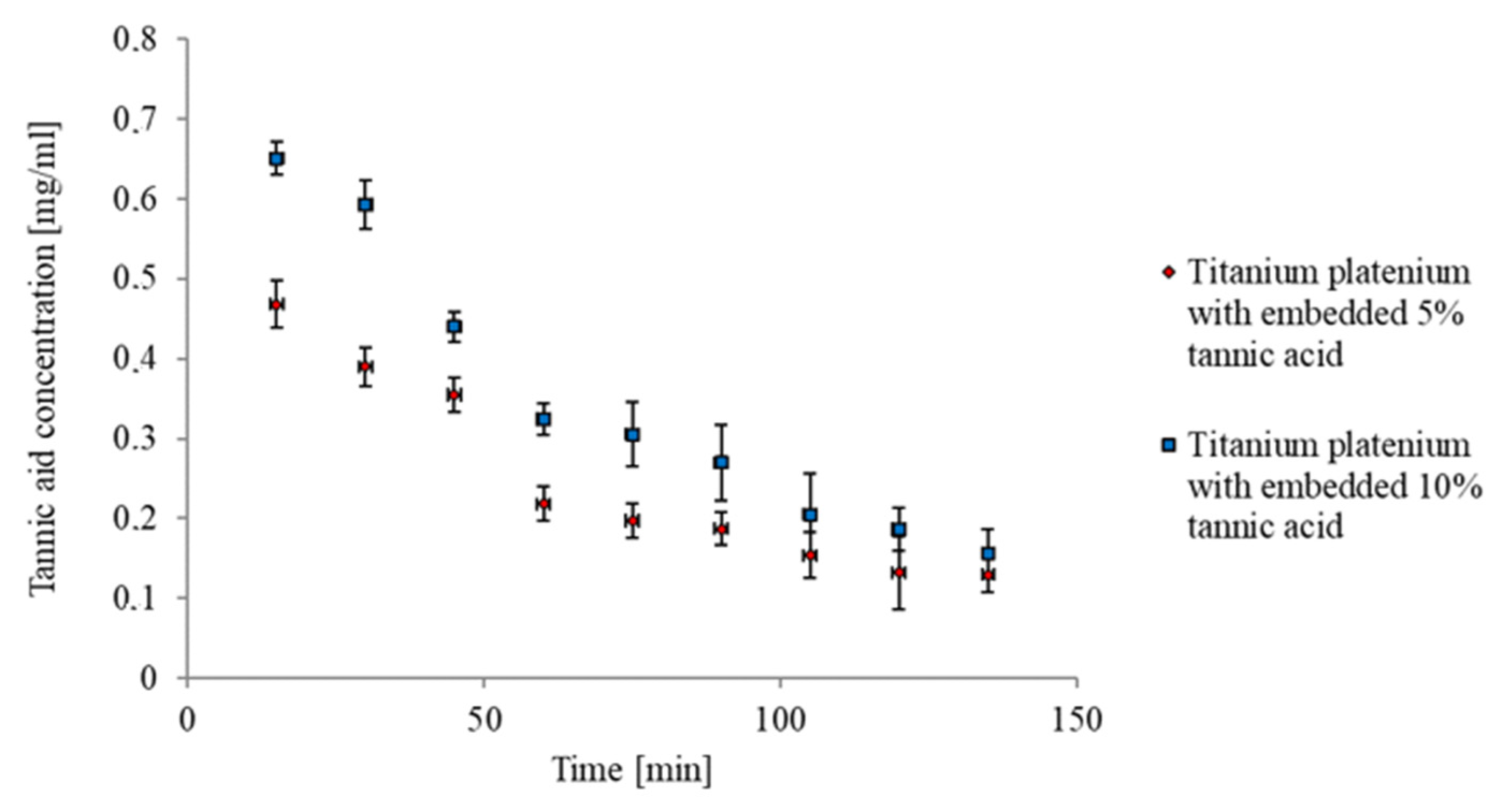
The tannic acid concentration was determined through the Folin—Ciocalteu method. A total of 0.5 mL of Folin—Ciocalteu reagent was mixed with 1 mL of Na2CO3, and the 1 mL of the sample was mixed with distilled water to reach 10 mL. The mixture was then stored at 40 °C for 30 min. The absorbance was measured by a UV—Vis spectrophotometer at 725 nm (UV-100, Shimadzu, Reinach, Switzerland), and the concentration of tannic acid released from the samples was calculated from the standard curve.
2.5. Blood Compatibility
The effect of materials on the hemolysis of red blood cells was studied through the use of anticoagulated whole sheep blood. Totals of 0.2 mL of blood and 10 mL of saline salt were placed in the tubes. The materials (10 mm × 10 mm) were added and left to make contact (60 min; 37 °C; ODspecimen). Tubes with the same amount of blood and saline salt without materials were left as negative controls (ODnegative). Totals of 0.2 mL of blood and 10 mL of distilled water were left as positive controls (ODpositive). All mixtures were centrifuged at 10,000 rpm for 10 min. The absorbance of the supernatants was measured using a microplate reader Multiscan FC (Thermo Fisher Scientific, Waltham, MA, USA) at 540 nm (n = 3). Then, the hemolysis rate was calculated based on the following equation:
where
ODspecimen,
ODnegative and
ODpositive denote the absorbance for the specimen, physiological saline and water, respectively.
2.6. Statistical Analysis
The statistical analysis of the data was performed using SigmaPlot 14.0 software (Systat Software, San Jose, CA, USA). The Shapiro–Wilk test was used to assess the normal distribution of the data, and all the results were calculated as means ± SD (standard deviations) and analyzed using a one-way ANOVA analysis of variance. Multiple comparisons versus the control group between means were carried out with the statistical significance set at p < 0.05 (using the Bonferroni t-test) and were compatible.
4. Discussion
The surface functionalization of various classes of biomaterials, including metals, ceramics and polymers, are required to improve their performance for potential medical applications. Titanium and its alloys play a crucial role in orthopedic and dental procedures, mostly due to their improved mechanical properties and biocompatibility [
15]. Despite being biocompatible, the antibacterial and antioxidant characteristics of titanium are inferior and can thereby lead to implant-related infections. One of the causes of implant failures can be attributed to allergic reactions associated with titanium ion release. There have been reports of hypersensitive reactions, such as erythema, urticaria, eczema, swelling, pain, necrosis and bone loss, due to titanium dental implants. In order to circumvent these limitations, surface functionalization various techniques have been extensively studied.
The present work outlines the use of a facile, cost effective and sustainable technique to develop tannic acid-based surfaces on Ti metallic surfaces via a vacuum deposition technique. Tannic acid is a natural phenolic compound that has antibacterial and long-term antioxidant properties, a fact that has been widely documented [
16,
17,
18]. It has been studied as a dental implant coating and considered to be a bio-safe additive [
19]. The presence of phenolic hydroxyl groups allows tannic acid to eliminate free-radicals and display good antioxidant properties. Tannic acid is able to adhere to the titanium surfaces of various metal oxides to form a thin coating through metal–ligand coordination [
20,
21,
22]. As a result, a tannic acid–metal complex is formed. Tannic acid is capable of combining with the metal ions present in the defects and the dangling bonds present on the native oxide layer of titanium by means of a coordinate bond. It is also evident from morphological studies that the increase in tannic acid concentration has led to a reduced homogenization of the developed tannic acid on the titanium surface. This could be due to the fact that the abovementioned defects and dangling bonds available on the native oxide layer surface will be reduced during direct contact after the initial layer of tannic acid is deposited onto the surface.
Surface free energy is one of the most important surface properties that dictates cell attachment, cellular proliferation and differentiation; moreover, cells generally prefer to grow on high energy surfaces. As a result of the deposition of tannic acid, the polar component of the surface energy was increased, which can be attributed to the presence of numerous hydroxyl groups in the acid structure. The increased polar components can effectively interact with the surface groups present on the cell walls and can generate chemical bonds, whereas non-polar groups are capable of developing non-specific short range interactions, such as in case of van der Waals interactions. In addition, the presence of a large number of phenolic hydroxyl groups allows them to play the role of donor in the process of forming hydrogen bonds. This is advantageous, as the presence of these large numbers of hydrogen bond donors from phenolic groups present in tannic acid attract individual water molecules. Hydrophilic surfaces have a positive effect on the adhesion of cells, e.g., osteoblasts, which means an increase in the compatibility of the material [
23]. It was reported by Cheng et al. [
24] that tannic acid can be utilized to immobilize glucose oxidase on the surface of biomaterials.
The published studies of tannic acid show that the initial “burst effect” that is observed may be important in the first minutes after the material implantation. In order to reduce this effect, tannic acid could, for instance, be mixed with a polymer and then embedded on the titanium surface [
25]. The titanium plates with and without tannic acid are non-hemolytic and do not cause the lysis of erythrocytes, indicating no disruption of the erythrocyte membrane and no subsequent release of hemoglobin. Thereby, they are safe for use on implant surfaces that will come into contact with blood. It has also been reported that methods used to fabricate biomaterials should involve safe techniques and raw materials [
26,
27]. Tannic acid has antimicrobial and antioxidant properties [
16]. Tannic acid embedded on the titanium surface has great potential to enhance the application properties of such materials for implant applications.
Plant polyphenols are an emerging class of antimicrobial molecules with the ability to form coatings on many materials, including titanium. They represent an environmentally friendly and economical approach toward sustainable surface functionalization. Based on these interactions with biomolecules, polyphenols have shown to possess broad spectrum antibacterial and antifungal properties by cell wall and cell membrane interactions, the inhibition of enzymes and the complexation of ions vital for regular cell metabolism. The presence of polyphenolic compounds on the titanium surfaces reduce the growth and biofilm formation of bacteria and fungi related to oral infections [
28]. Since polyphenolic molecules can be deposited as coating on almost any surface, they are able to control microbial colonization [
29]. Overall, this approach has promise in terms of improving the surface free energy without harming erythrocytes. More studies pertaining to the development of polyphenolic surfaces on various surfaces, such as those with a native oxide layer, with a well-developed grown oxide layer or on an etched Ti surface, can provide more insights into the application of polyphenols on metallic Ti surfaces. However, it has been reported that polyphenolic surface modifications were not able to prevent
C. albicans colonization through a reduction in growth [
30]. Thus, more studies focused on testing the underlying mechanisms of the antibacterial activity of these surfaces against specific types of bacteria needs to be explored.

 ,
, 


