Article

Synthesis and Physicochemical Properties of Cefepime Derivatives Suitable for Labeling with Gallium-68

by
Przemysław Koźmiński
*,
Kinga Żelechowska-Matysiak
and
Ewa Gniazdowska
*
Centre of Radiochemistry and Nuclear Chemistry, Institute of Nuclear Chemistry and Technology, 03-195 Warsaw, Poland
*
Authors to whom correspondence should be addressed.
Appl. Sci. 2023, 13(8), 5019; https://doi.org/10.3390/app13085019
Submission received: 8 March 2023 / Revised: 13 April 2023 / Accepted: 13 April 2023 / Published: 17 April 2023

Abstract

:
Bone and soft tissue infections are potentially life-threatening and require immediate and intensive treatment. However, there is still no single diagnostic method that can reliably confirm or rule out such conditions. Imaging with radiopharmaceuticals (i.e., scintigraphy) is a powerful diagnostic tool in the management of patients with infectious or inflammatory diseases. In this work, a new and efficient way to modify the thiazole ring of the cefepime molecule has been proposed and experimentally verified. The developed organic synthesis routes allow for the coupling of the appropriate complexing ligand of the gallium-68 radionuclide with cefepime. The new NODAGA-Glu-CFM conjugate was radiolabeled with gallium-68 with a high yield and showed full stability in human serum. In addition, the [68Ga]Ga-NODAGA-Glu-CFM radioconjugate was hydrophilic and positively charged. Therefore, on the basis of these results, the [68Ga]Ga-NODAGA-Glu-CFM radioconjugate might be considered as a new promising radioconjugate for the diagnosis of bacterial infections.

1. Introduction

Nowadays, one of the biggest problems in medicine is the time from the correct diagnosis to the start of treatment. Fortunately, there are more and more diagnostic techniques and treatment methods that allow the fastest and best possible response. One of the most desired methods is positron-emission tomography (PET), which allows for personalized diagnosis [1]. The advantages of using PET outweigh the high cost of the devices, thus increasing their use in diagnostics [2]. Due to the growing role of PET, there is a huge necessity to search and test new radiopharmaceuticals for PET diagnostics to help clinicians effectively treat patients [3]. Therefore, there are a significant number of publications on PET radiopharmaceuticals that have been tested as diagnostics of bacterial infections [4,5,6,7,8,9,10].
The spread of this imaging technique is resulting in the increasing use of gallium-68, for PET diagnostics, which has a better resolution and sensitivity than SPECT [3,4]; however, it will be difficult or impossible to replace the significance of technetium-99m in diagnosis [11].
In addition to the growing need for oncological diagnostics, there is a strong clinical need for quantitative and specific imaging of inflammation and bacterial infections [3]. As the diagnosis of infection or inflammation can be ambiguous, clinicians often struggle with many doubts or wrong diagnoses [12]. Such infections, which can be diagnosed using the PET technique, include diabetic foot syndrome (DFS). This is one of the most serious complications of diabetes, which has been recognized as one of the diseases of civilization according to a WHO report. According to WHO estimates, 422 million people suffered from diabetes in 2014, representing 8.5% of the total population. This is a fourfold increase compared to 1980 (108 million), primarily associated with a global lifestyle change related to being overweight or obese. Unfortunately, the predictions are not optimistic and there will be more and more people with diabetes, with an estimated 578 million people affected by 2030 [13]. DFS involves deterioration of the blood supply to the foot, which becomes hypoxic and susceptible to infection. The spreading soft tissue infection results in bacterial infection of the bones. This is an indication for amputation, but there is difficulty in distinguishing between bone infections and non-infectious neuropathic bone lesions, which is why a clear diagnosis is so important [14]. At the time of diagnosis, more than 10% of people with DFS are accompanied by peripheral neuropathy and/or vascular disease [12]. The lifetime risk of foot ulceration in a person with diabetes may be as high as 25% [15,16]. It is important to be aware that a lower limb amputation occurs due to diabetes every 30 seconds [16,17,18].
Osteomyelitis is often overlooked; thus, the correct diagnosis and prompt treatment of the infected area are important in DFS [19]. For this purpose, white blood cell (WBC) scintigraphy is used as the gold standard, but due to the time-consuming nature of this method, other approaches are being searched for [20,21]. In the current literature, publications exist which suggest a similar diagnostic accuracy for the use of [18F]fluorodeoxyglucose [22,23], or alternatively, radiolabeled ciprofloxacin derivatives such as N4′-3-[18F]fluoropropylciprofloxacin [24], [18F]F-ciprofloxacin [25] and 99mTc-ciprofloxacin [13]. In the present study, we attempted to label another antibiotic—cefepime (CFM)—which is widely used in the treatment of diabetic foot infections caused by Gram-positive and Gram-negative bacteria [26,27]. Cefepime is a fourth-generation cephalosporin that is registered for use above the age of two months according to the Food and Drug Administration (FDA). It is active against S. pyogenes, S. pneumoniae, E. coli, H. influenzae, M. catarrhalis, N. gonorrhoeae, P. aeruginosa, M. morganii, Proteusmirabilis, some Citrobacter, Enterobacter, Klebsiella, Providencia and Serratia. It is used for pneumonia, complicated urinary tract infections, complicated abdominal infections, peritonitis associated with dialysis, acute biliary infections and bacterial meningitis [28].
The aim of this work was to design and study a new synthesis route for the cefepime derivatives, as well as the synthesis and study of the physicochemical properties of cefepime labeled with the radionuclide 68Ga, a radiopharmaceutical which can be used for PET diagnosis of diabetic foot syndrome.

2. Materials and Methods

Cefepime was a gift from the Public Central Teaching Hospital of Medical University of Warsaw. Fmoc-Asp-OH was purchased from Bachem AG (Bubendorf, Switzerland), Boc-Glu-anhydride was purchased from Fluorochem Ltd (Hadfield, UK). Macrocyclic chelator 2,2′-(7-(1-carboxy-4-((2,5-dioxopyrrolidin-1-yl)oxy)-4-oxobutyl)-1,4,7-triazonane-1,4-diyl)diacetic acid (NODAGA-NHS) was purchased from CheMatech (Dijon, France), other chemicals and solvents were purchased from Merck (Darmstadt, Germany) and used as obtained. 68Ga radionuclide (emitter β+, t1/2 = 67.7 min, Eβmax = 1.9 MeV, in the form of [68Ga]GaCl3 in 0.1 M HCl solutions) was eluted from the commercially available GalliaPharm® 68Ge/68Ga generator (Eckert and Ziegler, Berlin, Germany). Deionized water was prepared in a Hydrolab water-purification system (Hydrolab, Straszyn, Poland). Human serum was a gift from the Regional Centre for Blood Donation and Blood Treatment in Warsaw, Poland.
HPLC analyses, separations and purifications were carried out using a VWR-Hitachi LaChrom Elite HPLC system which consisted of a pump L2130, column thermostat L-2350, UV diode array detector (DAD) L-2455 and the EZChrom Elite data system. The radioactivity was monitored using a 3 × 3” NaI(Tl) scintillation detector Raytest Gabi Star (Straubenhardt, Germany). The separation of the obtained products was accomplished on a LiChrospher® 100 RP-18 analytical column (5 µm particle size, 100 Å pore size, 4.6 mm × 250 mm) from Merck (Darmstadt, Germany) and a SUPELCOSIL™ LC-18 semi preparative column (5 µm particle size, 120 Å pore size, 10 mm × 250 mm) from Sigma-Aldrich Co. LLC (Darmstadt, Germany). The solvents were as follows: solvent A, 0.1% (v/v) trifluoroacetic acid (TFA) in water; solvent B, 0.1% (v/v) trifluoroacetic acid (TFA) in acetonitrile. The gradient conditions are presented in Table 1. The chromatograms were performed using UV (220–400 nm) or γ-detection.
The radiochemical purity (RPC) of the synthesized [68Ga]Ga-NODAGA-Glu-CFM radioconjugate was analyzed by instant thin-layer chromatography (ITLC) using a glass microfiber chromatography paper impregnated with a silica gel (Agilent, Santa Clara, CA, USA), and 0.2 M citrate buffer, pH 5.0, was used as the developing solvent. After completing development, the distribution of radioactivity on the strips was determined using a Storage Phosphor System Cyclone Plus (Perkin-Elmer Life and Analytical Sciences, Downers Grove, IL, USA).
1H NMR spectra were registered with a Bruker Avance Neo 400 spectrometer in DMSO-d6 solvent. During the measurements, the temperature was stabilized at 295 K and the 1H signals were calibrated on the residual solvent signal (2.5 ppm).
The IR spectrum was recorded on a Thermo Scientific Nicolet iS10 FT-IR spectrometer in the range of 4000–400 cm−1 using a KBr pellet.

2.1. Syntheses

The low reactivity of the amine group attached to the thiazole ring (as in the CFM molecule) in the coupling reaction of the chelator in the form of active ester such as NHS or SCN and CFM molecule results in a very low reaction yield [29]. In the absence of ligand in the anhydride form for a direct coupling reaction, the use of a linker in the anhydride form becomes necessary for the coupling reaction of the ligand with the CFM molecule. Other synthetic pathways which allow the conjugation of chelators with beta-lactam antibiotic cores are described in the literature [30,31].

2.1.1. Preparation of Linker-CFM Derivatives

The aim of the modification of the CFM molecule was to obtain a linker-CFM derivative. The selected linker should play the role of a spacer between the cefepime molecule and a radionuclide chelator, e.g., such as NODAGA-NHS. Taking into account the structure of the cefepime molecule and the pharmacophore fragment responsible for the biological properties of this antibiotic, the most suitable site for linker attachment appears to be the primary amino group of the thiazole ring as suggested in [29] and the papers cited therein. Thus, we used two homologues of dicarboxylic acids as linkers: aspartic acid, Asp, (Scheme 1) and glutamic acid, Glu, (Scheme 2). In both cases, dicarboxylic acid was used in the anhydride form, and the primary amino group was protected with an Fmoc (9-fluorenylmethoxycarbonyl group) or Boc (tert-butyloxycarbonyl group) protecting group, respectively. After removing the protecting group, the free amino group of the linker was used for the coupling reaction with the radionuclide chelator. The modification processes were carried out in several steps described in Section 3. Results and Discussion.

2.1.2. Preparation of [68Ga]Ga-NODAGA-Glu-CFM Radioconjugate

The 68Ge/68Ga generator was eluted with an NE-1000 programmable single syringe pump, and the eluate was collected every 500 μL. The most radioactive fraction (containing the most 68Ga radionuclide) was used for the synthesis of radioconjugates without additional concentration. The 68Ga radiolabeling of the NODAGA-Glu-CFM conjugate was achieved as follows: 200 µL of [68Ga]GaCl3 (50.4 MBq) in 0.1 M HCl from the 68Ge/68Ga generator was added into a solution of 50 µg of the NODAGA-Glu-CFM conjugate previously dissolved in 300 µL of a 0.4 M acetate buffer (pH = 5), and the reaction mixture was allowed to stand at room temperature for 15 min. The radiochemical yield and purity were determined by the HPLC and ITLC methods (as an average value from at least three independent measurements).

2.1.3. Preparation of Ga-NODAGA-Glu-CFM-‘Cold’ Reference Compound

The Ga-NODAGA-Glu-CFM reference compound was obtained using 4 µL (0.776 µmol) of GaCl3 solution in 0.65 M HCl added to 0.5 mg (0.517 µmol) NODAGA-Glu-CFM conjugate previously dissolved in 75 µL of a 0.4 M acetate buffer (pH = 5). The obtained crude products were purified by HPLC before lyophilization and characterization by mass spectrometry.

2.2. Physicochemical Properties Studies of [68Ga]Ga-NODAGA-Glu-CFM

Physicochemical properties studies of the obtained radioconjugate were performed using a radioconjugate previously isolated from the radiolabeling reaction mixture by the HPLC method (gradient 2, γ-detection).

2.2.1. Lipophilicity Studies

The lipophilicity of the studied radioconjugate was characterized by the logarithm of its distribution coefficient, LogD7.4, in the n-octanol/PBS (pH 7.40) system, which mimics the physiological conditions (Product Properties Test Guidelines OPPTS 830.7550, 1996).
The 0.5 mL of the isolated radioconjugate in PBS solution was introduced to a test tube containing 0.5 mL of n-octanol and vortexed for 1 min. After that the test tube was centrifuged for 5 min at 5800 rpm to ensure complete separation of layers. The radioactivity of each phase was determined, using the well-type NaI(Tl) detector (ISOMED 2100, NUVIA Instruments, Dresden, Germany). The LogD was determined as an average value from three independent measurements.

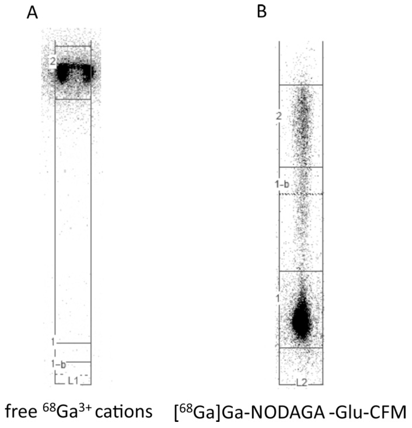
2.2.2. Paper Electrophoresis Studies

Paper electrophoresis studies have been performed for the [68Ga]Ga-NODAGA-Glu-CFM radioconjugate using the gallium eluate [68Ga]GaCl3 ([68Ga]Ga3+ cation as a reference radiocompound).
Paper electrophoresis experiments were performed on 20 × 1 cm chromatographic paper strips, Paper Chromedia GF 83 (Whatman, Little Chalfont, UK), pre-treated with phosphate buffer (0.01 M, pH 7.40), using a midi horizontal electrophoresis unit (Sigma-Aldrich, St. Louis, MO, USA) device. The experimental conditions were as follows: 200 V (10 V cm−1) for 45 min.
The developed paper strips were left to dry, and then the distribution of radioactivity on the strips was determined using a Storage Phosphor System Cyclone Plus (Perkin-Elmer Life and Analytical Sciences, Downers Grove, IL, USA).

2.2.3. Radioconjugate Stability Study in Human Serum

The stability of the [68Ga]Ga-NODAGA-Glu-CFM radioconjugate separated from the reaction mixture was studied by incubation at 37 °C in human serum for up to a few half-lives of the gallium-68 radionuclide. In order to study the stability of the radioconjugate in human serum, a 0.1 mL aliquot of the solution of radioconjugate was added to 0.9 mL of human serum and incubated at 37 °C. After time periods of 0.5 and 2 h, an appropriate volume of the incubated serum mixture was withdrawn, mixed in an Eppendorf tube with ethanol and vigorously shaken to precipitate proteins. After centrifugation, the radioactivity of both phases (precipitate and supernatant) was measured and, additionally, aliquots of the supernatant were analyzed by the HPLC method to check whether the radioconjugate had converted into another water-soluble radioactive form. The radioconjugate stability study was conducted in triplicate.

3. Results and Discussion

The CFM derivatives were synthesized according to Scheme 1 and Scheme 2, and the progress of reaction was monitored by the HPLC method.

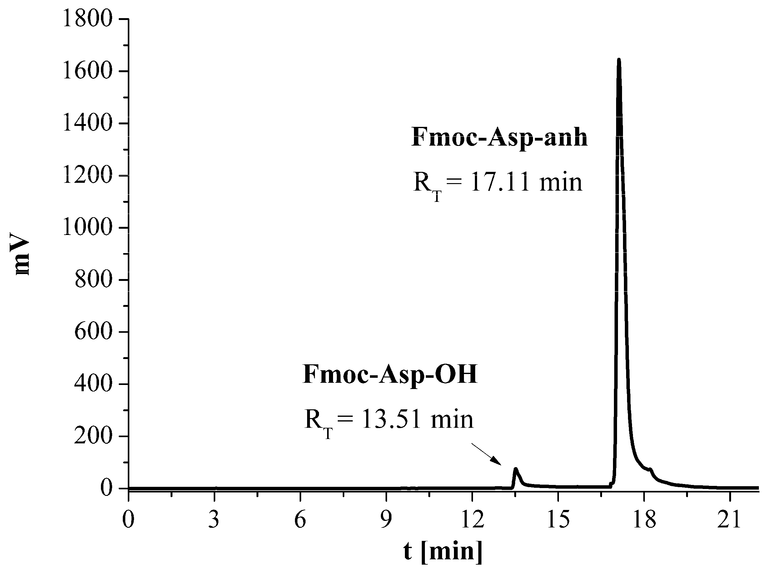
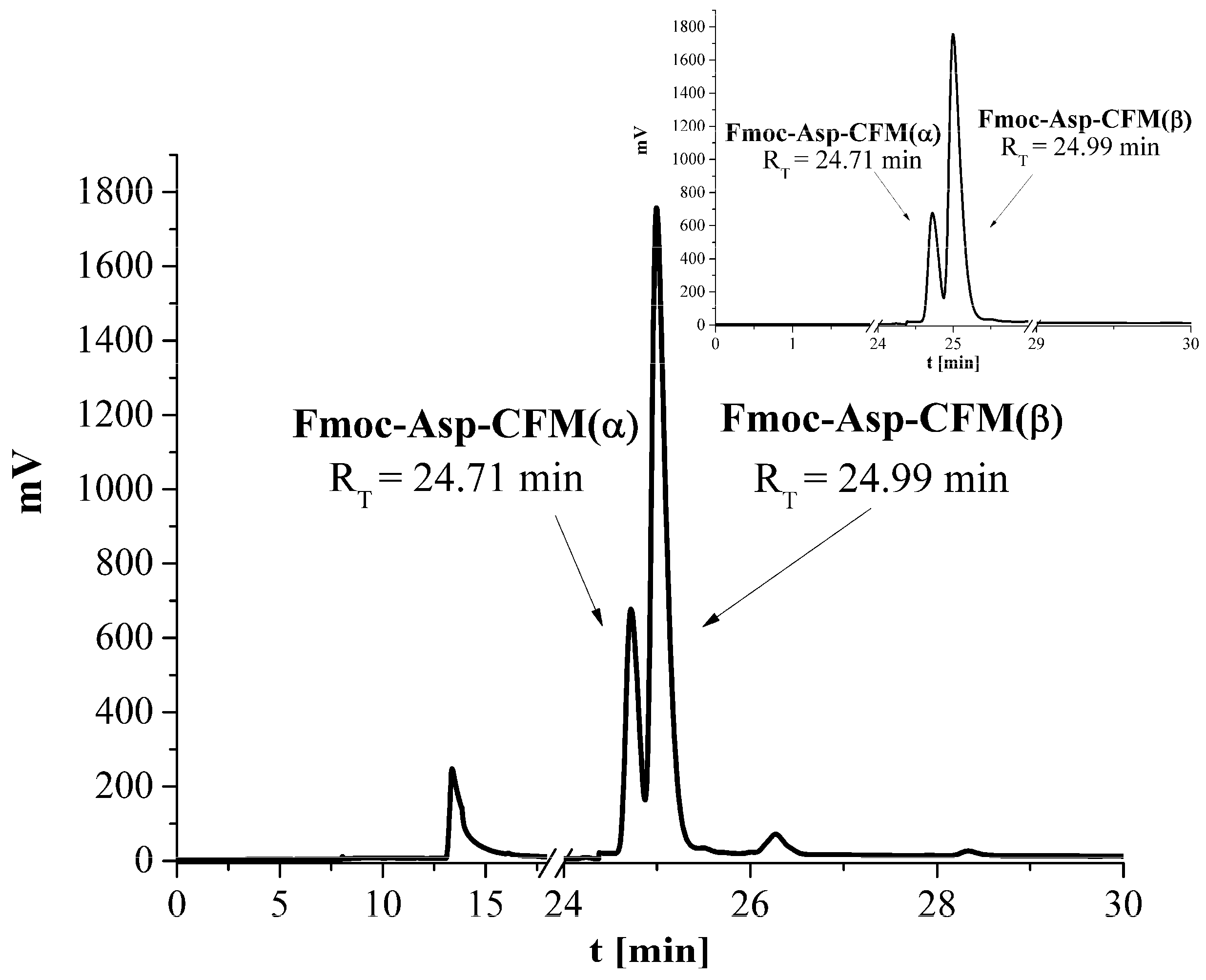
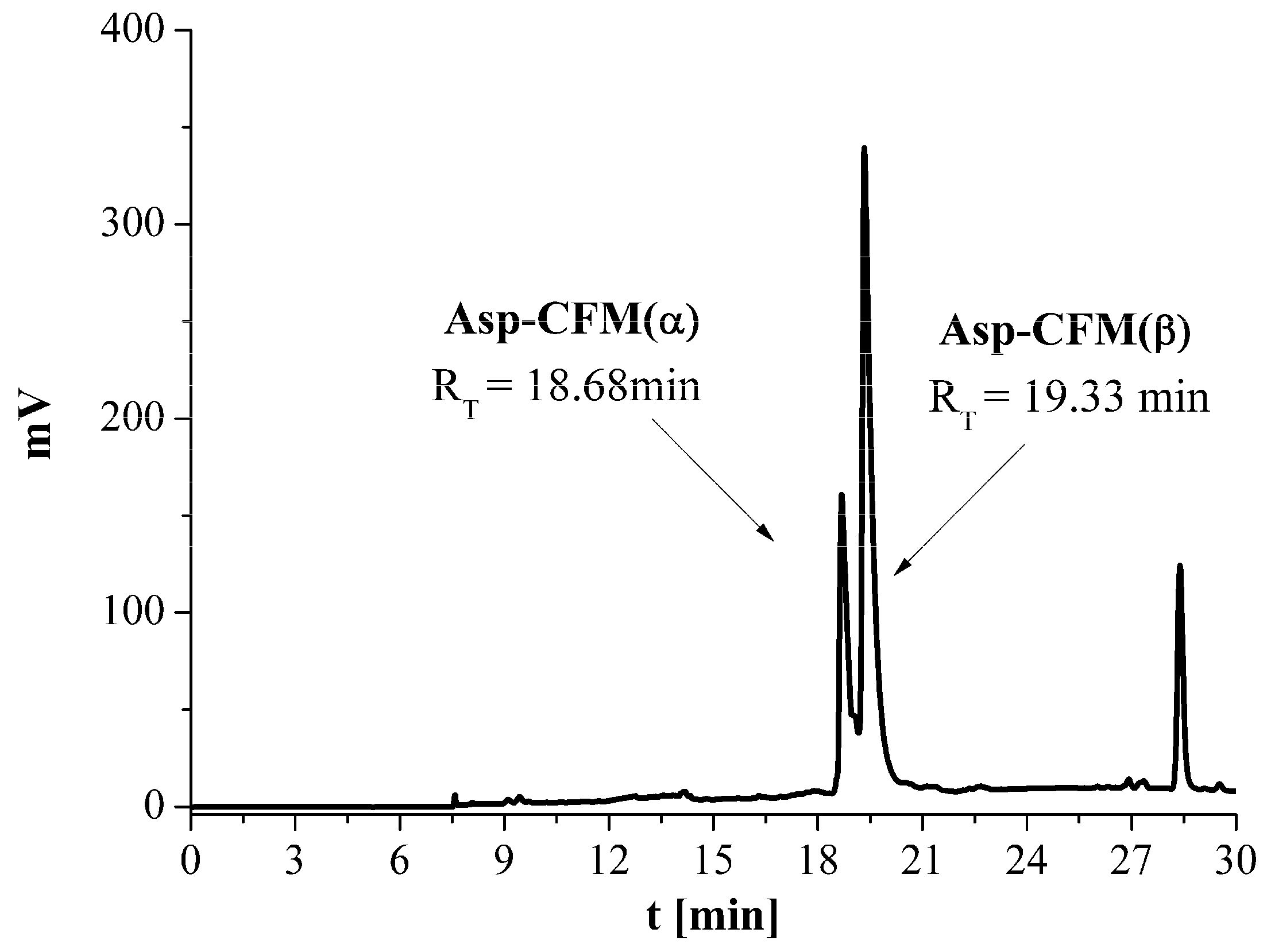
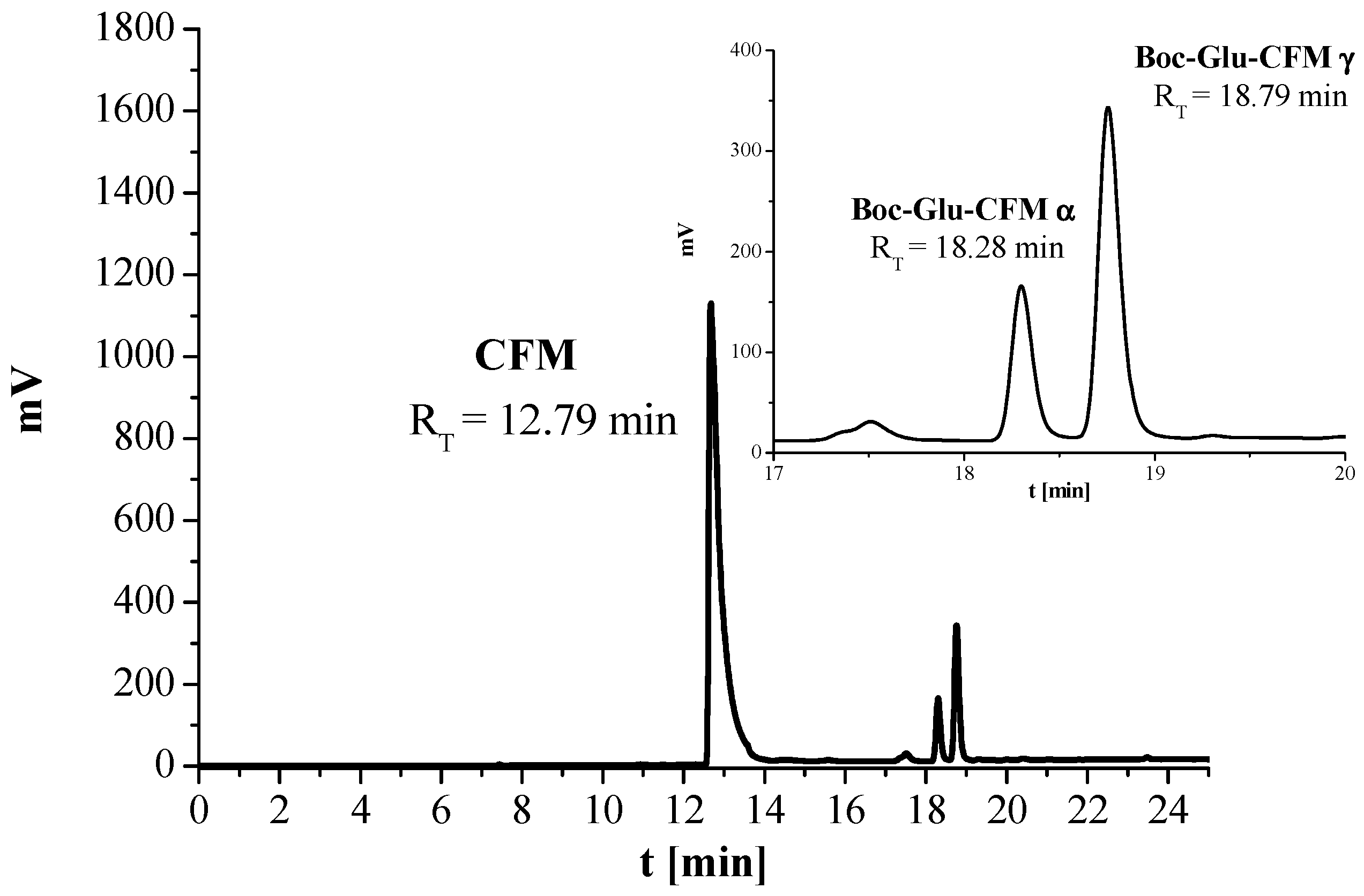
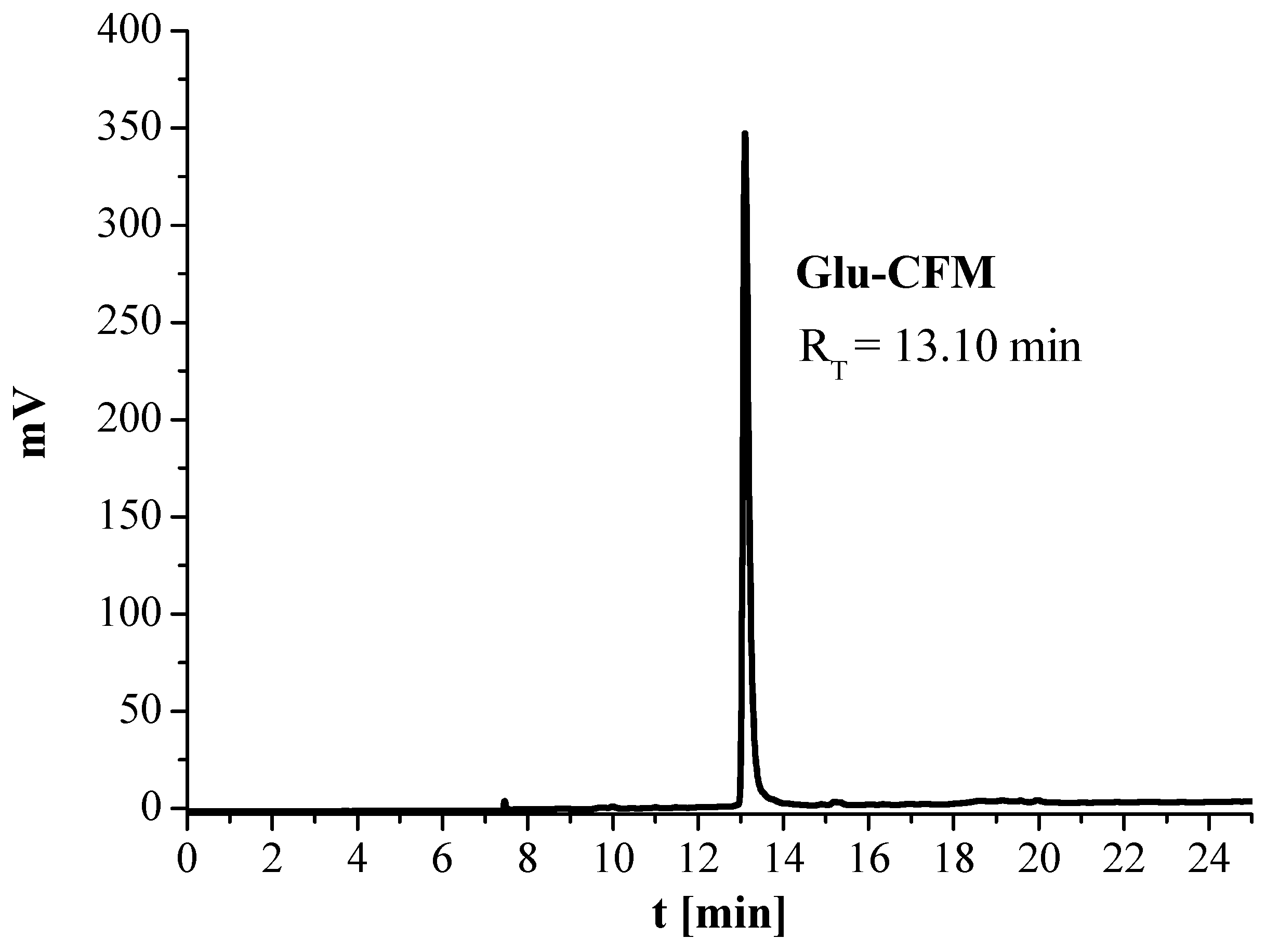
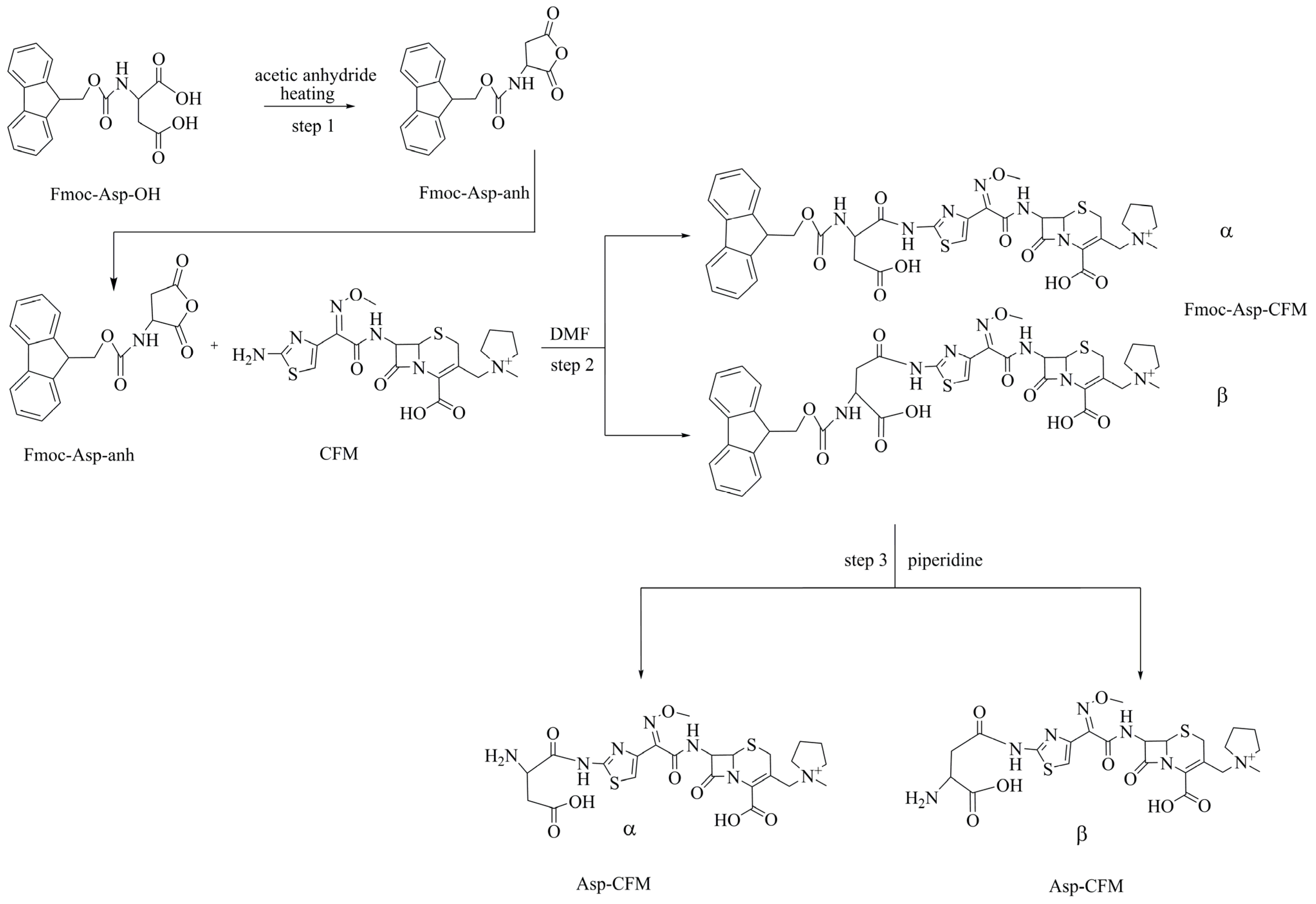
3.1. Synthesis of Asp-CFM Molecule

In the first step, Fmoc aspartic anhydride (Fmoc-Asp-anh) was synthetized (Figure 1). A total of 135 mg (0.38 mmol) of Fmoc-Asp-OH (linker) was added to the flask with 500 μL (5.30 mmol) of acetic anhydride. The flask was then heated in an oil bath to 85 °C. An additional 500 μL of acetic anhydride (1 mL in total) was added. The mixture was heated for about 20 min until the linker was completely dissolved. The flask was then cooled rapidly to room temperature, and the product was filtered and washed with diethyl ether. The resulting white dusty precipitate was dried in vacuo.
To the Eppendorf tube with 8.40 mg (25 µmol) of Fmoc-Asp-anh, 10 mg (20.83 µmol) of CFM dissolved in 200 μL of DMF was added. The synthesis was carried out overnight at room temperature, and the progress of the reaction was checked by HPLC. After completion of the reaction, the solvent was removed under vacuum and the residue dissolved in acetonitrile. The crude product Fmoc-Asp-CFM, consisting of two structural (constitutional) isomers (Scheme 1, products of step two), was purified by HPLC (gradient two, Figure 2, RT = 24.71 and RT = 24.99 min) and lyophilized: yield 75% and purity over 98%. The presence of the product was confirmed by mass spectrometric analysis.
MS for C38H40N7O10S2+, TOF MS ES+ m/z: (calculated: 818.23); found: 818.36.
In order to remove the Fmoc-protecting group, 5 mg (6.11 µmol) of Fmoc-Asp-CFM was used, dissolved in 50 µL of 2.5% piperidine in DMF (Scheme 1, step three). The reaction mixture was maintained for 30 min at room temperature. The final products (isomer Asp-CFM(α), RT = 18.67 min, and isomer Asp-CFM(β), RT = 19.33 min) were isolated from the reaction mixture by HPLC (Figure 3, gradient two) and analyzed by MS. For further syntheses, the beta isomer obtained with a higher yield was used.
MS for C23H30N7O8S2+, TOF MS ES+ m/z: (calculated: 596.16); found: 596.29.

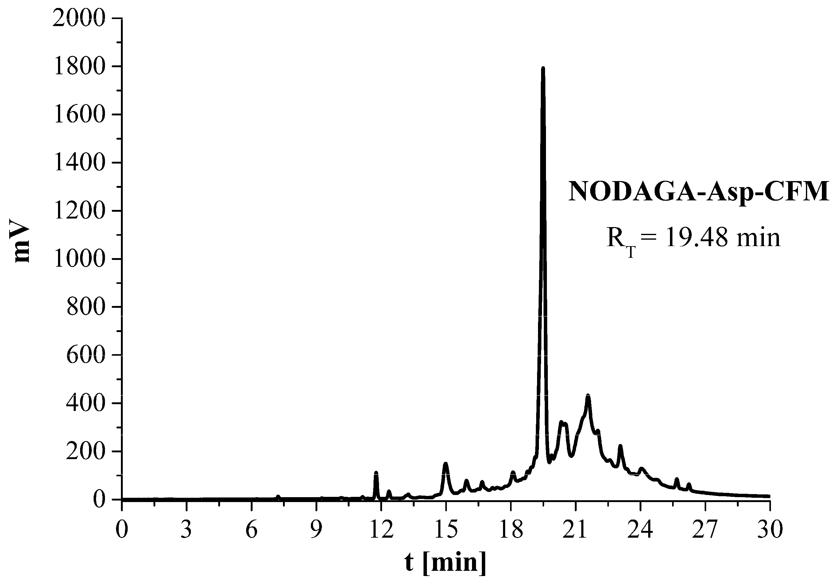
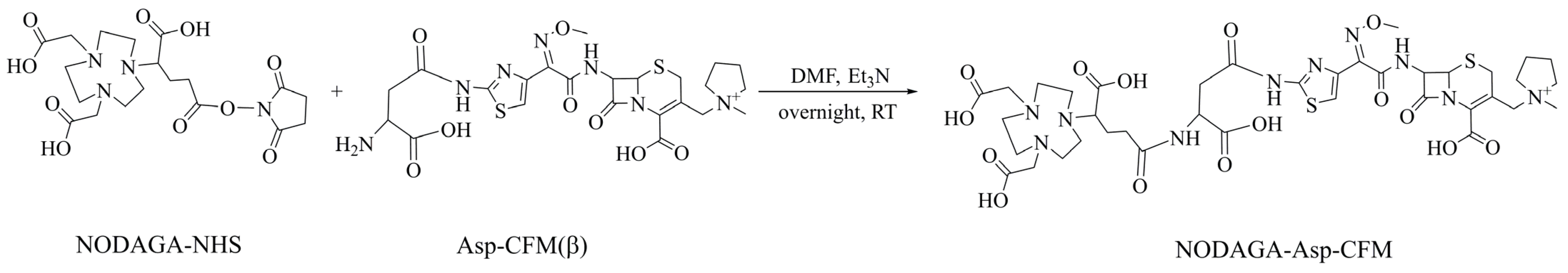
3.2. Synthesis of NODAGA-Asp-CFM Conjugate

A total of 1 mg (1.58 µmol) of Asp-CFM(β) was dissolved in 25 µL of DMF. Then, the solution was transferred to an Eppendorf tube with 1.5 mg (2.04 µmol) NODAGA-NHS before 0.86 µL (6.21µmol) of triethylamine (Et3N) was immediately added (Scheme 3). The tube was closed and left overnight on the magnetic stirrer at room temperature. The final conjugate was purified by HPLC (Figure 4) and lyophilized for later use: yield 90–95%, purity 95%. The presence of the conjugate was confirmed by MS analysis.

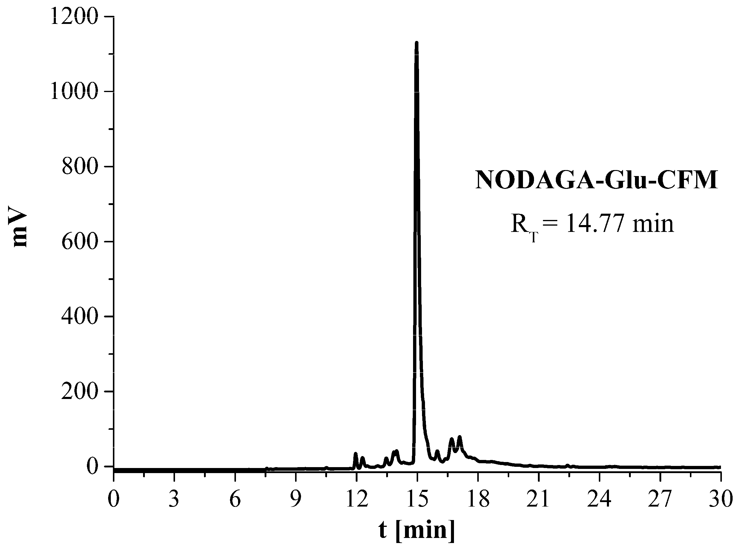
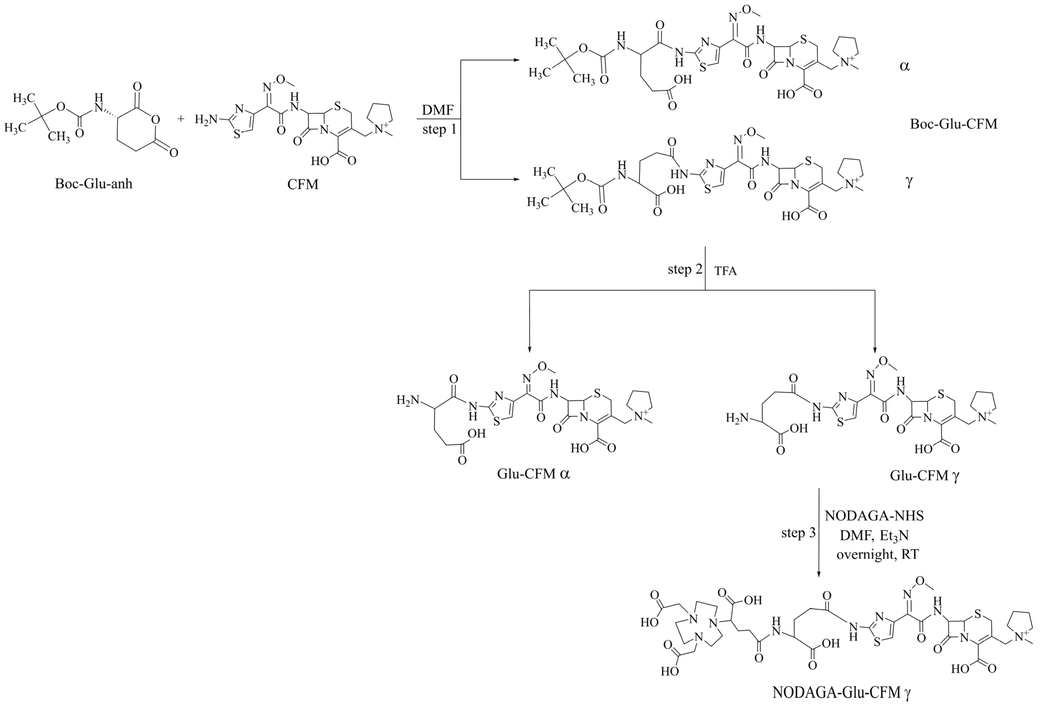
3.3. Synthesis of NODAGA-Glu-CFM Conjugate

The above-described synthesis of NODAGA-Asp-CFM indicates a problem with the separation of the two isomers Fmoc-Asp-CFM(α) and Fmoc-Asp-CFM(β). Therefore, it was decided to carry out an analogous synthesis of the NODAGA-linker-CFM conjugate using glutamic acid as a linker, which, compared to aspartic acid, contains an additional -CH2 group, which should result in a better separation of the products. The stepwise synthesis of the NODAGA-Glu-CFM conjugate (Scheme 2) is analogous to that of the NODAGA-Asp-CFM, with the exception of the Boc-Glu-anh preparation step as this reagent was commercially available.
For the synthesis of the Boc-Glu-CFM derivative, 13.50 mg (28 µmol) of CFM and 6.42 mg (28 µmol) of Boc-Glu-anh in 200 µL of DMF were used. The Eppendorf tube was closed and left for 24 h on the magnetic stirrer at room temperature, and the progress of the reaction was checked by HPLC. The obtained final products with RT values of 18.28 min and 18.79 min (Figure 5) were isolated from the reaction mixture by HPLC (gradient two). The results of mass spectrometry confirmed the obtainment of the Boc-Glu-CFM derivative in the form of two isomers: Boc-Glu-CFM(α) (RT =18.29 min) and Boc-Glu-CFM(γ) (RT =18.79 min).
MS for C29H40N7O10S2+, TOF MS ES+ m/z: (calculated: 710.80); found: 710.23.
The chemical yield of this reaction was surprisingly low; therefore, in order to check the purity of the purchased reagent Boc-Glu-anh, mass spectrometry analysis was performed. The results of MS analysis showed that the signal from the active anhydride form (Boc-Glu-anh, m/z-228.14) was several times smaller than that of the hydrated form of the reagent (246.12 m/z). The active form of Boc-Glu-anh in the purchased reagent was present at only about 10%, which can provide an explanation for the low yield of synthesis of the Boc-Glu-CFM product obtained in the form of the two isomers. For further syntheses, only the gamma isomer obtained with a higher yield was used. The purity of the final product was higher than 98%.
In the second step, the Boc-protecting group was removed. In this case, 4 mg (5.64 µmol) of γ isomer of Boc-Glu-CFM was used and dissolved in 40 µL of 2.5% TFA in DMF. The reaction mixture was incubated for 45 min. The final product Glu-CFM was isolated from the reaction mixture by HPLC (purity 98%) (gradient two, Figure 6, RT = 13.10) and analyzed by MS.
MS for C24H32N7O8S2+, TOF MS ES+ m/z: (calculated: 610.17); found: 610.32.
In the last step, 1 mg (1.64 µmol) Glu-CFM (γ isomer) dissolved in 40 µL DMF was transferred to a tube containing 1.44 mg (1.97 µmol) of NODAGA-NHS, and then 0.823 µL (5.9 µmol) of Et3N was added. The vial was left for 24 h on the magnetic stirrer at room temperature, and the final product was isolated from the reaction mixture by HPLC (gradient two, purity 98%). The recorded chromatogram (Figure 7) showed a single peak with a retention time of 14.77 min. The obtained NODAGA-Glu-CFM conjugate was characterized by the methods dedicated for the analysis of radiopharmaceutical precursors recommended by the European Pharmacopoeia 11.0, monograph 07/2022:2902 (nuclear magnetic resonance spectrometry, infrared absorption spectrophotometry, mass spectrometry and chromatographic methods). The results of infrared absorption spectrophotometry and mass spectrometry are shown below. However, we do not present the results of the 1H NMR analysis. The presence of the four-membered ring (lactam) adjacent to a six-membered ring in the synthetized conjugate resulted in the broadening of the signals, leading to a poor resolution of the multiplets. Moreover, the obtained 1H NMR spectrum was complicated by overlapping signals arising from many protons and was difficult to analyze. For this reason, the NMR spectrum was not essential to confirm that the conjugate was successfully obtained, and the NMR data are not shown.
IR (KBr) νmax/cm−1: 3435 s, br (OH, NH), 2942 w, br (CH) 2853 sh (CH), 1777 w (C=O), 1677 vs. (C=O), 1629 sh (C=O), 1562 sh (C=O), 1462 w, 1393 w, br, 1352 w, sh, 1286 w, 1204 m, 1183 sh, 1135 w, 1072 vw sh, 1043 w, 923 vw, 836 vw, 801 vw, 722 vw, 594 vw, 518 vw.
MS for C39H55N10O15S2+, TOF MS ES+ m/z: (calculated: 967.33); found: 967.36.

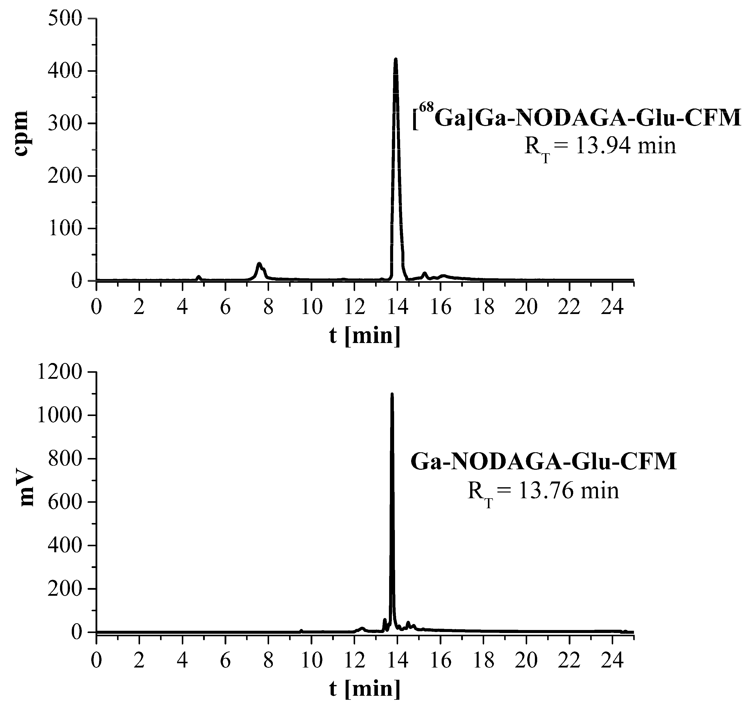
3.4. Synthesis of [68Ga]Ga-NODAGA-Glu-CFM Radioconjugate and Ga-NODAGA-Glu-CFM ‘Cold’ Reference Compound

For the radiolabeling reaction, NODAGA-Glu-CFM conjugate was selected. The conditions have been described previously in Section 2.1.2. Under these conditions, [68Ga]Ga-NODAGA-Glu-CFM was synthesized with a radiochemical yield (RCY) higher than 90%, as is shown in Figure 8. The radiochemical purity (RPC) of the [68Ga]Ga-NODAGA-Glu-CFM radioconjugate was checked using the ITLC method, where free 68Ga3+ cations moved with the solvent front (Rf ≈ 1), while the [68Ga]Ga-NODAGA-Glu-CFM radioconjugate remained at the origin (Rf ≈ 0.0–0.1) (Figure 9). The HPLC chromatogram of the ‘cold’ reference compound Ga-NODAGA-Glu-CFM (Figure 8), obtained with a high yield (95%) and final purity over 98%, showed a peak with RT = 13.76. The slight shift of the peak position compared to the RT of the [68Ga]Ga-NODAGA-Glu-CFM radioconjugate (RT = 13.94 min) results from the arrangement in the series of the gamma detector after the DAD detector. The peak position of the ‘cold’ reference compound (characterized by MS) confirmed that the corresponding radioconjugate was obtained in the labeling reactions.
MS for C39H52GaN10O15S2+, TOF MS ES+ m/z: (calculated: 1033.23); found: 1033.22.
The 1H NMR data for the reference compound Ga-NODAGA-Glu-CFM are not shown for the same reason as the NODAGA-Glu-CFM conjugate.

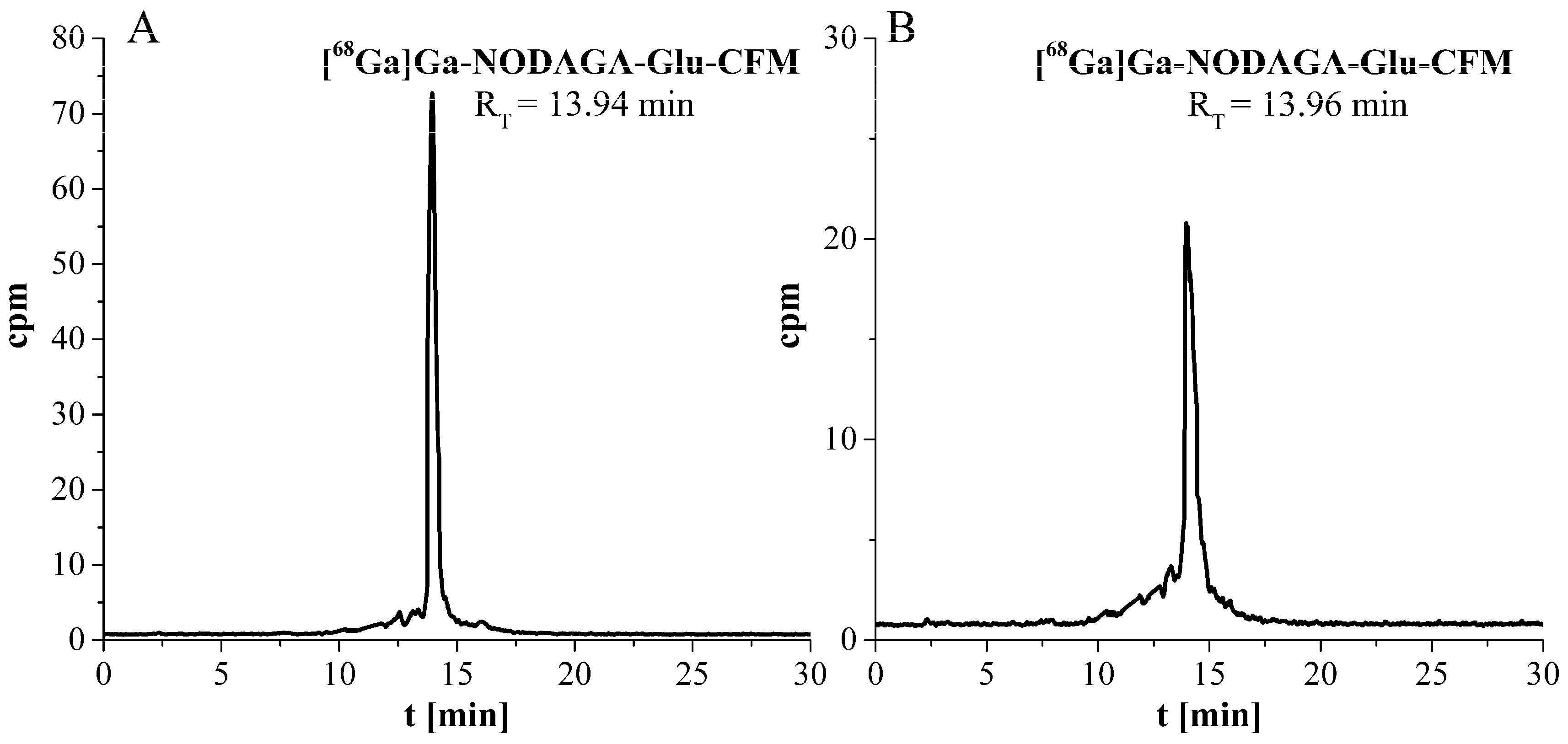
3.5. Characterization of [68Ga]Ga-NODAGA-Glu-CFM Radioconjugate

The obtained [68Ga]Ga-NODAGA-Glu-CFM radioconjugate was purified by HPLC (gradient two) and tested for its lipophilicity, as well its stability in human serum. The lipophilicity (LogD7.4 value) of the [68Ga]Ga-NODAGA-Glu-CFM radioconjugate was determined to be −3.68 ± 0.12, which proves its highly hydrophilic nature.
The electrophoretic diagrams of the [68Ga]Ga-NODAGA-Glu-CFM radioconjugate and gallium eluate [68Ga]GaCl3 (with the [68Ga]Ga3+ cation as a reference radiocompound) showed only single peaks. In both cases, the peaks were shifted toward the cathode. These results confirmed the expected positive charge of the [68Ga]Ga-NODAGA-Glu-CFM radioconjugate, which may have a beneficial effect on the interaction of the radioconjugate with bacterial cells. Based on the literature data, most bacterial cells possess an overall negative charge due to the presence of peptidoglycan, which is rich in carboxyl and amino groups [32,33,34]. For this reason, better binding of the designed radioconjugate to the wall of bacterial cells can be expected.
The HPLC radiochromatograms of the isolated [68Ga]Ga-NODAGA-Glu-CFM radioconjugate recorded after 0.5 h and 2 h of incubation at 37 °C in human serum are shown in Figure 10. These studies showed that the tested radioconjugate is completely stable in human serum. The measurements of radioactivity of both the supernatant and precipitate fractions showed that the percentage of 68Ga-radioconjugate bound to serum components ranged from 7% to 10% and was independent of incubation time.

4. Conclusions

In this study, a new and efficient way to synthesize the cefepime derivatives Asp-CFM and Glu-CFM has been designed and experimentally confirmed. It was shown that modification of the cefepime molecule can be achieved by a direct coupling reaction between Fmoc-Asp-anh or Boc-Glu-anh and the amine group of the thiazole ring of the CFM molecule. Modification of the CFM molecule at this site (via the thiazole ring) does not affect the biological activity of the antibiotic (its affinity to bacterial cells), which has already been determined [29]. Mild acidic/basic conditions can be used for the deprotection reaction of the amine group (removal of the Fmoc or Boc groups) which is crucial for the possibility of further coupling with any chelator in the ester (-NHS or –SCN) form.
The new NODAGA-Glu-CFM conjugate was radiolabeled with gallium-68. The obtained [68Ga]Ga-NODAGA-Glu-CFM radioconjugate was formed with a high yield and specific activity within the range of 0.68–0.87 MBq/nmol, as well as exhibiting full stability in human serum.
In conclusion, on the basis of these results, it can be considered that the [68Ga]Ga-NODAGA-Glu-CFM radioconjugate meets all the physicochemical requirements for radiopharmaceuticals. Bearing in mind that the modification of cefepime via the amine group of the thiazole ring does not affect its affinity to bacterial cells [29], the newly developed and tested radioconjugate can be considered as a new promising radioconjugate for the diagnosis of bacterial infections which, despite its promise, requires further biological research. This is the first report on the labeling of the antibiotic cefepime with gallium-68 and a cefepime-based diagnostic 68Ga-radiopharmaceutical with potential use in PET imaging of bacterial infections in diabetic foot syndrome.

Author Contributions

Conceptualization, P.K. and E.G.; methodology, P.K. and K.Ż.-M.; investigation, P.K. and K.Ż.-M.; writing—original draft preparation, P.K., K.Ż.-M. and E.G.; writing—review and editing, P.K., K.Ż.-M. and E.G.; supervision, P.K. and E.G.; project administration, P.K.; funding acquisition, P.K.; All authors have read and agreed to the published version of the manuscript.

Funding

This research was funded by the National Science Center (Poland), grant number 2017/01/X/ST4/00925.

Institutional Review Board Statement

Not applicable.

Informed Consent Statement

Not applicable.

Data Availability Statement

Not applicable.

Acknowledgments

The contribution of student Kinga Żelechowska-Matysiak was made within the framework of the National Centre for Research and Development Project No. POWR.03.02.00-00-I009/17 (radiopharmaceuticals for molecularly targeted diagnosis and therapy, RadFarm, Operational Project Knowledge Education Development 2014–2020, co-financed by the European Social Fund).

Conflicts of Interest

The authors declare no conflict of interest.

References

  1. Velikyan, I. Prospective of 68Ga-Radiopharmaceutical Development. Theranostics 2014, 4, 47–80. [Google Scholar] [CrossRef]
  2. Ranachowska, C.; Lass, P.; Korzon-Burakowska, A.; Dobosz, M. Diagnostic Imaging of the Diabetic Foot. Nucl. Med. Rev. 2010, 13, 18–22. [Google Scholar]
  3. Velikyan, I. 68Ga-Based Radiopharmaceuticals: Production and Application Relationship. Molecules 2015, 20, 12913–12943. [Google Scholar] [CrossRef] [PubMed]
  4. Signore, A.; Glaudemans, A.W.J.M. The Molecular Imaging Approach to Image Infections and Inflammation by Nuclear Medicine Techniques. Ann. Nucl. Med. 2011, 25, 681–700. [Google Scholar] [CrossRef]
  5. Welling, M.M.; Hensbergen, A.W.; Bunschoten, A.; Velders, A.H.; Roestenberg, M.; van Leeuwen, F.W.B. An update on radiotracer development for molecular imaging of bacterial infections. Clin. Transl. Imaging 2019, 7, 105–124. [Google Scholar] [CrossRef]
  6. Gouws, A.C.; Kruger, H.G.; Gheysens, O.; Zeevaart, J.R.; Govender, T.; Naicker, T.; Ebenhan, T. Antibiotic-Derived Radiotracers for Positron Emission Tomography: Nuclear or “Unclear” Infection Imaging? Angew. Chem. Int. Ed. 2022, 61, e202204955. [Google Scholar] [CrossRef]
  7. Velikyan, I. Prospective of 68Ga Radionuclide Contribution to the Development of Imaging Agents for Infection and Inflammation. Contrast Media Mol. Imaging 2018, 2018, 9713691. [Google Scholar] [CrossRef]
  8. Petrik, M.; Umlaufova, E.; Raclavsky, V.; Palyzova, A.; Havlicek, V.; Pfister, J.; Mair, C.; Novy, Z.; Popper, M.; Hajduch, M.; et al. 68Ga-labelled desferrioxamine-B for bacterial infection imaging. Eur. J. Nucl. Med. Mol. Imaging 2021, 48, 372–382. [Google Scholar] [CrossRef]
  9. Chopra, S.; Singh, B.; Koul, A.; Mishra, A.K.; Robu, S.; Kaur, A.; Ghai, A.; Caplash, N.; Wester, H.J. Radiosynthesis and pre-clinical evaluation of [68Ga] labeled antimicrobial peptide fragment GF-17 as a potential infection imaging PET radiotracer. Appl. Radiat. Isot. 2019, 149, 9–21. [Google Scholar] [CrossRef]
  10. Kumar, V.; Boddeti, D.K. 68Ga-radiopharmaceuticals for PET imaging of infection and inflammation. Recent Results Cancer Res. 2013, 194, 189–219. [Google Scholar] [CrossRef]
  11. OECD/NEA. The Supply of Medical Isotopes: An Economic Diagnosis and Possible Solutions; OECD Publishing: Paris, France, 2019. [Google Scholar] [CrossRef]
  12. Signore, A.; Glaudemans, A.W.J.M.; Galli, F.; Rouzet, F. Imaging Infection and Inflammation. BioMed Res. Int. 2015, 2015, 615150. [Google Scholar] [CrossRef] [PubMed]
  13. Koźmiński, P.; Gawęda, W.; Rzewuska, M.; Kopatys, A.; Kujda, S.; Dudek, M.K.; Halik, P.K.; Królicki, L.; Gniazdowska, E. Physicochemical and Biological Study of 99mTc and 68Ga Radiolabelled Ciprofloxacin and Evaluation of [99mTc]Tc-CIP as Potential Diagnostic Radiopharmaceutical for Diabetic Foot Syndrome Imaging. Tomography 2021, 7, 829–842. [Google Scholar] [CrossRef] [PubMed]
  14. Lipsky, B.A. Osteomyelitis of the Foot in Diabetic Patients. Clin. Infect. Dis. 1997, 25, 1318–1326. [Google Scholar] [CrossRef] [PubMed]
  15. Singh, N.; Armstrong, D.G.; Lipsky, B.A. Preventing Foot Ulcers in Patients with Diabetes. JAMA 2005, 293, 217–228. [Google Scholar] [CrossRef]
  16. Boulton, A.J.M.; Vileikyte, L.; Ragnarson-Tennvall, G.; Apelqvist, J. The Global Burden of Diabetic Foot Disease. Lancet 2005, 366, 1719–1724. [Google Scholar] [CrossRef]
  17. Das, A.K.; Shashank, R.J. Put Feet First: Prevent Amputations–Diabetes and Feet. J. Assoc. Physicians India 2005, 53, 929–930. [Google Scholar]
  18. Howarth, D. Putting feet first: Preventing avoidable amputations. J. Diabetes Nurs. 2017, 21, 235. [Google Scholar]
  19. Palestro, C.J.; Love, C. Nuclear Medicine and Diabetic Foot Infections. Semin. Nucl. Med. 2009, 39, 52–65. [Google Scholar] [CrossRef]
  20. Auletta, S.; Riolo, D.; Varani, M.; Lauri, C.; Galli, F.; Signore, A. Labelling and Clinical Performance of Human Leukocytes Labelled with 99mTc-HMPAO Using Leukokit® with Gelofusine versus Leukokit® with HES as Sedimentation Agent. Contrast Media Mol. Imaging 2019, 2019, 4368342. [Google Scholar] [CrossRef]
  21. Wagner, T.; Payoux, P.; Simon, J.; Anne, J.; Tafani, M.; Esquerré, J.P.; Bonnet, E. Discordance between labelled white blood cell scintigraphy and bone scan following suspicion of bone infection: What should be done about it? Nucl. Med. Cent. East. Eur. 2010, 13, 5–7. [Google Scholar]
  22. Familiari, D.; Glaudemans, A.W.J.M.; Vitale, V.; Prosperi, D.; Bagni, O.; Lenza, A.; Cavallini, M.; Scopinaro, F.; Signore, A. Can Sequential 18F-FDG PET/CT Replace WBC Imaging in the Diabetic Foot? J. Nucl. Med. 2011, 52, 1012–1019. [Google Scholar] [CrossRef]
  23. Treglia, G.; Sadeghi, R.; Annunziata, S.; Zakavi, S.R.; Caldarella, C.; Muoio, B.; Bertagna, F.; Ceriani, L.; Giovanella, L. Diagnostic Performance of Fluorine-18-Fluorodeoxyglucose Positron Emission Tomography for the Diagnosis of Osteomyelitis Related to Diabetic Foot: A Systematic Review and a Meta-Analysis. Foot 2013, 23, 140–148. [Google Scholar] [CrossRef] [PubMed]
  24. Sachin, K.; Kim, E.M.; Cheong, S.J.; Jeong, H.J.; Lim, S.T.; Sohn, M.H.; Kim, D.W. Synthesis of N 4′-[18F]Fluoroalkylated Ciprofloxacin as a Potential Bacterial Infection Imaging Agent for PET Study. Bioconjug. Chem. 2010, 21, 2282–2288. [Google Scholar] [CrossRef]
  25. Langer, O.; Brunner, M.; Zeitlinger, M.; Ziegler, S.; Müller, U.; Dobrozemsky, G.; Lackner, E.; Joukhadar, C.; Mitterhauser, M.; Wadsak, W.; et al. In Vitro and in Vivo Evaluation of [18F]Ciprofloxacin for the Imaging of Bacterial Infections with PET. Eur. J. Nucl. Med. Mol. Imaging 2005, 32, 143–150. [Google Scholar] [CrossRef] [PubMed]
  26. So, W.; Kuti, J.L.; Shepard, A.; Nugent, J.; Nicolau, D.P. Tissue Penetration and Exposure of Cefepime in Patients with Diabetic Foot Infections. Int. J. Antimicrob. Agents 2016, 47, 247–248. [Google Scholar] [CrossRef] [PubMed]
  27. Motaleb, M.A.; El-Kolaly, M.T.; Ibrahim, A.B.; El-Bary, A.A. Study on the preparation and biological evaluation of 99mTc–gatifloxacin and 99mTc–cefepime complexes. J. Radioanal. Nucl. Chem. 2011, 289, 57–65. [Google Scholar] [CrossRef]
  28. Harrison, C.J.; Bratcher, D. Cephalosporins: A Review. Pediatr. Rev. 2008, 29, 264–273. [Google Scholar] [CrossRef]
  29. Koźmiński, P.; Rzewuska, M.; Piądłowska, A.; Halik, P.; Gniazdowska, E. Synthesis, physicochemical and in vitro biological evaluation of 99mTc-cefepime radioconjugates, and development of DTPA-cefepime single vial kit formulation for labelling with technetium-99m. J. Radioanal. Nucl. Chem. 2022, 331, 2883–2894. [Google Scholar] [CrossRef]
  30. Peters, B.K.; Reddy, N.; Shungube, M.; Girdhari, L.; Baijnath, S.; Mdanda, S.; Chetty, L.; Ntombela, T.; Arumugam, T.; Bester, L.A.; et al. In Vitro and In Vivo Development of a β-Lactam-Metallo-β-Lactamase Inhibitor: Targeting Carbapenem-Resistant Enterobacterales. ACS Infect. Dis. 2023, 9, 486–496. [Google Scholar] [CrossRef]
  31. Reddy, N.; Girdhari, L.; Shungube, M.; Gouws, A.C.; Peters, B.K.; Rajbongshi, K.K.; Baijnath, S.; Mdanda, S.; Ntombela, T.; Arumugam, T.; et al. Neutralizing Carbapenem Resistance by Co-Administering Meropenem with Novel β-Lactam-Metallo-β-Lactamase Inhibitors. Antibiotics 2023, 12, 633. [Google Scholar] [CrossRef]
  32. Kapoor, G.; Saigal, S.; Elongavan, A. Action and resistance mechanisms of antibiotics: A guide for clinicians. J. Anaesthesiol. Clin. Pharmacol. 2017, 33, 300–305. [Google Scholar] [CrossRef] [PubMed]
  33. Wilson, W.W.; Wade, M.M.; Holman, S.C.; Champlin, F.R. Status of methods for assessing bacterial cell surface charge properties based on zeta potential measurements. J. Microbiol. Methods 2001, 43, 153–164. [Google Scholar] [CrossRef] [PubMed]
  34. Da Costa, D.; Exbrayat-Héritier, C.; Rambaud, B.; Megy, S.; Terreux, R.; Verrier, B.; Primard, C. Surface charge modulation of rifampicin-loaded PLA nanoparticles to improve antibiotic delivery in Staphylococcus aureus biofilms. J. Nanobiotechnol. 2021, 19, 12. [Google Scholar] [CrossRef] [PubMed]
Scheme 1. Modification scheme of the CFM molecule with aspartic acid as a linker.
Applsci 13 05019 sch001
Scheme 2. Synthesis route of the NODAGA-Glu-CFM conjugate.
Applsci 13 05019 sch002
Figure 1. HPLC chromatogram (at 280 nm) of the reaction mixture of Fmoc-Asp-anh synthesis (RT = 17.11 min, gradient 1).
Applsci 13 05019 g001
Figure 2. HPLC chromatogram (at 270 nm) of the reaction mixture of Fmoc-Asp-CFM derivative synthesis (products of step 2: isomer α (RT = 24.71 min) and isomer β (RT = 24.99 min) (gradient 2).
Applsci 13 05019 g002
Figure 3. HPLC chromatogram (at 270 nm) of deprotection reaction mixture of Fmoc-Asp-CFM derivative in 2.5% piperidine solution after 30 min; isomer Asp-CFM(α) with RT = 18.68 min and isomer Asp-CFM(β) with RT = 19.33 min (gradient 2).
Applsci 13 05019 g003
Scheme 3. Coupling reaction of NODAGA-NHS with Asp-CFM(β).
Applsci 13 05019 sch003
Figure 4. HPLC chromatogram (at 270 nm) of the reaction mixture of NODAGA-Asp-CFM (RT = 19.48 min) conjugate synthesis (gradient 2).
Applsci 13 05019 g004
Figure 5. HPLC chromatogram (at 270 nm) of the reaction mixture of Boc-Glu-CFM derivative synthesis: Boc-Glu-CFM(α) (RT = 18.28 min) and Boc-Glu-CFM(γ) (RT = 18.79 min) (gradient 2).
Applsci 13 05019 g005
Figure 6. HPLC chromatogram recorded at 270 nm of the deprotection reaction of Boc-Glu-CFM in 2.5% TFA solution (gradient 2).
Applsci 13 05019 g006
Figure 7. HPLC chromatogram at 270 nm of the reaction mixture of NODAGA-Glu-CFM conjugate (Rt = 14.77 min) synthesis (gradient 2).
Applsci 13 05019 g007
Figure 8. HPLC analysis of reaction mixtures after synthesis of [68Ga]Ga-NODAGA-Glu-CFM (gradient 2) and the ‘cold’ reference compound Ga-NODAGA-Glu-CFM (270 nm, gradient 2).
Applsci 13 05019 g008
Figure 9. ITLC analyses of free 68Ga3+ (A) and the reaction mixture after synthesis of the [68Ga]Ga-NODAGA-Glu-CFM radioconjugate (B) using citrate buffer as a developing solvent.
Applsci 13 05019 g009
Figure 10. The HPLC radiochromatograms of the [68Ga]Ga-NODAGA-Glu-CFM radioconjugate recorded after (A) 0.5 h and (B) 2 h of incubation at 37 °C in human serum.
Applsci 13 05019 g010
Table 1. Gradient profiles used in HPLC.
Gradient ConditionsFlow [mL/min]Time
[min]
Eluent Content
%A%B
Gradient profile 1 used in HPLC with analytical column10595
116040
136040
168020
208020
22595
Gradient profile 2 used in HPLC with semi-preparative column300100
226040
266040
288020
308020
320100
Disclaimer/Publisher’s Note: The statements, opinions and data contained in all publications are solely those of the individual author(s) and contributor(s) and not of MDPI and/or the editor(s). MDPI and/or the editor(s) disclaim responsibility for any injury to people or property resulting from any ideas, methods, instructions or products referred to in the content.

