Article

Variations in Gold Nanoparticle Size on DNA Damage: A Monte Carlo Study Based on a Multiple-Particle Model Using Electron Beams

by
Christine A. Santiago
1 and
James C. L. Chow
2,3,*
1
Department of Physics, Toronto Metropolitan University, Toronto, ON M5B 2K3, Canada
2
Radiation Medicine Program, Princess Margaret Cancer Centre, University Health Network, Toronto, ON M5G 1X6, Canada
3
Department of Radiation Oncology, University of Toronto, Toronto, ON M5T 1P5, Canada
*
Author to whom correspondence should be addressed.
Appl. Sci. 2023, 13(8), 4916; https://doi.org/10.3390/app13084916
Submission received: 28 March 2023 / Revised: 7 April 2023 / Accepted: 13 April 2023 / Published: 14 April 2023
(This article belongs to the Special Issue Next Steps for the Production of Nanoparticles for Nanomedicine)

Abstract

:
Research is currently focused on maximizing cancer cell death while minimizing harm to healthy cells. Gold nanoparticles (GNPs) have been extensively studied as a radiosensitizer to improve cancer cell death while sparing normal tissue. Previous research and simulations have demonstrated that the presence of a single GNP increases DNA damage and dose. In this study, a Monte Carlo simulation using the Geant4-DNA code was used to investigate the effects of multiple GNPs on DNA damage when exposed to electron beams with energies of 50, 100, 150, and 200 keV. The study examined DNA damage caused by 1–4 GNPs of the same total volume by analyzing both single- and double-strand breaks. The results indicate that increasing the number of GNPs and decreasing the electron beam energy increases the total number of strand breaks. Although DNA damage increased, the proportion of double-strand breaks remained unchanged in relation to the total number of strand breaks.

1. Introduction

Over the course of past centuries, cancer has affected many people. With over 100 types, it continues to be one of the leading causes of death worldwide. In 2020, there was an estimated 18.1 million cases [1]. This is about 0.225 percent of the entire population. A Canadian study released in 2021 predicted that 1 in 4 people will die from cancer [2]. In the United States of America alone, it is estimated there will be about 1.8 million new cases of cancer a year, and 600,000 will die from it. With the progress of healthcare and treatment, the projected number of cancer survivors will increase by 5.3 million by 2030 [3]. While the number of cancer survivors is increasing, there continues to be an anticipated increase in the number of cancer diagnoses and deaths [4].
Cancer can be treated by one or more treatments such as surgery, chemotherapy, or radiation therapy. Given the different types of cancer, there are many factors to consider when creating a treatment plan. Radiation therapy is a common and effective treatment that can be used as the primary treatment or in combination with other forms of treatments. It is estimated that about half of cancer patients will receive treatment involving radiation therapy [5]. Radiation therapy is a form of treatment where cancer cells or tumors are irradiated by an ionizing radiation source such as X-rays, electrons, or protons. In hospitals, patients are irradiated using a linear accelerator (LINAC). The LINACs head rotates around the patient to irradiate the tumor to avoid unnecessary damage to healthy tissue. The energy deposited kills the cancer cells by damaging their deoxyribonucleic acid (DNA), which leads to cell death or tumor shrinkage. The main goal of cancer treatment is to kill cancer cells without compromising the surrounding healthy tissue. Because of the effects of radiation on cells, minimizing the damage to normal cells has become important in medical physics research, and radiosensitizers have been shown to be effective in minimizing the damage done to healthy tissue. Radiosensitizers are chemical or pharmaceutical agents that increase cancer cell death through the production of free radicals and secondary electrons [6,7,8]. Radiosensitizer candidates that exhibit a high biocompatibility with cancer cells and are non-toxic to healthy cells are considered favorable [9,10]. Compared to other traditionally used agents, gold has a low biological toxicity. GNPs deposit preferentially at tumor sites due to their enhanced permeability and retention or enhanced permeability and retention effect. The tumor vasculature system is leaky and has a low permeability to normal vasculature [11]. Furthermore, GNPs can be used as an imaging contrast [12,13], and using gold as a contrast agent has shown that it exhibits high X-ray attenuation [14]. Another characteristic that makes a favorable candidate is an agent with a high atomic number. During radiation therapy, the target is irradiated, and the radiation particle interacts with molecules in cells such as water and undergoes Compton scattering, during which there is a loss of energy in the conversion into an electron and a scattered photon. The scattered photon has lower energy relative to the recoiling electron, resulting in the electron being ejected from the atom [15,16]. When the radiation particle interacts with a molecule with a high atomic number, such as gold (Z = 79), there is a larger total absorption cross-section. Gold’s total absorption cross-section is 100 times greater than for water, so it is more effective in absorbing photon energy [17,18,19]. The production of secondary electrons is important for causing indirect damage to cancer cell DNA.
Two types of DNA damage can occur: single-strand breaks (SSBs) and double-strand breaks (DSBs). SSBs occur when one DNA strand breaks, and DSBs occur when both strands break simultaneously. One nucleotide sequence is broken in an SSB while a DSB results in two broken nucleotide sequences, and a disturbed or broken sugar–phosphate backbone. The frequency with which SSBs occur is much higher than for DSBs, which are more difficult to repair than SSBs because of the severity of the damage. During radiation therapy, DSBs are more favorable as they lead to mutations and cell death. To repair an SSB, the strand that has not been broken is used as a template to repair the DNA. DSBs are repaired through homologous recombination, which is not as direct as both strands are damaged and they are not able to use a template. The repair is achieved by exchanging the genetic material between another DNA molecule that is identical to the damaged DNA [20]. If there are enough strand breaks, cancer cell death will occur.
Different cancers require different treatment plans. For example, external beam radiation therapies (EBRTs) that use photon beams are used to treat head and neck cancer while EBRTs that use electron beams are used for superficial cancers such as cutaneous T-cell lymphoma, also known as mycosis fungoides, which requires total skin electron irradiation [21]. Electron therapy has been used as an important treatment method for over 50 years. Emerging research on radiosensitizers has focused on photon beams because of their novelty. However, fewer studies have been conducted on the enhancement of electron therapy when gold nanoparticles are present.
Earlier research into Monte Carlo simulations has demonstrated that the presence of a single gold nanoparticle results in dose enhancement [22,23,24]. Nonetheless, these investigations do not provide insight into the effects of nanoparticle size of the amount of DNA damage. This study examines the influence of multiple gold nanoparticles, with an equivalent total volume on DNA damage, the magnitude of which is assessed in relation to both SSBs and DSBs.
A nanoparticle is a small particle with at least one dimension measuring between 1 and 100 nanometers [15]. They can be made of various materials, such as metals, ceramics, or polymers. Nanoparticles have unique properties due to their size, including a high surface-to-volume ratio, increased reactivity, and unique optical and magnetic properties [9]. In radiation therapy, nanoparticles are used to enhance the effectiveness of radiation treatment. When a nanoparticle is irradiated, it releases secondary electrons that can cause more damage to cancer cells [16,24]. Additionally, nanoparticles can be designed to accumulate preferentially in cancer cells due to their enhanced permeability and retention, resulting in a higher concentration of the particles in the tumor. This allows for a more targeted and precise delivery of radiation to cancer cells, thereby sparing healthy surrounding tissues [6,8].
Nanoparticle radiation therapy has shown promise for a variety of cancer types. For example, heavy-atom nanoparticles such as GNPs can be used to enhance the effectiveness of radiation therapy for prostate cancer by improving the distribution of radiation within the tumor and providing dose enhancement [25]. In addition, nanoparticles have been used to deliver radiation therapy directly to lung tumors, improving the effectiveness of treatment and minimizing damage to healthy surrounding tissues [26]. The use of GNPs as a radiosensitizer has been shown to be effective in enhancing the dose for skin lesions when using orthovoltage beams in skin therapy [27].

2. Materials and Methods

2.1. Monte Carlo Simulation: Geant4-DNA

Monte Carlo simulations are based on the statistical Monte Carlo method, which is the statistical process of predicting probability of radiation transport using random sampling. It is a stochastic method that uses random numbers and probability to solve complicated physical and mathematical problems that cannot be computed by hand, such as multi-dimensional integrals [28,29]. The results of Monte Carlo simulations increase in confidence when there are many random numbers. Depending on the computer’s central processing unit, the simulation may take some time to process.
The pathway of one individual radiation particle is known as radiation transport, and every interaction the particle undergoes is tracked from its interactions with tissue, water, etc. The simulation of radiation transport is important when computing dosimetry quantities [30]. One simulation of an individual particle’s pathway is also known as a history. The more histories or simulations there are, the more accurate the results and the less variance there is. Each calculation is made in each voxel. After the code runs, the deposited energy values in each voxel can be used to calculate dosimetric quantities or strand breaks. The number of histories used in this simulation is 10,000,000.
The Monte Carlo code used was Geant4 (GEometry ANd Tracking), which is a free Monte Carlo simulation toolkit. It is used for simulations of particles interacting with matter. It is used in different areas of physics such as high-energy physics, nuclear physics, and medical physics [31]. Geant4 is accessed through downloading source code files from the Geant4 Download section on their website using the terminal and instructions in the user guide documentation. The programming language used is C++, and an integrated development environment (IDE), such as Xcode on MacBooks, is used to create and edit the codes. To simulate the DNA and gold nanoparticles, an extension, Geant4-DNA, was used [32]. The files that were downloaded contain examples of different physics applications. There are basic examples containing models with simple detector geometry and shapes. There is an advanced section for more specific and difficult simulations. The last folder is the extended folder containing the medical physics examples.

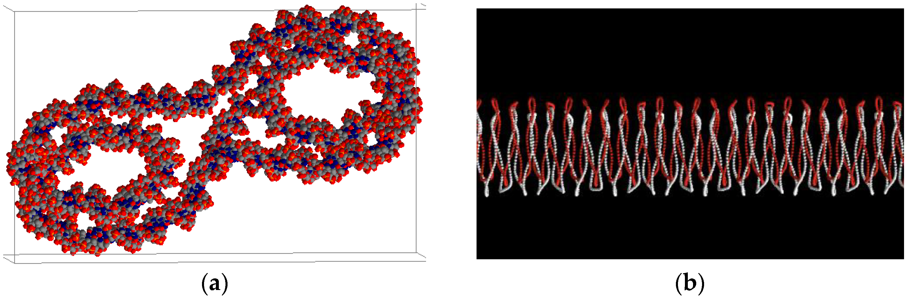
2.2. Realistic DNA Model and Radial Electron Beams

The extended examples folder in the downloaded Geant4 files contains an example called PDB4DNA which uses a free open-source C++ library called PDBlib [33]. The Protein Data Bank (PDB) file represents the molecular geometry of the DNA molecule. The PDB4DNA code is able to evaluate the DNA damage calculated through energy deposition in the sugar–phosphate groups. Compared to the original simulation of the DNA molecule, the PDB4DNA DNA model is shorter to account for the size of a real cell, which has about 3.1 × 10⁹ base pairs. The sugar–phosphate groups were arranged as prisms using parametric equations, which allowed for computations for up to 1.2 × 10⁸ base pairs [34]. The code of the DNA model was created in the DetectorConstruction source file, which contains the geometry, including the addition of gold nanoparticles. In the Monte Carlo simulation, there is an EventAction source file that contains the code that calculates the strand breaks, which occur when the secondary electron interacts with a phosphate group. The energy threshold for one SSB to occur is 8.22 eV. When two SSBs occur, they can either be in neighboring sugar–phosphate groups or in a phosphate group near another sugar–phosphate group with an SSB. To improve the accuracy of our simulation, we used the realistic DNA model depicted in Figure 1a, replacing the simpler DNA model previously used in Figure 1b.
In a clinical setting, LINACs have a rotating beam, so in order to simulate this, a radial electron beam was used as seen in Figure 2. The electron beam energies that were used were 50, 100, 150, and 200 keV, and the radius of the gold nanoparticle used was 5 nm. The distance between the GNPs and the DNA molecule was 5 nm. Reviews on electron therapy have shown that the beam energy is between 6 and 20 MeV, but since energy is lost when interacting with tissue or water, the energy examined was between 50 and 200 keV [16].

2.3. Single and Multiple Gold Nanoparticles
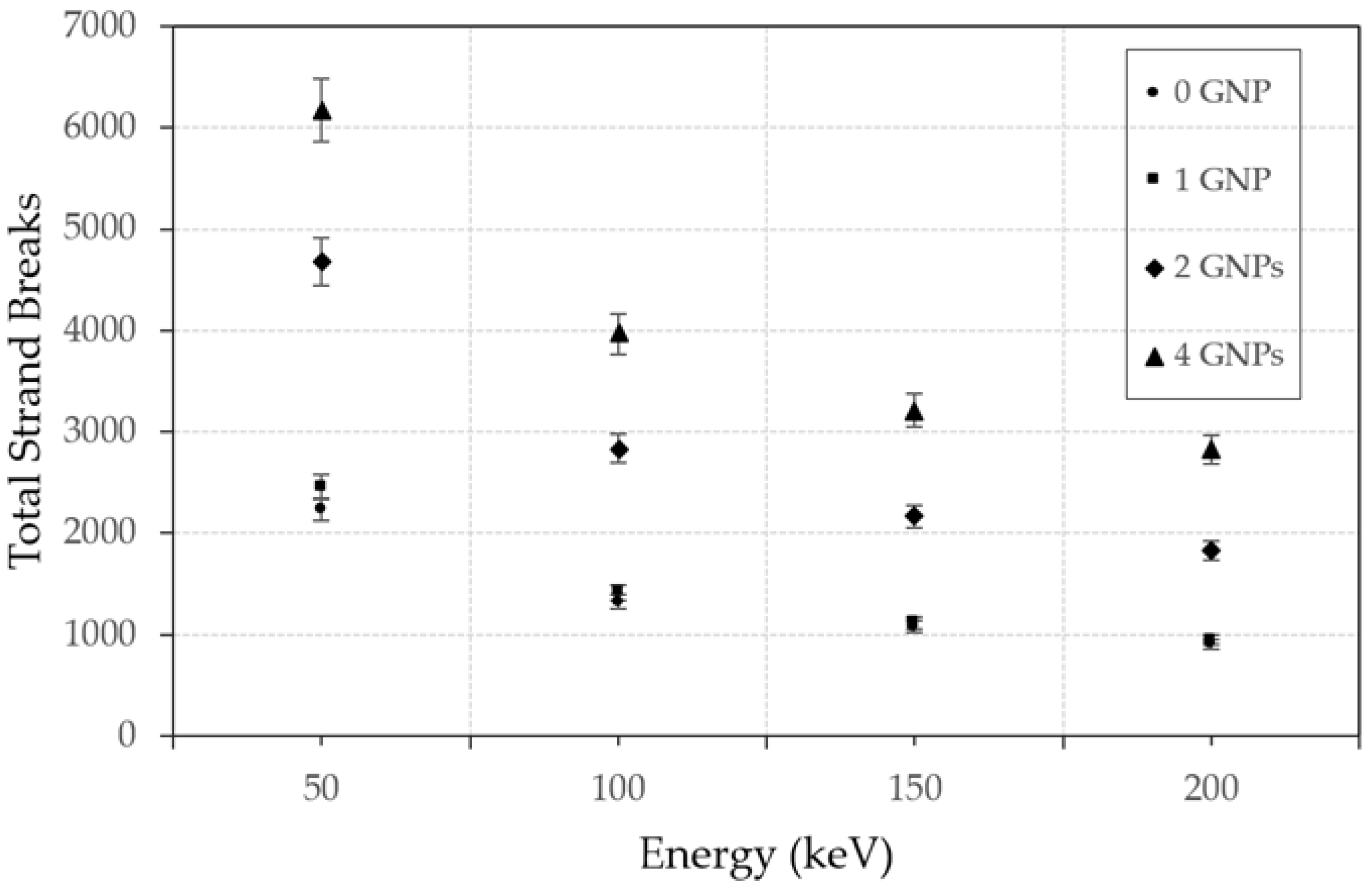
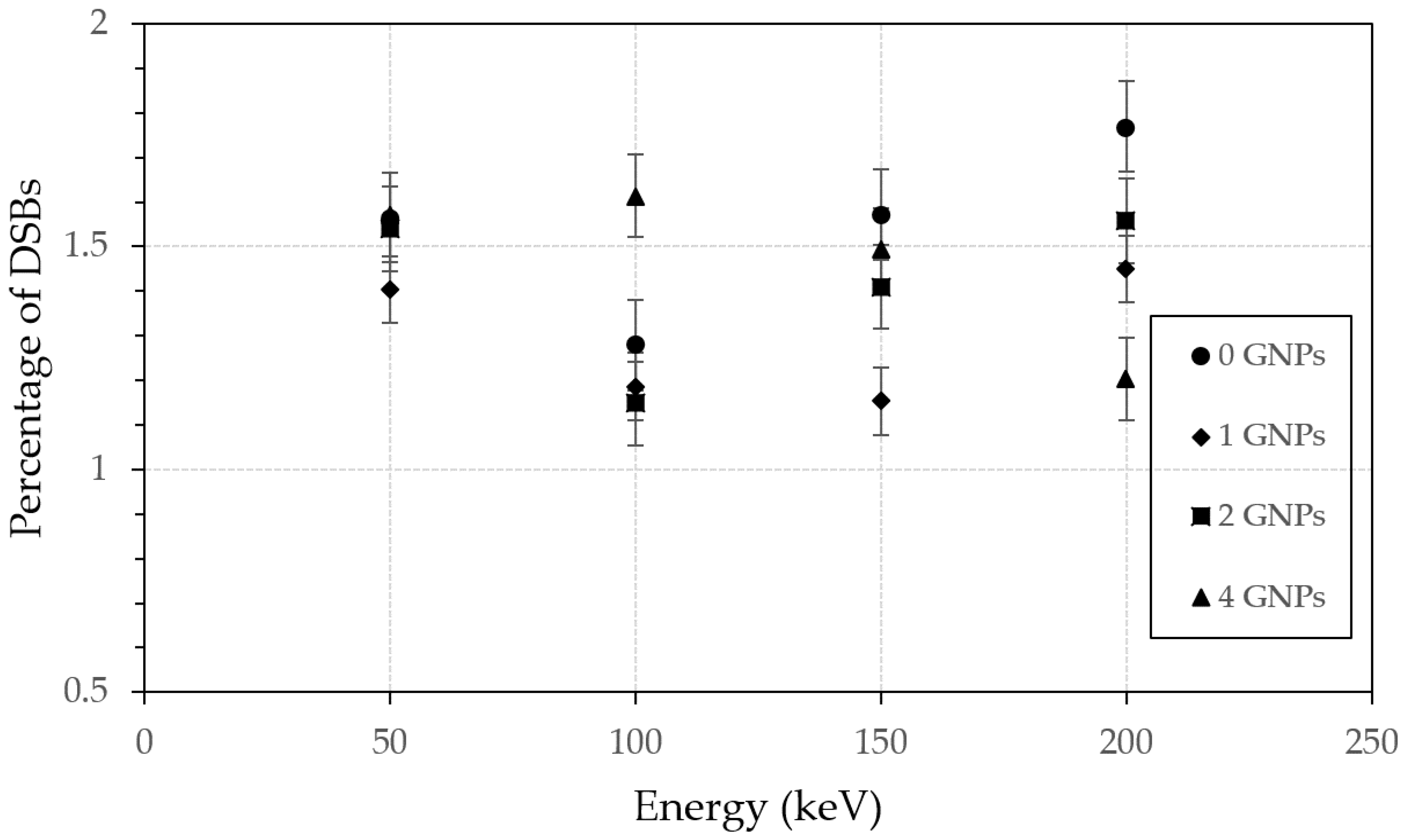
The PDB4DNA model was modified by adding GNPs, and the code was modified for each quantity, from 0, 1 (Figure 3a), 2 (Figure 3b), to 4 GNPs (Figure 3c). The largest nanoparticle had a radius of 5 nm and a volume of 523.6 nm3. The parameters used for 2 GNPs were a radius of 3.97 nm and a volume of 262.1 nm3. With both nanoparticles, the volume would equal about the same as 1 GNP. The last simulation trial with 4 gold nanoparticles had a radius of 3.15 nm and a volume of 130.92 nm3. The total volume of gold used in each scenario was 523.6 nm3.

3. Results

Figure 4 illustrates the correlation between the electron beam energy (50–200 keV) and the total number of strand breaks, as influenced by the quantity of GNPs used in the simulation. The volume of gold in each simulation was held constant, ensuring a consistent nanoparticle concentration across all geometries. The data clearly show that increasing the number of GNPs results in a higher number of strand breaks, while reducing the electron beam energy amplifies the strand break count. Table 1 provides an overview of the number of SSBs and DSBs detected in the Monte Carlo simulations using various quantities of nanoparticles that combine all the different energy levels. Interestingly, the number of DSBs was consistently lower than the SSBs, primarily due to the former being a more complex process requiring two SBs occurring in separate strands in close proximity. It was also evident that both SSBs and DSBs increased with the number of GNPs, assuming the same volume (concentration) of gold in the simulation. Figure 5 highlights the percentages of DSBs present in simulations using different quantities of nanoparticles at different energy levels. These findings offer insight into how the number and distribution of nanoparticles affect the proportion of DSBs during irradiation.

4. Discussion

4.1. Dependence of DNA Damege on Number of GNPs

In Figure 4, it is evident that the most extensive DNA damage and the greatest number of strand breaks occurred when four GNPs were present. Increasing the number of GNPs from 0 to 4 resulted in an increase in the total number of strand breaks. When the simulations held the volume of gold constant, adding more nanoparticles led to a reduction in nanoparticle size or an increase in the surface-to-volume ratio of the GNPs. This change intensified interactions between the nanoparticles and the radiation beam, thereby increasing the secondary electron yield. These secondary electrons could then be transported to the DNA to cause strand breaks. Another important factor to consider is the self-absorption of the GNP. It was observed that the self-absorption effect was more significant when the nanoparticle size increased. This effect resulted in a reduction in the secondary electrons generated by the nanoparticle, which subsequently reduced DNA damage. However, in the simulation geometry of the four GNPs, each nanoparticle was smaller than in the single GNP geometry. Therefore, the self-absorption effect was less significant, resulting in the generation of more secondary electrons from the four GNPs geometry. This caused more strand breaks and DNA damage.
Figure 4 also demonstrates that the total number of strand breaks for zero and one GNP was almost identical, while the differences between the number of strand breaks for zero and multiple GNPs were more significant. This discrepancy was primarily due to the single GNP geometry, in which all the gold volume contributed to a single nanoparticle, resulting in a larger nanoparticle size than in other GNP geometries. This caused an enhanced self-absorption effect, which offset the dose enhancement. Additionally, the presence of a single GNP acted as a “shield”, attenuating the electron beams that irradiated the DNA, thereby further reducing the enhancement of DNA damage.
Table 1 shows that the number of DSBs for a single GNP was slightly lower than that for the water NP or no GNP. This is due to the self-absorption effect of the single GNP, which reduced the number of secondary electrons that moved to the DNA. This effect was less pronounced when the GNP size was smaller. Since the total gold volume in each simulation with different nanoparticle quantities was constant, the amount of gold used in the clinic would remain the same if the number of gold nanoparticles increased. Even with smaller sizes, the same amount of gold would lead to enhanced DNA damage. Regarding the average number of DSBs per GNP, larger GNPs still had more DSBs than the smaller ones (e.g., 79 vs. 61 for one vs. four GNPs). On the other hand, increasing the number of GNPs and decreasing their size led to a higher total number of DSBs, as shown by the simulations with the four GNPs. This suggests that to enhance DNA damage at the same nanoparticle concentration, increasing the number of GNPs and decreasing their size is recommended. However, when comparing GNPs of different sizes while keeping the nanoparticle number constant, larger GNPs produce more DSBs than smaller ones do.

4.2. Dependence of DNA Damege on the Electron Beam Energy

The amount of DNA damage is dependent on the energy of the electrons used to irradiate the DNA. Increasing the electron energy from 50 to 200 keV resulted in a decrease in the total number of strand breaks. Specifically, at 50 keV, there were 15,576 strand breaks, while at 200 keV, there were 6559. Since electrons are charged particles, multiple scattering occurred when they entered the GNP, causing them to lose energy and generate secondary electrons. Maximizing the DNA damage required the production of the maximum number of secondary electrons through the interaction between the incident electron and the GNP. However, if the electron energy is too high, the incident electron will likely only penetrate the nanoparticle or generate a small number of scatterings within the nanoparticle, resulting in a reduced number of secondary electrons. In such cases, the incident electron may pass through the DNA without any further interaction. It is worth noting that in the simulation geometry used in this study, all incident electrons were directed toward the center of the nanoparticle to ensure that all electrons from the beam could interact with the GNP. Our findings suggest that the lowest electron beam energy of 50 keV is optimal for achieving maximum DNA damage with the four GNPs geometry.

4.3. Efficiency Depending on Double- to Single-Strand Break Ratio

The enhancement of DNA damage can be further analyzed by investigating the ratio of DSBs compared to SSBs. SSBs and DSBs are both types of damage that can occur to the DNA molecule. An SSB occurs when one of the strands of the DNA molecule is broken, but the other strand remains intact. SSBs can be caused by a variety of factors, including exposure to radiation. SSBs can be repaired by a process called base excision repair, in which the damaged base is removed and replaced with a new one. In contrast, a DSB occurs when both strands of the DNA molecule are broken. DSBs can be caused by exposure to ionizing radiation. DSBs are more severe than SSBs because they can lead to the loss of genetic information, chromosomal rearrangements, and cell death.
Ionizing radiation is used in radiotherapy to deposit energy in the DNA of cancer cells, resulting in both SSBs and DSBs that cause DNA damage. DSBs are more detrimental than SSBs as they cause the DNA to break into two pieces, making recovery and repair extremely challenging. Since DNA is vital for cancer cell reproduction, lethal DNA damage prevents cell replication and leads to cell death, enabling tumor control through radiotherapy. By increasing the secondary electron yield transport to the DNA, the addition of GNPs can enhance damage. Therefore, it is worth exploring whether altering the nanoparticle size and volume of gold would generate more DSBs during irradiation. Figure 5 displays the percentage of DSBs to total strand breaks. The graph demonstrates that regardless of energy or the number of gold nanoparticles used, the amount of DSBs produced remains consistent. On average, the proportion of DSBs is 1.43%, with a population standard deviation of 0.188. These percentages display minimal variation, suggesting that the pattern of DSB damage remains unchanged. Thus, it appears that reducing nanoparticle size and increasing their number, while keeping the gold amount constant, does not increase the proportion of DSBs in the total number of strand breaks.

4.4. Future Research

This study is a step toward increasing the enhancement of DNA damage. There is an increase in the number of total strand breaks when the energy is low and there are multiple GNPs, but there is room for improvement in increasing the favorable DNA damage. In the future, other parameters may be modified to find a way to improve the percentage of favorable strand breaks. Parameters that can be tested and affect DNA damage are the GNP radius, the distance between the GNP and the DNA, and greater numbers of DNA molecules. With the evidence of multiple nanoparticle enhancement, other radiation sources can be tested. With the improvements found through simulations, testing through live cells is the next step.
Ongoing research aims to construct a more realistic simulation model based on biological tissues that can account for the interactions between nanoparticles and incident electrons, thereby reflecting the complexities of a cancer cell in real-world settings. Moreover, as soon as a more realistic DNA model becomes available, it will be incorporated into the simulation.

5. Conclusions

This study employed Monte Carlo simulation to investigate the impact of different numbers of GNPs on DNA damage, marking the first time that single and multiple nanoparticle geometries had been compared. The study established the relationship between electron beam energy, the number of GNPs, and DNA damage in SSBs and DSBs. The results indicate that GNPs intensify the lethality of radiation by increasing the particle number while maintaining the same amount of gold. The 50 keV electron beams produced the highest number of DNA strand breaks due to the greater dose associated with lower electron energy. Notably, changing the number and size of GNPs did not alter the proportion of DSBs in DNA damage. These findings provide valuable insight into how DNA damage varies with GNP distribution compared to the single nanoparticle approach. Future work should be conducted on enhancing the ratio of DSBs to SSBs and the efficiency of DNA damage.

Author Contributions

Conceptualization, J.C.L.C.; methodology, J.C.L.C.; software, C.A.S. and J.C.L.C.; validation, C.A.S. and J.C.L.C.; formal analysis, C.A.S.; investigation, C.A.S.; resources, C.A.S. and J.C.L.C.; data curation, C.A.S.; writing—original draft preparation, C.A.S.; writing—review and editing, C.A.S. and J.C.L.C.; visualization, C.A.S. and J.C.L.C.; supervision, J.C.L.C.; project administration, J.C.L.C. All authors have read and agreed to the published version of the manuscript.

Funding

This research received no external funding.

Institutional Review Board Statement

Not Applicable.

Informed Consent Statement

Not Applicable.

Data Availability Statement

Not Applicable.

Acknowledgments

The authors would like to thank Chun He and Kaden Kujanpaa from the University of Toronto, Canada, and Mehwish Jabeen from the Toronto Metropolitan University, Canada, for their assistance in the Monte Carlo simulation using the Geant4-DNA code.

Conflicts of Interest

The authors declare no conflict of interest.

References

  1. World Cancer Research Fund International. Worldwide Cancer Data. Available online: https://www.wcrf.org/cancer-trends/worldwide-cancer-data/#:~:text=Find%20information%20about%20world%20cancer,and%208.8%20million%20in%20women (accessed on 8 March 2023).
  2. Canadian Cancer Statistics. Health Promotion and Chronic Disease Prevention in Canada; Public Health Agency of Canada: Ottawa, ON, Canada, 2021; Volume 41, p. 399.
  3. National Cancer Institute. Cancer Statistics. Available online: https://www.cancer.gov/about-cancer/understanding/statistics (accessed on 10 March 2023).
  4. Statistics Canada. New Cancer Estimates for 2022. Available online: https://www.statcan.gc.ca/o1/en/plus/1181-new-cancer-estimates-2022 (accessed on 10 March 2023).
  5. Baskar, R.; Lee, K.A.; Yeo, R.; Yeoh, K. Cancer and Radiation Therapy: Current Advances and Future Directions. Int. J. Med. Sci. 2012, 9, 193–199. [Google Scholar] [CrossRef] [PubMed] [Green Version]
  6. Gong, L.; Zhang, Y.; Lui, C.; Zhang, M.; Han, S. Applications of Radiosensitizer in Cancer Radiotherapy. Int. J. Nanomed. 2021, 16, 1083–1102. [Google Scholar] [CrossRef]
  7. Siddique, S.; Chow, J.C.L. Recent advances in functionalized nanoparticles in cancer theranostics. Nanomaterials 2022, 12, 2826. [Google Scholar] [CrossRef] [PubMed]
  8. Wang, H.; Mu, X.; He, H.; Zhang, X.D. Cancer radiosensitizers. Trends Pharmacol. Sci. 2018, 39, 24–48. [Google Scholar] [CrossRef] [PubMed]
  9. Siddique, S.; Chow, J.C.L. Application of Nanomaterials in Biomedical Imaging and Cancer Therapy. Nanomaterials 2020, 10, 1700. [Google Scholar] [CrossRef]
  10. Siddique, S.; Chow, J.C.L. Gold nanoparticles for drug delivery and cancer therapy. Appl. Sci. 2020, 10, 3824. [Google Scholar] [CrossRef]
  11. Chen, Y.; Yang, J.; Fu, S.; Wu, J. Gold nanoparticles as radiosensitizers in cancer radiotherapy. Int. J. Nanomed. 2020, 15, 9407–9430. [Google Scholar] [CrossRef]
  12. Abdulle, A.; Chow, J.C.L. Contrast enhancement for portal imaging in nanoparticle-enhanced radiotherapy: A Monte Carlo phantom evaluation using flattening-filter-free photon beams. Nanomaterials 2019, 9, 920. [Google Scholar] [CrossRef] [Green Version]
  13. Albayedh, F.; Chow, J.C.L. Monte Carlo simulation on the imaging contrast enhancement in nanoparticle-enhanced radiotherapy. J. Med. Phys. 2018, 43, 195–199. [Google Scholar]
  14. Mututantri-Bastiyange, D.; Chow, J.C.L. Imaging dose of cone-beam computed tomography in nanoparticle-enhanced image-guided radiotherapy: A Monte Carlo phantom study. AIMS Bioeng. 2020, 7, 1–11. [Google Scholar] [CrossRef]
  15. Chow, J.C.L. Characteristics of secondary electrons from irradiated gold nanoparticle in radiotherapy. In Handbook of Nanoparticles; Mahmood, A., Ed.; Springer International Publishing: Cham, Switzerland, 2015; Chapter 10; pp. 1–18. [Google Scholar]
  16. Chow, J.C.L.; Leung, M.K.K.; Jaffrey, D.A. Monte Carlo simulation on a gold nanoparticle irradiated by electron beams. Phys. Med. Biol. 2012, 47, 3323. [Google Scholar] [CrossRef] [PubMed]
  17. Penninckx, S.; Heuskin, A.; Michiels, C.; Lucas, S. Gold Nanoparticles as a Potent Radiosensitizer: A Transdisciplinary Approach from Physics to Patient. Cancers 2021, 12, 2021. [Google Scholar] [CrossRef] [PubMed]
  18. Moore, J.; Chow, J.C.L. Recent progress and applications of gold nanotechnology in medical biophysics using artificial intelligence and mathematical modeling. Nano Express 2021, 2, 022001. [Google Scholar] [CrossRef]
  19. Shrestha, S.; Cooper, L.N.; Andreev, O.A.; Reshetnyak, Y.K.; Antosh, M.P. Gold Nanoparticles for Radiation Enhancement in Vivo. Jacobs J. Radiat. Oncol. 2016, 3, 026. [Google Scholar]
  20. Byjus’s. Difference between Single-Strand Break and Double-Strand Break. Available online: https://byjus.com/biology/difference-between-single-strand-break-and-double-strand-break/ (accessed on 27 March 2023).
  21. Hogstrom, K.; Almond, P. Review of electron beam therapy physics. Phys. Med. Biol. 2006, 51, R455–R489. [Google Scholar] [CrossRef] [Green Version]
  22. Jabeen, M.; Chow, J.C.L. Gold Nanoparticle DNA Damage by Photon Beam in a Magnetic Field: A Monte Carlo Study. Nanomaterials 2021, 11, 1751. [Google Scholar] [CrossRef]
  23. Chun, H.; Chow, J.C.L. Gold nanoparticle DNA damage in radiotherapy: A Monte Carlo study. AIMS Bioeng. 2016, 3, 352–361. [Google Scholar]
  24. Leung, M.K.K.; Chow, J.C.L.; Chithrani, B.D.; Lee, M.J.G.; Oms, B.; Jaffray, D.A. Irradiation of gold nanoparticles by x-rays: Monte Carlo simulation of dose enhancements and the spatial properties of the secondary electrons production. Med. Phys. 2011, 38, 624–631. [Google Scholar] [CrossRef]
  25. Martelli, S.; Chow, J.C.L. Dose enhancement for the flattening-filter-free and flattening-filter photon beams in nanoparticle-enhanced radiotherapy: A Monte Carlo phantom study. Nanomaterials 2020, 10, 637. [Google Scholar] [CrossRef] [Green Version]
  26. Carrasco-Esteban, E.; Domínguez-Rullán, J.A.; Barrionuevo-Castillo, P.; Pelari-Mici, L.; Leaman, O.; Sastre-Gallego, S.; López-Campos, F. Current role of nanoparticles in the treatment of lung cancer. J. Clin. Transl. Res. 2021, 7, 140. [Google Scholar]
  27. Sadiq, A.; Chow, J.C.L. Evaluation of Dosimetric Effect of Bone Scatter on Nanoparticle-Enhanced Orthovoltage Radiotherapy: A Monte Carlo Phantom Study. Nanomaterials 2022, 12, 2991. [Google Scholar] [CrossRef] [PubMed]
  28. Rogers, D.W. Fifty years of Monte Carlo simulations for medical physics. Phys. Med. Biol. 2006, 51, R287. [Google Scholar] [CrossRef]
  29. Chow, J.C.L. Recent progress in Monte Carlo simulation on gold nanoparticle radiosensitization. AIMS Biophys. 2018, 5, 231–244. [Google Scholar] [CrossRef]
  30. Jabbari, K. Review of Fast Monte Carlo Codes for Dose Calculation in Radiation Therapy Treatment Planning. J. Med. Signals Sens. 2011, 1, 72–86. [Google Scholar] [CrossRef] [Green Version]
  31. Documentation. Geant4. Available online: https://geant4.web.cern.ch/docs/ (accessed on 11 March 2023).
  32. Incerti, S.; Baldacchino, G.; Bernal, M.; Capra, R.; Champion, C.; Francis, Z.; Gueye, P.; Mantero, A.; Mascialino, B.; Moretto, P.; et al. The geant4-dna project. Int. J. Model. Simul. Sci. Comput. 2010, 1, 157–178. [Google Scholar] [CrossRef]
  33. Delage, E.; Pham, Q.T.; Karamitros, M.; Paynom, H.; Stepan, V.; Incerti, S.; Maigne, L.; Perrot, Y. PDB4DNA: Implementation of DNA geometry from the Protein Data Bank (PDB) description for Geant4-DNA Monte-Carlo simulaitons. Comput. Phys. Commun. 2015, 192, 282–288. [Google Scholar] [CrossRef] [Green Version]
  34. Ngoc, H.H.; Chow, C.L. DNA Dosimetry with Gold Nanoparticle Irradiated by Proton Beams: A Monte Carlo Study on Dose Enhancement. Appl. Sci. 2021, 11, 10856. [Google Scholar]
Figure 1. (a) Visualization of the realistic PDB4DNA DNA model on the default application programming interface, OpenGL. (b) Visualization of the original DNA model used in previous work [22] on OpenGL.
Applsci 13 04916 g001
Figure 2. Diagram of a DNA simulation with radial electron beams. Electron beam energies used are equal to 50, 100, 150, and 200 keV. The radius of the gold nanoparticle is 5 nm. This diagram does not represent the exact scale or the ratio between the GNP and DNA molecule.
Applsci 13 04916 g002
Figure 3. (a) OpenGL visualization of the irradiated single gold nanoparticle with a radius of 5 nm and a DNA molecule. (b) OpenGL visualization of the irradiated single gold nanoparticle with a radius of 3.97 nm and a DNA molecule. (c) OpenGL visualization of the irradiated single gold nanoparticle with a radius of 3.15 nm and a DNA molecule. The red tracks represent the secondary electron paths in the simulation.
Applsci 13 04916 g003
Figure 4. Relationship between the total strand breaks and electron energy (keV) with a dependence on the number of GNPs present.
Applsci 13 04916 g004
Figure 5. Plot of the percentage of DSBs present at different energies when 0, 1, 2, and 4 GNPs are present.
Applsci 13 04916 g005
Table 1. Comparison of the number of SSBs to DSBs with increasing number of gold nanoparticles for all energies of 50, 100, 150, and 200 keV combined.
Number of GNPsSingle-Strand BreaksDouble-Strand Breaks
0546585
1594179
211,446165
415,932243
Disclaimer/Publisher’s Note: The statements, opinions and data contained in all publications are solely those of the individual author(s) and contributor(s) and not of MDPI and/or the editor(s). MDPI and/or the editor(s) disclaim responsibility for any injury to people or property resulting from any ideas, methods, instructions or products referred to in the content.

