Article

Influence of Thermal and Mechanical Load Cycling on Fracture Resistance of Premolars Filled with Calcium Silicate Sealer

1
Department of Restorative Dentistry, Faculty of Dentistry, University of Malaya, Kuala Lumpur 50603, Malaysia
2
Department of Restorative and Prosthetic Dental Sciences, College of Dentistry, Dar Al Uloom University, Riyadh 13314, Saudi Arabia
3
Periodontics and Community Dentistry, College of Dentistry, King Saud University, Riyadh 12372, Saudi Arabia
4
Department of Prosthodontics, College of Dentistry, Ibb University, Ibb 70270, Yemen
*
Author to whom correspondence should be addressed.
Appl. Sci. 2023, 13(7), 4388; https://doi.org/10.3390/app13074388
Submission received: 28 February 2023 / Revised: 20 March 2023 / Accepted: 21 March 2023 / Published: 30 March 2023
(This article belongs to the Special Issue Dental Biomaterials: Evaluation and Clinical Applications)

Abstract

:
The aim of this study was to evaluate the aging effect of thermomechanical cyclic load on fracture resistance of lower premolars obturated with AH Plus and BioRoot RCS root canal sealers. Forty-eight single-rooted premolars teeth were instrumented with REVO-S files up to SU/0.06 taper. The teeth were randomly assigned into 2 main groups (n = 24) according to the selected two root canal sealers (AH Plus or BioRoot RCS). All teeth were obturated using matching gutta-percha. Each main group were further divided into 3 subgroups (A, B and C) (n = 8). Group A acted as the negative control group (non-Thermomechanical aging). Whereas Group B and C were subjected first to thermal variations in a thermal cycling machine (7500 and 15,000 thermal cycles), then two different dynamic loading periods namely 3 × 10⁵ and 6 × 10⁵ in a masticatory simulator with a nominal load of 5 kg at 1.2 Hz which simulate approximately 1 ½ and 3 years of clinical function respectively. The roots were decoronated and fracture resistance were measured using a universal testing machine. After thermal-mechanical aging, BioRoot RCS showed significantly higher fracture resistance (p < 0.05) than AH Plus. As the thermal-mechanical cycles increased both AH Plus and BioRoot RCS exhibited a significant decrease in fracture resistance (p < 0.05). It could be concluded that thermomechanical aging had a significant impact on the outcome of the fracture resistance of AH Plus and BioRoot RCS.

1. Introduction

The removal of intracanal posts, excessive instrumentation, past endodontic treatment, internal root resorption, and/or dehydration from the use of irrigating solutions can all result in the thinning or weakening of root canal walls [1]. Consequently, this will make teeth less resistant to functional loads and more prone to fracture, resulting in tooth loss [2]. Vertical root fracture (VRF) is a root-canal treatment complication [3] that is considered the third main cause of extraction among root-canal-treated teeth, after caries and periodontal disease [4]. The frequency of extracted vertically fractured teeth among endodontically treated patients ranges from 11% to 20%. VRFs were detected in 10.9% of removed endodontically treated teeth (ETT) in the root canal system [5]. Endodontically treated premolars have the lowest survival rates due to the high frequency of vertical root fracture [6] because of their roots with a cross-section of a smaller mesiodistal diameter and with a deep oval or flattened shape [7]. The comparatively small size of their crown and the high occlusal forces they are subjected to explain their fragility [8] Therefore, one of the primary objectives of endodontic treatment is to strengthen the remaining tooth structure with root canal filling materials following chemical and mechanical preparation [9].
It is reported that the use of gutta percha (GP) as a core material along with root canal sealers are considered the standard root filling of endodontically treated teeth (ETT) [10]. However, since the GP has a lower elastic modulus than dentine, it has a little effect in reinforcing roots after root canal treatment [11]. Furthermore, GP cannot fill the whole three-dimensional canal space due to the existence of lateral canals, accessory canals, canal irregularities and small discrepancies that present between root dentinal walls and the obturating material [12]. Consequently, when selecting root canal sealers, some factors must be considered to seal the gap between the dentinal wall and the obturating material and to reinforce the remaining structure of the teeth [9]. It has been suggested that sealers with stable adhesion to the radicular dentine and have a Young modulus of elasticity similar to dentine will strengthen the remaining tooth structure, thus contributing to the long-term success of an ETT [9].
The epoxy resin-based AH Plus has been considered the “gold-standard” endodontic sealer [13,14] because of its low solubility and excellent flow, apical seal, biocompatibility, and adhesion to root dentine [15]. When compared to other sealers, AH Plus has shown promising outcomes. It demonstrated the lowest weight loss among the various root canal sealers in water and artificial saliva with varying pH values, regardless of the solubility medium used. In addition, compared to traditional sealers, AH Plus demonstrated the highest level of solution durability [16]. However, it has drawbacks, including the possibility of mutagenicity, cytotoxicity, and an inflammatory reaction. Furthermore, due to its hydrophobicity, the hydrophilic channel is unable to completely fill with fluid. Retained dental moisture, in particular, can cause problems in AH Plus adhesion to canal walls [17]. Thus, sealers or endodontic obturation materials providing higher fracture resistance and improved seal have been developed.
Sealers based on calcium silicates have recently gained popularity due to their favorable biological properties. They are biocompatible and non-cytotoxic, are dimensionally stable, chemically bond to root dentin, produce an alkaline pH, have antimicrobial properties, and promote enhanced root strength after obturation [18]. BioRoot RCS which exists in both liquid and powder form, is one of the calcium silicate-based root canal sealers. The primary ingredients of the powder, according to the manufacturer, are tricalcium silicate, povidone (a sticky agent), and zirconium dioxide (a contrast medium). Calcium chloride, a curing accelerator, and polycarboxylate, superplasticizers, are combined to form the liquid, which is an aqueous solution [19]. The manufacturer claims that this sealer is composed similarly to Biodentine (Septodont), probably in an effort to incorporate Biodentine’s desired characteristics into a root canal sealer [20]. BioRoot RCS provides biocompatibility and bioactivity and displays an appropriate seal with dentine [21]. The bioactive component can promote hard tissue deposit, providing a significant clinical benefit [22].
The mechanical behavior of teeth restored with different root canal sealers has been tested by applying monotonic loads to determine the load at which catastrophic fracture occurs [23]. However, It is debatable whether the results of a monotonic analysis have clinical significance because they do not accurately reflect actual occlusal loads and because the stress reactions to dynamic and static loads differ [24]. Consequently, testing the samples under cyclic loading and thermal cycling will be more clinically relevant and will improve predictions of how dental filling materials would perform when used in vivo [25]. Moreover, when evaluating the fracture or fatigue strength of dental materials, controlled temperature and moisture addition to the environment have been found to be significant [25]. It is not known if there is an undesirable effect of cyclic aging on fracture resistance of teeth obturated with epoxy resin-based and calcium silicate-based sealers.
This study analyzed the effects of thermomechanical cycling at two different periods (7500/3 × 105 and 15,000/6 × 105, simulating approximately 1 ½ and 3 years of clinical function, on the vertical root fracture VRF of ETT obturated with epoxy resin based and calcium silicate-based sealers. The null hypothesis was that the different root canal sealers would have similar fracture strength after different thermomechanical cycling.

2. Materials and Methods
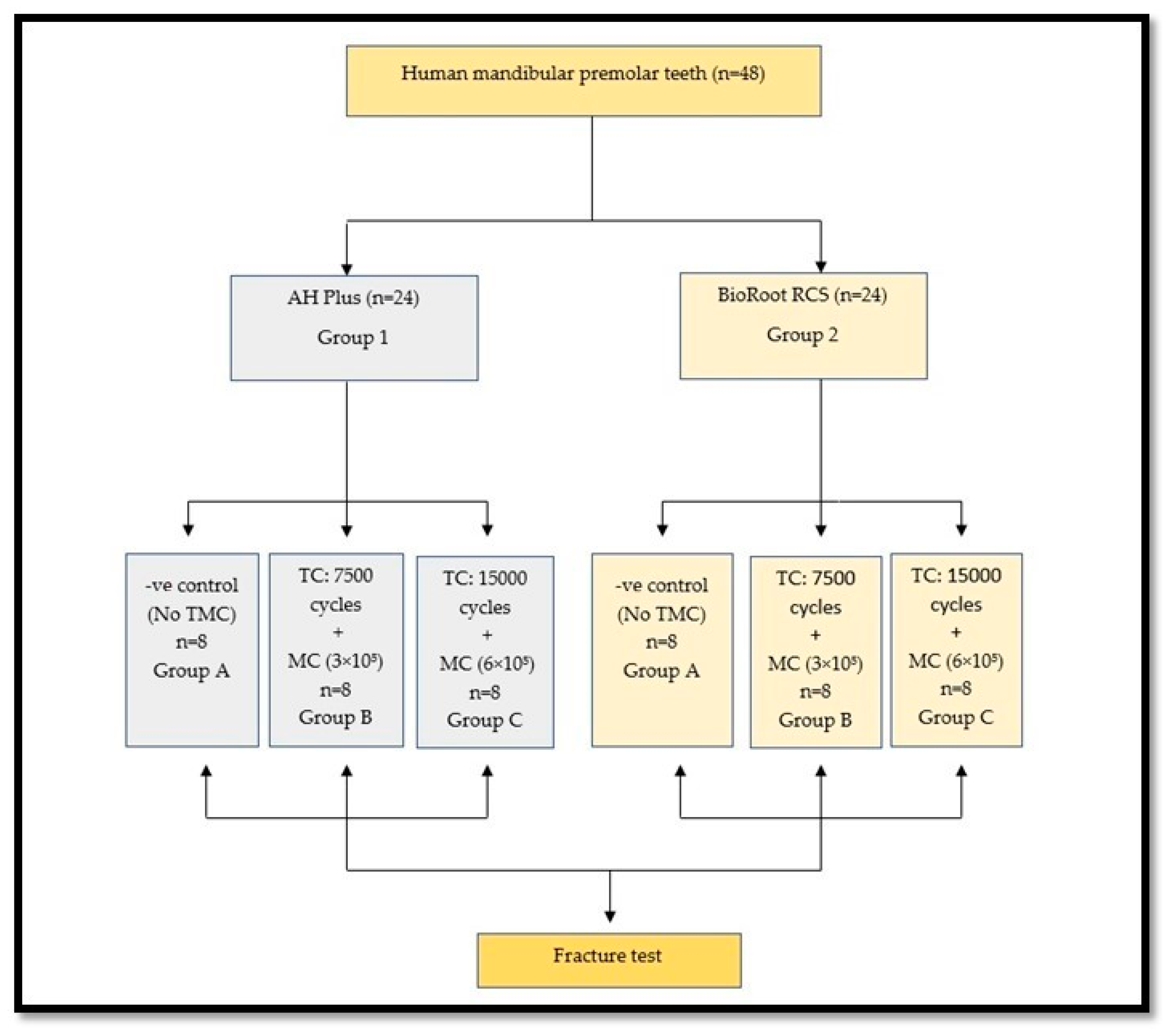
2.1. Sample Size

The sample size was determined through an a-priori power analysis performed with the G*Power software (G*Power V 3.1.9.7 Franz Faul, Universität Kiel, Kiel, Germany). This experiment was designed to detect statistical significance among groups in effect size of 0.65 with 82% power at a = 0.05. According to this calculation 24 extracted teeth were needed (8 per each group).

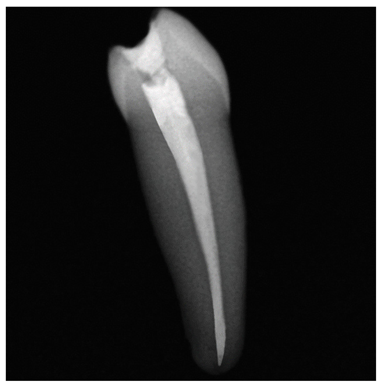
2.2. Specimen Preparation

The local research and ethics committee’s ethical approval was obtained before the study was carried out (DF-RD-1912/0032-P). Forty-eight single-rooted mandibular premolars extracted for orthodontic reasons were collected from patients between 18 and 30 years old. Using a digital caliper (Measuring Tool Enterprise, Shanghai, China), teeth with similar buccolingual (BL), mesiodistal (MD), and root lengths of 22.0 mm (1.0 mm) from the tip of the buccal cusp were compared to control for biological variances. Tooth with caries, open apex or previous root canal treatment were excluded. BL and MD periapical radiographs were made to confirm the presence of a straight single root canal without calcifications or resorption. The chosen teeth were kept in a 0.12% thymol solution and used within three months of storage.
With a round bur (Dentsply Maillefer, Ballaigues, Switzerland) the pulp chamber was accessed. The working length was determined to be 1 mm short of the apex by using K-file size 10 (MANI Inc., Utsunomiya, Tochigi, Japan). All teeth were instrumented using REVO-S rotary files (MICRO-MEGA, Besancon Cedex, France). The Revo-S three-step crown-down sequence was used according to the manufacturer’s instructions; SC1 (25, 0.06) tapered instrument was used with slow downward movement in a free progression and without pressure to enlarge the coronal two-thirds of the canal, SC2 (25, 0.04) tapered instrument was used with a progressive three-wave movement (up and down movement) to the working length, and SU (25, 0.06) tapered instrument was used to the working length with a slow downward movement in a free progression and without pressure. The root canals were irrigated with 3 mL of 2.5% NaOCl after each file. After completion of instrumentation, the root canals were rinsed with 2 mL of 5.25% NaOCl for 60 s, with 1 mL of 17% EDTA (Pulpdent Corp., Watertown, MA, USA) for 60 s, and with a final rinse of 5 mL normal saline. Just before obturation, the canals were dried with paper points (Brasseler USA, Savannah, GA, USA) and randomly assigned to 2 groups (n = 24): Group 1, AH Plus + GP (Dentsply Maillefer NA, Tulsa, OK, USA) and Group 2, BioRoot RCS + GP.
The root canals were obturated with matched-taper single-cone (SC) gutta percha coated with the tested sealers, which were handled according to the manufacturers’ instructions (Table 1). BL and MD radiographs were made to confirm the adequacy of the root filling in terms of appropriate length, density, and taper (Figure 1). The access opening was sealed with a composite resin (MultCore Flow; Ivoclar AG, Schaan, Liechtenstein). The teeth were stored at 37 °C with 100% humidity for 7 days to allow the sealers to set.

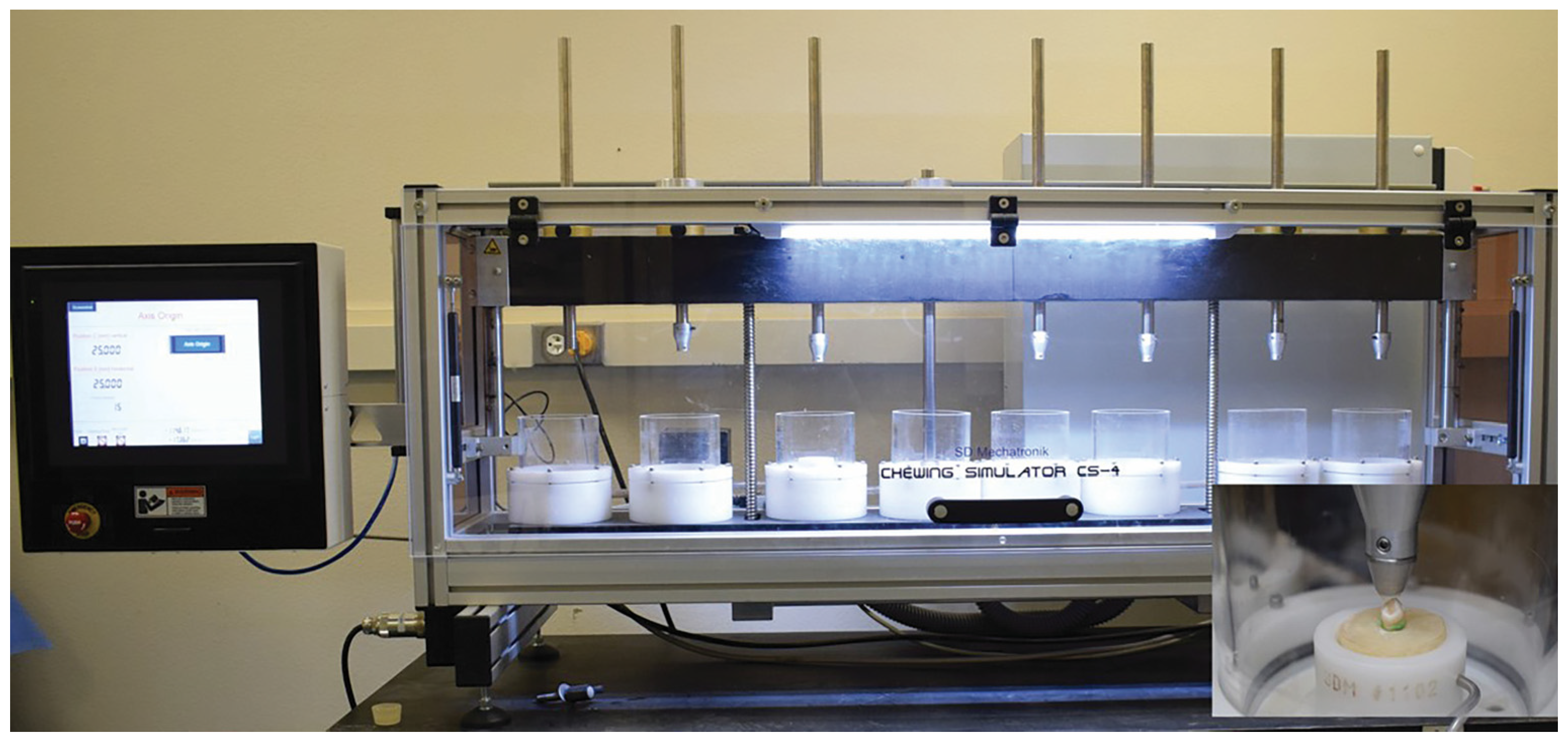
2.3. Thermomechanical Cycling
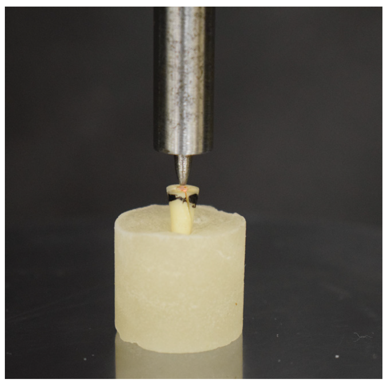
The teeth were immersed in molten wax (Horus; Herpo Produtos Dentarios, Petropolis, RJ, Brazil) up to 2 mm apical to the cementoenamel junction to simulate the periodontal ligament. After cooling, a 0.2- to 0.3-mm-thick wax layer was produced. Specimens from each group were placed in polyvinyl chloride cylinders containing an autopolymerized acrylic resin (Dencrilay, Dencril, SP, Brazil). To mimic the physiologic connection between the bone crest and the tooth, a 2-mm space was allowed between the top of the acrylic and the cemento-enamel junction on the facial and lingual aspects. Following resin polymerization, wax was washed away for two seconds from the surface of the roots with warm water. An impression syringe was used to inject a polyether impression material (Impregum Soft; 3M ESPE) into the resin cylinders. Then, the specimens were reinserted into their respective cylinder sockets. Each main group was further divided into 3 subgroups (A, B and C, n = 8 each) (Figure 2).
Group A was not thermomechanically cycled and acted as the negative control group. Samples from groups B and C were exposed to thermal changes in a thermal cycling machine (MCT2-AMM2, Saõ Paulo, SP, Brazil). The samples were set in a wire basket and exposed to 7500 and 15,000 thermal cycles in two separate water baths, one at 5 °C (2) and the other at 55 °C (2), with dwell times of one minute at each temperature and transfer times of five seconds.For mechanical cycling (MC), all test specimens from groups B and C were screwed into the upper sample holder, and the material specimens, along with their resin blocks, were fixed in the lower sample holder. At the masticatory simulator, the test specimens underwent two different dynamic loading periods, namely 3 × 10⁵ and 6 × 10⁵, simulate approximately 1 ½ and 3 years of clinical function [26] with a nominal load of 49 N at 1.2 Hz. The masticatory simulator has eight identical sample chambers. In each chamber there are two stepper motors, a bar, a weight, and two antagonistic specimens made of a 6 mm ceramic ball (Steatite Hoechst Ceram Tec, Wunsiedel, Germany) that can be moved vertically and horizontally by computer control (Figure 3). The ceramic ball has a Vicker hardness that is comparable to enamel and strikes the lingual incline of the buccal cusp at 2 mm from the central fossa of the crown. Three stages made up the masticatory cycle in this study: contact with a vertical load, horizontal sliding millimeters, and separation of the teeth and their antagonistic material.
The chewing simulator’s test parameters were changed to include: vertical movement of 6 mm, horizontal movement of 0.3 mm, rising speed of 55 mm/s, forward speed of 30 mm/s, descending speed of 30 mm/s, and backward speed of 55 mm/s; weight per sample of 5 kg; cycle frequency of 1.2 Hz; and kinetic energy of: 2250 × 10−6 [27]. These tests ran at room temperature under 37 ± 3 °C water irrigation.

2.4. Fracture Test

The roots were decoronated using a slow-speed diamond saw (Isomet 1000; Buhler, Lake Bluff, NY, USA) to achieve a 13-mm root length and loaded in a universal testing machine (Zwick Z010/TN2A; Zwick, Ulm, Germany). Using a spherical stainless-steel tip with a diameter of 2.8 mm attached to the top movable compartment of the testing machine and moving at a crosshead speed of 1 mm/min, each sample was subjected to gradually rising vertical force applied perpendicular to the long axis of the tooth at the junction of the buccal wall and root canal spaced until the root fractured (Figure 4).
The “fracture moment” was identified as the time when there was a dramatic drop in force, as shown on the testing machine display. In most samples, the fracture period was accompanied by an audible sound. Data were recorded using computer software (Instron® Bluehill Lite Software), and the maximum force necessary to fracture each specimen was recorded in Newtons (N).

2.5. Statistical Analysis

All fracture and dislodgment resistance data were analyzed with IBM SPSS statistical software version 25. The Shapiro–Wilk normality test and the Levene variance homogeneity tests were applied to the data and showed that the data were normally distributed and homogenous. Therefore, two-way ANOVA was used to analyze the effect of aging on the root canal sealers. One-way ANOVA was used to compare sealers within different time periods followed by the Tukey post hoc comparisons test, revealing the existence of significant differences between different time periods for each sealer. An independent t test (α = 0.05) was used to compare the mean of fracture for each group at different times.

3. Results

The mean values for fracture resistance and standard deviations (SD) for each main group and sub-group are show in Table 2. For AH Plus group, 3 years of thermomechanical aging resulted in the lowest fracture resistance values (53.69 ± 24.082) whereas the fracture resistance increased significantly to 98.41 ± 19.34 after 1 ½ of thermomechanical aging (p = 0.001). There were no significant differences between control group of AH Plus, which was not subjected to any thermomechanical aging, and 3 years of thermal-mechanical aging (p = 0.919) (Table 2).
In the BioRoot RCS group, as the number of thermomechanical aging increased to 1 ½ and 3 years, the mean force required for fracture was decreased, 730.36 ± 133.30 and 672.92 ± 131.11 respectively with significant differences between 1 ½ and 3 years of thermomechanical aging and control group, which was not subjected to any thermomechanical aging (p = 0.000 and p = 0.001).
Based on independent T-test (Table 2) (Figure 5), thermomechanical aging for 1 ½ and 3 years result has significantly higher resistance to fracture in all the tested groups obturated with BioRoot RCS when compared with AH Plus of (p = 0.000). Similarly, the control group of BioRoot RCS showed higher resistance to fracture compared to control group in AH Plus (p = 0.000).

4. Discussion

VRFs in ETT are inconvenient for both the patient and the dentist. VRFs typically develop slowly and go undetected by patients until clinical signs and symptoms arise [28] at which point root resection and tooth extraction are usually required [29]. In order to enhance the fracture resistance of ETT, clinicians have long worked to strengthen the remaining tooth structure [30]. In this situation, it would be advantageous to employ a root canal filling material or sealer that also reinforces the root against fracture [31].
The current study investigated and compared the fracture resistance of AH Plus and BioRoot RCS following thermomechanical ageing. The null hypothesis was rejected since all sealers demonstrated significant changes after different time periods of the thermomechanical cycling.
The purpose of the aging techniques is to simulate the challenges that chewing, clenching, and/or bruxing habits place on teeth and restorative materials in vivo by applying repeated and continuous mechanical pressures [32]. The aging method’s objective is not to immediately cause fractures, but rather to demonstrate that these may occur over time as the surrounding teeth and restorative material become more brittle. The spread of cracks could ultimately combine to raise the risk of chipping or catastrophic fractures [33]. By creating expansion and contraction stresses as a result of alternate immersions in liquid media at low and high temperatures, thermal cycling leads to stresses in the obturation material, the root canal wall, and the bonded interface [34]. The repeated temperature variations and chewing loads may impact the mechanical performance of obturation. Root canal sealers may experience accelerated mechanical deterioration and fatigue if mechanical and thermal cycling are used simultaneously (thermomechanical cycling) [35]. For a better understanding of mechanical material behavior, an in vitro methodology that can replicate some in vivo challenges must be developed.
After repeated loading, mechanical cycling (MC), a fatigue test that can replicate masticatory activity, may cause structural fractures, which can be explained as the result of minute fissures spreading from concentrated areas of strain [36]. MC provides an excellent tool for predicting the clinical efficacy of various materials and restoration procedures [37]. The chewing simulator in a computer-controlled mastication simulation is the most widely used fatigue device. It can mimic the masticatory pressures and the environment that dental restorations are exposed to while in use [36].
The BioRoot RCS manufacturer instructs using a single cone or cold lateral condensation (LC) methods for obturation [38]. Because warm vertical compaction is not recommended for the bioceramic sealers tested in this study, it would affect the adhesion properties of these materials, which would have an impact on the outcomes [39]. According to Moinzadeh et al. [40], cold lateral compaction of gutta-percha produced noticeably more voids, especially coronally, than a single gutta-percha cone obturation. Additionally, the matched-taper SC technique avoids the spreaders’ wedging pressures during the LC technique, which lowers fracture resistance [41]. Taking into account these details, the single-cone technique was employed in the current research.
The anatomical variations, age, and timing of tooth extraction that may affect the findings make it difficult to standardize human teeth for evaluation of fracture strength [42]. It is important to standardize dimensions of the MD, BL, and root canal diameter and length of the extracted samples [43]. In our study, the BL and MD coronal plane diameters were measured using a digital caliper. The preparation size, root length, and root width were standardized for all teeth. Due to the evenly distributed stress in roots during filling, creating canals with a round cross-section reduces the risk of root fracture [44]. To accomplish this, rotary files were used for root canal preparation.
Moreover, the testing device’s parameters in this experiment were restricted to the physiological values reported in the literature [27]. Prior to loading, the specimens are exposed to thermal cycling between 5 and 55 °C according to ISO standardized bath temperature [45]. The forces that are generated by chewing or swallowing, which frequently range from 2 to 50 N Thus, a clinically relevant condition was approached using a cyclic loading force of 49 N [25].
To simulate bony sockets that may alter the force distributions around the teeth when external pressures like lateral compaction are used, many studies have replicated the periodontal ligament during root canal filling [46]. Elastomeric materials, according to Soros et al. [46], are unable to react to compaction forces in the same way as normal ligament, which collapses under pressure. In the present experiment, after filling and before the thermal-mechanical cycling began, the roots were surrounded with a polyether impression material. This method tried to mimic the periodontal ligament, which effectively improves stress distribution during fatigue loading [47].
In the current study, the samples were decoronated at the cement-enamel junction in accordance with a prior study [48], for the sake of simplification and to eliminate the access cavity as a variable, the compressive load was applied directly to the root canal filling in this study [49]. The roots protruded 9 mm above the embedding substance. This is to mimic the clinical situation in which no 50% bone support is required for premolars to have a good periodontal prognosis if all other variables are favourable [50].
The present study revealed that thermomechanical aging resulted in statistically significant differences, BioRoot RCS showed decreased fracture resistance after different periods of aging, while AH Plus showed increased fracture resistance after a simulated 1 ½ years and then a dramatic reduction in fracture resistance after 3 years of aging. However, all different time periods of thermomechanical aging with BioRoot RCS showed significantly higher fracture resistance than AH Plus, in contrast with previous studies [51,52,53]. There may not have been any differences in VRF between BioRoot RCS and AH Plus in prior experiments because of the absence of fatigue stress.
The increased fracture resistance of BioRoot RCS compared to AH Plus sealers may be caused by the fact that BioRoot RCS acquired lower flow and higher film thickness values than those recommended for AH Plus in ISO 6876 [54]. As BioRoot RCS has a low contact angle and is hydrophilic material, it should enable these sealers to penetrate the difficult aspects of the root canal more effectively [55]. Furthermore, zirconium oxide, one of the components of BioRoot RCS has properties that include high fracture resistance, high tensile strength, and a low Young modulus [56]. Calcium silicate sealers have a higher modulus of elasticity than epoxy resin-based sealers, which are closer to dentine, improving fracture resistance [36].
Another explanation for the high fracture resistance of BioRoot RCS compared to AH Plus in all periods of thermomechanical aging may be the higher bond strength of Calcium silicate sealers due to the formation of apatite layer, intrafibrillar apatite deposition and tag-like structures that form mineral plugs along with the dentine bonding interface, known as the mineral infiltration zone [57]. Thus, increased mechanical interlocking and improve the adaptation to dentin may increase resistance to fracture [54]. Besides that, bioceramic sealers go through a bioactivity process that results in apatite nucleation, which protects the gutta-percha from dislodging from the canal [57].
A reduction was found in the fracture resistance values of BioRoot RCS after two different periods of thermomechanical aging, possibly because of the adverse effects of the high alkaline pH of calcium silicate sealers [58]. The calcium silicate weakens the collagenous component of the interfacial dentine, increasing permeability by breaking down the intermolecular bonds of the collagen fibrils and negatively impacting the mechanical properties of the root [59]. In addition, the degradation may affect the mechanical properties of the calcium silicate sealer after prolonged thermomechanical cycling.
The findings with AH Plus were noteworthy. After thermomechanical aging for a simulated 1 ½ years, AH Plus showed increasing in resistance to fracture, although this sealer led to reduced fracture resistance in the unaged control group. The rise could be related to the beneficial effects of AH Plus initial expansion, which might balance out or overcome the adverse effects of the sealer’s polymerization shrinkage in the control group [60]. Longer thermomechanical cycling might have weakened the covalent bonds between the epoxy resin and amino groups, resulting in a lower resistance to fracture of AH Plus after 3 years.

5. Conclusions

The results suggest that increased thermomechanical aging negatively affected the fracture resistance of both sealers. However, BioRoot RCS provided improved mechanical performance than AH Plus. To more properly estimate and evaluate the performance of root canal material in laboratory tests, an accurate simulation of the oral environment may be required. Clinical trials, as well as studies involving other groups of teeth, different testing methods (such as stairwell), different loading assemblies (such as sliding), or different endodontic treatment techniques, should be conducted.

Author Contributions

Concept—A.S. (Ahlam Smran), M.A. and N.A.-M.; Design—A.S. (Abdulaziz Samran); Supervision—M.A. and N.A.A.; Materials—A.S. (Ahlam Smran) and A.S. (Abdulaziz Samran); Data Collection and/or Processing—A.S. (Ahlam Smran); Analysis and/or Interpretation—A.S.(Ahlam Smran) and M.A.; Literature Review—A.S. (Ahlam Smran) and A.S. (Abdulaziz Samran); Writing—A.S. (Ahlam Smran) and A.S. (Abdulaziz Samran); Critical Review—A.S. (Ahlam Smran) and M.A. All authors have read and agreed to the published version of the manuscript.

Funding

This research received no external funding.

Institutional Review Board Statement

The study was conducted in accordance with the Declaration of Helsinki, and approved by the Research Ethics Committee (Approval No: DF-RD-1912/0032).

Informed Consent Statement

Informed consent was obtained from all subjects involved in the study.

Data Availability Statement

The data used to support the findings of this study are available from the corresponding author upon request.

Acknowledgments

The authors extend their appreciation to the Deanship of Postgraduate and Scientific Research at Dar Al-ULoom University for their support for this work.

Conflicts of Interest

The authors declare no conflict of interest.

References

  1. Uzunoglu, E.; Aktemur, S.; Uyanik, M.O.; Durmaz, V.; Nagas, E. Effect of Ethylenediaminetetraacetic Acid on Root Fracture with Respect to Concentration at Different Time Exposures. J. Endod. 2012, 38, 1110–1113. [Google Scholar] [CrossRef]
  2. Al-Omiri, M.K.; Mahmoud, A.A.; Rayyan, M.R.; Abu-Hammad, O. Fracture Resistance of Teeth Restored with Post-Retained Restorations: An Overview. J. Endod. 2010, 36, 1439–1449. [Google Scholar] [CrossRef]
  3. Mikrogeorgis, G.; Eirinaki, E.; Kapralos, V.; Koutroulis, A.; Lyroudia, K.; Pitas, I. Diagnosis of Vertical Root Fractures in Endodontically Treated Teeth Utilising Digital Subtraction Radiography: A Case Series Report. Aust. Endod. J. 2018, 44, 286–291. [Google Scholar] [CrossRef]
  4. Seo, D.-G.; Yi, Y.-A.; Shin, S.-J.; Park, J.-W. Analysis of Factors Associated with Cracked Teeth. J. Endod. 2012, 38, 288–292. [Google Scholar] [CrossRef] [PubMed]
  5. Yoshino, K.; Ito, K.; Kuroda, M.; Sugihara, N. Prevalence of Vertical Root Fracture as the Reason for Tooth Extraction in Dental Clinics. Clin. Oral Investig. 2015, 19, 1405–1409. [Google Scholar] [CrossRef]
  6. Almasri, M. Assessment of Extracting Molars and Premolars after Root Canal Treatment: A Retrospective Study. Saudi Dent. J. 2019, 31, 487–491. [Google Scholar] [CrossRef] [PubMed]
  7. Lertchirakarn, V.; Palamara, J.E.; Messer, H.H. Patterns of Vertical Root Fracture: Factors Affecting Stress Distribution in the Root Canal. J. Endod. 2003, 29, 523–528. [Google Scholar] [CrossRef] [PubMed] [Green Version]
  8. Ghoddusi, J.; Bagherpour, A.; Mahmudabadi, F.; Forghani, M.; Sarmad, M. Residual Dentin Thickness of Bifurcated Maxillary Premolars Following Two Post Space Preparation Methods. Iran. Endod. J. 2013, 8, 94. [Google Scholar]
  9. Johnson, M.E.; Stewart, G.P.; Nielsen, C.J.; Hatton, J.F. Evaluation of Root Reinforcement of Endodontically Treated Teeth. Oral Surg. Oral Med. Oral Pathol. Oral Radiol. Endodontol. 2000, 90, 360–364. [Google Scholar] [CrossRef]
  10. Gulsahi, K.; Cehreli, Z.; Kuraner, T.; Dagli, F. Sealer Area Associated with Cold Lateral Condensation of Gutta-percha and Warm Coated Carrier Filling Systems in Canals Prepared with Various Rotary NiTi Systems. Int. Endod. J. 2007, 40, 275–281. [Google Scholar] [CrossRef]
  11. Ribeiro, F.C.; Souza-Gabriel, A.E.; Marchesan, M.A.; Alfredo, E.; Silva-Sousa, Y.T.C.; Sousa-Neto, M.D. Influence of Different Endodontic Filling Materials on Root Fracture Susceptibility. J. Dent. 2008, 36, 69–73. [Google Scholar] [CrossRef]
  12. Lin, G.S.S.; Ghani, N.R.N.A.; Noorani, T.Y.; Kamarudin, A. Apical Sealing Ability of Different Endodontic Sealers Using Glucose Penetration Test: A Standardized Methodological Approach. Cumhur. Dent. J. 2020, 23, 79–87. [Google Scholar] [CrossRef]
  13. Lee, S.-J.; Monsef, M.; Torabinejad, M. Sealing Ability of a Mineral Trioxide Aggregate for Repair of Lateral Root Perforations. J. Endod. 1993, 19, 541–544. [Google Scholar] [CrossRef]
  14. Neelakantan, P.; Subbarao, C.; Subbarao, C.V.; De-Deus, G.; Zehnder, M. The Impact of Root Dentine Conditioning on Sealing Ability and Push-out Bond Strength of an Epoxy Resin Root Canal Sealer. Int. Endod. J. 2011, 44, 491–498. [Google Scholar] [CrossRef] [Green Version]
  15. Hergt, A.; Wiegand, A.; Hülsmann, M.; Rödig, T. AH Plus Root Canal Sealer–an Updated Literature. Endod. Pract. Today 2015, 9, 245–265. [Google Scholar]
  16. McMichen, F.; Pearson, G.; Rahbaran, S.; Gulabivala, K. A Comparative Study of Selected Physical Properties of Five Root-canal Sealers. Int. Endod. J. 2003, 36, 629–635. [Google Scholar] [CrossRef]
  17. Azar, N.G.; Heidari, M.; Bahrami, Z.S.; Shokri, F. In Vitro Cytotoxicity of a New Epoxy Resin Root Canal Sealer. J. Endod. 2000, 26, 462–465. [Google Scholar] [CrossRef] [PubMed]
  18. Antunovic, M.; Vukmanovic, L.; Budimir, A.; Kabil, E.; Anic, I.; Bago, I. Evaluation of Sealing Ability of Four Bioceramic Root Canal Sealers and an Epoxy Resin-Based Sealer: An in Vitro Study. Saudi Endod. J. 2021, 11, 66. [Google Scholar]
  19. Prüllage, R.-K.; Urban, K.; Schäfer, E.; Dammaschke, T. Material Properties of a Tricalcium Silicate–Containing, a Mineral Trioxide Aggregate–Containing, and an Epoxy Resin–Based Root Canal Sealer. J. Endod. 2016, 42, 1784–1788. [Google Scholar] [CrossRef]
  20. Guneser, M.B.; Akman, M.; Kolcu, İ.B.; Eldeniz, A.U. Fracture Resistance of Roots Obturated with a Novel Calcium Silicate-Based Endodontic Sealer (BioRoot RCS). J. Adhes. Sci. Technol. 2016, 30, 2420–2428. [Google Scholar] [CrossRef]
  21. Siboni, F.; Taddei, P.; Zamparini, F.; Prati, C.; Gandolfi, M.G. Properties of BioRoot RCS, a Tricalcium Silicate Endodontic Sealer Modified with Povidone and Polycarboxylate. Int. Endod. J. 2017, 50, e120–e136. [Google Scholar] [CrossRef] [PubMed] [Green Version]
  22. Seo, D.-G.; Lee, D.; Kim, Y.-M.; Song, D.; Kim, S.-Y. Biocompatibility and Mineralization Activity of Three Calcium Silicate-Based Root Canal Sealers Compared to Conventional Resin-Based Sealer in Human Dental Pulp Stem Cells. Materials 2019, 12, 2482. [Google Scholar] [CrossRef] [Green Version]
  23. Sterzenbach, G.; Rosentritt, M.; Frankenberger, R.; Paris, S.; Naumann, M. Loading Standardization of Postendodontic Restorations in Vitro: Impact of Restorative Stage, Static Loading, and Dynamic Loading. Oper. Dent. 2012, 37, 71–79. [Google Scholar] [CrossRef] [Green Version]
  24. Ou, K.; Chang, C.; Chang, W.; Lin, C.; Chang, K.; Huang, H. Effect of Damping Properties on Fracture Resistance of Root Filled Premolar Teeth: A Dynamic Finite Element Analysis. Int. Endod. J. 2009, 42, 694–704. [Google Scholar] [CrossRef] [PubMed]
  25. Att, W.; Kurun, S.; Gerds, T.; Strub, J.R. Fracture Resistance of Single-Tooth Implant-Supported All-Ceramic Restorations: An in Vitro Study. J. Prosthet. Dent. 2006, 95, 111–116. [Google Scholar] [CrossRef]
  26. Baldissara, P.; Zicari, F.; Valandro, L.F.; Scotti, R. Effect of Root Canal Treatments on Quartz Fiber Posts Bonding to Root Dentin. J. Endod. 2006, 32, 985–988. [Google Scholar] [CrossRef]
  27. Habib, S.R.; Alotaibi, A.; Al Hazza, N.; Allam, Y.; AlGhazi, M. Two-Body Wear Behavior of Human Enamel versus Monolithic Zirconia, Lithium Disilicate, Ceramometal and Composite Resin. J. Adv. Prosthodont. 2019, 11, 23–31. [Google Scholar] [CrossRef] [Green Version]
  28. Tay, F.R.; Loushine, R.J.; Monticelli, F.; Weller, R.N.; Breschi, L.; Ferrari, M.; Pashley, D.H. Effectiveness of Resin-Coated Gutta-Percha Cones and a Dual-Cured, Hydrophilic Methacrylate Resin-Based Sealer in Obturating Root Canals. J. Endod. 2005, 31, 659–664. [Google Scholar] [CrossRef] [PubMed]
  29. Lam, P.P.; Palamara, J.E.; Messer, H.H. Fracture Strength of Tooth Roots Following Canal Preparation by Hand and Rotary Instrumentation. J. Endod. 2005, 31, 529–532. [Google Scholar] [CrossRef] [PubMed]
  30. Trope, M.; Ray Jr, H.L. Resistance to Fracture of Endodontically Treated Roots. Oral Surg. Oral Med. Oral Pathol. 1992, 73, 99–102. [Google Scholar] [CrossRef]
  31. Demiriz, L.; Bodrumlu, E.H. Fracture Resistance of Simulated Incomplete Vertical Fractured Teeth Treated with Different Bioceramic Based Root Canal Sealers. Int. J. Appl. Dent. Sci. 2018, 4, 25–30. [Google Scholar]
  32. Arola, D. Fatigue Testing of Biomaterials and Their Interfaces. Dent. Mater. 2017, 33, 367–381. [Google Scholar] [CrossRef]
  33. da Silva, G.R.; da Silva, N.R.; Soares, P.V.; Costa, A.R.; Fernandes-Neto, A.J.; Soares, C.J. Influence of Different Load Application Devices on Fracture Resistance of Restored Premolars. Braz. Dent. J. 2012, 23, 484–489. [Google Scholar] [CrossRef] [Green Version]
  34. Georgiev, Z.; Kovacevska, I.; Sotirovska-Ivkovska, A.; Zabokova-Bilbilova, E.; Dimova, C. Methods of Deciduous Dental Pulp Research. Int. Dent. J. 2013, 63, 151–153. [Google Scholar]
  35. Lima, V.P.; Machado, J.B.; Zhang, Y.; Loomans, B.A.; Moraes, R.R. Laboratory Methods to Simulate the Mechanical Degradation of Resin Composite Restorations. Dent. Mater. 2022, 38, 214–229. [Google Scholar] [CrossRef]
  36. Nawafleh, N.; Hatamleh, M.; Elshiyab, S.; Mack, F. Lithium Disilicate Restorations Fatigue Testing Parameters: A Systematic Review. J. Prosthodont. 2016, 25, 116–126. [Google Scholar] [CrossRef]
  37. Yusufoglu, S.İ.; Akman, M.; Akbulut, M.B.; Eldeniz, A.Ü. Fracture Resistance of Roots Enlarged with Various Rotary Systems and Obturated with Different Sealers. J. Dent. Res. Dent. Clin. Dent. Prospect. 2019, 13, 215. [Google Scholar] [CrossRef] [Green Version]
  38. Viapiana, R.; Moinzadeh, A.T.; Camilleri, L.; Wesselink, P.R.; Tanomaru Filho, M.; Camilleri, J. Porosity and Sealing Ability of Root Fillings with Gutta-percha and BioRoot RCS or AH Plus Sealers. Evaluation by Three Ex Vivo Methods. Int. Endod. J. 2016, 49, 774–782. [Google Scholar] [CrossRef]
  39. Al-Hiyasat, A.S.; Alfirjani, S.A. The Effect of Obturation Techniques on the Push-out Bond Strength of a Premixed Bioceramic Root Canal Sealer. J. Dent. 2019, 89, 103169. [Google Scholar] [CrossRef]
  40. Moinzadeh, A.T.; Zerbst, W.; Boutsioukis, C.; Shemesh, H.; Zaslansky, P. Porosity Distribution in Root Canals Filled with Gutta Percha and Calcium Silicate Cement. Dent. Mater. 2015, 31, 1100–1108. [Google Scholar] [CrossRef]
  41. Pişkin, B.; Aydın, B.; Sarıkanat, M. The Effect of Spreader Size on Fracture Resistance of Maxillary Incisor Roots. Int. Endod. J. 2008, 41, 54–59. [Google Scholar] [CrossRef]
  42. Özyürek, T.; Ülker, Ö.; Demiryürek, E.Ö.; Yılmaz, F. The Effects of Endodontic Access Cavity Preparation Design on the Fracture Strength of Endodontically Treated Teeth: Traditional versus Conservative Preparation. J. Endod. 2018, 44, 800–805. [Google Scholar] [CrossRef]
  43. Sagsen, B.; Er, O.; Kahraman, Y.; Akdogan, G. Resistance to Fracture of Roots Filled with Three Different Techniques. Int. Endod. J. 2007, 40, 31–35. [Google Scholar] [CrossRef]
  44. Bonessio, N.; Arias, A.; Lomiento, G.; Peters, O.A. Effect of Root Canal Treatment Procedures with a Novel Rotary Nickel Titanium Instrument (TRUShape) on Stress in Mandibular Molars: A Comparative Finite Element Analysis. Odontology 2017, 105, 54–61. [Google Scholar] [CrossRef]
  45. Xie, C.; Han, Y.; Zhao, X.-Y.; Wang, Z.-Y.; He, H.-M. Microtensile Bond Strength of One-and Two-Step Self-Etching Adhesives on Sclerotic Dentin: The Effects of Thermocycling. Oper. Dent. 2010, 35, 547–555. [Google Scholar] [CrossRef] [Green Version]
  46. Soros, C.; Zinelis, S.; Lambrianidis, T.; Palaghias, G. Spreader Load Required for Vertical Root Fracture during Lateral Compaction Ex Vivo: Evaluation of Periodontal Simulation and Fracture Load Information. Oral Surg. Oral Med. Oral Pathol. Oral Radiol. Endodontol. 2008, 106, e64–e70. [Google Scholar] [CrossRef]
  47. Ausiello, P.; Ciaramella, S.; Martorelli, M.; Lanzotti, A.; Zarone, F.; Watts, D.C.; Gloria, A. Mechanical Behavior of Endodontically Restored Canine Teeth: Effects of Ferrule, Post Material and Shape. Dent. Mater. 2017, 33, 1466–1472. [Google Scholar] [CrossRef] [Green Version]
  48. Rippe, M.P.; Santini, M.F.; Bier, C.A.S.; Borges, A.L.S.; Valandro, L.F. Root Canal Filling: Fracture Strength of Fiber-Reinforced Composite-Restored Roots and Finite Element Analysis. Braz. Dent. J. 2013, 24, 619–625. [Google Scholar] [CrossRef] [Green Version]
  49. Ulusoy, Ö. Fracture Resistance of Roots with Simulated Internal Resorption Defects and Obturated Using Different Hybrid Techniques. J. Dent. Sci. 2017, 12, 121–125. [Google Scholar] [CrossRef] [Green Version]
  50. Nagpal, A.; Annapoorna, B.M.; Prashanth, M.B.; Prashanth, N.T.; Singla, M.; Deepak, B.S.; Singh, A.; Tavane, P.N. A Comparative Evaluation of the Vertical Root Fracture Resistance of Endodontically Treated Teeth Using Different Root Canal Sealers: An in Vitro Study. J. Contemp. Dent. Pract. 2012, 13, 351–355. [Google Scholar]
  51. Saba, A.A.; Al-Zaka, I.M. Fracture Resistance of Endodontically Treated Teeth Obturated with Different Root Canal Sealers (in Vitro Study). J. Contemp. Dent. Pract. 2019, 65, 1567–1575. [Google Scholar] [CrossRef] [Green Version]
  52. Osiri, S.; Banomyong, D.; Sattabanasuk, V.; Yanpiset, K. Root Reinforcement after Obturation with Calcium Silicate–Based Sealer and Modified Gutta-Percha Cone. J. Endod. 2018, 44, 1843–1848. [Google Scholar] [CrossRef] [PubMed]
  53. Khalil, I.; Naaman, A.; Camilleri, J. Properties of Tricalcium Silicate Sealers. J. Endod. 2016, 42, 1529–1535. [Google Scholar] [CrossRef] [PubMed]
  54. Zhang, W.; Li, Z.; Peng, B. Assessment of a New Root Canal Sealer’s Apical Sealing Ability. Oral Surg. Oral Med. Oral Pathol. Oral Radiol. Endodontol. 2009, 107, e79–e82. [Google Scholar] [CrossRef]
  55. Chakraborty, J.; Basu, D. Bioceramics—A New Era. Trans. Indian Ceram. Soc. 2005, 64, 171–192. [Google Scholar] [CrossRef]
  56. Duarte, M.A.H.; Marciano, M.A.; Vivan, R.R.; Tanomaru Filho, M.; Tanomaru, J.M.G.; Camilleri, J. Tricalcium Silicate-Based Cements: Properties and Modifications. Braz. Oral Res. 2018, 32, e70. [Google Scholar] [CrossRef] [Green Version]
  57. Cirstea, A.C.; Gheorghita, L.M.; Diaconu, O.A.; Bataiosu, M.; Georgescu, R.V.; Dascalu, I.T. Bioceramic-Based Root Canal Sealers: A Review. Rom. J. Oral Rehabil. 2020, 12, 48–54. [Google Scholar]
  58. Shetty, S.; Kahler, B. Alkaline Material Effects on Roots of Teeth. Materials 2017, 10, 1412. [Google Scholar] [CrossRef] [PubMed] [Green Version]
  59. Resende, L.M.; Rached-Junior, F.J.A.; Versiani, M.A.; Souza-Gabriel, A.E.; Miranda, C.E.S.; Silva-Sousa, Y.T.C.; Sousa Neto, M.D. de A Comparative Study of Physicochemical Properties of AH Plus, Epiphany, and Epiphany SE Root Canal Sealers. Int. Endod. J. 2009, 42, 785–793. [Google Scholar] [CrossRef]
  60. Atmeh, A.R.; Chong, E.Z.; Richard, G.; Festy, F.; Watson, T.F. Dentin-Cement Interfacial Interaction: Calcium Silicates and Polyalkenoates. J. Dent. Res. 2012, 91, 454–459. [Google Scholar] [CrossRef]
Figure 1. Post operative periapical radiograph showing the adequacy of the root filling.
Applsci 13 04388 g001
Figure 2. A flow chart of the experimental study. TMC: Thermomechanical cycling, TC: Thermal cycling, MC: Mechanical cycling.
Applsci 13 04388 g002
Figure 3. Masticatory simulation machine.
Applsci 13 04388 g003
Figure 4. Representative sample mounted on universal testing machine for vertical root fracture test.
Applsci 13 04388 g004
Figure 5. Mean force strength (N) of sealers at different time intervals.
Applsci 13 04388 g005
Table 1. Manufacturers and composition of the tested sealers.
GroupManufacturersComposition
AH PlusDentsply-De Trey, Constance, GermanyPaste A: diepoxide, calcium tungstate, zirconium oxide, aerosil, pigment (Fe oxide)
Paste B: 1-adamantane amine
BioRoot RCSSeptodont, St. Maur-des-Fossés, FrancePowder: tricalcium silicate, zirconium oxide and povidone
Liquid: aqueous solution of calcium chloride and polycarboxylate
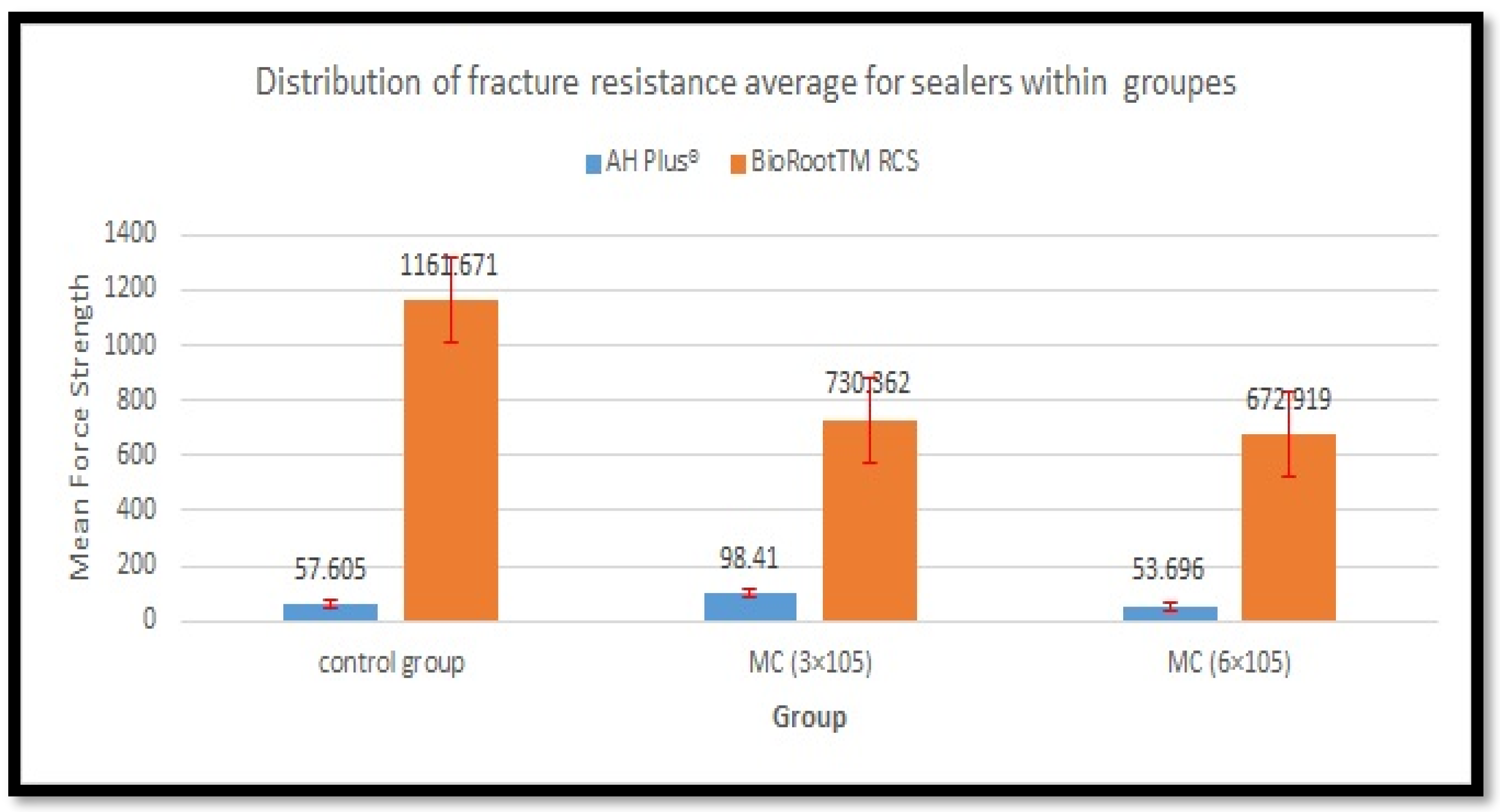
Table 2. Mean and SD of force required for fracture of tooth of tested sealers at two different thermomechanical time periods. Within same row, different superscript lowercase letters mean statistical difference between groups (p < 0.05). Within same column, different superscript uppercase letters mean statistical difference between groups (p < 0.05).
SealerControl GroupMC (3 × 10⁵)MC (6 × 10⁵)p-Value
AH Plus57.605 ± 15.207 A,a98.41 ± 19.335 A,b53.696 ± 24.082 A,a0.000
BioRoot RCS1161.671 ± 214.948 B,a730.362 ± 133.296 B,b672.919 ± 131.109 B,b0.000
T-test p-value0.0000.0000.000
Disclaimer/Publisher’s Note: The statements, opinions and data contained in all publications are solely those of the individual author(s) and contributor(s) and not of MDPI and/or the editor(s). MDPI and/or the editor(s) disclaim responsibility for any injury to people or property resulting from any ideas, methods, instructions or products referred to in the content.

