Review

Bisphosphonates and Their Influence on the Implant Failure: A Systematic Review

by
Cristiana Gomes Rebelo
1,
Juliana Campos Hasse Fernandes
2,
Nuno Bernardo
1,
Patrícia Couto
1 and
Gustavo Vicentis Oliveira Fernandes
1,3,*
1
Center for Interdisciplinary Research in Health (CIIS), Universidade Católica Portuguesa—Faculty of Dental Medicine (FMD-UCP), 3504-505 Viseu, Portugal
2
Private Practice, Ann Arbor, MI 48109, USA
3
Periodontics and Oral Medicine Department, University of Michigan School of Dentistry, Ann Arbor, MI 48109, USA
*
Author to whom correspondence should be addressed.
Appl. Sci. 2023, 13(6), 3496; https://doi.org/10.3390/app13063496
Submission received: 24 February 2023 / Revised: 6 March 2023 / Accepted: 7 March 2023 / Published: 9 March 2023
(This article belongs to the Special Issue New Techniques, Materials and Technologies in Dentistry)

Abstract

:
Objective: The goal of this systematic review was to study the relationship between the use of bisphosphonates (oral or intravenous) and its effect on implant osseointegration. Methods: The focused question was “In patients medicated with bisphosphonates and who underwent surgery to place dental implants, what is the influence of that medication (of different generations) on the failure of dental implants (O)?” Following specific eligibility criteria, four databases (PubMed/MEDLINE, Scopus, Web of Science, DOAJ) were electronically screened to search the articles. Specific MeSH terms were used in combinations with Boolean Operators “AND” and “OR” for the research. In addition, a manual search was done. The data extracted were the (i) author, (ii) year of publication, (iii) country, (iv) research question, (v) study design, (vi) patient information, (vii) the number of patients included, (viii) patient/implant status, (ix) the number of implants evaluated, (x) type of implant, (xi) risk factors, and (xii) findings obtained. Moreover, the following were also registered: the (i) type, generation, duration, and route for BP administrated; (ii) the presence of any systemic condition and drug treatment; (iii) follow-up (months); and (iv) implant failure rate (%). The quality assessment of the included studies was carried out using the Modified Newcastle–Ottawa scale. Results: A total of 491 articles were found (183 in PubMed/MEDLINE, 171 in Scopus, 65 in Web of Science, and 72 articles in DOAJ), and 17 articles were considered for full-text reading. After the exclusion of 3 articles, 14 were included in this systematic review (11 case reports, 2 retrospective, and 1 prospective study). The reasons for the bisphosphonates intaking included osteoporosis, multiple myeloma, breast cancer, knee cancer, and osteogenesis imperfecta. The oral administration involved Alendronato (eight studies), Risedronate (three studies), and Ibandronate (three studies); whereas the intravenous administrations were Zoledronate (seven studies), Clodronato (one study), and Pamidronato (three studies). The duration of use of bisphosphonates at the time of implant placement was diverse; it ranged from no interruption of bisphosphonate intaking up to its discontinuation for 2, 3, or 6 months before surgery, with respective use being resumed 1, 3, or 8 months after surgery. Antibiotic treatment (amoxicillin + clavulanic acid) was performed before the intervention in two cases and after the intervention in three cases. Finally, the percent of implant failure rate when intaking BPs had an average of 49.96%. Conclusions: Within the limitation of this systematic review, it was possible to conclude that a high mean failure rate of implant osseointegration (49.96%) was found, regardless of the generation of bisphosphonates used. Moreover, the failure rate was lower in patients using second generation bisphosphonates (Alendronate and Pamidronate) and was higher with the IV administration compared to the oral administration of bisphosphonates.

1. Introduction

Osseointegration is defined as a connection between the living bone and the implant surface in a stable and functional way. This involves anchoring the implant by forming bone tissue around the implant without fibrous tissue growth at the bone–implant interface [1]. This direct contact between the implant surface and the bone is decisive for the success of the implant, since it decreases the risk of failure, improves stability, and promotes implant longevity. In order to enhance osseointegration, it is possible to change the roughness of the implant surface [2].
Over the years, studies have been carried out to find the most suitable material for the composition of the implants and the consequent success or failure of their osseointegration [3,4,5]. The most scientifically studied implants are titanium implants, and more recently, investigations and comparisons with zirconia material have emerged [3,4,5]. According to Hanawa (2020) [6], titanium has shown excellent biocompatibility, corrosion-resistance, and high fracture toughness based on high strength and elongation. Compared to titanium, zirconia reported a significantly reduced bacterial biofilm formation and increased microcirculation in the peri-implant soft tissues [3]. Regarding peri-implant soft tissues, both materials have similar integration properties. However, titanium appears to have a faster initial osseointegration process when compared to zirconia. Survival rates of more than 96% for titanium implants with microrough surfaces have been reported after being followed for 10 years [3]. On the other hand, Sivaraman et al. (2018) [7] reported higher success rates (95.8%) for titanium and zirconia implants (90.9%) in the mandible compared to the maxilla, at 71.9% and 55%, respectively.
Nevertheless, the use of bisphosphonate (BP) may impair the osseointegration. It is a class of drugs that are frequently selected when there is an alteration in the bone metabolism, which are utilized to prevent bone loss [8]. It can be administered orally (e.g., daily, weekly, or monthly) for treatment of osteoporosis and Paget’s disease, or intravenously (every 3 months or annually) to treat malignant skeletal oncological diseases [9]. BPs can also be classified according to generations, with the first generation being non-nitrogenous and including drugs such as Clodronate, Etidronate, and Tiludronate. The second and third generation, on the other hand, contain nitrogen. Regarding the second generation, it includes drugs such as Alendronate, Neridronate, and Pamidronate and the third generation includes drugs such as Risedronate, Minodronate, Zoledronate, and Ibandronate [9].
Despite the fact that they increase the quality of life of the patients, there is an elevated risk that BPs can cause osteonecrosis of the jaw. This is characterized by an exposure of necrotic bone in the mandibular region that normally persists for 8 or more weeks [10]. Patients who have been treated with BPs intravenously have shown a greater chance of developing medication-related osteonecrosis of the jaw (MRONJ) or implant loss compared to oral-intaking therapy [11].
In a study developed by Gelazius et al. [10], patients taking BPs who were treated intravenously lost 6 implants out of 68, which yielded an 8.82% for failure rate; patients treated with intraoral therapy had a failure of 5 implants in 423 (1.18%), which was more than 7-fold less. They considered the dental implant a failure if the implant had mobility, active inflammation for more than 8 weeks without healing with antibiotic therapy, drainage of purulent secretion near the implant, the presence of necrotic bone or unhealed soft tissue, or implant loss [10].
A study by Chen et al. [12] showed that Zoledronate (the third generation of BPs) and Alendronate (the second-generation BPs) improved titanium implant osseointegration in ovariectomized rats. In this case, a single dose injection of Zoledronate (0.1 mg/kg) was shown to be able to increase bone implant contact (BIC), osseointegration, more than the oral administration of Alendronate (7 mg/kg/week) [12]. However, in oral mucosa cells, which provide the first physical and immunological barrier to prevent bacterial invasion, these BPs have been shown to have a difficult adhesion and metabolism [13]. In addition, the potential risk of medication-related osteonecrosis of the jaw (MRONJ) or loss of the implant associated with BP therapy cannot be disregarded, and more standardized studies are needed to provide more accurate information on this subject.
Therefore, the aim of this review was to systematically study, in the literature, the association of bisphosphonates (oral or intravenous) and its effect on implant osseointegration. The null hypothesis was that there is an impairment of the bone formation/osseointegration around implants when the patient is intaking bisphosphonates (BPs).

2. Material and Methods

2.1. Focused Question

A focused question was constructed according to the Preferred Reporting Items for Systematic Reviews and Meta-Analyses (PRISMA) guidelines and Participants Intervention Control Outcomes (PICO) protocol. The focused question presented in this systematic review was “In patients medicated with bisphosphonates (P) and who underwent surgery to place dental implants (I), what is the influence of that medication (of different generations) (C) on the failure of dental implants (O)?” (Table 1).

2.2. Eligibility Criteria

Inclusion criteria were (i) patients undergoing therapy with BPs (oral or intravenous), (ii) patients undergoing a dental implant placement procedure, (iii) using different generations and routes of BPs, (iv) that studied the influence of BPs intake on implant failure, (v) random controlled trials, case-control, case series, retrospective studies, prospective studies, (vi) trials conducted between 2000–2021, and (vii) in English language. The exclusion criteria included the following: (i) patients undergoing drug therapy other than BPs, (ii) patients whose surgical procedure was not the placement of dental implants, (iii) studies that did not refer to the type of BP used, (iv) studies that did not assess the relationship between dental implants and local/systemic therapy with BPs, (v) reviews, meta-analysis, commentaries, editorial, in vitro or preclinical studies, letters to the editor, duplicated articles, and (vi) other languages.

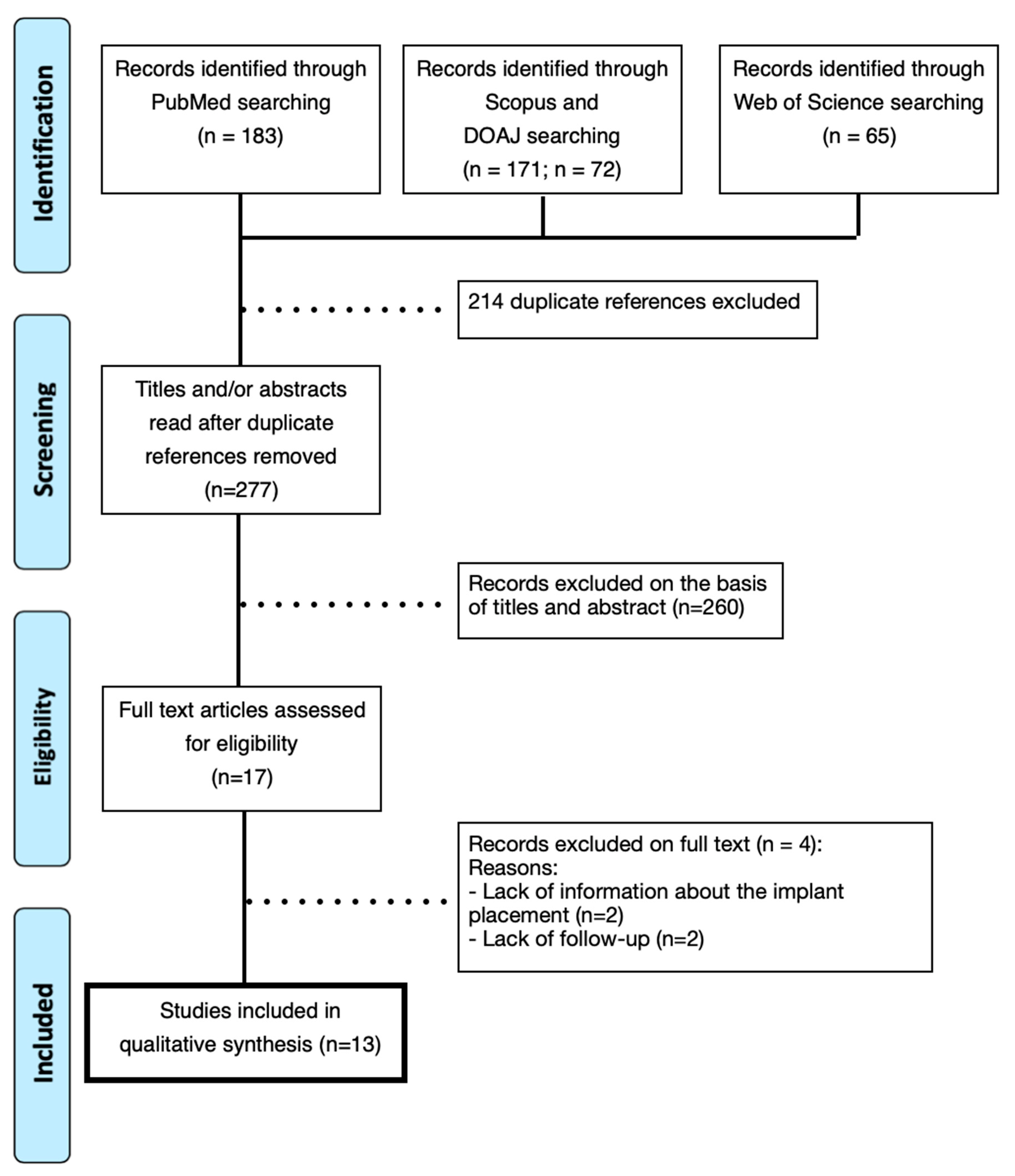
2.3. Literature Search and Screening

Four databases (PubMed/MEDLINE, Scopus, Web of Science, DOAJ) were used to electronically search for the articles. The following MeSH (Medical Subject Headings) terms were used: “Diphosphonates”, “Bisphosphonates”, “Clodronate”, “Etidronate”, “Alendronate”, “Pamidronate”, “Risedronate”, “Ibandronate”, “Dental Implants”, “Bisphosphonate-Associated Osteonecrosis of the Jaw” and their related entry terms were used in different combinations using the Boolean Operators “AND” and “OR” for the research and specific related-terms with the theme of this study. In addition, a manual search was made by each one of the researchers with the terms: “Osseointegration”, “Tiludronate”, “Neridronate”, “Minodronate”, “Zolendronate” (Table 2).

2.4. Data Extraction

The data collected from the included articles were inserted in an Excel spreadsheet (Microsoft Excel® for Mac, v. 16, Redmond, WA, USA, USA). They were the following: (i) author, (ii) year of publication, (iii) country, (iv) research question, (v) study design, (vi) patient information, (vii) number of patients included, (viii) patient/implant status, (ix) number of implants evaluated, (x) type of implant, (xi) risk factors, and (xii) findings obtained. Moreover, the following were also registered: the (i) type, generation, duration, and route for BP administrated; (ii) the presence of any systemic condition and drug treatment; (iii) follow-up (months); and (iv) implant failure rate (%).

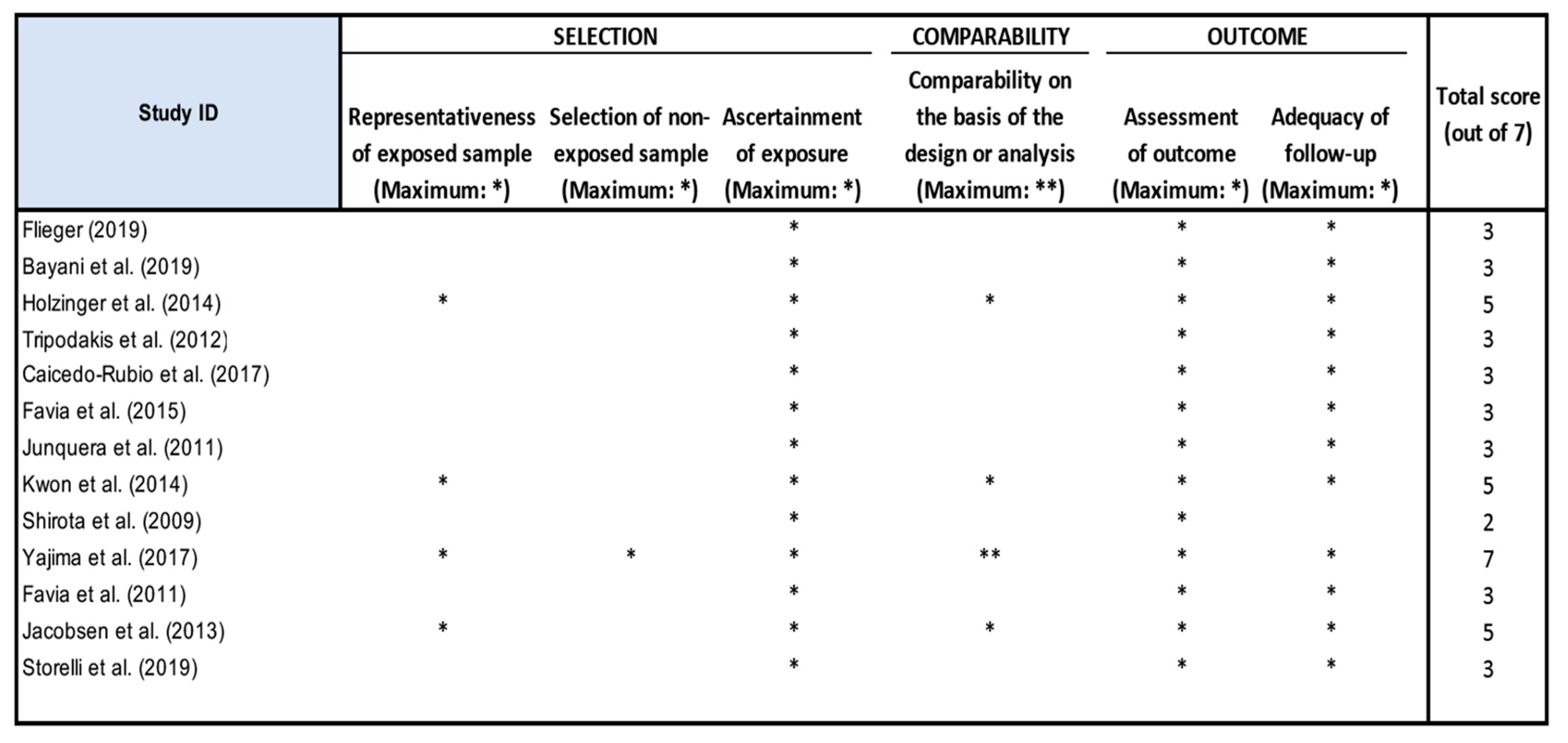
2.5. Quality Assessment

After the selection of articles, an assessment of their quality was carried out. For this, we used the Modified Newcastle–Ottawa scale, in which the following parameters were evaluated: representativeness, selection, comparability, blinding and, finally, the follow up. All parameters had a maximum score of 1 value, except for comparability, which can be evaluated up to a score of 2 values, totaling 7 points. From 0 to 3 points, the study was considered to have a low level of quality; between 4–6 was considered a moderate level; and a 7 score was considered a high level of quality.

3. Results

After carrying out the search, 491 articles were found. Of these, 183 articles were identified in PubMed/MEDLINE, 171 in Scopus, 65 in Web of Science, and 72 articles in DOAJ. Then, the articles that were duplicated (n = 214) were eliminated, which resulted in 277 studies. After analyzing the title and abstract, another 260 articles were excluded. Then, a total of 17 articles remained for full-text reading. After performing the full reading, four more articles were excluded due to lack of information and detail on the patient follow up. Thus, 13 articles were included in this systematic review (10 case reports, 2 retrospective studies, and 1 prospective study). The agreements between reviewers were, for initial assessment, k = 0.97 and, for assessment of the final inclusion, k = 0.91 (Figure 1).
The demographic data for the patients/implants and studies included are summarized in Table 3 and Table 4. A total of 67 patients were analyzed and 163 dental implants were placed in the studies included. All of them were Caucasian, with a mean age of 62 years old (58 female and 9 male). The mean follow-up period was 28.9 months (ranging from 12 months to 48 months). The risk factors reported were hypertension, tobacco, poor oral hygiene, and diabetes, and all of these factors were respectively linked to higher implant failure rates of 8%, 19%, 2%, and 7%. Most of the diseases for which BPs were taken were osteoporosis, multiple myeloma, breast cancer, lung cancer, prostate cancer, knee cancer, and osteogenesis imperfecta.
The routes of BP administration were oral and intravenous. A total of 42 patients (63%) were on therapy with BPs administered orally, and 25 patients (37%) received BPs through IV. For oral administration, the drugs were Alendronate [14,15,16,17,18,19,20,21], Risedronate [16,17,21], and Ibandronate [14,17,19], whereas those administered intravenously were Zoledronate [14,17,19,22,23,24,25,26], Clodronate [27], and Pamidronate [14,19,26]. The duration of the use of BPs at the time of implant placement was diverse. It varied from no interruption of BP intake to its discontinuation from 2, 3, or for 6 months before surgery, with respective resumption 1, 3, or 8 months after surgery. Antibiotic treatment (amoxicillin + clavulanic acid) was performed before the intervention in two cases and before and after surgical treatment in three cases. Use only after intervention was not found. Antibiotic treatment proved to be effective in cases where it was used as a pre- and post-surgical therapy. Finally, a percentage of implant survival in the group taking BPs had an average failure rate of 42.27%. Four studies did not have any implant failure [15,16,22,24].
The most prevalent BPs corresponded to the second generation (Alendronate and Pamidronate), which were used in 61% of the cases, followed by the third generation (Zoledronate, Ibandronate, and Risedronate), which were found in 38% of the cases; only one case utilized first generation BPs (Clodronate). Regarding this variable, a lower failure rate was noted when patients used the second-generation BPs (37%), followed by third generation (38%) and first generation (100%). Moreover, when patients discontinued BP therapy (45% of the cases), lower failure rates were obtained than patients with continuous use (55%). The quality assessment of the study was considered to be of low/moderate level, with all studies excepting Yajima et al. presenting values between 2 and 5 (Figure 2).
Table 3. Characteristics of the patients and implants studied.
Authors (Year)CountryResearch QuestionPatient InformationDesign (n)Pacient Status/ImplantN. of Implants Assessed (Site)Risk FactorsResults
1Flieger (2019) [15]PolandPlacement of two missing teeth with insertion of immediate implants in a patient medicated with BPsF, 56 yon = 1; case reportAbsence of bone loss in both implants; normal peri-implant soft tissue condition (no signs of inflammation)2 (15, 24)Hypertensionimplant survival
2Bayani et al. (2019) [21]IranReport of implant placement in a patient with MRONJM, 54 yon = 1; case reportMinimal bone loss1 (14)Non-smoking, good oral hygieneimplant survival
3Holzinger et al. (2014) [14]AustriaDevelopment of MRONJ in patients treated with BPs who received implants.F; average of 65.7 ± 8.5 yon = 13; retrospective study NR1 (47)7 former smokers; and 5 smokersImplant failure (MRONJ)
4Tripodakis et al. (2012) [16]GreeceCare in the placement of implants and prevention of MRONJ in patients with BP therapyF, 70 and 65 yon = 2; case reportNo observed complications3 (14, 15, 17)
14 (13, 14, 16, 17, 25, 26, 27, 28, 36, 37, 38, 46, 47, 48)
1 with hypertension and hyperlipidemiaimplant survival
5Caicedo-Rubio et al. (2017) [23]Spain Insertion of implants in a patient treated with IV bisphosphonatesM, 61 yon = 1; case reportGeneralized gingival inflammation; peri-implant tissues without inflammation; loss of 1.25 mm of crestal bone in the implant area 363 (36, 37, 46)Smoker (20 cigarettes/day); Stroke prior to 2007; poor oral hygieneimplant survival
6Favia et al. (2015) [22]ItalyPatient with breast cancer affected by MRONJF, 66 yon = 1; case reportPain; purulent secretion; right-sided inferior alveolar nerve paresthesia7 (16, 31, 35, 36, 41, 44, 46)Poor oral hygieneImplant failure (MRONJ)
7Junquera et al. (2011) [24]SpainMandibular dental implant placement in a patient with MRONJM, 59 yon = 1; case reportLeft lower lip paresthesia; purulent discharge; necrotic bone2NRImplant failure (MRONJ)
8Kwon et al. (2014) [17]Korea Analysis of MRONJ characteristics around dental implants2 M, 17 F; 42 to 85 yon = 19; prospective studyNecrotic bone exposure, purulent discharge; fistula; swelling for more than 8 weeks23Hypertension; and DiabetesImplant failure (MRONJ)
9Shirota et al. (2009) [25]Japan MRONJ around implants in maxillary molarsF, 54 yon = 1; case reportPain; bone exposure; redness; swelling3 (15, 25, 27)NRImplant failure (MRONJ)
10Yajima et al. (2017) [18]JapanBMD and influence of the use of BPs on early implant failureF, >60 yo n = 11; retrospective studyNR25Diabetes, smoking, steroid, poor oral hygiene were excludedImplant survival and failure cases
11Favia et al. (2011) [26]ItalyOccurrence of MRONJ after implant insertionF, 65 yon = 1; case reportnon-loading2 (35, 36)No pre-existing bone lesionsImplant failure (MRONJ)
12Jacobsen et al. (2013) [19]SwitzerlandReport of 14 patients with mandibular osteopathology associated with BP therapy and dental implant insertion11 F and 3 Mn = 14; case seriespurulent; periapical radiolucency surrounding the implants23NRImplant failure (MRONJ)
13Storelli et al. (2019) [20]ItalyMRONJ after implant placement in a patient undergoing oral BF therapyF, 77 years oldn = 1; case reportNecrotic bone; pain; abscess; nerve paresthesia; fistula; exposed bone; lack of healing8Non-smoking, hypothyroidism, hypercholesterolemia, hypertension, arterial fibrillationImplant failure (MRONJ)
BMD = bone mineral density; BP = bisphosphonates; F = female; M = male; wk = weeks; y = years; yo = years old; NR = not reported.
Table 4. Comparison of the characteristics of the variables studied.
Authors (Year)BP TypeBP GenerationRoute of AdministrationAntibiotic TreatmentDurationPatient/Implant StatusType of the Dental ImplantRisk FactorsImplant Failure Rate (%)
1Flieger (2019) [15]Alendronate2ndOral 70 mg/week for 24 monthsBefore surgery amoxicillin + clavulanic acid. 1000 mg day/7 daysNo discontinuation Absence of bone loss; normal condition of peri-implant soft tissue (no signs of inflammation)ICX-plus (3.45 × 10 mm) at bone level Hypertension0%
2Bayani et al. (2019) [21]Zoledronate3rdIV 3.5 mg/month for 22 monthsBefore surgery 2g amoxicillin/clavulanic acid. After: 1000 mg 2×/day/7 daysDiscontinued 6 months before surgery and resumed 8 months afterMinimal bone lossSuperline; Dentium (3.6 × 10 mm)Non-smoking, good oral hygiene0%
3Holzinger et al. (2014) [14]Zolendronate (n = 7)
Alendronate (n = 3)
Pamidronate (n = 2)
Ibandronate (n = 1)
3rd (n = 8)
2nd (n = 5)
7—IV 4 mg/month
3—Oral 70 mg/week
2—Oral 90 mg/month
1—IV 3 mg each 3 months
NR3—BPs after implant placement
3—BPs before implant placement
7—BPs before and after implant placement
NRNR7 former smokers; and 5 smokers63.8%
4Tripodakis et al. (2012) [16]Risedronate
Alendronate
2nd
3rd
1—Oral for 2 months
1—Oral for 4 years
24h before surgery 500 mg of amoxicillin up to 10 days after surgeryDiscontinued 3 months before surgery and resumed 3 months after surgeryNo complications observedBranemark System Mk III Groovy a 13 mm; SPI, Alpha Bio 16 mm1 with hypertension and hyperlipidemia0%
5Caicedo-Rubio et al. (2017) [23]Zoledronate 3rdIV 5 mg each 6 months for 4 yearsBefore surgery, amoxicillin 500 mg every 8 h until 6 days after surgeryDiscontinued 2 months before surgery and resumed 1 month afterGeneralized gingival inflamation; peri-implants good health; 1.25 mm loss of of crestal bone in the implant area 36MIS Implants Technologies LTD 7.5 × 10 mm and 75 × 11.5 mm subcrestalSmoker (20 cigarettes/day); Stroke prior to 2007; poor oral hygiene0%
6Favia et al. (2015) [22]Zoledronate3rdIV 4 mg monthly for 33 monthsNR6 months after implant placementPain; pus secretion; right inferior alveolar nerve paresthesiaNRPoor oral hygiene57.1%
7Junquera et al. (2011) [24]Zoledronate 3rdIV 4 mg monthlyNR9 months after implant placementLower left labial paraesthesia; purulent secretion; necrotic boneEndosseous dental implantsNR50%
8Kwon et al. (2014) [17]Zolendronate Alendronate
Ibandronate Risedronate
3rd
2nd
Oral ou IVNRStarted before surgery (n = 16) and after (n = 3)Necrotic bone exposure,
pus secretion; fistula; swelling for more than 8 weeks
NRHypertension; and Diabetes15.8%
9Shirota et al. (2009) [25]Pamidronate
Zolendronate
2nd
3rd
IV (P 17 times and Zolendronate 9 times) in 16 monthsNR4 years after implant placementPain; bone exposure; redness; swellingNRNR66.7%
10Yajima et al. (2017) [18]Alendronate2ndOralNRNo discontinuation. Using BF: 3.8 + 2.1 yearsNRNRDiabetes, smoking, steroid, poor oral hygiene were excluded12%
11Favia et al. (2011) [26]Clodronate1stIV 300 mg twice a month NRDiscontinuation 3 months before surgeryPurulent secretion; periapical radioluscency surrounding the implantsNRNo pre-existing bone lesions100%
12Jacobsen et al. (2013) [19]9—Zoledronate
2—Alendronate
1—Ibandronate
2—Pamidronate
2nd
3rd
IV e Oral in 3 monthsNRNRNecrotic bone; ache; abscess; nerve paraesthesia; fistula; exposed bone; no healingNRNR100%
13Storelli et al. (2019) [20]Alendronate2ndOral 70 mg once a week NRNo discontinuation. Use started 3 years before surgeryInflamed peri-implant tissues; bleeding on probing; bone resorption < 2 mm around implants; purulent secretions; exposure of necrotic bone; mobilityNRNon-smoking, hypothyroidism, hypercholesterolemia, hypertension, arterial fibrillation100%
BP, bisphosphonate; BRONJ, bisphosphonate-related osteonecrosis of the jaw; IV, intravenous.

4. Discussion

The objective of the present systematic review was to analyze the negative influence of BPs on dental implant osseointegration. Of the 67 Caucasian patients who were included, there was a predominance of females (58 patients) most of whom were in an age for menopause, over 50 years old, and had an elevated risk for osteoporosis [27]. On the other hand, male patients constituted the minority (9 individuals). That pathology can be diagnosed in other age groups and also in men. Worldwide, this pathology affects a total of 200 million women, with a growing trend in North America and Europe [28].

4.1. BP Use and Dental Implants

BPs are prescribed in several pathologies, whether they affect the bone (osteoporosis, OI, Paget’s disease) or in malignant pathologies (malignant hypercalcemia, bone metastases, lung and breast cancer, and MM), because they prevent bone resorption. Of the studies included in this systematic review, 44 patients had osteoporosis, 8 had MM, 10 had breast cancer, 2 had lung cancer, 1 had prostate cancer, 1 had Langerhans cell histiocytosis, and 1 had OI. The administration of BPs is more prevalent in patients with osteoporosis, since, as reported in the literature, this is one of the most common bone pathologies in developed countries and one which has the most indication for the prescription of these drugs due to the risk of occurrence of bone fractures [29].
Of all the studies included, the presence of patients undergoing therapy with second generation (Alendronate, Pamidronate) [14,15,16,17,18,19,20,25] and third generation BPs (Risedronate, Zoledronate and Ibandronate) [14,16,17,19,21,22,23,24,25] were greater than the use of first generation (Clodronate) [26]. The first generation seems to show a decreasing trend in use nowadays. On the other hand, the failure rate for osseointegration proved to be lower in patients who used therapy with second generation of BPs (about 37%) compared to patients who had therapy with first and third generations. Second generation BPs have been shown to be a well-tolerated drug, with low side effects. This fact has been shown through their growing use in recent years [30].
The interruption of therapy with BPs was a parameter with varied results in this systematic review, from patients who did not discontinue to patients who discontinued for 2, 3, or 6 months before surgery, with respective resumption for 1, 3, or 8 months after surgery. Tripodakis et al. [16] reported the case of two female patients, both in their seventh decade of life, who requested rehabilitation with implant placement. The patients were medicated with second and third generation BPs (Alendronate and Risedronate). After consultation with the attending physician, the patients discontinued BPs 3 months before and resumed 3 months after implant placement. They received antibiotic therapy after surgical interventions, and the treatment plan was completed uneventfully and without complications during a 2-year follow up. In another study, Flieger [15] reported the case of a female patient (56 years old), who intended to carry out the prosthetic reconstruction of the crown of two molars lost in the maxilla with the placement of two implants. She was medicated with Alendronate (a second-generation BP) for osteoporosis. There was no bone loss around both implants, and it was observed that the peri-implant soft tissue did not show any signs of inflammation. Bayani et al. [21] reported that the placement of dental implants in patients with MM undergoing therapy with third generation BPs (Zoledronate) can be performed. Therefore, a meticulous selection of cases, an adequate medical consultation, and a minimally invasive surgery should be considered.
Flieger [15], Yajima et al. [18], and Storelli et al. [20] recommended that patients (n = 13) not interrupt their therapy with BPs during implant placement surgery. Fliger [15] and Yajima et al. [18] obtained a low failure rate in the implant placement procedure of 0% and 12% respectively. On the contrary, Storelli et al. [20] had a complete failure rate (100%). Similarly, in the study carried out by Kwon et al. [17], a complete failure of implant placement was observed in patients who started therapy with BPs before implant placement surgery.
Otherwise, Bayani et al. [21] reported the discontinuation of BP therapy for6 months before surgery that was resumed therapy 8 months after surgery. The failure rate was 0%, and no complications were observed. The same happened with Tripodakis et al. [16] who interrupted therapy 3 months before the surgery and resumed it for 3 months after. After 17 implants were placed, none of them failed. Caicedo-Rubio et al. [23] discontinued the therapy 2 months before the surgery and resumed it 1 month later, and they also obtained 0% for implant failure rate. This fact suggests an association between discontinuing BP therapy with a low rate of dental implant failure (around 45%) than for non-interruption therapy (around 55%). These data may still be different depending on the involvement of risk factors. Moreover, the cumulative dose and duration of drug exposure, medical comorbidities (corticosteroids, diabetes, immunosuppressive conditions), and dental comorbidities (extractions, implant placement, invasive procedures, periodontal disease, trauma, infection) must be verified. In this way, all the most invasive dental procedures constitute a risk when we are facing patients who use BPs.
According to Holzinger et al. [14], the occurrence of complications seems to be delayed when dental implants are inserted before starting BP therapy. However, the incidence of complications seems to be higher when implants are placed after BP treatment or during its therapy. Thus, it is suggested as ideal to proceed with implant placement before initiating BPs therapy; once therapy is started, the risk becomes higher.
Specifically, for four studies without implant failure [15,16,21,23], all cases reported types of study that must be carefully interpreted, due to the low level of scientific weight, Bayani et al. [21] found excellent results after a 1-year follow up in a 54-year-old man patient with multiple myeloma (MM) who complained of difficulty in mastication and esthetical concern for his upper anterior teeth. He received a monthly infusion of 3.5 mg of the IV BP drug Zoledronate for a period of 22 months, which is considered a long period and a high-risk treatment. The other two studies, Flieger [15] and Tripodakis et al. [16], had 2-year follow up periods without complications and bone loss. Similar results were obtained by Caicedo-Rubio et al. [23], after 4-year follow up, which showed no evidence of pathology in the peri-implant tissues.

4.2. Dental Implants Characteristics

Flieger [15] performed a surgical procedure using two implants with widths of 3.45 mm and lengths of 10 mm at the tissue level. Bayani et al. [21] opted for the placement of a bone-level implant that was 3.6 mm in diameter and 10 mm in length. Tripodakis et al. [16] placed a total of 17 implants that were 13 mm long at the bone level. Caicedo-Rubio et al. [23] placed three implants of 3.75 × 10 mm and 3.75 × 11.5 mm at the subcrestal level; Junquera et al. [24] placed two subcrestal implants. All these implants showed a significantly acceptable success rate, except for the implants placed by Junquera et al. [24], which resulted in severe complications and implant failure due to the MRONJ. The literature showed in the Hammerle et al.’s study [31] that the placement of implants at the subcrestal level was not recommended for these types of patients, who can achieve greater marginal bone loss [32].

4.3. Implants Associated with Risk Factors

Implant placement can also be influenced by risk factors, local or systemic, which can lead to complications. This includes cases of smoker patients, patients with pathologies (diabetes), with poor oral hygiene, and with a history of recent stroke (first 6 months after the episode) [33]. According to several authors, the risk of implant failure is greater with the increase in the number of cigarettes smoked per day; therefore, this factor is considered a real risk factor for implant placement [14]. On the other hand, Caicedo-Rubio et al. [23] reported that smoker patients and those with poor oral hygiene had favorable results for the implants. These data must be carefully analyzed due to the reduced sample size present in the study. This fact has led researchers to exclude from their studies all smoker patients, patients with diabetes, those using steroids, or those with poor oral hygiene, precisely because of the higher implant failure risk [18].
In our study, we found a somewhat significant failure rate in the case of smoker patients (19%), patients who had diabetes (7%), hypertensive patients (8%), and those who had poor oral hygiene (2%). However, even though the patients did not present any risk factor, they had very similar failure rates to those with risk factors. In the case of diabetes mellitus, this was closely related to oral health. From the data available to date, it increases the susceptibility to infection and impairs the tissue healing. In addition, there is evidence that patients with diabetes are more likely to develop complications than patients without this pathology [17].

4.4. MRONJ and Route of Administration

Several studies have focused on the risk factors for MRONJ development with the treatment of IV BPs (nitrogenated) and performing tooth extractions (identified as important risk factors) [10]. There is scientific evidence showing that drugs (Pamidronate and Zolendronate) whose route of administration is exclusively IV have been strongly associated with cases of MRONJ [19]. This can be explained because these drugs are more potent and have greater bioavailability due to the type of administration (IV).
For this purpose, Shirota et al. [25] described a case of a 54-year-old woman with gum ulceration, bone exposure, and intense spontaneous pain around implants. The patient in question had undergone IV therapy with BPs (Pamidronate and Zoledronate) for 2 years to treat bone metastases from breast cancer. The authors reported MRONJ related to BPs, with symptoms of necrotic bone for more than 8 weeks; the patient did not undergo radiotherapy in the maxillofacial area.
Drugs, such as Alendronate and Risedronate, are administered exclusively orally. It has been reported that these drugs are safer and have a lower risk of MRONJ [16]. This was observed in the Flieger’s study [15] of a a 56-year-old woman who underwent rehabilitation of two missing molars in the maxilla. She was taking oral Alendronate and, during the time of osteoporosis treatment with Alendronate, there were no episodes of MRONJ.
Upon analyzing the studies included in this systematic review, it was not possible to be precise in presenting the failure rates for both routes of administration, due to the lack of data provided by the studies. Nevertheless, there was a consensus among authors that the IV route of administration results in a high number of failure cases. Thus, the oral route of administration still seems to be the safest route.

4.5. MRONJ and Implant Failure

MRONJ can be manifested through several signs and symptoms. Its development may present clinical manifestations such as the presence of pain, necrotic bone, bone exposure, the presence of purulent secretion, redness, abscess, swelling, paresthesia of the right inferior alveolar nerve, an ill-defined radiolucent area, bleeding upon probing, bone resorption around the implants, and the presence of mobility. These symptoms can persist for more than 8 weeks. It is a problem with a multifactorial origin; it is difficult to predict its occurrence.
Favia et al. [22] showed failure in four of the seven implants placed in the same patient that were related to the occurrence of MRONJ. In this case, the reported symptoms were essentially pain, the presence of purulent secretion, and paresthesia of the inferior alveolar nerve on the right side associated with an ill-defined radiolucent area that extended from the right posterior mandible to the opposite region of the premolar. These data were attributed to the patient’s poor oral hygiene. As for the remaining implants that still showed acceptable osseointegration, it was not possible to conclude what would be the long-term prognosis, since the follow-up only occurred after 18 months.
Similar results happened with Junquera et al. [24]. The patient had two implants presenting features compatible with MRONJ (necrotic bone, left lower lip paresthesia, and purulent secretion in only one of the implants). Also, Shirota et al. [25] reported a case with three implants placed; two of them presented pain, bone exposure, redness, and swelling. On the other hand, we had cases, in this study, where there was complete failure of the implants, and all patients developed MRONJ. Kwon et al. [17] and Jacobsen et al. [19] obtained the same results from evaluating a total of 23 implants, which all failed with reports of necrotic bone exposure, purulent secretion, pain, abscess, paresthesia, fistula, and swelling for more than 8 weeks.
Storelli et al. [20] reported a case of MRONJ in a 77-year-old female patient. After receiving oral implant rehabilitation and an immediate-load fixed prosthesis in the maxilla, she began to report pain and purulent secretions, which were neglected by the responsible professional. She returned to see the same professional after another episode of acute pain. The fixed prosthesis was removed and exposure of necrotic bone around the implants was observed. In this case, all implants failed. The patient was submitted to surgery to remove necrotic bone blocks. This was the most severe case analyzed in this systematic review.

4.6. Study Limitations

One of the main limitations of this study was the non-inclusion of randomized clinical trials. This occurred because there is scarce and limited literature. It was confirmed by the analysis of the quality of the included studies, in which the majority were classified as low and moderate quality. Several variables were studied that likely caused bias in analyzing the influence of BPs on implant placement. However, we presented the most clinically relevant results that can be interpreted from a trend perspective. Some of the studies included in this systematic review did not include all information regarding the influence of the route of administration on the implant failure rate. This situation made a statistical treatment of the variable under analysis impossible. Thus, it is suggested that, in a future investigation, the exploration of this theme be continued and that a longitudinal cohort study be developed.

5. Conclusions

Within the limitation of this systematic review, it was possible to conclude that a high mean for failure rate of implant osseointegration (49.96%) was found, regardless of the generation of BPs used. Moreover, the failure rate was lower in patients using second generation BPs (Alendronate and Pamidronate) and when there was an interruption of the BP therapy when placing implants when compared, respectively, with third generation and the continuous administration. Otherwise, it was higher with the IV administration compared to the oral administration of BPs. Furthermore, if the patients were smokers, diabetic, had hypertension, or poor oral hygiene, they were more prone to failure of the implants placed. However, more studies must be conducted to better understand the clinical findings associated with BPs and implant therapy.

Funding

This research received no external funding.

Conflicts of Interest

The authors declare no conflict of interest.

References

  1. Jayesh, R.S.; Dhinakarsamy, V. Osseointegration. J. Pharm. Bioallied Sci. 2015, 7, 226–229. [Google Scholar]
  2. Albrektsson, T.; Wennerberg, A. On osseointegration in relation to implant surfaces. Clin. Implant Dent. Relat. Res. 2019, 21, 4–7. [Google Scholar] [CrossRef] [Green Version]
  3. Borges, H.; Correia, A.R.M.; Castilho, R.M.; Fernandes, G.V.O. Zirconia Implants and Marginal Bone Loss: A Systematic Review and Meta-Analysis of Clinical Studies. Int. J. Oral Maxillofac. Implants 2020, 35, 707–720. [Google Scholar] [CrossRef]
  4. Fernandes, G.V.O.; Costa, B.M.G.N.; Trindade, H.F.; Castilho, R.M.; Fernandes, J.C.H. Comparative analysis between extra-short implants (≤6 mm) and 6 mm-longer implants: A meta-analysis of randomized controlled trial. Aust. Dent. J. 2022, 67, 194–211. [Google Scholar] [CrossRef]
  5. Fernandes, P.R.E.; Otero, A.I.P.; Fernandes, J.C.H.; Nassani, L.M.; Castilho, R.M.; Fernandes, G.V.O. Clinical Performance Comparing Titanium and Titanium-Zirconium or Zirconia Dental Implants: A Systematic Review of Randomized Controlled Trials. Dent. J. 2022, 10, 83. [Google Scholar] [CrossRef]
  6. Hanawa, T. Zirconia versus titanium in dentistry: A review. Dent. Mater. J. 2020, 39, 24–36. [Google Scholar] [CrossRef] [Green Version]
  7. Sivaraman, K.; Chopra, A.; Narayan, A.I.; Balakrishnan, D. Is zirconia a viable alternative to titanium for oral implant? A critical review. J. Prosthodont. Res. 2018, 62, 121–133. [Google Scholar] [CrossRef]
  8. Cremers, S.; Drake, M.T.; Ebetino, F.H.; Bilezikian, J.P.; Russell, R.G.G. Pharmacology of bisphosphonates. Br. J. Clin. Pharmacol. 2019, 85, 1052–1062. [Google Scholar] [CrossRef]
  9. Nayak, S. Application of bisphosphonates in dentistry: A review of literature. Indian J. Public Health Res. Dev. 2019, 10, 299–303. [Google Scholar] [CrossRef]
  10. Gelazius, R.; Poskevicius, L.; Sakavicius, D.; Grimuta, V.; Juodzbalys, G. Dental Implant Placement in Patients on Bisphosphonate Therapy: A Systematic Review. J. Oral Maxillofac. Res. 2018, 9, e2. [Google Scholar] [CrossRef]
  11. Freitas, N.R.; Lima, L.B.; Moura, M.B.; Veloso-Guedes, C.C.F.; Simamoto-Júnior, P.C.; Magalhães, D. Bisphosphonate treatment and dental implants: A systematic review. Med. Oral Patol. Oral Cir. Bucal 2016, 21, e644–e651. [Google Scholar]
  12. Chen, B.; Li, Y.; Yang, X.; Xu, H.; Xie, D. Zoledronic acid enhances bone-implant osseointegration more than alendronate and strontium ranelate in ovariectomized rats. Osteoporos Int. 2013, 24, 2115–2121. [Google Scholar] [CrossRef]
  13. Basso, F.G.; Pansani, T.N.; Soares, D.G.; Cardoso, L.M.; Hebling, J.; de Souza Costa, C.A. Influence of bisphosphonates on the adherence and metabolism of epithelial cells and gingival fibroblasts to titanium surfaces. Clin. Oral Investig. 2018, 22, 893–900. [Google Scholar] [CrossRef] [Green Version]
  14. Holzinger, D.; Seemann, R.; Matoni, N.; Ewers, R.; Millesi, W.; Wutzl, A. Effect of dental implants on bisphosphonate-related osteonecrosis of the jaws. J. Oral Maxillofac. Surg. 2014, 72, e1–e8. [Google Scholar] [CrossRef]
  15. Flieger, R. Bilateral bone ridge splitting in maxilla with immediate implant placement in a patient with osteoporosis: A clinical report with 2-year follow-up. Case Rep. Dent. 2019, 6, 1458571. [Google Scholar] [CrossRef] [Green Version]
  16. Tripodakis, A.P.; Kamperos, G.; Nikitakis, N.; Sklavounou-Andrikopoulou, A. Implant therapy on patients treated with oral bisphosphonates. J. Osseointegration 2012, 4, 9–14. [Google Scholar]
  17. Kwon, T.G.; Lee, C.O.; Park, J.W.; Choi, S.Y.; Rijal, G.; Shin, H.I. Osteonecrosis associated with dental implants in patients undergoing bisphosphonate treatment. Clin. Oral Implants Res. 2014, 25, 632–640. [Google Scholar] [CrossRef]
  18. Yajima, N.; Munakata, M.; Fuchigami, K.; Sanda, M.; Kasugai, S. Influence of bisphosphonates on implant failure rates and characteristics of postmenopausal woman mandibular jawbone. J. Oral Implantol. 2017, 43, 345–349. [Google Scholar] [CrossRef]
  19. Jacobsen, C.; Metzler, P.; Rössle, M.; Obwegeser, J.; Zemann, W.; Grätz, K.W. Osteopathology induced by bisphosphonates and dental implants: Clinical observations. Clin. Oral Investig. 2013, 17, 167–175. [Google Scholar] [CrossRef]
  20. Storelli, S.; Storelli, S.; Palandrani, G.; Dondi, C.; Tagliatesta, L.; Rossi, A. Severe case of Osteonecrosis following implant placement in a patient in therapy with bisphosphonates: A case report. J. Oral Implantol. 2019, 45, 139–144. [Google Scholar] [CrossRef]
  21. Bayani, M.; Anooshirvani, A.A.; Keivan, M.; Mohammad-Rabei, E. Dental implant in a multiple myeloma patient undergoing bisphosphonate therapy: A case report. Clin. Case Rep. 2019, 7, 1043–1048. [Google Scholar] [CrossRef] [Green Version]
  22. Favia, G.; Tempesta, A.; Limongelli, L.; Crincoli, V.; Piattelli, A.; Maiorano, E. Metastatic breast cancer in medication-related osteonecrosis around mandibular implants. Am. J. Case Rep. 2015, 16, 621–626. [Google Scholar] [CrossRef] [Green Version]
  23. Caicedo-Rubio, M.; Ferrés-Amat, E.; Ferrés-Padró, E. Implant-supported fixed prostheses in a Patient with Osteogenesis Imperfecta: A 4-year follow-up. J. Clin. Exp. Dent. 2017, 9, e1482–e1486. [Google Scholar] [CrossRef]
  24. Junquera, L.; Gallego, L.; Pelaz, A. Multiple Myeloma and Bisphosphonate-Related Osteonecrosis of the Mandible Associated with Dental Implants. Case Rep. Dent. 2011, 2011, 568246. [Google Scholar] [CrossRef]
  25. Shirota, T.; Nakamura, A.; Matsui, Y.; Hatori, M.; Nakamura, M.; Shintani, S. Bisphosphonate-related osteonecrosis of the jaw around dental implants in the maxilla: Report of a case: Case Report. Clin. Oral Implants Res. 2009, 20, 1402–1408. [Google Scholar] [CrossRef]
  26. Favia, G.; Piattelli, A.; Sportelli, P.; Capodiferro, S.; Iezzi, G. Osteonecrosis of the Posterior Mandible after Implant Insertion: A Clinical and Histological Case Report. Clin. Implant Dent. Relat. Res. 2011, 13, 58–63. [Google Scholar] [CrossRef]
  27. Kanis, J.A.; Cooper, C.; Rizzoli, R.; Reginster, J.Y. Correction to: European guidance for the diagnosis and management of osteoporosis in postmenopausal women. Osteoporos Int. 2020, 31, 209. [Google Scholar] [CrossRef] [Green Version]
  28. Lane, N.E. Epidemiology, etiology, and diagnosis of osteoporosis. Am. J. Obstet. Gynecol. 2006, 194, S3–S11. [Google Scholar] [CrossRef]
  29. Serrano, A.J.; Begoña, L.; Anitua, E.; Cobos, R.; Orive, G. Systematic review and meta-analysis of the efficacy and safety of alendronate and zoledronate for the treatment of postmenopausal osteoporosis. Gynecol. Endocrinol. 2013, 29, 1005–1014. [Google Scholar] [CrossRef]
  30. Aghaloo, T.; Pi-Anfruns, J.; Moshaverinia, A.; Sim, D.; Grogan, T.; Hadaya, D. The Effects of Systemic Diseases and Medications on Implant Osseointegration: A Systematic Review. Int. J. Oral Maxillofac. Implants 2019, 34, s35–s49. [Google Scholar] [CrossRef]
  31. Hämmerle, C.H.; Brägger, U.; Bürgin, W.; Lang, N.P. The effect of subcrestal placement of the polished surface of ITI implants on marginal soft and hard tissues. Clin. Oral Implants Res. 1996, 7, 11–119. [Google Scholar] [CrossRef]
  32. Pellicer-Chover, H.; Peñarrocha-Diago, M.; Peñarrocha-Oltra, D.; Gomar-Vercher, S.; Agustín-Panadero, R.; Peñarrocha-Diago, M. Impact of crestal and subcrestal implant placement in peri-implant bone: A prospective comparative study. Med. Oral Patol. Oral Cir. Bucal 2016, 21, e103–e110. [Google Scholar] [CrossRef]
  33. Hwang, D.; Wang, H.L. Medical contraindications to implant therapy: Part I: Absolute contraindications. Implant Dent. 2006, 15, 353–360. [Google Scholar] [CrossRef]
Figure 1. Article selection strategy, according to the PRISMA Flowchart (Preferred Reporting Items for Systematic Reviews and Meta-Analyses).
Applsci 13 03496 g001
Figure 2. Qualitative assessment of the studies by the Modified Newcastle-Ottawa Quality Assessment scale for Cohort and Case-Control Studies (m-NOS). (Flieger (2019) [15]; Bayani et al. (2019) [21]; Holzinger et al. (2014) [14]; Tripodakis et al. (2012) [16]; Caicedo-Rubio et al. (2017) [23]; Favia et al. (2015) [22]; Junquera et al. (2011) [24]; Kwon et al. (2014) [17]; Shirota et al. (2009) [25]; Yajima et al. (2017) [18]; Favia et al. (2011) [26]; Jacobsen et al. (2013) [19]; Storelli et al. (2019) [20]). * 1 point for the score; ** 2 points for the score.
Applsci 13 03496 g002
Table 1. PICO characterization.
Participants (P) Patients using bisphosphonates
Intervention (I)Placement of one or more dental implants
Control (C)Different generations and administration routes of BFs
Outcomes (O)Dental implant failure rate
Table 2. Search strategy.
DatabaseEquation ImplementedFilters
Pubmed/
MEDLINE
((((((((((((diphosphonates [MeSH Terms]) OR bisphosphonates [MeSH Terms]) OR clodronate [MeSH Terms]) OR etidronate [MeSH Terms]) OR alendronate [MeSH Terms]) OR pamidronate [MeSH Terms]) OR risedronate [MeSH Terms]) OR ibandronate [MeSH Terms]) OR “bisphosphonate- associated osteonecrosis of the jaw” [MeSH Terms]) OR tiludronate) OR neridronate) OR minodronate) OR zoledronate AND (osseointegration OR dental implants [MeSH Terms]) In English, from January 2000 to December 2021, humans
Scopus ALL ((diphosphonates OR bisphosphonates OR clodronate alendronate OR risedronate OR bisphosphonate-associated AND osteonecrosis AND jaw) OR tiludronate OR neridronate OR etidronate OR pamidronate OR ibandronate OR (minodronate OR zoledronate) AND (osseointegration OR dental AND implants) AND (failure AND rate)) AND PUBYEAR > 1999 AND PUBYEAR < 2022 AND (LIMIT-TO (PUBSTAGE, “final”)) AND (LIMIT-TO (DOCTYPE, “ar”)) AND (LIMIT-TO (EXACTKEYWORD, “Humans”)) AND (LIMIT-TO (LANGUAGE, “English”)) AND (LIMIT-TO (SRCTYPE, “j”))In English, from January 2000 to December 2021, humans, final stage
Web of Science (diphosphonates OR bisphosphonates OR clodronate OR etidronate OR alendronate OR pamidronate OR risedronate OR ibandronate OR (bisphosphonate-associated AND osteonecrosis AND of AND the AND jaw) OR tiludronate OR neridronate OR minodronate OR zoledronate) AND (osseointegration OR dental implants) AND failure (All Fields) In English, from January 2000 to December 2021
DOAJ (diphosphonates OR bisphosphonates OR clodronate OR etidronate OR alendronate OR pamidronate OR risedronate OR ibandronate OR (bisphosphonate-associated AND osteonecrosis AND of AND the AND jaw) OR tiludronate OR neridronate OR minodronate OR zoledronate) AND (osseointegration OR dental implants) AND failure (All Fields) In English, from January 2000 to December 2021, humans
Disclaimer/Publisher’s Note: The statements, opinions and data contained in all publications are solely those of the individual author(s) and contributor(s) and not of MDPI and/or the editor(s). MDPI and/or the editor(s) disclaim responsibility for any injury to people or property resulting from any ideas, methods, instructions or products referred to in the content.

