Article

Comparative Analysis of the Effect of Different Concentrations of Dextran Sodium Sulfate on the Severity and Extent of Inflammation in Experimental Ulcerative Colitis

by
Abdulmajeed G. Almutary
1,*,
Abdullah M. Alnuqaydan
1,
Saleh A. Almatroodi
2 and
Murtaza M. Tambuwala
3,4,*
1
Department of Medical Biotechnology, College of Applied Medical Sciences, Qassim University, Buraydah 52571, Saudi Arabia
2
Department of Medical Laboratories, College of Applied Medical Sciences, Qassim University, Buraydah 51452, Saudi Arabia
3
Lincoln Medical School, Brayford Pool Campus, University of Lincoln, Lincoln LN6 7TS, UK
4
School of Pharmacy and Pharmaceutical Sciences, Ulster University, Coleraine BT52 1SA, UK
*
Authors to whom correspondence should be addressed.
Appl. Sci. 2023, 13(5), 3233; https://doi.org/10.3390/app13053233
Submission received: 30 January 2023 / Revised: 28 February 2023 / Accepted: 1 March 2023 / Published: 2 March 2023
(This article belongs to the Special Issue Gut Microbiota and Chronic Diseases)

Abstract

:
Several chemicals, such as dextran sulfate sodium (DSS), oxazolone, acetic acid, and trinitrobenzene sulphonic acid (TNBS), have been used for establishing animal models of ulcerative colitis. These animal models help us to study or explore several factors involved in the etiology or pathogenesis of ulcerative colitis. They are also useful tools to design and develop effective drug delivery strategies. DSS is the most widely used tool to induce colitis in animals. The model of ulcerative colitis developed by this method effectively mimics the colitis condition in humans. The amount of DSS in drinking water can be adjusted to control the severity of colitis, such as acute or chronic inflammation. However, a miscalculation in the amount of DSS produces severe inflammation, which may lead to the death of mice. DSS has been shown to rupture the epithelial lining and induce infiltration of inflammatory markers such as TNF, interferons, and interleukins. The current study aims to study the effects of different amounts of DSS on weight loss, changes in colon length, and histological scoring. Furthermore, the main objective of this study was to find an optimum concentration of DSS to establish a mouse model for ulcerative colitis. Based on the disease index, weight loss, bleeding, histological studies, and colon length, 2.5% w/v DSS for 7 days in water was found to be adequate for the DSS-induced colitis model for a moderate level of colitis, and 3.5% w/v DSS could be used to study severe experimental colitis.

1. Introduction

Ulcerative colitis is the prolonged inflammation of the epithelial tissue of the colon with common symptoms such as diarrhoea, weight loss, abdominal pain, and rectal bleeding [1]. UC is three times more common in European and American populations than in Asian populations [2]. Despite the developed therapeutic options currently available, chronic inflammation of the colon can lead to bowel surgery or cancer risk. The major challenge in inflammatory bowel disease (IBD) research is the complex and poorly understood nature of the disease, that may be caused by a combination of environmental, genetic, and immune system factors. Additionally, these heterogeneity of IBD patient populations and the difficulty in accurately diagnosing and monitoring the disease also pose significant challenges in IBD research.
All of the molecular events involved in the pathology of ulcerative colitis are complex and are a combination of changes in genetic, microbial, and other unknown factors. The hereditary component was confirmed by the observation that a higher percentage of IBD patients have a family history of IBD [3]. The incidence was greater in the population of children of mothers with IBD. This indicates that maternal inheritance factors dominate ulcerative colitis-related genetic risk [4]. This same meta-analysis study also confirmed the 47 rick loci in ulcerative colitis [4]. Another major component of ulcerative colitis is lifestyle and environmental factors. Late-stage protection against ulcerative colitis is linked with early-life exposure to microbes. This is in sync with the positive correlation between better sanitation in developed countries and a lower incidence of ulcerative colitis [5]. Although the exact cause of the disease has not been determined, it is believed that overexpression of inflammatory markers such as TNF-, interferons, and interleukins cause upregulation of nuclear factor kappa beta sub-proteins [6,7]. These overexpressed inflammatory cytokines and NF-kB cause weight loss, diarrhoea, bleeding, epithelial distortion, cryptic abscesses, and damage to the mucosal layer of the colon in ulcerative colitis [8,9,10]. Therefore, research scientists are working on different synthetic and natural nano-formulations to inhibit the over-activation of NF-Kb sub-molecules to cure ulcerative colitis.
Conducting human studies is not possible for academic researchers and access to human tissue samples is a huge challenge. Thus, researchers are dependent on in vitro and in vivo models for laboratory-scale research and drug testing.
There are several in vitro models of ulcerative colitis that are commonly used for research purposes:
  • Organoid culture: This involves the growth of colonic crypts in vitro to generate 3D structures that mimic the native tissue architecture [11].
  • Cell line models: These models use immortalised cell lines such as Caco-2 or HT-29 cells, which are commonly used to study epithelial cell biology and inflammation [12].
  • Co-culture models: These models use combinations of different cell types, such as epithelial cells and immune cells, to study interactions and inflammation in a more complex environment [13].
  • Microfluidic models: These models utilise microfluidic devices to create controlled microenvironments for the study of cellular responses in a controlled and reproducible manner [14].
  • Tissue-engineered models: These models utilise scaffolds or hydrogels to engineer 3D tissue structures that can be used to study various aspects of ulcerative colitis [15,16].
Each of these models has its own advantages and limitations, and researchers can choose the most appropriate model based on the specific research question they are trying to answer. The major disadvantage of in-vitro models of IBD is reproducibility and lack of microbiota which is a key characteristic in the development of intestinal inflammation [17,18,19]. Hence, researchers heavily depend on in vivo models to conduct studies related to IBD research.
There are several chemical agents available to induce inflammatory conditions in animals that mimic ulcerative colitis conditions in patients, such as dextran sulfate sodium (DSS), oxazolone, and acetic acid [2]. The selection of the chemical agents is based on their simplicity, economic value, and reproducibility characteristics. DSS is readily accessible and easy to promote inflammation since it is taken orally by drinking water. The regrowth of the colon after discontinuing DSS therapy for a few weeks was also documented [20]. This model produces more resemblance to disease conditions in ulcerative colitis patients, such as loss of weight, diarrhoea, bleeding epithelial distortion, cryptic abscess, and epithelial distortion [21]. DSS injured the lining of the epithelial monolayer of the intestine and overexpressed inflammatory markers of the epithelia; however, the exact mechanism is still not clear. Acute and chronic intestinal inflammation can be attained by adjusting the amount and frequency of DSS [22]. This is the first study that attempts to correlate the level of the development of experimental colitis with the concentration of DSS administered.

2. Material and Methods

2.1. Animals and Dextran Sodium Sulfate

Dextran sulfate sodium (DSS): molecular weight 40,000 (MP Biomedicals, Illkirch-Graffenstaden, France). We acquired 32 female BALB/c mice aged 4 to 6 weeks and which weighed 20 to 24 g. Two weeks were spent maintaining the mice in cages at the Ulster University (Northern Ireland) Animal Care Facility at 22–24 °C, 49% humidity, and a 12/12 h light/dark cycle. Fresh drinking water and feed were provided every day. The Affiliates of Ulster University’s Ethics Committee for Animal Care and Use authorised all of the animal research (approval number PL-2784).

2.2. Establishment of a Colitis Model

Different concentrations of DSS, such as 1.5% (w/v), 2.5% (w/v), and 3.5% (w/v), were given to mice in drinking water for seven days. Clinical symptoms such as weight loss, changes in stool consistency, rectal bleeding, and diarrhoea were recorded to calculate the disease activity index (DAI) [23].

2.3. Dextran Sodium Sulfate Model of Colitis

Charles River UK provided a total of 32 mice. The mice were acclimatised for two weeks and then divided into four different groups. Group A, consisting of healthy mice, received plain drinking water. Different concentrations of DSS were given to groups B, C, and D, such as 1.5%, 2.5%, and 3.5%, respectively, in drinking water for seven days. The parameters, such as a change in body weight or stool consistency or faecal bleeding, were recorded. The disease activity index was calculated based on the clinical parameters. The mice were sacrificed on the seventh day by cervical dislocation. The colon was removed, and a change in colon length of was recorded from the first day of the study; all of the mice were appropriately labelled into the control and experiment groups. Features such as stool consistency, bleeding, and weight loss were observed daily [24].

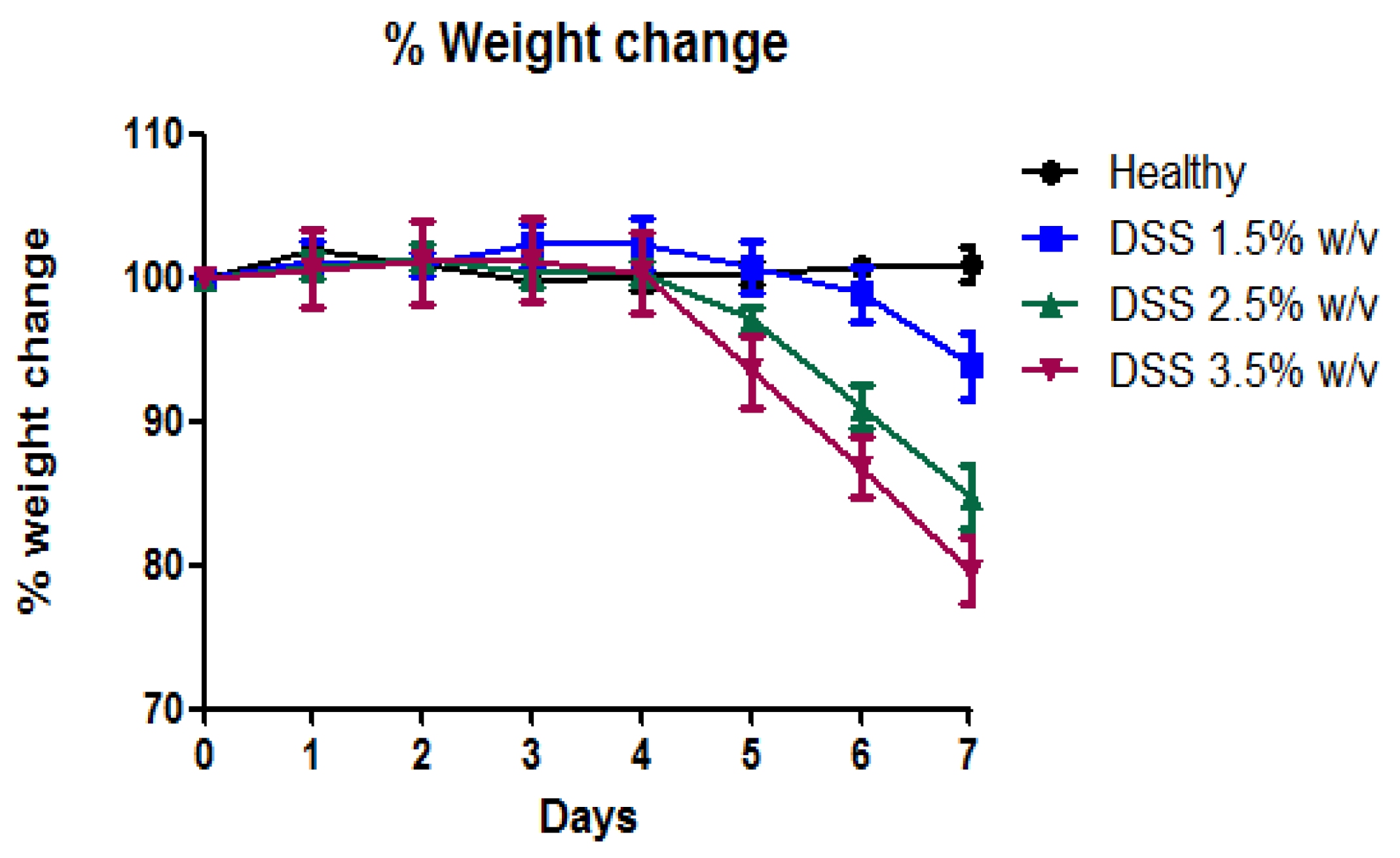
2.4. Percentage Weight Change

Progress in colitis inflammation was assessed by examining body weight. The change in body weight was observed daily from the first days of administration until the time of sacrifice. The weight of each animal on the first day was considered 100% to calculate the percentage weight change of each mouse.

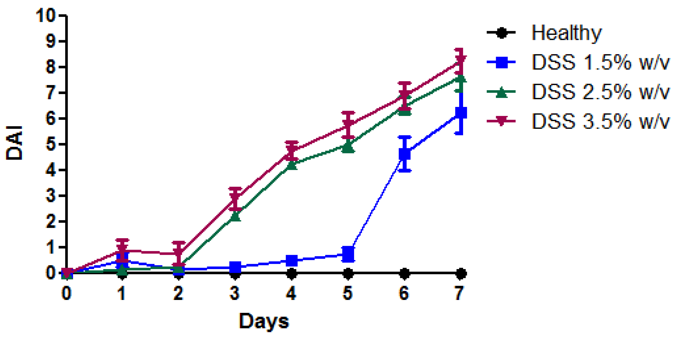
2.5. Disease Activity Index (DAI)

The disease activity index (DAI) is used to evaluate and determine the clinical progression of the disease in experimental colitis. The Friedman et al. method was used to determine the DAI score [25]. The DAI score was determined by adding the scores for weight loss, stool consistency, and hematochezia. The daily variations in growth rate, stool consistency, and the occurrence of gross bleeding or occult blood in the faeces were rated on a scale of 0 to 4 for each animal, as shown in Table 1. The overall score was equal to the sum of the subscores [26].

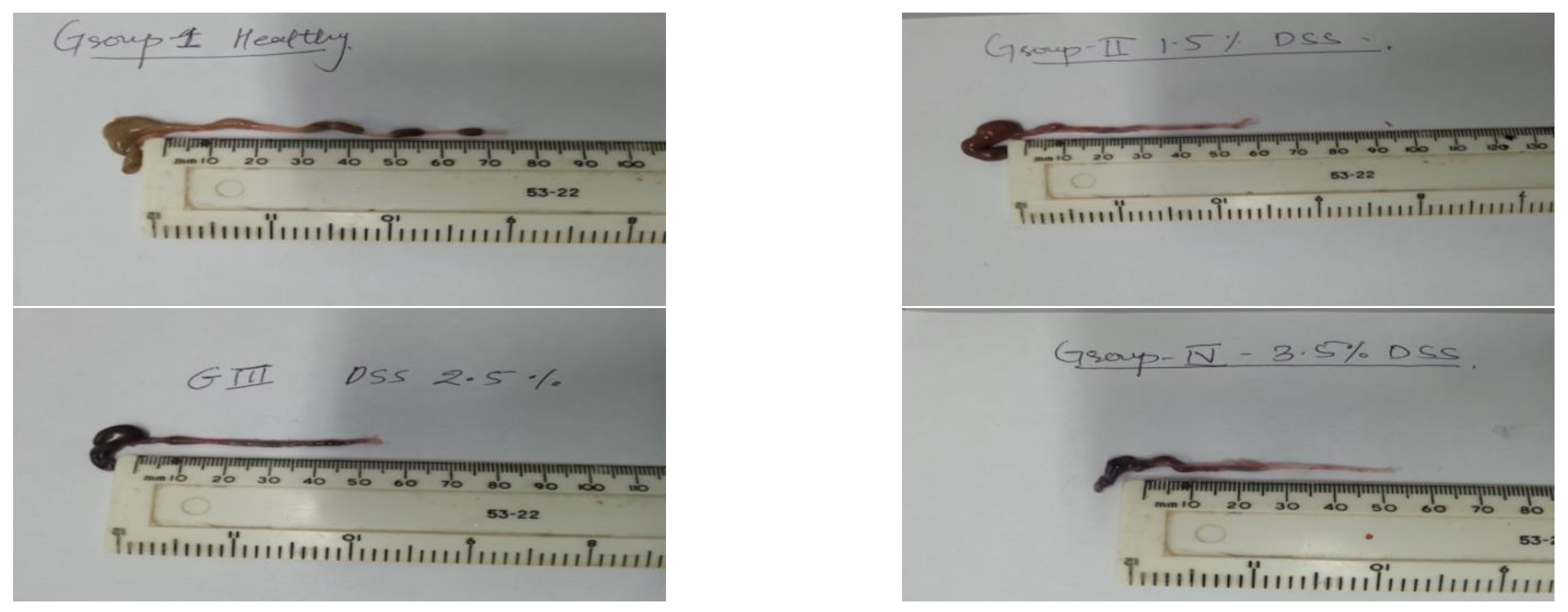
2.6. Change in Colon Length

Mice were sacrificed on the 7th day by cervical dislocation. Each mouse’s colon was surgically excised (post-mortem). The faeces and blood were removed with the help of cotton and washed with PBS to remove blood clots from the colon. The length of each colon was measured with the help of a measuring ruler [27].

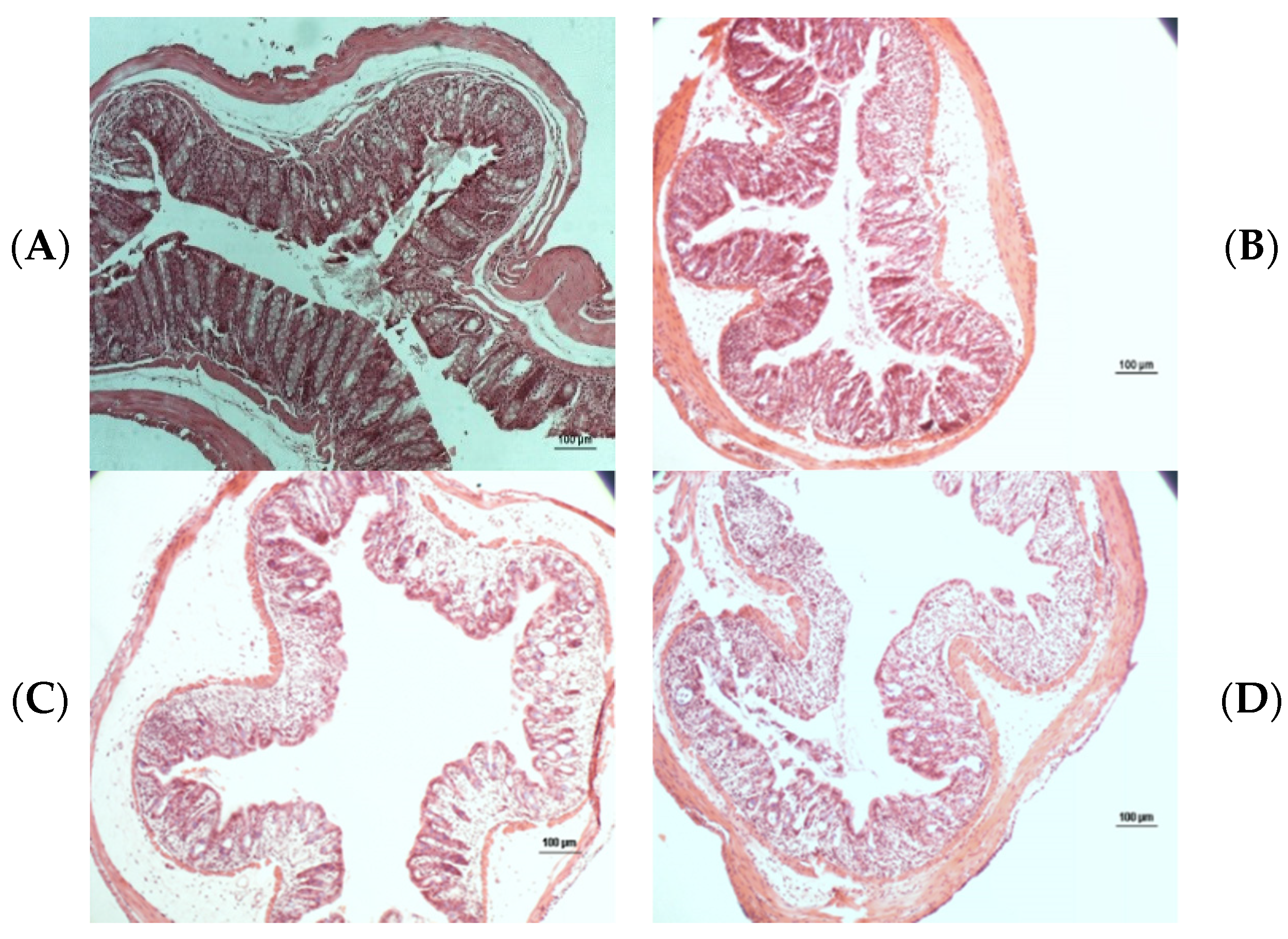
2.7. Histopathological Analysis

The animals were sacrificed on the seventh day, and the colon was removed. After the removal of the distal colon section, it was stored in 4% paraformaldehyde solution. It was then embedded in paraffin wax and sliced with the help of a microtome [28]. For further pathological analysis, a 5 mm tissue section was stained with haematoxylin and eosin. The epithelial damage score was allocated based on the histological observation. Using blinded analysis, the samples were graded according to the criteria outlined by Boirivant et al. [29] Samples were independently graded by two pathologists, who were blinded to one another’s scoring, and the mean of the two histology scores was calculated [30].

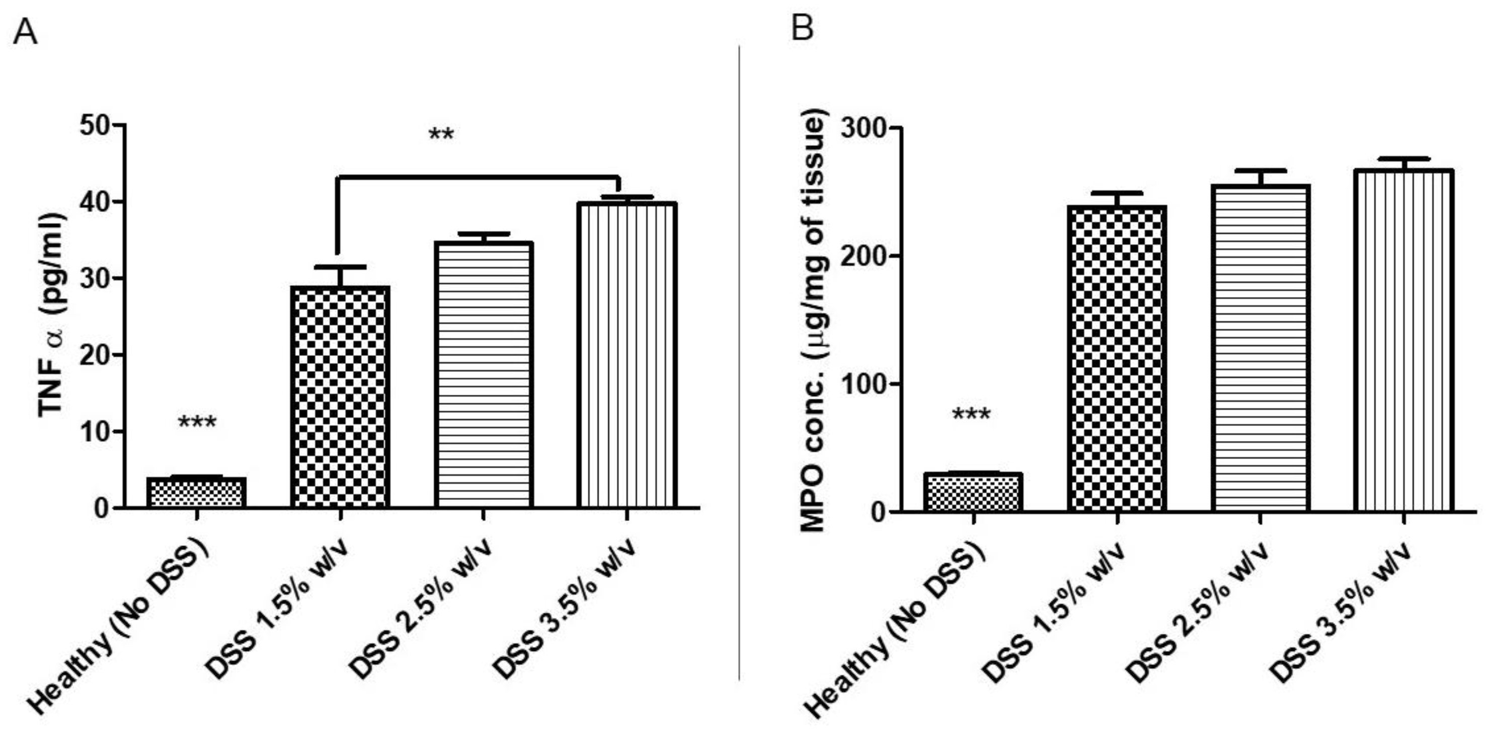
2.8. Colon Cytokine and Myeloperoxidases Measurements

Post-mortem colon tissue (middle section) was snap-frozen and stored in cryo vials at −80 degrees until further analysis. The snap-frozen tissue was thawed to room temperature and then homogenised using a method adapted from processing lung tissue used in our laboratory. TNF- levels were measured using mouse cytokine V-Plex Assay Plates (Meso Scale Diagnostics; Rockville, MD, USA) in accordance with the manufacturer’s instructions. MPO activity was measured utilising o-phenylenediamine dihydrochloride as a substrate, and the findings were extrapolated from an MPO standard curve (Sigma). TNF and MPO levels were reported as pg per mg or g per mg, respectively, relative to colon protein concentration.

3. Result and Discussion

It has been documented that weight loss is one of the clinical observations during colitis [31]. In the present work, a decrease in weight was documented when the mice were treated with different concentrations of DSS, as shown in Figure 1. Mice treated with 1.5%, 2.5%, and 3.5% DSS demonstrated a decrease in weight after the 4th day of the experiment. The extent of weight loss was different in each group. The mice treated with 3.5% w/v DSS weighed significantly less than those treated with 2.5% and 1.5% DSS. Moreover, 3.5% and 2.5% w/v DSS exhibited a similar pattern of weight loss; however, the 3.5% DSS-treated mice had a slightly greater decrease in weight. Thus, the extent of weight loss increases with the increasing concentrations of DSS.
To evaluate the severity of inflammation, DAI scores were monitored from day 0 to day 7. The DAI scores were assigned based on the composite score of weight loss, stool consistency, and the appearance of blood in the faeces. The mice treated with 1.5% DSS started losing weight after the 4th day of the experiment, as shown in Figure 2. Their stools became loose, and the appearance of blood was seen on the 6th day of the experiment, whereas the weight of the mice treated with 2.5% and 3.5% DSS showed a decrease in weight on the 2nd day of the experiment. However, diarrhoea and blood in the faeces were seen on the 5th and 3rd days of the experiment for 2.5% and 3.5% DSS, respectively. Therefore, the clinical signs of colitis progress as the concentration of DSS increases.
On the 7th day, all of the mice were sacrificed by cervical dislocation. The colon was removed, and the length was measured for each group. It has been reported that shortening of the colon is one of the clinical signs of colitis [21]. In the current work, a reduction in colon length was reported, as shown in Figure 3. The length of the healthy colon was 80 mm, as visible in the figure. In comparison to 1.5% and 3.5% DSS, the mice treated with 2.5% DSS had the shortest colon lengths.
According to Figure 4, the mice treated with 2.5% DSS show a significantly greater (p-value < 0.0001) decrease in colon length than the mice treated with 1.5% DSS. Furthermore, the 2.5% DSS group had significantly shorter colon lengths (p-value < 0.001) than the 3.5% DSS group. The colons of all the groups of mice showed blood and loose stools, whereas the colons of the healthy group showed normal stools. Overall, the mean colon length of all the DSS groups was shorter than that of the control group.
Figure 5 represents the colon histology of healthy individuals and mice treated with different concentrations of DSS. The mucosa of a healthy colon shows an intact epithelial cell layer. It also demonstrates a straight and unbroken cryptographer. The mice treated with 1.2% DSS (C) had less crypt damage than the mice treated with 2.5% DSS and 3.5% DSS. The appearance of neutrophils is more evident in C (2.5% DSS), and its epithelial structure has maintained its position. However, the extent of cryptic abscess and epithelial tissue damage is more prominent in D (3.5% DSS). This higher extent of epithelial damage may cause death due to chronic inflammatory conditions in mice and may cause irreversible chronic inflammation [32].
The microscopic histological damage score was assigned based on the observation of epithelial damage and infiltration of inflammatory cells, as shown in Figure 6. The mice given 2.5% DSS had significantly more epithelial damage (p-value < 0.0001) than the mice given 1.5% DSS. However, the 3.5% DSS-treated mice had higher epithelial damage than the 2.5% DSS group. To avoid a higher extent of inflammation, which may cause the death of mice, 2.5% DSS is the best suited to develop the colitis model.
Finally, we investigated the effect of different levels of DSS treatment on the expression of markers of colonic inflammation namely, tumor necrosis factor alpha (TNF α) and myeloperoxidase (MPO) as these markers could provide a better understanding how changing/increasing levels of DSS increase the intensity of inflammation in the colon (Figure 7). We observed that there was a statistically significant increase in levels of both TNF α and MPO in the DSS-treated mice as compared to the healthy control mice which is the expected outcome. We also see a gradual increase in levels of both TNF α and MPO as the dose of DSS is increased. This finding provides further evidence that the increase in the level of DSS is directly proportional to the increase in the intensity of inflammation.

4. Discussion

The entry of bacteria or their antigens into the mucosa due to the disruption of the epithelial layer of the intestine is considered the causative event in the prognosis of enterocolitis [33]. As shown previously, intestinal inflammation can be easier or more readily induced in animal models where the factors that damage the epithelial layer are clear. Hermiston et al., in a landmark study, confirmed that mucosal inflammation could be prevented with a healthy, intact epithelium, where cadherins perform a very crucial role in their adhesion [34].
As discussed before, several types of animal models have been evaluated to study intestinal inflammation. Proper selection of an animal model that can mimic human disease is crucial for the development of new drugs.
Dextran sulfate sodium (DSS) is known to induce colitis in animal models by disrupting the intestinal epithelial barrier and causing an inflammatory response. One consequence of this disruption is alteration in the constitution of the gut microbiota.
Experimental studies have shown that DSS treatment leads to a decrease in the diversity of gut microbial communities in mice. Specifically, DSS treatment is associated with a decrease in the relative abundance of beneficial bacteria such as Lactobacillus and Bifidobacterium, while increasing the relative abundance of potentially pathogenic bacteria such as Enterococcus and Proteobacteria [35].
These changes in microbial composition and diversity can contribute to the development and progression of colitis by altering host–microbe interactions and promoting inflammation. However, it is important to note that the effects of DSS on the gut microbiota can vary depending on the DSS concentration used, the duration of treatment, and other experimental factors [36].
In the present work, we examined DSS because of its easy availability, low cost, and short time to induce inflammation in mice. DSS-induced colitis causes a series of events that damage the colon epithelium. These events result in an innate immune response that alters the function of the epithelial barrier. The effect of the different concentrations on the degree of damage to the epithelial barrier was carefully evaluated in this study. We have selected three different concentrations of DSS based on previous studies reported in the literature and studies carried out in our laboratory [23,27]. The administration of 1.5% DSS w/v DSS in drinking water causes cryptic structures, cytokine damage, and cryptic abscesses. For a short period of time, 1.5% DSS w/v of DSS could cause reproducible acute inflammation limited to the colon. With 3.5% DSS w/v in drinking water, we observed a more damaged cryptic structure, infiltration of cytokines, and cryptic abscesses.
DSS-induced colitis inflammation is independent of T-cell-mediated immunity, as demonstrated in immunodeficient mice such as SCID4 and Rag1. This indicates that the cytokines produced by the innate immune cells are sufficient to produce inflammation in colitis [37,38]. Several studies have confirmed the crucial roles of macrophages and neutrophils in mucosal inflammation [39,40,41]. This makes the DSS-induced colitis models more useful for the investigation of the innate immune response. These findings are crucial, which indicates that the cytokines produced by the macrophages are very crucial in regulating the functions of epithelial cells. The DSS models are also found to be very crucial in exploring the molecular events of the innate immune system in colitis, especially those of TLR [42,43]. The effect of DSS on body weight loss was observed; an initial small increase in body weight for up to 3 days, which began to decrease with bleeding and diarrhoea. The treatment group showed significant differences in weight loss when treated with 1.5%, 2.5%, and 3.5% DSS. The loss in weight in the 2.5% and 3.5% DSS groups had been observed since the 4th day, whereas the loss in weight in the 1.5% group was seen on the 6th day of the experiment. The disease activity index score graph demonstrates a noticeable difference between the 1.5%, 2.5%, and 3.5% DSS-treated groups. The DAI score was observable on the 2nd day for the 2.5% and 3.5% DSS-treated mice. In contrast, it was visible on the 5th day of the experiment for the 1.5% DSS-treated group. The length of the colon reduced as the disease developed and was significantly shorter on day 4. The 2.5% DSS-treated mice had the shortest colon lengths when compared with the 1.5% and 3.5% DSS-treated mice.
A histological study of the colon sections stained by haematoxylin and eosin showed marked inflammation. BALB/c mice were treated with three different concentrations of DSS for 7 days, and the mice were sacrificed to collect their colons. The extent of inflammation in the 2.5% DSS-treated mice was significantly greater than in the 1.5% group. The crypt and epithelial layer were almost fully damaged, and severe infiltration of inflammatory cells into the mucosa was seen in the 3.5% DSS-treated group. The group treated with 1.2% DSS (C) showed less crypt damage than the 2.5% and 3.5% DSS-treated groups. The appearance of neutrophils is more evident in C (2.5% DSS), and its epithelial structure maintained its position. However, the extent of cryptic abscesses and epithelial tissue damage is more prominent in D (3.5% DSS). This higher extent of epithelial damage may cause death due to chronic inflammatory conditions in mice and may cause irreversible chronic inflammation [23]. Therefore, 2.5% could be a suitable concentration to study the colitis model and to recover inflammation by stopping the administration of DSS.
DSS induces colitis and involves the disruption of the protective epithelial barrier. This has led to the investigation of the factors that help maintain and reclaim cellular integrity after the damage. However, the healing of the colon depends on the extent of epithelial damage. Excessive damage to the epithelium layer leads to loss of life. In this case, the use of a 2.5% DSS solution offers the advantage of reversing the cellular damage to the epithelial layer of the colon. Several studies have investigated the role of TLR, cytokines, and proteins derived from the epithelial layer itself on damage reversal. The TLR2 ligand was found to reduce the severity of DSS on epithelial cells, while its deficiency promotes colitis. In addition to this, TLR4 activation reduces the DSS-mediated action on the epithelial cells by releasing growth factors that stimulate the cells positively [44,45]. In yet another study, it was shown that the Wnt pathway inhibited by Dkk1 was positively correlated with colonic renewal [46].
As it appears from the above discussion, this study could be useful for selecting the right concentration of DSS to induce inflammation. However, a crucial point to be noted here is that DSS induces inflammation but does not accurately mimic ulcerative colitis in humans. The most significant difference is the involvement of the T-cell proinflammatory cytokine response in chronic DSS colitis and polarised T-cell-mediated responses in human IBD.

5. Conclusions

The current work shows the induction of mild to severe levels of colitis upon administration of different doses of DSS. Figure 1 depicts the variation in weight loss patterns when the mice were given different concentrations of DSS in their drinking water. The figure depicts the extent of weight loss, which was found to increase upon increasing the DSS concentration. Different doses of DSS, such as 1.5%, 2.5%, and 3.5%, had different effects on the DAI, as shown in the figure. The amounts of 2.5%, 2.5%, and 3.5% DSS showed clinical signs of the DAI in the early days of the experiment, whereas 1.5% DSS appears nearly normal on the last day of the investigation. The mice treated with DSS had shorter colon lengths than the healthy group, as shown in Figure 3. Blood and loose stools were observed in the colons treated with 2.5% DSS. The DSS-treated mice had significantly shorter colon lengths than the 1.5% and 3.5% DSS-treated mice, as depicted in Figure 4. The colon of a mouse was examined under 100× fluorescent microscopy to confirm epithelial layer distortion, cryptic structure damage, and neutrophil infiltration. This damage was proportional to the increase in the DSS concentration. The microscopic histological scores assigned to the colons of the healthy mice and the 1.5%, 2.5%, and 3.5% DSS-treated mice are shown in Figure 6. It confirms that the microscopic scores increase with the increase in DSS concentration. Based on the above clinical signs, the severity of inflammation, and observations, 2.5% may be the most suitable concentration of DSS to induce reversible colitis in mice.
Overall, the major benefit of using the dextran sulfate sodium (DSS)-induced model of colitis in inflammatory bowel disease (IBD) research is its ability to mimic the key features of human IBD, such as its chronic relapsing nature, mucosal inflammation, and the development of symptoms such as diarrhoea, abdominal pain, and rectal bleeding. This makes DSS a useful tool for investigating the underlying mechanisms of IBD, testing potential therapeutic strategies, and evaluating their efficacy prior to clinical trials in humans.
Our work makes a very specific addition to the available literature by showing that changing the level of DSS can alter the disease’s intensity from mild to severe; this can be very useful for researchers as ulcerative colitis is a dynamic disease that changes from remission to flare-ups in humans. Thus, considering the optimal dose and duration of DSS exposure for the induction of colitis can allow for better mechanistic studies and the development of effective therapeutics for ulcerative colitis.

6. Future Work

A combination of the DSS model with other models: The DSS model can be combined with other models, such as adoptive transfer of T cells or infection with gut microbes, to study the interplay between different factors in IBD.
Improved characterisation of disease phenotype: The characterisation of the disease phenotype can be improved through the use of more comprehensive tools, such as histological, transcriptomic, or proteomic analysis.
Clinical relevance: The model can be improved by increasing its clinical relevance through the use of human-derived cells or the addition of gut microbes.
By implementing these modifications, the DSS model can provide a more accurate representation of the complex etiology of IBD and provide valuable insights for IBD research.

Author Contributions

Conceptualization, M.M.T.; methodology, M.M.T.; software, M.M.T., A.G.A., A.M.A. and S.A.A.; validation, M.M.T., A.G.A., A.M.A. and S.A.A.; formal analysis, M.M.T., A.G.A., A.M.A. and S.A.A.; investigation, M.M.T. and A.G.A., resources, M.M.T. and A.G.A., data curation, M.M.T. and A.G.A., writing—original draft preparation, M.M.T. and A.G.A., writing—review and editing, M.M.T., A.G.A., A.M.A. and S.A.A.; visualization, M.M.T., supervision, M.M.T., project administration, M.M.T. and A.G.A., funding acquisition, M.M.T. and A.G.A. All authors have read and agreed to the published version of the manuscript.

Funding

This research was funded by Deanship of Scientific Research, Qassim University grant number [10166-cams1-2020-1-3-1].

Institutional Review Board Statement

The Affiliates of Ulster University’s Ethics Committee for Animal Care and Use authorised all of the animal research (approval number PL-2784).

Informed Consent Statement

Not applicable.

Data Availability Statement

All the raw data are stored in the University of Lincoln’s OneDrive storage folder behind a two-factor authentication wall. All data will be available upon request.

Acknowledgments

The authors gratefully acknowledge Qassim University, represented by the Deanship of Scientific Research, on the financial support for this research under the number (10166-cams1-2020-1-3-1) during the academic year 1441 AH/2020 AD.

Conflicts of Interest

The authors declare no conflict of interest.

References

  1. Feagan, B.G.; Rutgeerts, P.; Sands, B.E.; Hanauer, S.; Colombel, J.-F.; Sandborn, W.J.; Van Assche, G.; Axler, J.; Kim, H.-J.; Danese, S.; et al. Vedolizumab as Induction and Maintenance Therapy for Ulcerative Colitis. N. Engl. J. Med. 2013, 369, 699–710. [Google Scholar] [CrossRef] [PubMed] [Green Version]
  2. Mizoguchi, E.; Nguyen, D.; Low, D. Animal models of ulcerative colitis and their application in drug research. Drug Des. Dev. Ther. 2013, 7, 1341–1357. [Google Scholar] [CrossRef] [Green Version]
  3. Akolkar, P.N.; Gulwani-Akolkar, B.; Heresbach, D.; Lin, X.Y.; Fisher, S.; Katz, S.; Silver, J. Differences in risk of Crohn’s disease in offspring of mothers and fathers with inflammatory bowel disease. Am. J. Gastroenterol. 1997, 92, 2241–2244. [Google Scholar]
  4. Anderson, C.A.; Boucher, G.; Lees, C.W.; Franke, A.; D’Amato, M.; Taylor, K.D.; Lee, J.C.; Goyette, P.; Imielinski, M.; Latiano, A.; et al. Meta-analysis identifies 29 additional ulcerative colitis risk loci, increasing the number of confirmed associations to 47. Nat. Genet. 2011, 43, 246–252. [Google Scholar] [CrossRef] [PubMed] [Green Version]
  5. Mahid, S.S.; Minor, K.S.; Soto, R.E.; Hornung, C.A.; Galandiuk, S. Smoking and Inflammatory Bowel Disease: A Meta-analysis. Mayo Clin. Proc. 2006, 81, 1462–1471. [Google Scholar] [CrossRef] [PubMed]
  6. Strober, W.; Fuss, I. Pro-Inflammatory Cytokines in the Pathogenesis of IBD. Gastroenterology 2011, 140, 1756–1767. [Google Scholar] [CrossRef] [PubMed] [Green Version]
  7. Aggarwal, B.B.; Harikumar, K.B. Potential therapeutic effects of curcumin, the anti-inflammatory agent, against neurodegenerative, cardiovascular, pulmonary, metabolic, autoimmune and neoplastic diseases. Int. J. Biochem. Cell Biol. 2009, 41, 40–59. [Google Scholar] [CrossRef] [Green Version]
  8. Neurath, M.F. New targets for mucosal healing and therapy in inflammatory bowel diseases. Mucosal Immunol. 2014, 7, 6–19. [Google Scholar] [CrossRef] [Green Version]
  9. Qin, X. Why is damage limited to the mucosa in ulcerative colitis but transmural in Crohn’s disease? World J. Gastrointest. Pathophysiol. 2013, 4, 63–64. [Google Scholar] [CrossRef]
  10. Chen, H.; Fang, Y.; Li, W.; Orlando, R.C.; Shaheen, N.; Chen, X.L. NFkB and Nrf2 in esophageal epithelial barrier function. Tissue Barriers 2013, 1, e27463. [Google Scholar] [CrossRef] [Green Version]
  11. D’Aldebert, E.; Quaranta, M.; Sébert, M.; Bonnet, D.; Kirzin, S.; Portier, G.; Duffas, J.-P.; Chabot, S.; Lluel, P.; Allart, S.; et al. Characterization of Human Colon Organoids From Inflammatory Bowel Disease Patients. Front. Cell Dev. Biol. 2020, 8, 363. [Google Scholar] [CrossRef]
  12. Dieleman, L.A.; Palmen, M.J.H.J.; Akol, H.; Bloemena, E.; Peña, A.S.; Meuwissen, S.G.M.; Van Rees, E.P. Chronic experimental colitis induced by dextran sulphate sodium (DSS) is characterized by Th1 and Th2 cytokines. Clin. Exp. Immunol. 1998, 114, 385–391. [Google Scholar] [CrossRef]
  13. Kämpfer, A.A.; Urbán, P.; Gioria, S.; Kanase, N.; Stone, V.; Kinsner-Ovaskainen, A. Development of an in vitro co-culture model to mimic the human intestine in healthy and diseased state. Toxicol. Vitr. 2017, 45, 31–43. [Google Scholar] [CrossRef] [PubMed]
  14. Yu, Y.; Wen, H.; Li, S.; Cao, H.; Li, X.; Ma, Z.; She, X.; Zhou, L.; Huang, S. Emerging microfluidic technologies for microbiome research. Front. Microbiol. 2022, 13, 3116, Erratum in: Front Microbiol. 2022, 13, 1038682. [Google Scholar] [CrossRef] [PubMed]
  15. Roh, T.T.; Chen, Y.; Paul, H.T.; Guo, C.; Kaplan, D.L. 3D bioengineered tissue model of the large intestine to study inflammatory bowel disease. Biomaterials 2019, 225, 119517. [Google Scholar] [CrossRef] [PubMed]
  16. Hendow, E.K.; Guhmann, P.; Wright, B.; Sofokleous, P.; Parmar, N.; Day, R.M. Biomaterials for hollow organ tissue engineering. Fibrogenesis Tissue Repair 2016, 9, 3. [Google Scholar] [CrossRef] [PubMed] [Green Version]
  17. Joshi, A.; Soni, A.; Acharya, S. In vitro models and ex vivo systems used in inflammatory bowel disease. Vitr. Model. 2022, 1, 213–227. [Google Scholar] [CrossRef]
  18. Khan, R.; Roy, N.; Ali, H.; Naeem, M. Fecal Microbiota Transplants for Inflammatory Bowel Disease Treatment: Synthetic- and Engineered Communities-Based Microbiota Transplants Are the Future. Gastroenterol. Res. Pract. 2022, 2022, 9999925. [Google Scholar] [CrossRef]
  19. Hartman, K.G.; Bortner, J.J.D.; Falk, G.; Ginsberg, G.G.; Jhala, N.; Yu, J.; Martín, M.G.; Rustgi, A.K.; Lynch, J.P. Modeling human gastrointestinal inflammatory diseases using microphysiological culture systems. Exp. Biol. Med. 2014, 239, 1108–1123. [Google Scholar] [CrossRef] [Green Version]
  20. Randhawa, P.K.; Singh, K.; Singh, N.; Jaggi, A.S. A Review on Chemical-Induced Inflammatory Bowel Disease Models in Rodents. Korean J. Physiol. Pharmacol. 2014, 18, 279–288. [Google Scholar] [CrossRef] [Green Version]
  21. Chassaing, B.; Aitken, J.D.; Malleshappa, M.; Vijay-Kumar, M. Dextran Sulfate Sodium (DSS)-Induced Colitis in Mice. Curr. Protoc. Immunol. 2014, 104, 15.25.1–15.25.14. [Google Scholar] [CrossRef] [PubMed] [Green Version]
  22. Egger, B.; Bajaj-Elliott, M.; Macdonald, T.T.; Inglin, R.; Eysselein, V.E.; Büchler, M.W. Characterisation of Acute Murine Dextran Sodium Sulphate Colitis: Cytokine Profile and Dose Dependency. Digestion 2000, 62, 240–248. [Google Scholar] [CrossRef]
  23. Okayasu, I.; Hatakeyama, S.; Yamada, M.; Ohkusa, T.; Inagaki, Y.; Nakaya, R. A novel method in the induction of reliable experimental acute and chronic ulcerative colitis in mice. Gastroenterology 1990, 98, 694–702. [Google Scholar] [CrossRef]
  24. Friedman, D.J.; Künzli, B.M.; A-Rahim, Y.I.; Sevigny, J.; Berberat, P.O.; Enjyoji, K.; Csizmadia, E.; Friess, H.; Robson, S.C. From the cover: CD39 deletion exacerbates experimental murine colitis and human polymorphisms increase susceptibility to inflammatory bowel disease. Proc. Natl. Acad. Sci. USA 2009, 106, 16788–16793. [Google Scholar] [CrossRef] [PubMed] [Green Version]
  25. Chen, Y.; Si, J.-M.; Liu, W.-L.; Cai, J.-T.; Du, Q.; Wang, L.-J.; Gao, M. Induction of experimental acute ulcerative colitis in rats by administration of dextran sulfate sodium at low concentration followed by intracolonic administration of 30% ethanol. J. Zhejiang Univ. B 2007, 8, 632–637. [Google Scholar] [CrossRef] [Green Version]
  26. Ogawa, A.; Andoh, A.; Araki, Y.; Bamba, T.; Fujiyama, Y. Neutralization of interleukin-17 aggravates dextran sulfate sodium-induced colitis in mice. Clin. Immunol. 2003, 110, 55–62. [Google Scholar] [CrossRef]
  27. Tambuwala, M.M.; Manresa, M.C.; Cummins, E.P.; Aversa, V.; Coulter, I.S.; Taylor, C.T. Targeted delivery of the hydroxylase inhibitor DMOG provides enhanced efficacy with reduced systemic exposure in a murine model of colitis. J. Control. Release 2015, 217, 221–227. [Google Scholar] [CrossRef]
  28. Boirivant, M.; Cossu, A. Inflammatory bowel disease. Oral Dis. 2011, 18, 1–15. [Google Scholar] [CrossRef]
  29. Kim, J.; Shajib, S.; Manocha, M.M.; Khan, W.I. Investigating Intestinal Inflammation in DSS-induced Model of IBD. J. Vis. Exp. 2012, e3678. [Google Scholar] [CrossRef] [Green Version]
  30. Taghipour, N.; Molaei, M.; Mosaffa, N.; Rostami-Nejad, M.; Agdaei, H.A.; Anissian, A.; Azimzadeh, P.; Zali, M.R. An experimental model of colitis induced by dextran sulfate sodium from acute progresses to chronicity in C57BL/6: Correlation between conditions of mice and the environment. Gastroenterol. Hepatol. Bed Bench 2015, 1, 45–52. [Google Scholar] [CrossRef]
  31. Perše, M.; Cerar, A. Dextran Sodium Sulphate Colitis Mouse Model: Traps and Tricks. J. Biomed. Biotechnol. 2012, 2012, 718617. [Google Scholar] [CrossRef] [Green Version]
  32. Neu, J.; Walker, W.A. Necrotizing Enterocolitis. N. Engl. J. Med. 2011, 364, 255–264. [Google Scholar] [CrossRef] [Green Version]
  33. Hermiston, M.L.; Gordon, J.I. Inflammatory Bowel Disease and Adenomas in Mice Expressing a Dominant Negative N-Cadherin. Science 1995, 270, 1203–1207. [Google Scholar] [CrossRef]
  34. Krieglstein, C.F.; Cerwinka, W.H.; Sprague, A.G.; Laroux, F.S.; Grisham, M.B.; Koteliansky, V.E.; Senninger, N.; Granger, D.N.; De Fougerolles, A.R. Collagen-binding integrin α1β1 regulates intestinal inflammation in experimental colitis. J. Clin. Investig. 2002, 110, 1773–1782. [Google Scholar] [CrossRef] [Green Version]
  35. Weng, Y.-J.; Jiang, D.-X.; Liang, J.; Ye, S.-C.; Tan, W.-K.; Yu, C.-Y.; Zhou, Y. Effects of Pretreatment with Bifidobacterium bifidum Using 16S Ribosomal RNA Gene Sequencing in a Mouse Model of Acute Colitis Induced by Dextran Sulfate Sodium. Med. Sci. Monit. Int. Med. J. Exp. Clin. Res. 2021, 27, e928478-1–e928478-12. [Google Scholar] [CrossRef]
  36. Lobionda, S.; Sittipo, P.; Kwon, H.Y.; Lee, Y.K. The Role of Gut Microbiota in Intestinal Inflammation with Respect to Diet and Extrinsic Stressors. Microorganisms 2019, 7, 271. [Google Scholar] [CrossRef] [Green Version]
  37. Dieleman, L.A.; Ridwan, B.U.; Tennyson, G.S.; Beagley, K.W.; Bucy, R.; Elson, C.O. Dextran sulfate sodium-induced colitis occurs in severe combined immunodeficient mice. Gastroenterology 1994, 107, 1643–1652. [Google Scholar] [CrossRef]
  38. Fukata, M.; Michelsen, K.S.; Eri, R.; Thomas, L.S.; Hu, B.; Lukasek, K.; Nast, C.C.; Lechago, J.; Xu, R.; Naiki, Y.; et al. Toll-like receptor-4 is required for intestinal response to epithelial injury and limiting bacterial translocation in a murine model of acute colitis. Am. J. Physiol. Liver Physiol. 2005, 288, G1055–G1065. [Google Scholar] [CrossRef] [Green Version]
  39. Pull, S.L.; Doherty, J.M.; Mills, J.C.; Gordon, J.I.; Stappenbeck, T.S. Activated macrophages are an adaptive element of the colonic epithelial progenitor niche necessary for regenerative responses to injury. Proc. Natl. Acad. Sci. USA 2004, 102, 99–104. [Google Scholar] [CrossRef] [Green Version]
  40. Chami, B.; Yeung, A.W.S.; van Vreden, C.; King, N.J.C.; Bao, S. The Role of CXCR3 in DSS-Induced Colitis. PLoS ONE 2014, 9, e101622. [Google Scholar] [CrossRef] [Green Version]
  41. Dupaul-Chicoine, J.; Yeretssian, G.; Doiron, K.; Bergstrom, K.S.; McIntire, C.R.; LeBlanc, P.M.; Meunier, C.; Turbide, C.; Gros, P.; Beauchemin, N.; et al. Control of Intestinal Homeostasis, Colitis, and Colitis-Associated Colorectal Cancer by the Inflammatory Caspases. Immunity 2010, 32, 367–378. [Google Scholar] [CrossRef] [PubMed] [Green Version]
  42. Sokol, H.; Conway, K.L.; Zhang, M.; Choi, M.; Morin, B.; Cao, Z.; Villablanca, E.; Li, C.; Wijmenga, C.; Yun, S.H.; et al. Card9 Mediates Intestinal Epithelial Cell Restitution, T-Helper 17 Responses, and Control of Bacterial Infection in Mice. Gastroenterology 2013, 145, 591–601.e3. [Google Scholar] [CrossRef] [PubMed] [Green Version]
  43. Cario, E.; Gerken, G.; Podolsky, D. Toll-Like Receptor 2 Controls Mucosal Inflammation by Regulating Epithelial Barrier Function. Gastroenterology 2007, 132, 1359–1374. [Google Scholar] [CrossRef]
  44. Hsu, D.; Fukata, M.; Hernandez, Y.G.; Sotolongo, J.P.; Goo, T.; Maki, J.; Hayes, L.A.; Ungaro, R.C.; Chen, A.; Breglio, K.J.; et al. Toll-like receptor 4 differentially regulates epidermal growth factor-related growth factors in response to intestinal mucosal injury. Lab. Investig. 2010, 90, 1295–1305. [Google Scholar] [CrossRef] [Green Version]
  45. Krzywinska, E.; Sobecki, M.; Nagarajan, S.; Zacharjasz, J.; Tambuwala, M.M.; Pelletier, A.; Cummins, E.; Gotthardt, D.; Fandrey, J.; Kerdiles, Y.M.; et al. The transcription factor HIF-1α mediates plasticity of NKp46+ innate lymphoid cells in the gut. J. Exp. Med. 2022, 219, e20210909. [Google Scholar] [CrossRef] [PubMed]
  46. Koch, S.; Nava, P.; Addis, C.; Kim, W.; Denning, T.L.; Li, L.; Parkos, C.A.; Nusrat, A. The Wnt Antagonist Dkk1 Regulates Intestinal Epithelial Homeostasis and Wound Repair. Gastroenterology 2011, 141, 259–268.e8. [Google Scholar] [CrossRef] [PubMed] [Green Version]
Figure 1. Percentage change in body weight in the mice at different concentrations of DSS. The mice exhibited noticeable differences in weight loss when they were treated with 1.5% and 2.5% DSS rather than 3.5% DSS. The loss in weight of the 2.5% and 3.5% DSS groups was observed from the 4th day, whereas the loss in weight for 1.5% was seen on the 6th day of the experiment.
Applsci 13 03233 g001
Figure 2. Effect of different concentrations of DSS on the disease activity index. The DAI score graph demonstrates a noticeable difference between the 1.5%, 2.5%, and 3.5% DSS-treated group. The DAI score was observable on the 2nd day for the 2.5% and 3.5% DSS-treated mice, whereas it was visible on the 5th day of the experiment for the 1.55 DSS-treated group.
Applsci 13 03233 g002
Figure 3. Change in colon length at different concentrations of DSS. Group I is the colon of the healthy mice, Group II is the colon of the 1.5% DSS-treated mice, group III is the colon of the 3.5 DSS-treated mice, and group IV is the colon of the 3.5% DSS = treated mice. The colon in group II has a shorter length as compared to the entire group.
Applsci 13 03233 g003
Figure 4. Effect of different concentrations of DSS on colon length. The colons of the entire DSS-treated group had significantly shorter lengths than the healthy group. Furthermore, the 2.5% DSS group had a significantly shorter colon length than the 1.5% and 3.5% DSS-treated groups. N = five mice per group (*** p < 0.0001).
Applsci 13 03233 g004
Figure 5. Histological section of the colons representing (100×) the healthy colon (A), the 1.5% DSS treated colon (B), the 2.5% DSS-treated colon (C), and the 3.5% DSS-treated colon (D). The extent of inflammation was more noticeable in the 2.5% DSS-treated mice as compared to the 1.5% group. The crypt was fully damaged in the 3.5% DSS group. The epithelial layer was distorted more in the 3.5% DSS-treated mice.
Applsci 13 03233 g005
Figure 6. Microscopic damage score. The mice treated with 2.5% DSS gained significantly higher colitis scores than the 1.5% DSS group. Although, the 3.5% DSS-treated mice had an even higher score than the 2.5% and 1.5% DSS-treated mice. N = five mice per group (*** p < 0.0001 and ** p < 0.001).
Applsci 13 03233 g006
Figure 7. Effect of DSS on the expression levels of TNF α (graph (A)) and MPO (graph (B)) in colon tissue. N = five mice per group (*** p < 0.0001 and ** p < 0.001).
Applsci 13 03233 g007
Table 1. Scoring of the disease activity index.
ScoreWeight LossStool ConsistencyFaecal Blood
0NoneNormalNone
11–5%
25–10%LooseHemoccult +
310–20%
4>20%DiarrhoeaGross bleeding
Disclaimer/Publisher’s Note: The statements, opinions and data contained in all publications are solely those of the individual author(s) and contributor(s) and not of MDPI and/or the editor(s). MDPI and/or the editor(s) disclaim responsibility for any injury to people or property resulting from any ideas, methods, instructions or products referred to in the content.

