Article

Does the Nature of Added Bioactive Lipids Affect the Biological Properties of Yogurts?—Case Study Coconut and Avocado Oils

by
Manuela Machado
,
Luís M. Rodríguez-Alcalá
,
Ana Maria Gomes
and
Manuela Pintado
*
CBQF Centro de Biotecnologia e Química Fina—Laboratório Associado, Escola Superior de Biotecnologia, Universidade Católica Portuguesa, Rua Diogo Botelho 1327, 4169-005 Porto, Portugal
*
Author to whom correspondence should be addressed.
Appl. Sci. 2023, 13(5), 3101; https://doi.org/10.3390/app13053101
Submission received: 21 January 2023 / Revised: 23 February 2023 / Accepted: 25 February 2023 / Published: 28 February 2023
(This article belongs to the Special Issue Editorial Board Members' Collection Series: Functional Foods)

Abstract

:
Bioactive lipids play an important role in human health and their benefits are linked to their chemical nature; for example, medium-chain fatty acids can have an important contribution to body weight management. This work aimed to test a strategy to enhance the quality profile and gastrointestinal tract resistance of previously developed vegetable oil-functionalized yogurts and further probe the biological potential of functionalized yogurts. Fortification with coconut and avocado vegetable oils led to increased nutritional value through an increase in essential fatty acids content, particularly in yogurts with vegetable oil bigels. One of the main problems with bioactive fatty acids fortification is their poor stability during in vitro digestion and consequently poor bioavailability. Despite this, the digested samples decreased lipid accumulation in Hep G2 and 3T3-L1 cells Functionalized yogurts were also responsible for high improvements in adiponectin secretion (35% for COY, 46% for CBY, 53% for AOY, and 48% ABY) compared with control yogurt. Moreover, in the inflammatory model, a reduction between 30% (for control yogurt) and 70% (for CBY) was observed for IL-6 secretion in LPS-stimulated cells. Considering these results, yogurt’s fortification with vegetable oils can be a viable alternative strategy to be scaled up for obesity management.

1. Introduction

Although the primary function of bioactive fatty acids is as an energy source, they also play an important role in human health, namely in cell structure and signalling [1,2]. Moreover, they help maintain homeostasis and prevent metabolic disorders, and some fatty acids are essential as precursors in the production of hormones and vitamins [1,2,3,4]. The more well-established health effects of bioactive fatty acids are related to cardiovascular disease. However, many other effects have been studied including the positive impact on atherosclerosis, cancer, mental health, brain function, diabetes, and obesity [2,4,5,6,7,8,9,10]. This wide range of benefits has increased the interest of the food industry in producing functional foods with bioactive lipids [11,12]. As referred by several authors, milk and dairy products appear as preferred matrices for bioactive lipid fortification. Within the range of dairy products, yogurt has been the most widely used, due to its high consumption and associated benefits [11,13,14]. Avocado (AO) and coconut oils (CO) can be valuable sources of fatty acids with a positive impact on lipid accumulation, thermogenesis, and modulation of adipokines and insulin resistance [15,16,17,18,19,20]. Moreover, the presence of vegetable oils can have a positive effect on gut microbiota; for example, coconut oil has been responsible for an increase in the abundance of probiotic bacteria, such as Lactobacillus, Allobaculum, and Bifidobacterium species [21]. On the other hand, polyunsaturated fatty acids (PUFAs) are responsible for maintaining the Firmicutes/Bacteroidetes [22]. However, as reported in previous works, the fatty acids profile of vegetable oil is significantly affected by gastrointestinal tract conditions [17,18]. Resistance to such conditions is one of the most important factors in a food’s functionalization with bioactive fatty acids and is directly linked to their potential bioactivity. As discussed in previous works, bigels can be a valuable strategy to enhance vegetable oil’s resistance. Bigels are semi-solid formulations produced by mixing an organogel and hydrogel at a certain temperature. These formulations possess interesting properties such as cooling and moisturizing effects, delivery of both hydrophobic and hydrophilic compounds, and improved spreadability [23]. Despite these benefits, the presence of these soft materials does not always have a positive effect on the visual appearance of the products [17,18]. Taking into consideration the above rationale and the drawbacks identified in the previous studies, this work aimed to improve the previous functionalized yogurts formulations by enhancing their in vitro digestion resistance and visual appearance. Furthermore, the effect of the nature of different fatty acids profiles was evaluated though a more detailed evaluation of yogurt’s biological potential.

2. Materials and Methods

2.1. Materials and Reagents

Geleol was kindly donated by Gatefossé (France). Avocado oil was obtained from Fula (Portugal), Coconut oil, Origens Bio, was obtained from Continente (Portugal), and powdered skim milk from Nestlé (Portugal); natural yogurts were obtained from Mimosa (Lactogal, Portugal). Carboxymethylcellulose was obtained from Merck (USA), 3-(4,5-dimethylthiazol-2-yl)-2,5-diphenyltetrazolium bromide (MTT), sodium alginate, α-amylase, pepsin, hydrochloric acid, sodium hydroxide, porcine pancreatin, bile salts, and dimethyl sulfoxide (DMSO) were obtained from Sigma (St. Louis, MO, USA). Lipase was obtained from Lipolytech (Marseille, France). Dulbecco’s Modified Eagle’s Medium (DMEM) and non-essential amino acids (NEAA) were obtained from Gibco, (Thermo Scientific, USA). Fetal bovine serum from Biowest (FBS; Nuaillé, France), penicillin-streptomycin-fungizone from Lonza (Basel, Switzerland), and calf bovine serum iron-fortified were obtained from ATCC (Virginia, USA). Lipopolysaccharides from Escherichia coli O111:B4 (LPS) were obtained from Invitrogen (USA). Adipolysis Assay Kit, Mouse Leptin ELISA kit, Mouse Adiponectin ELISA kit, and Hepatic Lipid Accumulation Kit were obtained from Abcam (ab133115, ab199082, ab18785, ab133131, Cambridge, UK). Legend Max Mouse Elisa Kit IL-6 and the Legend Max Mouse Elisa Kit TNF-α were obtained from BioLegend (San Diego, CA, USA) and BCA Pierce Assay Kit was obtained from ThermoScientific (Waltham, MA, USA).

2.2. Bigel Production

Bigels were produced as previously described by Machado et al., 2022 [17] with some modifications. In short, geleol was melted at 60 °C on a hotplate and mixed with tween 80. Following this, carboxymethyl cellulose (2% w/v) (for avocado bigels) or alginate (1% w/v) (for coconut bigels, new formulation) were added through continuous mixing for 2 min after which the respective vegetable oil was added and the mixtures homogenized for 1 min at 18,000 rpm in an ultra-turrax (IKA T 25 digital, Janke and Kunkel IKA, Germany). Mixtures were then sonicated (Sonics Vibra-Cell™ VCX 130) at 60% amplitude for 1 min and subsequently cooled to room temperature before use.

2.3. Yogurt Production

For yogurt production, commercial powdered skimmed milk was dissolved in water (13% w/v; Nestlé, Portugal). Then vegetable oils (1.5% (w/w) and respective bigels (3% (w/w), corresponding to the equivalent free oil 1.5% (w/w) were added and the mixture pasteurized (85 °C, 5 min) in a water bath. Plain reconstituted milk was used for the production of the control yogurt. Before fermentation, samples were cooled (1 h) to 45 °C, inoculated at 3% (w/w) with natural yogurt (Mimosa Natural, Lactogal, Portugal) as a source of starter culture, and then incubated at 43 °C, in plastic yogurt containers, until a final pH value of 4.6 was reached. Samples were stored at 4 °C until usage and per each yogurt type—plain yogurt control (CT), coconut oil fortified yogurt (COY), coconut bigel fortified yogurt (CBY), avocado oil fortified yogurt (AOY), and avocado bigel yogurt (ABY)—15 yogurt samples were prepared.

2.4. Fatty Acids Profile

The fatty acid profile was evaluated as described by Machado et al. (2022) [17]. Briefly, fatty acid methyl esters were analyzed in a gas chromatograph Agilent 8860 (Agilent, Santa Clara, CA, USA), equipped with a flame ionization detector and a BPX70 capillary column (60 m × 0.25 mm × 0.25 μm; SGE Europe Ltd., Milton Keynes, UK) with analysis conditions being as follows: injector (split 25:1; injection volume 1 µL), injector, and detector temperatures were 250 °C and 275 °C, respectively; hydrogen was used as a carrier gas at a flow rate of 1 mL/min. The initial oven temperature was 60 °C and was then increased to a final temperature of 225 °C. Sample identification was performed using Supelco 37 standard and each sample was analyzed in triplicate. Additionally, the nutritional quality indexes (atherogenic index (AI), thrombogenic index (TI), hypocholesterolemic/hypercholesterolemic ratio (HH), and health promoting index (HPI) were calculated according to equations 1 to 4 [24].
A I = C 12 : 0 + 4 × C 14 : 0 + C 16 : 0 M U F A + P U F A   n 6 + P U F A   n 3
T I = C 14 : 0 + C 16 : 0 + C 18 : 0 0.5 × M U F A + 0.5 × P U F A   n 6 + 3 × P U F A   n 3 + P U F A   n 3 P U F A   n 6
H H = C 18 : 1 n 9 + C 18 : 2 n 6 + C 18 : 3 n 3 + C 20 : 4 n 6 + C 20 : 5 n 3 C 14 : 0 + C 16 : 0
H P I = U F A C 12 : 0 + 4 × C 14 + C 16

2.5. In Vitro Digestion

The in vitro digestion was performed according to the INFOGEST method [25]. In brief, to mimic the oral phase, 5 g of yogurt was mixed with simulated salivary fluid (1:1 w/w), 75 U/ mL of α-amylase was added, and the mixture was incubated for 2 min at 37 °C under continuous shaking. In the gastric phase, samples were added to simulated gastric fluid (1:1 v/v), and the pH was set to 3 using HCl (1M). The enzymes pepsin and lipase were added (2000 U/mL and 60 U/mL, respectively), and the mixture was incubated for 120 min at 37 °C under continuous shaking. Finally, to mimic the intestinal phase, the pH was set to 7 using NaOH (1 M) and simulated intestinal fluid was added (1:1 v/v). Porcine pancreatin and bile were added to the digestion to achieve 100 U/mL and 10 mM, respectively. The mixture was incubated for another 120 min at 37 °C under continuous shaking. To screen the impact of digestion conditions on the fatty acids profile, yogurt samples were collected at different stages (after oral, gastric, and intestinal steps). The fatty acids profile was evaluated as described above using 500 µL of the sample. After in vitro digestion, samples were centrifuged at 14,000 rpm for 30 min, and the supernatant was collected for cell-based assays.

2.6. Biological Properties

2.6.1. Cell Lines

Human Caucasian colon carcinoma epithelial cells (Caco-2) were obtained from the European Collection of Authenticated Cell Cultures (ECACC 86010202). Human hepatocellular carcinoma (Hep G2), mouse macrophages RAW 264.7, and mouse pre-adipocytes 3T3-L1 were all obtained from American Type Culture Collection (ATCC; ATCC HB-8065, TIB-71 and CL-173, respectively). Hep G2 and RAW cell lines were cultured using DMEM supplemented with 10% (v/v) FBS and 1% (v/v) of Penicillin-Streptomycin-Fungizone. Caco-2′ was grown in the conditions previously referred to with the addition of 1% (v/v) NEAA. Pre-adipocytes were cultured in DMEM with 10% (v/v) of iron-fortified CBS and 1% (v/v) of Penicillin-Streptomycin-Fungizone. All cell lines were incubated at 37 °C under a humidified atmosphere comprised of 5% CO2 and 95% air.

2.6.2. Metabolic Activity

Metabolic activity assays were carried out as previously described by Machado et al. (2022) [17]. Briefly, cells were seeded (1.0 × 10⁴ cells/well) into 96-well tissue culture plates (Thermo Scientific, Denmark) and incubated. After 24 h, the media was carefully removed and replaced with 100 μL of digested samples (0.07 mg of fatty acid/mL), plain media (growth control), and media containing 40% (v/v) DMSO (death control) and re-incubated for 24 h. Following this, 100 μL of MTT solution (0.5 mg/mL) were added to each well, and the plates were incubated for 2 h following which the wells’ content was aspirated, 100 μL of DMSO were added, and the plates were shaken to ensure the stain dissolution. After 10 min, the absorbance was read (570 nm) using a microplate reader (Synergy H1, Biotek Instruments, Winooski, VT, USA). The percentage of metabolic inhibition was calculated according to the following equation:
%   m e t a b o l i c   i n h i b i t o n = A b s   g r o w t h   c o n t r o l A b s   s a m p l e A b s   g r o w t h   c o n t r o l × 100
In which the Abs growth control is the absorbance of cells treated with only growth medium, and the Abs sample is the absorbance of cells treated with each sample.

2.6.3. Hepatic Lipid Accumulation

Hepatic Lipid Accumulation was performed according to Machado et al. (2022) [17] using Abcam’s hepatic lipid accumulation assay kit. Hep G2 (1.0 × 10³ cells/well) were seeded in 96-well tissue culture plates and allowed to adhere. After 24 h, the media was removed and replaced with digested samples, plain media (negative control), or 25 μM chloroquine (positive control). After 72 h of exposure, media was removed, lipid droplets were stained with Oil Red O, and then dissolved using the dye extraction buffer. After gentle shaking, the absorbance at 490 nm was read using a microplate reader. To evaluate the possible effects of fatty acids on hepatic steatosis, another assay was conducted in which digested samples were diluted in media with 25 µM of chloroquine. All assays were performed in quadruplicate.

2.6.4. Adipolysis

Adipolysis assays were carried out according to the method described by Machado et al. (2022) [17] using Abcam’s adipolysis assay kit. Briefly, 3T3-L1 cells were seeded (3.0 × 10³ cells/well) in 96-well tissue culture plates and grown until confluent. Two days after confluence, differentiation was induced, and subsequent media renewals were conducted every 3 days using insulin media. When 80% differentiation was reached, cells were exposed to digested samples or exposed to 10 μM isoproterenol (positive control). After 24 h incubation, the glycerol concentration in the media was measured by adding a glycerol-free reagent to 25 μL of cell supernatants. This mixture was incubated for 15 min at room temperature, and the absorbance at 520 nm was read using a microplate reader (Synergy H1, Biotek Instruments, Winooski, VT, USA).

2.6.5. Adipokines Secretion

Adipokines secretion was evaluated according to Machado et al. (2022) [26]. Briefly, pre-adipocytes were seeded at 2.0 × 10⁴ cells/mL and differentiated. When cells reached 80% of differentiation, the medium was replaced with a medium with digested samples and incubated for 24 h. For basal activity, plain media was used as a control. At the end of the assay, supernatants were collected and centrifuged to remove debris. Adiponectin and leptin detection was performed by ELISA using Abcam’s Mouse Leptin ELISA kit and Mouse Adiponectin ELISA kit according to the manufacturer’s instructions. The protein content of samples was determined using the BCA Pierce Assay Kit. Leptin values were obtained in pg/µg of protein in the sample and adiponectin in ng/ng of protein.

2.6.6. Immunomodulation in Raw 264.7

Raw 264.7 cells were seeded (2 × 10⁵ cells/mL) into 24-well tissue culture plates. After 24 h, the culture media was carefully replaced with digested samples, plain culture media (basal expression control), media with 1 µg/mL of LPS (inflammation control), and samples with 1 µg/mL of LPS, and the plates were incubated. After 24 h of exposure, the supernatants were collected, centrifuged to remove cellular debris, aliquoted, and stored at −80 °C until analysis. Cytokine quantification was carried out using commercially available kits as per the manufacturer’s instructions. Interleukin (IL)-6 was determined using Biolegend’s LEGEND MAX Mouse IL-6 ELISA Kit, and Tumor Necrosis Factor (TNF)-α was determined using BioLegend’s LEGEND MAX Mouse TNF-α ELISA Kit. Total protein was determined using BCA Pierce Assay Kit and the cytokine values expressed in pg cytokine/µg of protein.

2.7. Statistical Analysis

Minitab 17 was used to carry out statistical analysis. All data was reported as mean ± standard deviation. Shapiro-Wilk’s test was used to confirm the normality of data distribution. The results obtained were tested at a 0.05 significance level using a one-way analysis of variance (ANOVA), followed by a multiple comparisons test (Tukey) to identify statistically significant differences between samples.

3. Results and Discussion

3.1. Fatty Acids Profile

The fatty acids profile of all produced yogurts is shown in Table 1. When considering only the data obtained for the coconut oil yogurts, the results obtained corroborated the trend already discussed by Machado et al. [18] for coconut oil yogurts produced at a pilot scale. Results showed that supplementation with coconut oil significantly (p < 0.05) enhanced the content of medium-chain fatty acids (81 times higher than the CT for COY samples and 52 times higher for CBY), and the amount of lauric acid (C12) significantly increased (p < 0.05) by more than 97%. This is particularly interesting as this fatty acid has a high biological potential as an anti-obesity ingredient due to its impact on fatty acid oxidation and thermogenesis [19,20]. Moreover, the amount of linoleic acid (C18:2), an essential fatty acid, was slightly improved in these yogurts. The high AI and TI values, which are related to the risk of coronary disease, can be attributed to the saturated nature of coconut oil, with the presence of a high concentration of saturated fatty acids (SFA), with myristic acid (C14) and palmitic acid (C16) in particular being linked to the thrombogenic and atherogenic effect, as these fatty acids favor the adhesion of lipids to cells of the circulatory and immune systems [24]. Additionally, the high amount of SFA was also responsible for the negative impact on cholesterol management, here demonstrated by the low HH ratio. From a nutritional perspective, the CBY yogurts seem to be a better option than COY, due to their significant (p < 0.05) lower AI and TI levels and slightly enhanced HH and HPI ratios, due to the lower amount of myristic acid (C14) and palmitic acid (C16) associated. In comparison with the results previously reported by Machado et al., 2022, the results showed an increase in AI and TI indexes for both coconut yogurts, due to the increased content of total fatty acids [18]. This increment can be due to the poor homogenization of the oil in the milk when compared with the homogenization achieved at the pilot scale as well as fewer pasteurization steps. Such observation further supports the interdependence between technological process, physico-chemical composition, and eventual biological role.
On the other hand, the supplementation with avocado oil significantly increased (p < 0.05) the amount of monounsaturated fatty acids (MUFAs) and polyunsaturated fatty acids (PUFAs), with MUFAs and PUFAs content being 9.9 times and 15 times higher, respectively, in AOY samples than in the CT. In the unsaturated fraction, the linoleic acid content increased by 93%, which is an important result, due to the biological importance of this essential fatty acid as a precursor of an important lipid mediator (such as leukotrienes) involved in inflammatory processes [27].
In the case of ABY samples, the increase in the MUFAs and PUFAs content was significantly (p < 0.05) higher than the ones reported for AOY samples, with MUFAs content being 2 times higher than the one reported for AOY and 19 times higher than the one obtained for CT. In relation to the PUFAs, a 55% increase was observed in ABY in comparison with AOY and a 96.6% increase when compared with CT. In addition to the significant (p < 0.05) increase (ca 96%) of the linoleic acid content, the addition of avocado bigel resulted in the incorporation of a small amount of α-linolenic acid (C18:3 c9c12c15), another essential fatty acid which is a precursor of anti-inflammatory lipid mediators [27]. In terms of the nutritional quality of the AOY and ABY fatty acids profile, a positive effect was observed on AI and TI values, with both showing significant (p < 0.05) reductions (ca. 92% for AI and ca. 86% for TI) in comparison with the CT. This result was justified by the high amount of unsaturated fatty acids present in the samples, which are recognized as anti-thrombogenic and anti-atherogenic molecules [24]. The high percentages (ca. 76%) of unsaturated fatty acids were responsible for the high HH and HPI ratios. Concerning the HH ratio, significant (p < 0.05) differences were observed between avocado oil yogurts (AOY and ABY) and the CT, with a 23% increase in the potential capacity to maintain normal cholesterol levels being registered. The positive effect of avocado oil was also demonstrated by a significant (p < 0.05) increase (ca 89%) in the HPI ratio, which is related to the global beneficial effects of a food product on human health [24]. The results obtained in this work helped corroborate with those reported by Machado et al. in 2022 and are aligned with the results obtained for flaxseed and olive oils [17,28,29].

3.2. Impact of In Vitro Digestion on Fatty Acids Profile

As can be seen in Figure 1, the fatty acid profile of fortified yogurts was significantly affected by gastrointestinal tract passage. In the COY samples (Figure 1A), small percentages (under 10%) of medium-chain fatty acids reached the intestine. In comparison, CBY samples showed higher recovery percentages, although no significant differences (p ˃ 0.05) were observed. In relation to the long-chain fatty acids (˃C14), higher recovery percentages were verified in both coconut oil yogurts. Significant differences (p < 0.05) were observed for myristic (C14) and oleic (C18:1 c9) acids in coconut yogurts. In these cases, CBY yogurts showed high recovery percentages (8 times higher than COY for myristic acid and 1.5 times for oleic acid). Indeed, the formulation with bigel continued to offer better gastrointestinal tract resistance in this study, and replacement of carboxymethylcellulose by alginate did not have a negative effect. More than 90% of the main coconut oil bioactive fatty acids were able to reach the intestine.
Regarding AOY and ABY yogurts (Figure 1B), significant differences (p < 0.05) were observed for myristic (C14), palmitic (C16), linoleic (C18:2), and γ-linolenic acid (C18:3 c6c9c12). In these cases, high recovery percentages were observed for ABY yogurts, except for myristic acid where AOY showed 1.5 times less degradation during the digestion. Although there were no significant differences (p ˃ 0.05), oleic acid the main bioactive fatty acid in avocado oil showed greater resistance to the gastrointestinal tract in ABY samples. The amount of this fatty acid was reduced by ca 53% in AOY and ca 37% in ABY yogurts. In summary, ABY showed less degradation during the digestion than the AOY yogurts. The recovery percentages of oleic acid obtained in this work were 2 times higher for AOY and 1.3 times higher for ABY when compared to the previous work [17].

3.3. Biological Properties

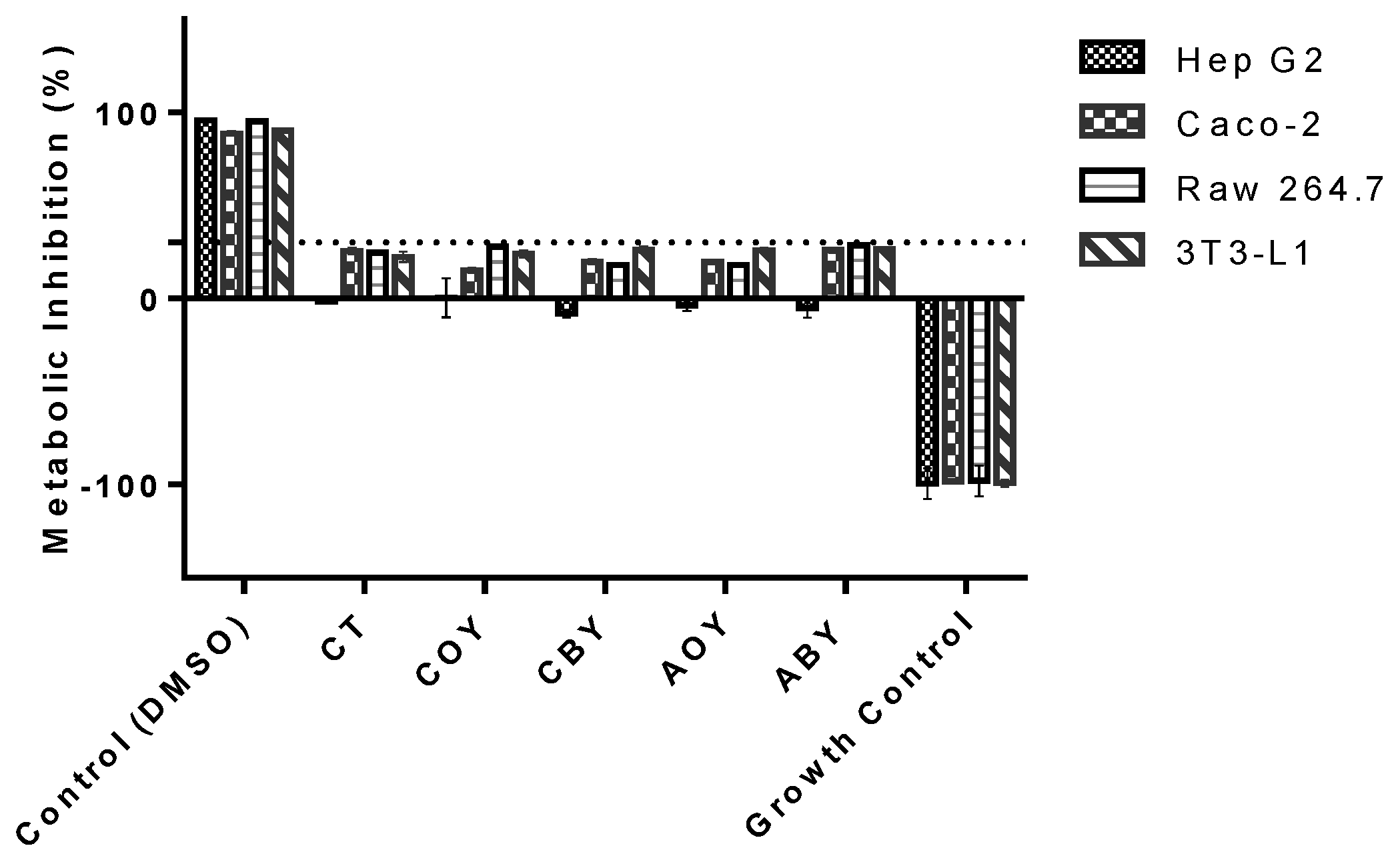
3.3.1. Metabolic Activity

As can be seen in Figure 2, the yogurt samples and the control did not exert significant inhibitory activity upon the cellular metabolism of all tested cell lines at the tested concentration (0.07 mg of fatty acid/mL). All samples showed metabolic inhibition percentages of less than 30%, the limit defined by ISO 10993-5:2009 [30]. Moreover, when considering the impact of yogurt samples upon Hep G2 metabolism, an apparent metabolic stimulation was found for CT, CBY, AOY, and ABY (negative value of metabolism inhibition). The same positive effect on Hep G2 metabolism was reported in previous work using avocado and coconut oils [17,18].

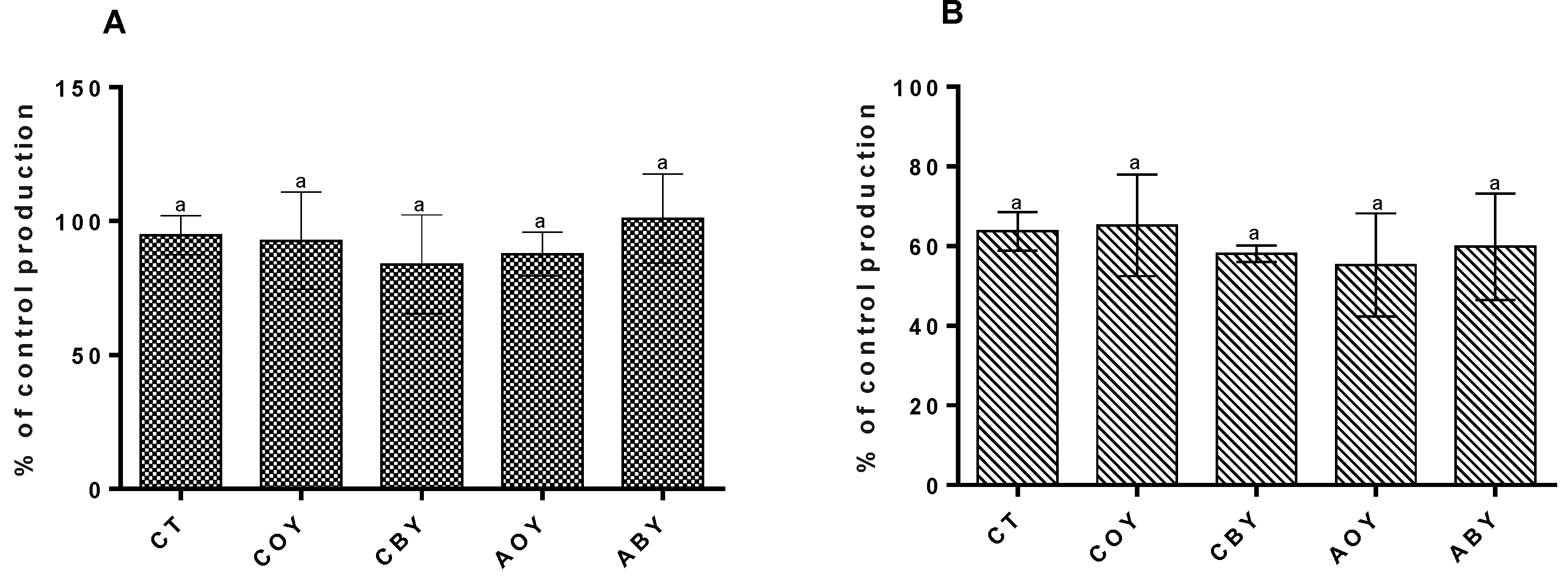
3.3.2. Impact on Hepatic Lipid Accumulation

Nonalcoholic fatty liver disease results from ectopic lipid accumulation, glucose intolerance, and hypertriglyceridemia [31]. Due to their continuous rise, the development of novel strategies for their prevention is needed. Dietary fatty acids can give an important contribution, because they are essential regulators of many cellular functions that exert an effect on lipogenesis and glucose metabolism [31].
The presence of vegetable oil-added did not increase the lipid accumulation in Hep G2 cells (Figure 3).
No significant differences (p ˃ 0.05) were observed between the control and fortified yogurts. These results showed an improvement (20%) in the bioactive fatty acids’ positive effect on lipid accumulation when compared with the previous works [17,18]. In the steatosis-induced Hep G2 cells, the presence of pre-digested yogurt samples reduced lipid accumulation; however, no statistically significant differences (p ˃ 0.05) were observed between fortified yogurts and their CT counterpart. Despite this, the avocado oil yogurts reduce 1.1 times more hepatic lipid accumulation (in the steatosis-induced model) than the control and 1.2 times more than the coconut yogurts. The positive effect of avocado and coconut oils on the steatosis-induced model was more evident in this work than in those previously reported [17,18]. Recall that the effect of coconut oil on lipid accumulation can be related to the capacity of medium-chain fatty acids to promote the up-regulation of adipose triglyceride lipase and hormone-sensitive lipase [32]. On the other hand, the presence of monounsaturated fatty acids (in avocado oil) can ameliorate the lipotoxicity induced by other fatty acids which have been demonstrated by in vivo and in vitro studies to be responsible for steatosis [33,34].

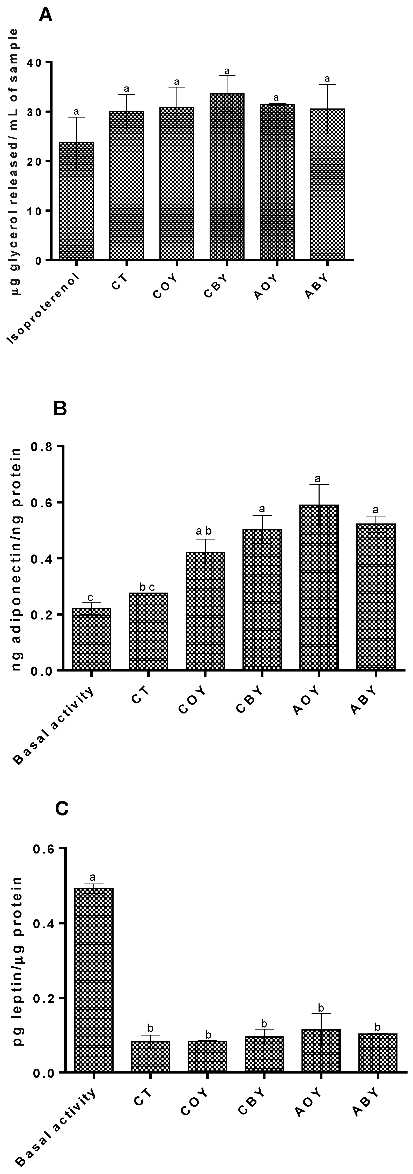
3.3.3. Impact on Adipolysis and Adipokines Secretion

Lipolysis is one of the most important metabolic processes occurring in the adipose tissue, and it is related to the decomposition of triglycerides [35]. The results obtained (Figure 4) showed that 3T3-L1 differentiated adipocytes treated with pre-digested yogurts showed a slight increase in adipolysis when compared with isoproterenol (the assay control); however, no significant differences (p ˃ 0.05) were verified (Figure 4A). The glycerol release values were similar to those reported in the previous works [17,18].
Despite the non-significant differences, the CBY samples increased the adipolysis 1.1 times more than ABY samples. Concerning the adipokines secretion (Figure 4B), the presence of pre-digested yogurt samples led to a significant (p < 0.05) increase in adiponectin secretion when compared with the basal activity. The presence of ABY samples increased by ca 62% the adiponectin secretion compared with the basal activity and by ca 53% when compared with CT yogurt. There were also significant (p < 0.05) increases in the presence of coconut oil yogurts. In this case, coconut yogurts promoted a ca 47% additional adiponectin secretion when compared with basal activity and 35% more when compared with the CT yogurt. A previous study reported that oleic acid (the major constituent of avocado oil) is responsible for an increase in the adiponectin secretion in 3T3-L1 cells [36]. In relation to coconut oil, an in vitro study showed that lauric acid (the main fatty acid in CO) did not affect adiponectin secretion in 3T3-L1 cells [37]. Regarding leptin secretion (Figure 4C), all yogurts promoted a significant (p < 0.05) reduction in its secretion. Comparing the fortified yogurts and control, no significant differences (p ˃ 0.05) were observed. Previous studies showed that medium-chain fatty acids present in coconut oil can down-regulate leptin secretion in rats [38,39], while for avocado oil yogurts, the effect can be related to the high amount of oleic acid which is responsible for the high reductions in leptin secretion in 3T3-L1 cells [40].

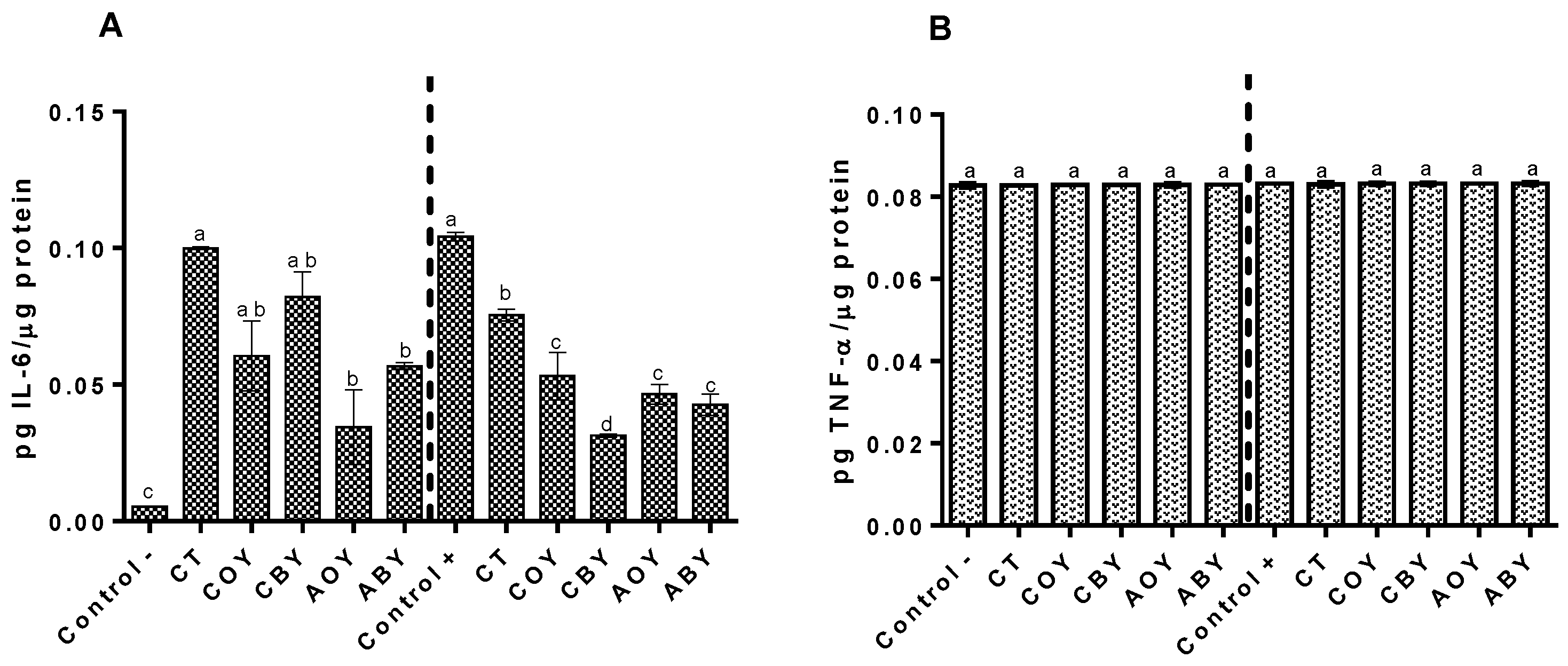
3.3.4. Immunomodulation Capacity

The immunomodulatory capacity of yogurts was assessed in RAW 264.7 cells. No effect was observed in TNF-α in basal and LPS-stimulated cells (Figure 5B). Regarding IL-6 secretion, in non-stimulated conditions, a pro-inflammatory effect was observed for all yogurt samples (Figure 5).
The presence of CT yogurt significantly (p < 0.05) increased (ca 95%) IL-6 secretion. Concerning the fortified yogurts, significant differences (p < 0.05) were observed between avocado fortified yogurts and the control. The CBY increased the IL-6 secretion by ca. 93%. This increase is higher than observed for ABY (ca. 90%). Conversely, in LPS-stimulated cells, all samples exhibited an anti-inflammatory effect with significant differences (p < 0.05) being observed between fortified yogurts and CT yogurts. The CBY yogurt showed the lowest value of IL-6 secretion, which corresponds to a ca. 75% reduction in comparison with the stimulated control (Control+). The presence of avocado oil led to a ca. 60% reduction in IL-6 secretion in the inflammation model. A possible explanation for these results can be attributed to the MUFAs activity, as previous in vivo and in vitro studies demonstrated that they can reduce IL-6 secretion due to their capacity to inhibit nuclear factor-kappaB (NF-kB) and the NOD-like receptor family pyrin domain activation through the direct binding to G-protein coupled receptor 120 (GRP120) and peroxisome proliferator-activated receptors [41]. On the other hand, coconut oil exhibits a capacity to down-regulate the secretion of pro-inflammatory cytokines through the downregulation of cyclooxygenase expression. Moreover, coconut oil has also been shown to be capable of inhibiting NF-kB expression and reducing the production of inflammatory lipid mediators [42,43,44,45,46]

4. Conclusions

Overall, the lab-scale formulations showed improved results compared to the previous formulations studied. These formulations showed high resistance during the in vitro digestion, particularly the formulations with avocado oil. The recovery percentage of oleic acids increased 2 times for AOY and 1.3 times for ABY. The use of alginate in the bigel preparation did not affect the coconut digestion resistance and maintained the high percentages of lauric acid recovery. The use of avocado oil in yogurt production allowed an improvement in the HH and HPI ratios, which are related to health benefits for human health, particularly in the management of normal cholesterol levels. The only drawback of the proposed fortified yogurts was the apparent decrease in their nutritional value, specifically in the case of coconut oil yogurts due to the high amount of saturated fatty acids; however, this may be compensated by the positive impacts upon the target cellular metabolism. In fact, all yogurt samples were capable of exerting positive modulation over various obesity-related key processes, such as reduction of lipid accumulation both in hepatocytes and adipocytes and leptin secretion, and they also showed anti-inflammatory potential as they reduced IL-6 secretion in LPS-stimulated macrophages. Comparing all yogurts, functionalized yogurts stand out positively in relation to the increase in adiponectin secretion and in the reduction of IL-6 in LPS-stimulated cells. In conclusion, the developed functional yogurt prototypes despite showing high potential as future obesity-modulating foodstuffs still require some optimization to improve their nutritional value, mainly the improvement of fatty acids stability during digestion.

Author Contributions

Methodology: M.M., formal analysis and investigation: M.M., writing—original draft preparation: M.M.; writing—review and editing: L.M.R.-A., A.M.G. and M.P.; funding acquisition: M.P.; resources: M.P.; supervision: A.M.G. and M.P. All authors have read and agreed to the published version of the manuscript.

Funding

This work was supported by National Funds from FCT—Fundação para a Ciência e Tecnologia through project UIDB/50016/2020 and through author M. Machado’s FCT doctoral grant (ref.: SFRH/BD/136701/2018).

Institutional Review Board Statement

Not applicable.

Informed Consent Statement

Not applicable.

Data Availability Statement

The data presented in this study are available on request from the corresponding author.

Conflicts of Interest

The authors declare no conflict of interest.

References

  1. Vázquez, L.; Corzo-martínez, M.; Arranz-martínez, P.; Barroso, E.; Reglero, G.; Torres, C. Bioactive Lipids. In Bioactive Molecules in Food; Mérillon, J.M., Ramawat, K.G., Eds.; Springer: Berlin/Heidelberg, Germany, 2017; pp. 1–61. ISBN 9783319545288. [Google Scholar]
  2. De Carvalho, C.C.; Caramujo, M.J. The various roles of fatty acids. Molecules 2018, 23, 2583. [Google Scholar] [CrossRef] [Green Version]
  3. Aluko, R. Bioactive Lipids 2 2.1. In Functional Foods and Nutraceuticals; Springer: Berlin/Heidelberg, Germany, 2012; pp. 23–35. ISBN 9781461434801. [Google Scholar]
  4. Chen, B.; McClements, D.J.; Decker, E.A. Design of Foods with Bioactive Lipids for Improved Health. Annu. Rev. Food Sci. Technol. 2012, 4, 35–56. [Google Scholar] [CrossRef] [PubMed]
  5. Zabielski, P.; Ksi, M. The Impact of OMEGA-3 Fatty Acids Supplementation on Insulin Resistance and Content of Adipocytokines and Biologically Active Lipids in Adipose Tissue of High-Fat Diet Fed Rats. Nutrients 2019, 11, 835. [Google Scholar] [CrossRef] [Green Version]
  6. Calder, P.C. The relationship between the fatty acid composition of immune cells and their function. Prostaglandins Leukot. Essent. Fat. Acids 2008, 79, 101–108. [Google Scholar] [CrossRef] [PubMed]
  7. Harris, W.S. Omega-3 fatty acids and cardiovascular disease: A case for omega-3 index as a new risk factor. Pharmacol. Res. 2007, 55, 217–223. [Google Scholar] [CrossRef] [Green Version]
  8. Kidd, P.M. Omega-3 DHA and EPA for Cognition, Behavior, and Mood: Clinical Findings and Structural-Functional Synergies with Cell Membrane Phospholipids. Altern. Med. Rev. 2007, 12, 207–227. [Google Scholar] [CrossRef] [Green Version]
  9. Gladine, C.; Newman, J.W.; Durand, T.; Pedersen, T.L.; Galano, J.M.; Demougeot, C.; Berdeaux, O.; Pujos-Guillot, E.; Mazur, A.; Comte, B. Lipid profiling following intake of the omega 3 fatty acid DHA identifies the peroxidized metabolites F4-neuroprostanes as the best predictors of atherosclerosis prevention. PLoS ONE 2014, 9, e89393. [Google Scholar] [CrossRef]
  10. Phillips, M.C. Molecular mechanisms of cellular cholesterol efflux. J. Biol. Chem. 2014, 289, 24020–24029. [Google Scholar] [CrossRef] [Green Version]
  11. Gumus, C.E.; Gharibzahedi, S.M.T. Yogurts supplemented with lipid emulsions rich in omega-3 fatty acids: New insights into the fortification, microencapsulation, quality properties, and health-promoting effects. Trends Food Sci. Technol. 2021, 110, 267–279. [Google Scholar] [CrossRef]
  12. Jamshidi, A.; Cao, H.; Xiao, J.; Simal-Gandara, J. Advantages of techniques to fortify food products with the benefits of fish oil. Food Res. Int. 2020, 137, 109353. [Google Scholar] [CrossRef]
  13. Donovan, S.M.; Rao, G. Health benefits of yogurt among infants and toddlers aged 4 to 24 months: A systematic review. Nutr. Rev. 2019, 77, 478–486. [Google Scholar] [CrossRef]
  14. Kok, C.R.; Hutkins, R. Yogurt and other fermented foods as sources of health-promoting bacteria. Nutr. Rev. 2018, 76, 4–15. [Google Scholar] [CrossRef] [PubMed] [Green Version]
  15. Tan, C.X.; Chong, G.H.; Hamzah, H.; Ghazali, H.M. Hypocholesterolaemic and hepatoprotective effects of virgin avocado oil in diet-induced hypercholesterolaemia rats. Int. J. Food Sci. Technol. 2018, 53, 2706–2713. [Google Scholar] [CrossRef] [Green Version]
  16. Cervantes-Paz, B.; Yahia, E.M. Avocado oil: Production and market demand, bioactive components, implications in health, and tendencies and potential uses. Compr. Rev. Food Sci. Food Saf. 2021, 20, 4120–4158. [Google Scholar] [CrossRef] [PubMed]
  17. Machado, M.; Sousa, S.; Morais, P.; Miranda, A.; Rodriguez-Alcalá, L.M.; Gomes, A.M.; Pintado, M. Novel avocado oil-functionalized yogurt with anti-obesity potential: Technological and nutraceutical perspectives. Food Biosci. 2022, 50, 101983. [Google Scholar] [CrossRef]
  18. Machado, M.; Sousa, S.; Morais, P.; Miranda, A.; Rodríguez-Alcalá, L.M.; Gomes, A.M.; Pintado, M.M. Medium-chain triglycerides and conjugated linolenic acids in functional yogurts: Impact of GIT and potential biological activities. Food Funct. 2022, 13, 10937–10946. [Google Scholar] [CrossRef] [PubMed]
  19. Rial, S.A.; Jutras-Carignan, A.; Bergeron, K.F.; Mounier, C. A high-fat diet enriched in medium chain triglycerides triggers hepatic thermogenesis and improves metabolic health in lean and obese mice. Biochim. Biophys. Acta–Mol. Cell Biol. Lipids 2020, 1865, 158582. [Google Scholar] [CrossRef]
  20. Thomas, D.D.; Stockman, M.C.; Yu, L.; Meshulam, T.; McCarthy, A.C.; Ionson, A.; Burritt, N.; Deeney, J.; Cabral, H.; Corkey, B.; et al. Effects of medium chain triglycerides supplementation on insulin sensitivity and beta cell function: A feasibility study. PLoS ONE 2019, 14, e0226200. [Google Scholar] [CrossRef] [PubMed] [Green Version]
  21. Djurasevic, S.; Bojic, S.; Nikolic, B.; Dimkic, I.; Todorovic, Z.; Djordjevic, J.; Mitic-Culafic, D. Beneficial Effect of Virgin Coconut Oil on Alloxan-Induced Diabetes and Microbiota Composition in Rats. Plant Foods Hum. Nutr. 2018, 73, 295–301. [Google Scholar] [CrossRef]
  22. Costantini, L.; Molinari, R.; Farinon, B.; Merendino, N. Impact of omega-3 fatty acids on the gut microbiota. Int. J. Mol. Sci. 2017, 18, 2645. [Google Scholar] [CrossRef] [Green Version]
  23. Shakeel, A.; Farooq, U.; Gabriele, D.; Marangoni, A.G.; Lupi, F.R. Bigels and multi-component organogels: An overview from rheological perspective. Food Hydrocoll. 2021, 111, 106190. [Google Scholar] [CrossRef]
  24. Chen, J.; Liu, H. Nutritional indices for assessing fatty acids: A mini-review. Int. J. Mol. Sci. 2020, 21, 5695. [Google Scholar] [CrossRef]
  25. Brodkorb, A.; Egger, L.; Alminger, M.; Alvito, P.; Assunção, R.; Ballance, S.; Bohn, T.; Bourlieu-Lacanal, C.; Boutrou, R.; Carrière, F.; et al. INFOGEST static in vitro simulation of gastrointestinal food digestion. Nat. Protoc. 2019, 14, 991–1014. [Google Scholar] [CrossRef] [PubMed]
  26. Machado, M.; Costa, E.M.; Silva, S.; Rodriguez-Alcalá, L.M.; Gomes, A.M.; Pintado, M. Pomegranate Oil’s Potential as an Anti-Obesity Ingredient. Molecules 2022, 27, 4958. [Google Scholar] [CrossRef] [PubMed]
  27. Costa, E.; Machado, M.; Pintado, M.; Silva, S. Biological macromolecules as immunomodulators. In Biological Macromolecules; Elsevier: Amsterdam, The Netherlands, 2022; pp. 273–287. [Google Scholar]
  28. Ribeiro, T.B.; Bonifácio-Lopes, T.; Morais, P.; Miranda, A.; Nunes, J.; Vicente, A.A.; Pintado, M. Incorporation of olive pomace ingredients into yoghurts as a source of fibre and hydroxytyrosol: Antioxidant activity and stability throughout gastrointestinal digestion. J. Food Eng. 2021, 297, 110476. [Google Scholar] [CrossRef]
  29. Ahmad, N.; Manzoor, M.F.; Shabbir, U.; Ahmed, S.; Ismail, T.; Saeed, F.; Nisa, M.U.; Anjum, F.M.; Hussain, S. Health lipid indices and physicochemical properties of dual fortified yogurt with extruded flaxseed omega fatty acids and fibers for hypercholesterolemic subjects. Food Sci. Nutr. 2020, 8, 273–280. [Google Scholar] [CrossRef] [Green Version]
  30. ISO-10993-5; Biological Evaluation of Medical Devices–Tests for In Vitro Cytotoxicity. International Organization Standardization: Geneva, Switzerland, 2009; Volume 2007, pp. 1–11.
  31. DeVito, L.M.; Dennis, E.A.; Kahn, B.B.; Shulman, G.I.; Witztum, J.L.; Sadhu, S.; Nickels, J.; Spite, M.; Smyth, S.; Spiegel, S. Bioactive lipids and metabolic syndrome—A symposium report. Ann. N. Y. Acad. Sci. 2022, 1511, 87–106. [Google Scholar] [CrossRef]
  32. Wang, B.; Fu, J.; Li, L.; Gong, D.; Wen, X.; Yu, P.; Zeng, Z. Medium-chain fatty acid reduces lipid accumulation by regulating expression of lipid-sensing genes in human liver cells with steatosis. Int. J. Food Sci. Nutr. 2016, 67, 288–297. [Google Scholar] [CrossRef]
  33. Giulitti, F.; Petrungaro, S.; Mandatori, S.; Tomaipitinca, L.; de Franchis, V.; D’Amore, A.; Filippini, A.; Gaudio, E.; Ziparo, E. Anti-tumor Effect of Oleic Acid in Hepatocellular Carcinoma Cell Lines via Autophagy Reduction. Front. Cell Dev. Biol. 2021, 9, 629182. [Google Scholar] [CrossRef]
  34. Zeng, X.; Zhu, M.; Liu, X.; Chen, X.; Yuan, Y.; Li, L.; Liu, J.; Lu, Y.; Cheng, J.; Chen, Y. Erratum: Oleic acid ameliorates palmitic acid induced hepatocellular lipotoxicity by inhibition of ER stress and pyroptosis. Nutr. Metab. 2020, 17, 11. [Google Scholar] [CrossRef] [Green Version]
  35. Kojta, I.; Chacińska, M.; Błachnio-Zabielska, A. Obesity, Bioactive Lipids, and Adipose Tissue Inflammation in Insulin Resistance. Nutrients 2020, 12, 1305. [Google Scholar] [CrossRef] [PubMed]
  36. Granados, N.; Amengual, J.; Ribot, J.; Palou, A.; Luisa Bonet, M. Distinct effects of oleic acid and its trans-isomer elaidic acid on the expression of myokines and adipokines in cell models. Br. J. Nutr. 2011, 105, 1226–1234. [Google Scholar] [CrossRef] [PubMed] [Green Version]
  37. Bueno, A.A.; Oyama, L.M.; De Oliveira, C.; Pisani, L.P.; Ribeiro, E.B.; Silveira, V.L.F.; Oller Do Nascimento, C.M. Effects of different fatty acids and dietary lipids on adiponectin gene expression in 3T3-L1 cells and C57BL/6J mice adipose tissue. Pflugers Arch. Eur. J. Physiol. 2008, 455, 701–709. [Google Scholar] [CrossRef] [PubMed]
  38. de Vasconcelos, M.H.A.; Tavares, R.L.; Torres Junior, E.U.; Dorand, V.A.M.; Batista, K.S.; Toscano, L.T.; Silva, A.S.; de Magalhães Cordeiro, A.M.T.; de Albuquerque Meireles, B.R.L.; da Silva Araujo, R.; et al. Extra virgin coconut oil (Cocos nucifera L.) exerts anti-obesity effect by modulating adiposity and improves hepatic lipid metabolism, leptin and insulin resistance in diet-induced obese rats. J. Funct. Foods 2022, 94, 105122. [Google Scholar] [CrossRef]
  39. Han, J.; Hamilton, J.A.; Kirkland, J.L.; Corkey, B.E.; Guo, W. Medium-chain oil reduces fat mass and down-regulates expression of adipogenic genes in rats. Obes. Res. 2003, 11, 734–744. [Google Scholar] [CrossRef]
  40. Scoditti, E.; Massaro, M.; Carluccio, M.A.; Pellegrino, M.; Wabitsch, M.; Calabriso, N.; Storelli, C.; De Caterina, R. Additive regulation of adiponectin expression by the mediterranean diet olive oil components oleic acid and hydroxytyrosol in human adipocytes. PLoS ONE 2015, 10, e0128218. [Google Scholar] [CrossRef]
  41. Ravaut, G.; Légiot, A.; Bergeron, K.F.; Mounier, C. Monounsaturated fatty acids in obesity-related inflammation. Int. J. Mol. Sci. 2021, 22, 330. [Google Scholar] [CrossRef]
  42. Rohman, A.; Irnawati; Erwanto, Y.; Lukitaningsih, E.; Rafi, M.; Fadzilah, N.A.; Windarsih, A.; Sulaiman, A.; Zakaria, Z. Virgin Coconut Oil: Extraction, Physicochemical Properties, Biological Activities and Its Authentication Analysis. Food Rev. Int. 2021, 37, 46–66. [Google Scholar] [CrossRef]
  43. Deen, A.; Visvanathan, R.; Wickramarachchi, D.; Marikkar, N.; Nammi, S.; Jayawardana, B.C.; Liyanage, R. Chemical composition and health benefits of coconut oil: An overview. J. Sci. Food Agric. 2021, 101, 2182–2193. [Google Scholar] [CrossRef]
  44. Vysakh, A.; Ratheesh, M.; Rajmohanan, T.P.; Pramod, C.; Premlal, S.; Girish Kumar, B.; Sibi, P.I. Polyphenolics isolated from virgin coconut oil inhibits adjuvant induced arthritis in rats through antioxidant and anti-inflammatory action. Int. Immunopharmacol. 2014, 20, 124–130. [Google Scholar] [CrossRef]
  45. Zicker, M.C.; Silveira, A.L.M.; Lacerda, D.R.; Rodrigues, D.F.; Oliveira, C.T.; de Souza Cordeiro, L.M.; Lima, L.C.F.; Santos, S.H.S.; Teixeira, M.M.; Ferreira, A.V.M. Virgin Coconut Oil Is Effective to Treat Metabolic and Inflammatory Dysfunction Induced by High Refined Carbohydrate-Containing Diet in Mice; Elsevier Inc.: Amsterdam, The Netherlands, 2019; Volume 63, ISBN 3134099853. [Google Scholar]
  46. Jose, S.P.; kumar, K.; Sandya, S.S.; Rajmohan, R.V. Polyphenolic fraction of virgin coconut oil inhibits the inflammatory response in oxidized LDL activated human peripheral blood mononuclear cells by modulating TLR/NF-κB signaling pathways. Eur. J. Integr. Med. 2017, 10, 59–65. [Google Scholar] [CrossRef]
Figure 1. Recovery percentages of fatty acids in fortified yogurts ((A) coconut oil yogurts, (B) avocado oil yogurts). Different letters mean significant differences between yogurts as determined by a one-way ANOVA test (p < 0.05). CT—control yogurt, COY—coconut oil yogurt; CBY—coconut bigel yogurt; AOY—avocado oil yogurt; ABY—avocado bigel yogurt.
Applsci 13 03101 g001
Figure 2. Effect of yogurt-digested samples on the metabolic activity of the target cell lines. The dotted line represents the 30% cytotoxicity limit as defined by ISO 10993-5:2009. CT—control yogurt, COY—coconut oil yogurt; CBY—coconut bigel yogurt; AOY—avocado oil yogurt; ABY—avocado bigel yogurt.
Applsci 13 03101 g002
Figure 3. Hepatic lipid accumulation results for the different yogurts (A) without induced steatosis; (B) steatosis induced. Different letters mean significant differences between yogurts as determined by a one-way ANOVA test (p < 0.05). CT—control yogurt, COY—coconut oil yogurt; CBY—coconut bigel yogurt; AOY—avocado oil yogurt; ABY—avocado bigel yogurt.
Applsci 13 03101 g003
Figure 4. Effect of yogurts on adipolysis and adipokines secretion. (A) adipolysis; (B) adiponectin; (C) leptin. Different letters mean significant differences as determined by a one-way ANOVA test (p < 0.05) between samples. CT—control yogurt, COY—coconut oil yogurt; CBY—coconut bigel yogurt; AOY—avocado oil yogurt; ABY—avocado bigel yogurt.
Applsci 13 03101 g004
Figure 5. Modulation of inflammatory response in RAW 264.7 cells ((A) IL-6 secretion; (B) TNF-α secretion). The left part of all graphs corresponds to the non-stimulated cell’s response, and the right is related to the anti-inflammatory effect. Different letters mean significant differences as determined by a one-way ANOVA test (p < 0.05) between samples. Control—is the basal activity, Control + is the basal activity in LPS-stimulated cells. CT—control yogurt, COY—coconut oil yogurt; CBY—coconut bigel yogurt; AOY—avocado oil yogurt; ABY—avocado bigel yogurt.
Applsci 13 03101 g005
Table 1. Fatty acids profile of yogurts and their nutritional quality indexes.
CTCOYCBYAOYABY
C6n.d.0.08 ± 0.04 a0.01 ± 0.01 b n.d.n.d.
C8n.d.1.36 ± 0.61 a0.20 ± 0.02 bn.d.n.d.
C100.02 ± 0.01 b1.27 ± 0.54 a0.20 ± 0.02 bn.d.n.d.
C120.03 ± 0.02 b1.47 ± 0.20 a2.16 ± 0.60 an.d.n.d.
C140.11 ± 0.07 b5.51 ± 2.05 a0.92 ± 0.08 b0.04 ± 0.001 b0.04 ± 0.001 b
C160.36 ± 0.22 b2.50 ± 0.85 a0.84 ± 0.08 b0.68 ± 0.36 b1.38 ± 0.56 ab
C16:1n.d.0.01 ± 0.001 bn.d.0.15 ± 0.09 ab0.29 ± 0.13 a
C180.12 ± 0.07 c1.01 ± 0.33 a0.60 ± 0.06 ab0.11 ± 0.04 c0.32 ± 0.11 bc
C18:1 c90.24 ± 0.14 b1.64 ± 0.58 ab0.65 ± 0.05 b2.08 ± 1.29 ab4.10 ± 1.78 a
C18:1 c11n.d.n.d.n.d.0.16 ± 0.10 a0.30 ± 0.14 a
C18:20.03 ± 0.01 b0.22 ± 0.19 b0.06 ± 0.01 b0.44 ± 0.28 ab0.85 ± 0.38 a
C18:3 c6c9c12n.d.0.09 ± 0.10 an.d.0.02 ± 0.01 a0.03 ± 0.01 a
C20:1n.d.n.d.n.d.n.d.0.02 ± 0.01
C18:3 c9c13c15n.d.n.d.n.d.n.d.0.02 ± 0.01
∑Fatty acids0.91 ± 0.53 b15.16 ± 4.99 a5.62 ± 0.88 b3.69 ± 2.16 b7.35 ± 3.13 b
Nutritional Quality Indexes
AI3.52 ± 0.07 c13.23 ± 0.11 a9.43 ± 0.76 b0.31 ± 0.05 d0.28 ± 0.02 d
TI4.37 ± 0.12 c9.15 ± 0.07 a7.30 ± 0.20 b0.61 ± 0.08 d0.60 ± 0.03 d
HH2.82 ± 0.13 b0.23 ± 0.02 c0.40 ± 0.01 c3.60 ± 0.42 a3.67 ± 0.16 a
HPI0.33 ± 0.01 b0.08 ± 0.001 b0.11 ± 0.01 b3.28 ± 0.58 a3.56 ± 0.26 a
Results are expressed in mg/g and are the means of three determinations ± standard deviation. Different letters in the same line mean significant differences between samples as determined by a one-way ANOVA test (p < 0.05). CT—control yogurt, COY—coconut oil yogurt; CBY—coconut bigel yogurt; AOY—avocado oil yogurt; ABY—avocado bigel yogurt. C6 caproic acid; C8 caprylic acid; C10 capric acid; C12 lauric acid; C14 myristic acid; C16 palmitic acid; C16:1 palmitoleic acid; C18 stearic acid; C18:1 c9 oleic acid; C18:1 c11 cis-vaccenic acid; C18:2 linoleic acid; C18:3 c6c9c12 γ-linolenic acid; C20:1 cis-gondoic acid, C18:3 α-linolenic acid. AI atherogenic acid, TI thrombogenic acid, HH hypocholesterolemic/hypercholesterolemic ratio, HPI health promoting index; n.d. not detected.
Disclaimer/Publisher’s Note: The statements, opinions and data contained in all publications are solely those of the individual author(s) and contributor(s) and not of MDPI and/or the editor(s). MDPI and/or the editor(s) disclaim responsibility for any injury to people or property resulting from any ideas, methods, instructions or products referred to in the content.

