Article

Analysis of Factors Associated with Charcot Neuroarthropathy following Pancreatic Transplantation

1
Department of Transplantation and Regenerative Medicine, School of Medicine, Fujita Health University, Toyoake 470-1192, Japan
2
Department of Endocrinology, Diabetes and Metabolism, School of Medicine, Fujita Health University, Toyoake 470-1192, Japan
*
Author to whom correspondence should be addressed.
Appl. Sci. 2023, 13(4), 2670; https://doi.org/10.3390/app13042670
Submission received: 19 December 2022 / Revised: 31 January 2023 / Accepted: 16 February 2023 / Published: 19 February 2023
(This article belongs to the Special Issue Diabetic Foot)

Abstract

:
Charcot neuroarthropathy (CN) is a progressive neuropathic complication of diabetes mellitus. Patients undergoing pancreatic transplantation are at risk of developing CN, and CN is known to be a poor prognostic factor for graft loss and patient death. This study aimed to investigate the factors associated with CN in patients who had undergone pancreatic transplantation. We analyzed the data of 61 patients who underwent pancreatic transplantations to investigate the relationship between patient background, nerve conduction velocity tests prior to transplantation, and CN onset. Of these patients, six developed CN. The cumulative incidence rates at 1, 3, and 5 years after transplantation were 3.3, 6.9, and 9.0%, respectively. Sensory neuropathy was severe in six patients with CN, with no sural nerve waveform detected. CN development was not observed when the sural nerve waveforms were visualized. However, when no sural nerve waveforms were observed, the incidence of CN significantly increased due to high-dose corticosteroid administration (p = 0.036). High-dose corticosteroids are associated with the development of CN in the presence of severe neuropathy. Corticosteroid administration is associated with bone metabolism; therefore, appropriate therapeutic intervention is required.

1. Introduction

Pancreatic transplantation is an effective treatment for patients with type 1 diabetes mellitus (DM) and has been shown to contribute to improved patient survival [1,2]. Pancreatic transplantation enables improved glycemic control and suppresses the development of complications because of hyperglycemia, thus helping to improve patient prognosis and quality of life (QOL). However, neuropathy remains a major challenge for patients with DM and pancreatic transplantation as neuropathy persists after transplantation [3]. Patients with neuropathy are at risk of developing Charcot neuroarthropathy (CN), which involves the progressive destruction of bones and joints. Rapid improvements in glycemic control have also been identified as a possible risk factor for developing CN [4,5], and patients who have undergone pancreatic transplantation are at a higher risk of developing CN. Moreover, a delayed diagnosis was found to exacerbate symptoms (Figure 1).
The two main theories concerning the development of CN are as follows. In terms of neurovascular theory, CN is a bone metabolic disorder resulting from increased blood flow to the bones due to autonomic neuropathy, which then results in bone mineral loss and an increased risk of bone fracture. In terms of neurotraumatic theory, which focuses on the failure of the defense mechanism due to sensory neuropathy, sensory disorders increase the risk of unrecognized trauma. In patients who do not experience pain, a continuous load is applied to the damaged bone, leading to progressive bone and joint destruction [6,7,8]. The incidence of CN is reportedly 29% in patients with DM-related neuropathy [9]. Matricali et al. reported that the incidence of CN in patients with pancreatic transplantation was 12% [10]. Other reports have also shown an association between CN and pancreatic transplantation. Rangel et al. reported that CN was diagnosed in 4.6% of patients within 12 months after transplantation, and that high-dose corticosteroids were found to be a risk factor for the development of CN [11]. One recent study reported that CN onset after pancreatic transplantation was a risk factor for graft loss and patient death [12], and that patient management was extremely important.
Corticosteroid dose reduction is considered useful for the prevention of CN. Corticosteroid withdrawal is recommended, which may contribute to improved metabolic parameters [13]. However, it is a key drug in the treatment of rejection, and its use cannot be avoided. Performing a risk assessment prior to transplantation is necessary. To our knowledge, data on risk factors for CN prior to pancreatic transplantation are lacking. Therefore, this study aimed to analyze the relationship between pre- and post-operative factors and CN development using data from 61 patients who underwent pancreatic transplantation.

2. Patients and Methods

2.1. Study Population

This study included data from 61 patients who had undergone pancreatic transplantation at Fujita Health University Hospital, Japan. Patients were included if: (i) they had undergone a pancreatic transplantation procedure before November 2021, (ii) their grafts had survived for at least 12 months, and (iii) they had undergone pre-operative nerve conduction velocity tests. No specific exclusion criteria were applied. The observation period spanned from the transplantation date to the date of graft loss for patients with graft loss and to November 2022 for all other patients. The criterion for graft loss was a C-peptide level < 0.3 ng/mL.

2.2. Immunosuppressive Protocol

All patients began maintenance immunotherapy with the triad of tacrolimus, mycophenolate mofetil (MMF), and corticosteroid medication. For induction therapy, thymoglobulin or basiliximab was used. Briefly, basiliximab was used in principle in SPK because of the national insurance system requirements. One immunologically high-risk patient who underwent SPK was allowed to take thymoglobulin. In PTA and PAK, induction therapy depends on the transplant date. Intraoperatively, methylprednisolone (250 mg) was administered, followed by tapering from prednisolone (50 mg) on day 1 after the operation to 5 mg within three weeks. A 5 mg dose of prednisolone was then continued, and none of the patients withdrew from the treatment. Patients received a total of 832.5 mg of prednisolone-equivalent corticosteroids in the first month after transplantation. Thereafter, 150 mg of prednisolone-equivalent corticosteroids was administered monthly. The target trough for tacrolimus was 5–8 ng/mL within 1 year after transplantation and 3–8 ng/mL thereafter. The MMF dose was adjusted according to each patient’s status. Of the 61 patients, 5 discontinued MMF treatment. Of these, one was changed to everolimus, and the remaining four were changed to mizoribine or azathioprine. Twelve of the fifty-six patients took both reduced MMF and everolimus. If rejection developed, pulse steroid therapy was administered (methylprednisolone, 250–500 mg/day for 3 days). When steroid pulse therapy was ineffective, thymoglobulin was administered for 5 days. To prevent side effects, 100 mg of hydrocortisone was administered before thymoglobulin administration.

2.3. Patient Characteristics and Other Relevant Data

Data concerning patient characteristics (age, sex, body mass index, duration of DM, dialysis history, surgical procedure, and HbA1c value) and the post-transplant course (the onset of rejection and history of high-dose corticosteroid administration) were collected. High-dose corticosteroids were defined by the total amount of prednisolone administered above the immunosuppression protocol of 50 mg. The results of nerve conduction velocity studies performed prior to transplantation and bone mineral density studies performed soon after transplantation were also investigated. The association between each of these factors and CN development was analyzed.

2.4. Nerve Conduction Velocity Study

Prior to transplantation, the tests were performed to assess the level and severity of peripheral neuropathy in the lower extremities. The tibial nerve was subjected to a motor nerve test. Stimulating electrodes were placed over the popliteal fossa and at the ankle. A recording electrode was placed on the abductor hallucis muscle. Nerve conduction velocity was calculated according to the distance between the stimulating electrodes and the difference in latency. The amplitude was measured with stimulation to the ankle. A sensory nerve test targeted the sural nerve. A stimulation electrode was attached to the lower leg, posterior to the lateral malleolus, which was used as the recording site. Nerve conduction velocity was calculated according to the distance and latency between stimulation and recording sites. Amplitude was also measured. As a measure of autonomic neuropathy, the variation coefficient of R-R intervals (CVR-R) was calculated using a resting electrocardiogram, which represented the coefficient of variation for 100 consecutive beats.

2.5. Perception Threshold Test

A Neurometer CPT®/C (Neuroton, Inc., Baltimore, MD, USA) was used to test the perception threshold. A stimulating electrode was placed on the plantar pedis. The intensity was manually increased to determine the stimulus threshold. The threshold was expressed as a current perception threshold (CPT) value, where 1 CPT represented 10 µA, and the maximum stimulus value was 999 (9.99 mA). Three stimuli at 5 Hz, 250 Hz, and 1000 Hz were measured. The cutoff value was the average ± standard deviation × 2, using healthy individuals’ data that was previously obtained at our hospital.

2.6. Bone Mineral Density Test

The bone mineral density T-score was calculated using the dual-energy X-ray absorptiometry of the proximal femur at 4 weeks after transplantation. Using the World Health Organization criteria, osteopenia was defined as a T-score ranging between −1.0 and −2.4. Osteoporosis was defined as a T-score ≤ −2.5 [14]. Oral vitamin D, anti-RANKL antibodies, and bisphosphonates are sometimes administered to patients with decreased bone mineral density; however, in this study, we did not include information on the presence or absence of these forms of treatment in this study.

2.7. CN Diagnosis and Treatment

All patients were checked monthly for leg swelling, redness, and pain. Radiographs were taken if these symptoms were evident. The onset of CN was diagnosed by an orthopedic surgeon according to radiographic findings. Resting or unloading was instructed in all patients to suppress the progression of the disease. No patients required surgery due to severe deformities. In addition, steroid doses remained unchanged after diagnosis.

2.8. Ethical Considerations

This study was conducted in accordance with the Declaration of Helsinki and approved by the Ethics Committee of Fujita Health University (HM22-388). The contents of this study were posted on the web page of Fujita Health University and in the outpatient clinic, and all study participants were provided with the opportunity to opt out.

2.9. Statistical Analysis
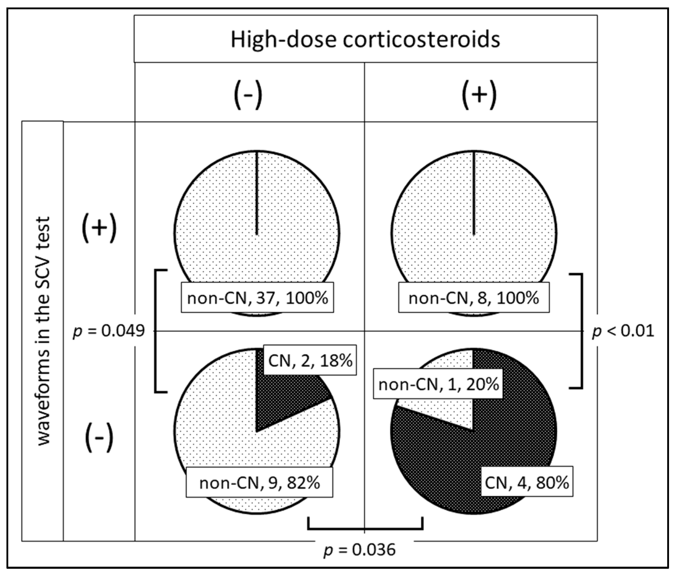
Continuous variables were expressed as median (minimum–maximum) values. The Mann–Whitney U test was used to compare the two groups, namely those with CN and those without CN. Fisher’s exact test was used to compare the frequencies. All statistical analyses were performed using EZR software [15]. Statistical significance was set at p < 0.05.

3. Results

3.1. Background Characteristics of All Patients

Table 1 shows the background characteristics of all 61 patients who underwent pancreatic transplantations. The median age was 44 years (range: 29–65), and 23 (37.7%) patients were men. The surgical procedures included 50 cases of simultaneous pancreas and kidney transplantation (SPK), 2 cases of pancreatic transplantation alone (PTA), and 9 cases of pancreatic transplantation after kidney transplantation (PAK). The median DM duration was 28.5 years (16.8–53.1), the median dialysis history of SPK was 3.7 years (0–19.2), and 50 (82.0%) patients had undergone SPK. There was no difference between induction and maintenance immunotherapies. Ten (16.4%) of the sixty-one patients developed rejection. High-dose corticosteroids were administered to 12 (19.7%) patients (rejection, 10 patients; sudden deafness, 1 patient). The remaining patients were administered with high-dose corticosteroids for cardiac arrest during their intensive care unit stay. CN onset during graft survival was observed in six patients.

3.2. The Cumulative Incidence of CN

Figure 2 shows the cumulative incidence of CN in the patients with graft survival. The onset of CN ranged from 93 to 2226 days after transplantation, with no trend in the time of onset. The cumulative incidence rate at 12 months after transplantation was 3.3%. The cumulative incidence rates 3, 5, and 10 years after transplantation were 6.9, 9.0, and 12.3%, respectively.

3.3. Background of Characteristics of Patients with CN

The characteristics of patients who developed CN are described in Table 1. No differences were observed in terms of age, sex, the duration of DM, or the duration of dialysis between patients with and without CN. The average pre-transplant HbA1c value in patients with CN was 8.8% (range: 7.3–9.8%), which was significantly higher than the value in patients without CN (7.1%) (range: 4.9–9.8%), indicating poor glycemic control. After transplantation, all patients rapidly withdrew insulin therapy and achieved euglycemia. The HbA1c value in patients at 1 month after transplantation with CN was 5.6 (5.0–6.9)%, which was similar to the value observed in patients without CN (5.6%) (4.3–6.9%) (p = 0.84). No difference was observed in terms of the frequency of rejection between patients with and without CN (2 of 6 vs. 7 of 55 patients, respectively; p = 0.21). However, the frequency of high-dose corticosteroid use in patients with CN had a significantly higher than in patients without CN (4 of 6 patients vs. 9 of 55 patients, respectively; p = 0.015). The median dose added to the immunosuppression protocol for 6 months before onset was 937.5 mg (0–3075 mg) of prednisolone-equivalent steroids in patients with CN, which was similar to the dose in patients without CN (2987.5 mg) (312.5–6000 mg) (p = 0.16).

3.4. The Neurological Examination Results

Table 2 shows the neurological examination results for patients with and without CN.
The CVR-R in patients with CN was 0.90% (range: 0.43–1.55%), which was similar to the CVR-R in patients without CN (1.07%) (0.36–4.79%) (p = 0.79). The values in both groups were extremely low and suggestive of severe autonomic neuropathy. The nerve conduction velocity of the tibial nerve was 32.7 m/s (26.0–36.0 m/s) in patients with CN and 37.0 m/s (21.0–45.0 m/s) in patients without CN, and it was significantly slower in patients with CN (p = 0.014). The amplitude was also significantly lower in patients with CN than those without CN (1.29 mV [0.4–8.02 mV] vs. 10.8 mV [0.15–28.11 mV]; p = 0.001). F waves were not detected in 3 of 6 patients with CN, compared with only 4 of the 54 patients without CN (p = 0.025). A significant difference was observed in the nerve conduction velocity of the sural nerve between the two patient groups. No waveforms were detected in any of the six patients with CN. No waveforms were detected in 10 of 55 patients without CN; however, the frequency was significantly higher in patients with CN (100% vs. 18.2%, respectively; p < 0.001).
In the CPT test, the percentage of abnormally high values was higher in patients with CN at all frequencies. Significant differences were observed at 5 Hz and 250 Hz. The bone mineral density test showed an overall decrease in bone mineral content; however, the results did not differ significantly between the two patient groups.

3.5. Severe Peripheral Neuropathy and CN Development

Our findings indicate that high-dose corticosteroid administration and severe peripheral neuropathy were related to CN development. Therefore, we compared the onset rates with and without the waveform visualization of sural nerves in patients with and without high-dose corticosteroid administration.
Figure 3 shows the incidence rate for each group. There was no onset with waveform visualization in the sensory nerve conduction velocity (SCV). In patients without waveform visualization, 2 of 11 (18%) patients developed CN without high-dose corticosteroids, and 4 of 5 (80.0%) patients developed CN when high-dose corticosteroids were used. In both groups, the frequency of onset was significantly higher when the waveforms were visualized (with high-dose corticosteroids, p = 0.049; without high-dose corticosteroids p < 0.01). Furthermore, the administration of high-dose corticosteroids only increased the incidence of CN in patients whose waveforms were absent (p = 0.036).

4. Discussion

This study examined the factors associated with CN development following pancreatic transplantation. Two important findings were identified in this study. First, while most patients who had undergone pancreatic transplantation had advanced neuropathy, those with CN had more severe neuropathy. Dyck et al. reported that >50% of patients with T1DM in their study had DM-related peripheral neuropathy [16], suggesting that DM-related neuropathy develops early during DM onset. Multiple pathological conditions, such as ischemic stress, inflammation, oxidative stress, mitochondrial damage, and microangiopathy, have been found to be involved in the development of DM-related neuropathy [17]. Aggressive glycemic control is required to suppress the progression of neuropathy [18]. Most patients who undergo pancreatic transplantation in Japan face challenges in relation to glycemic control and have a long-term history of DM. The median duration of DM in this study was 28.5 years, which was a sufficient timeframe to develop severe neuropathy. Neurological examination results in relation to CN development show that all the patients who developed CN had no visualized waveforms of the sural nerve and a marked prolongation in the perception threshold test. When such test results are observed pre-operatively, a patient should be identified as being at an increased risk of developing CN. Perception threshold testing is recommended as a pre-operative test for patients undergoing pancreatic transplantation, as it is a simple test to perform compared with nerve conduction velocity tests.
Second, the administration of high-dose corticosteroids was associated with the development of CN, but may not affect the incidence of CN when neuropathy is not severe. With corticosteroid administration as an immunosuppressant, bone mineral density decline progresses early after pancreas–kidney transplantation [19]. It has been suggested that immunosuppression protocols without steroid administration do not decrease bone density [20], but rather increase bone density due to the improved activities of daily living [21]. In addition to immunosuppressive protocols, high-dose corticosteroid administration increases the risk of further bone mineral density loss. Interestingly, four patients developed CN within 12 months of receiving high-dose corticosteroids. The remaining one patient developed CN within 12 months of transplantation. Five of the six patients developed CN within 12 months of high-dose corticosteroid administration (including the immunosuppression protocol); thus, it should be noted that a loss of bone mineral density soon after high-dose corticosteroid administration can increase CN development. In addition, in this study, most high-dose corticosteroid administration was intended for the treatment of rejection, but this treatment was also used for sudden deafness or cardiac arrest cases. High doses of corticosteroids may be required for unforeseen reasons. With severe neuropathy, meticulous bone density management is needed. In this study, there was no difference found in the T-scores between the CN and non-CN groups. However, DM-related peripheral neuropathy has been suggested to be associated with bone strength and stiffness [22], and may also affect bone metabolism.
Pancreatic transplantation can improve QOL and increase physical activity [23,24]. However, with an increase in physical activity, the combination of residual neuropathy and decreased bone density may promote the high incidence of CN following pancreatic transplantation. In this study, the cumulative incidence of CN increased almost linearly for up to 10 years after transplantation. As noted, high-dose corticosteroids are likely to have a significant effect on the development of CN; however, it should be recognized that CN can develop at any time. In particular, the graft survival rate has improved over the years, and the risk of developing CN must be reassessed as a necessary part of long-term follow-up.
This study had several limitations. First, this was a retrospective study, and the number of patients was limited. Due to the small number of patients with CN (n = 6), it was difficult to conduct multivariate analysis, which may have influenced our results. However, our study results on severe neuropathy and corticosteroid dosage accord with those reported elsewhere [11,12]. We consider that the findings of this study are likely to be useful.

5. Conclusions

Severe neuropathy and high-dose corticosteroid administration are associated with CN development. It is important to evaluate the degree of peripheral neuropathy prior to transplantation, and SCV and CPT measurements are useful assessment tests. In addition, given the risk of developing osteoporosis in the long-term post-transplantation period, therapeutic interventions for osteoporosis should be actively undertaken.

Author Contributions

Conceptualization, N.A., T.I. and T.K.; methodology, N.A. and T.I.; formal analysis, N.A.; investigation, N.A., T.I., K.K., I.H., M.S., A.S. and T.K.; writing—original draft preparation, N.A.; writing—review and editing, T.I., K.K., I.H., M.S., A.S. and T.K.; supervision, T.K. All authors have read and agreed to the published version of the manuscript.

Funding

This research received no external funding.

Institutional Review Board Statement

This study was conducted in accordance with the Declaration of Helsinki, and the protocol was approved by the Ethics Committee of the Fujita Health University (HM22-388).

Informed Consent Statement

Informed consent was obtained from all subjects involved in the study.

Data Availability Statement

The data presented in this study are available on request from the corresponding author. The data are not publicly available due to restrictions because displaying their information could compromise the privacy of research participants.

Acknowledgments

We would like to thank all members of the physiology laboratory at Fujita Health University Hospital for conducting nerve conduction velocity studies.

Conflicts of Interest

The authors declare no conflict of interest.

References

  1. Gruessner, R.W.; Sutherland, D.E.; Gruessne, A.C. Mortality assessment for pancreas transplants. Am. J. Transplant. 2004, 4, 2018–2026. [Google Scholar]
  2. Ito, T.; Kenmochi, T.; Aida, N.; Matsushima, H.; Kurihara, K.; Ishihara, T.; Shintani, A.; Asaoka, T.; Ito, T. Impact of pancreas transplantation on patient survival: An analysis of the Japanese pancreas transplant registry. J. Clin. Med. 2020, 9, 2134. [Google Scholar] [CrossRef] [PubMed]
  3. Albers, J.W.; Herman, W.H.; Pop-Busui, R.; Feldman, E.; Martin, C.; Cleary, P.; Waberski, B.; Lachin, J.; Diabetes Control and Complications Trial/Epidemiology of Diabetes Interventions and Complications Research Group. Effect of prior intensive insulin treatment during the Diabetes Control and Complications Trial (DCCT) on peripheral neuropathy in type 1 diabetes during the Epidemiology of Diabetes Interventions and Complications (EDIC) Study. Diabetes Care 2010, 33, 1090–1096. [Google Scholar] [CrossRef] [Green Version]
  4. Dardari, D.; Schuldiner, S.; Julien, C.-A.; Van, G.H.; M’Bemba, J.; Bourgeon, M.; Sultan, A.; Lepeut, M.; Grandperret-Vauthier, S.; Baudoux, F.; et al. Trends in the relation between hyperglycemia correction and active Charcot neuroarthropathy: Results from the EPICHAR study. BMJ Open Diabetes Res. Care 2022, 10, e002380. [Google Scholar] [CrossRef] [PubMed]
  5. Dardari, D.; Dardari, R. Why the Risk of Developing Neuroarthropathy Is Higher After Simultaneous Kidney and Pancreatic Transplantation Compared to Kidney Transplantation Only: The Role of Euglycemia. Ann. Transplant. 2021, 26, e928449. [Google Scholar] [CrossRef]
  6. Papanas, N.; Maltezos, E. Etiology, pathophysiology, and classifications of the diabetic Charcot Foot. Diabet. Foot Ankle 2013, 4. [Google Scholar] [CrossRef]
  7. Chantelau, E.; Onvlee, G.J. Charcot foot in diabetes: Farewell to neurotrophic theory. Horm. Metab. Res. 2006, 38, 361–367. [Google Scholar] [CrossRef] [PubMed]
  8. Christensen, T.M.; Bülow, J.; Simonsen, L.; Holstein, P.; Svendsen, O. Bone mineral density in diabetes mellitus patients with and without a Charcot foot. Clin. Physiol. Funct. Imaging 2010, 30, 130–134. [Google Scholar] [PubMed]
  9. Ergen, F.B.; Sanverdi, S.E.; Oznur, A. Charcot foot in diabetes and an update on imaging. Diabet. Foot Ankle 2013, 4. [Google Scholar] [CrossRef] [Green Version]
  10. Matricali, G.A.; Bammens, B.; Kuypers, D.; Flour, M.; Mathieu, C. High rate of Charcot foot attacks early after simultaneous pancreas-kidney transplantation. Transplantation 2007, 83, 245–246. [Google Scholar] [CrossRef]
  11. Rangel, É.B.; Sá, J.R.; Gomes, S.A.; Carvalho, A.; Melaragno, C.; Gonzalez, A.; Linhares, M.; Medina-Pestana, J. Charcot neuroarthropathy after simultaneous pancreas-kidney transplant. Transplantation 2012, 94, 642–645. [Google Scholar] [CrossRef] [PubMed]
  12. García, B.F.; Kuypers, D.R.; Matricali, G.A. Charcot neuroarthropathy after simultaneous pancreas-kidney transplantation: Risk factors, prevalence, and outcome. Clin. Transplant. 2015, 29, 712–719. [Google Scholar] [CrossRef] [PubMed]
  13. Boggi, U.; Vistoli, F.; Andres, A.; Arbogast, H.P.; Badet, L.; Baronti, W.; Bartlett, S.T.; Benedetti, E.; Branchereau, J.; Burke, G.W.; et al. First World Consensus Conference on pancreas transplantation: Part II-recommendations. Am. J. Transplant. 2021, 21, 17–59. [Google Scholar] [CrossRef] [PubMed]
  14. Cosman, F.; de Beur, S.J.; LeBoff, M.S. National Osteoporosis Foundation. Clinician’s guide to prevention and treatment of osteoporosis. Osteoporos. Int. 2014, 25, 2359–2381. [Google Scholar] [CrossRef] [PubMed] [Green Version]
  15. Kanda, Y. Investigation of the freely available easy-to-use software ‘EZR’ for medical statistics. Bone Marrow Transplant 2013, 48, 452–458. [Google Scholar]
  16. Dyck, P.J.; Kratz, K.M.; Karnes, J.L.; Litchy, W.J.; Klein, R.; Pach, J.M.; Wilson, D.M.; O’Brien, P.C.; Melton, L.J., 3rd; Service, F.J. The prevalence by staged severity of various types of diabetic neuropathy, retinopathy, and nephropathy in a population-based cohort: The Rochester Diabetic Neuropathy Study. Neurology 1993, 43, 817–824. [Google Scholar] [CrossRef]
  17. Feldman, E.L.; Callaghan, B.C.; Pop-Busui, R. Diabetic neuropathy. Nat. Rev. Dis. Primers 2019, 5, 42. [Google Scholar] [CrossRef]
  18. Callaghan, B.C.; Little, A.A.; Feldman, E.L.; Hughes, R.A.C. Enhanced glucose control for preventing and treating diabetic neuropathy. Cochrane Database Syst. Rev. 2012, 6, CD007543. [Google Scholar]
  19. Smets, Y.F.; de Fijter, J.W.; Ringers, J.; Lemkes, H.H.; Hamdy, N.A. Long-term follow-up study on bone mineral density and fractures after simultaneous pancreas-kidney transplantation. Kidney Int. 2004, 66, 2070–2076. [Google Scholar]
  20. Pereira, S.; Pedroso, S.; Martins, L.; Santos, P.; Almeida, M.; Freitas, C.; Dias, L.; Dores, J.; Almeida, R.; Henriques, A.C.; et al. Bone mineral density after simultaneous kidney-pancreas transplantation: Four years follow-up of 57 recipients. Transplant. Proc. 2010, 42, 555–557. [Google Scholar] [CrossRef] [PubMed]
  21. Rocha, A.; Martins, L.S.; Malheiro, J.; Dores, J.; Santos, C.; Henriques, C. Changes in bone mineral density following long-term simultaneous pancreas-kidney transplantation. J. Bone Miner. Metab. 2016, 34, 209–215. [Google Scholar] [CrossRef]
  22. Sewing, L.; Potasso, L.; Baumann, S.; Schenk, D.; Gazozcu, F.; Lippuner, K.; Kraenzlin, M.; Zysset, P.; Meier, C. Bone microarchitecture and strength in long-standing type 1 diabetes. J. Bone Miner. Res. 2022, 37, 837–847. [Google Scholar] [CrossRef] [PubMed]
  23. Gibbons, A.; Cinnirella, M.; Bayfield, J.; Watson, C.J.E.; Oniscu, G.C.; Draper, H.; Tomson, C.R.V.; Ravanan, R.; Johnson, R.J.; Forsythe, J.; et al. Changes in quality of life, health status and other patient-reported outcomes following simultaneous pancreas and kidney transplantation (SPKT): A quantitative and qualitative analysis within a UK-wide programme. Transpl. Int. 2020, 33, 1230–1243. [Google Scholar] [CrossRef] [PubMed]
  24. Suzuki, A.; Kenmochi, T.; Maruyama, M.; Saigo, K.; Akutsu, N.; Iwashita, C.; Otsuki, K.; Miyazaki, M. Evaluation of quality of life after simultaneous pancreas and kidney transplantation from living donors using short form 36. Transplant. Proc. 2008, 40, 2565–2567. [Google Scholar] [CrossRef] [PubMed]
Figure 1. Plain radiographic findings of CN. (a) Immediately after onset, no changes in the bone were visible; (b) 1 week after onset, a bone fracture was observed (arrowhead); and (c) 1 month after onset, joint deformity was observed (arrowhead). CN, Charcot neuroarthropathy.
Applsci 13 02670 g001
Figure 2. Cumulative incidence of CN in patients with pancreatic transplantation. The cumulative incidence rates at 1, 3, 5, and 10 years after transplantation were 3.3, 6.9, 9.0, and 12.3%, respectively.
Applsci 13 02670 g002
Figure 3. Comparison of incidence rates for CN based on severe peripheral neuropathy and a history of high-dose corticosteroids. No CN development was observed with or without high-dose corticosteroids in the presence of waveforms in the SCV test. In contrast, in the disappearance of the waveform in the SCV test, CN was observed in 2 of 11 patients (18%) without high-dose corticosteroids and in 4 of 5 patients (80.0%) with high-dose corticosteroids. In both instances, the frequency of onset was significantly higher in the group with visualized waveforms (with high-dose corticosteroids, p = 0.049; without high-dose corticosteroids, p < 0.01). Furthermore, the incidence rate was significantly higher when high-dose corticosteroids were used than when waveforms were not visualized in the SCV test (p = 0.036). CN, Charcot neuroarthropathy; SCV, sensory nerve conduction velocity.
Applsci 13 02670 g003
Table 1. Patient background characteristics.
All Patients
(n = 61)
Patients with CN
(n = 6)
Patients without CN
(n = 55)
p-Value
Age at Tx44 [29–65]38.5 [29–57]44 [31–65]0.42
Sex (male, %)23 (37.7)1 (16.7)22 (44.0)0.40
BMI (Kg/m2)19.9 [15.5–28.4]19.8 [18.1–22.8]20.2 [15.5–28.4]0.98
Duration of DM (years)28.5 [16.8–53.1]22.7 [18.7–36.6]29.2 [16.8–53.1]0.20
Duration of HD (years)3.7 [0–19.2]5.0 [0.4–11.3]3.6 [0–19.2]0.55
Procedure (%) 0.22
PTA2 (3.3)1 (16.7)1 (1.8)
PAK9 (14.8)0 (0)9 (16.4)
SPK50 (82.0)5 (83.3)45 (81.8)
Induction drug dose (%) 1.0
Basiliximab53 (86.9)6 (100)47 (85.5)
Thymoglobulin8 (13.1)0 (0)8 (14.5)
Everolimus (%)13 (21.3)1 (16.7)12 (21.8)1.0
MMF (%)56 (91.8)6 (100)50 (90.9)1.0
Pre-Tx HbA1c (%)7.3 [4.9–9.8]8.8 [7.3–9.8]7.1 [4.9–9.8]<0.001
HbA1c at 1M (%)5.5 [4.3–6.9]5.6 [5.0–6.9]5.6 [4.3–6.9]0.84
Rejection (%)10 (16.4)2 (33.3)8 (14.5)0.25
High-dose steroid dose (%)12 (19.7)4 (66.7)9 (16.4)0.015
Additional steroid dose (mg)1892.8
[0–6000]
937.5
[0–3075]
2987.5
[312.5–6000]
0.16
CN, Charcot neuroarthropathy: Tx, transplantation; BMI, body mass index; DM, diabetes mellitus; HD, hemodialysis; PTA, pancreatic transplantation alone; PAK, pancreas after kidney transplantation; SPK, simultaneous pancreas–kidney transplantation; MMF, mycophenolate mofetil; HbA1c, glycated hemoglobin.
Table 2. Neurological examination results.
Patients with CN
(N = 6)
Patients without CN
(N = 55)
p-Value
Autonomic nerve study
CVR-R (%)0.90 [0.43–1.55]1.07 [0.36–4.79]0.79
Nerve conduction study
Motor nerve velocity test (tibial)
Total disappearance (%)0/6 (0)1/55 (1.8)1.0
Velocity (m/s)32.7 [26.0–36.0]37.0 [21.0–45.0]0.014
Amplitude (mV)1.29 [0.4–8.02]10.8 [0.15–28.11]0.001
F wave (n = 60)
Total disappearance (%)3/6 (50.0)4/54 (7.3)0.025
Latency (s)58.3 [51.3–64.4]53.2 [45.1–72.9]0.19
Sensory nerve velocity test (sural)
Total disappearance (%)6/6 (100)10/55 (18.2)<0.001
Velocity (m/s)-40.5 [30.0–50.4]
Amplitude (mV)-0.004 [0.001–16.5]
Current perception threshold (n = 45)
5 Hz
>160 (%)3/33/420.020
250 Hz
>260 (%)3/34/420.033
2000 Hz
>550 (%)3/37/420.095
Bone density test (femur) (n = 52)
T-score 0.78
−1.0<0/4 (0)6/48 (12.5)
−2.5<, <−1.02/4 (50.0)26/48 (54.2)
<−2.52/4 (50.0)16/48 (33.3)
CN, Charcot neuroarthropathy.
Disclaimer/Publisher’s Note: The statements, opinions and data contained in all publications are solely those of the individual author(s) and contributor(s) and not of MDPI and/or the editor(s). MDPI and/or the editor(s) disclaim responsibility for any injury to people or property resulting from any ideas, methods, instructions or products referred to in the content.

