
Degradation Products Assessment of the Wooden Painted Surfaces from a XVIIth Heritage Monastery

 ,
,  ,
,  and
and 
Abstract
:1. Introduction
2. Materials and Methods
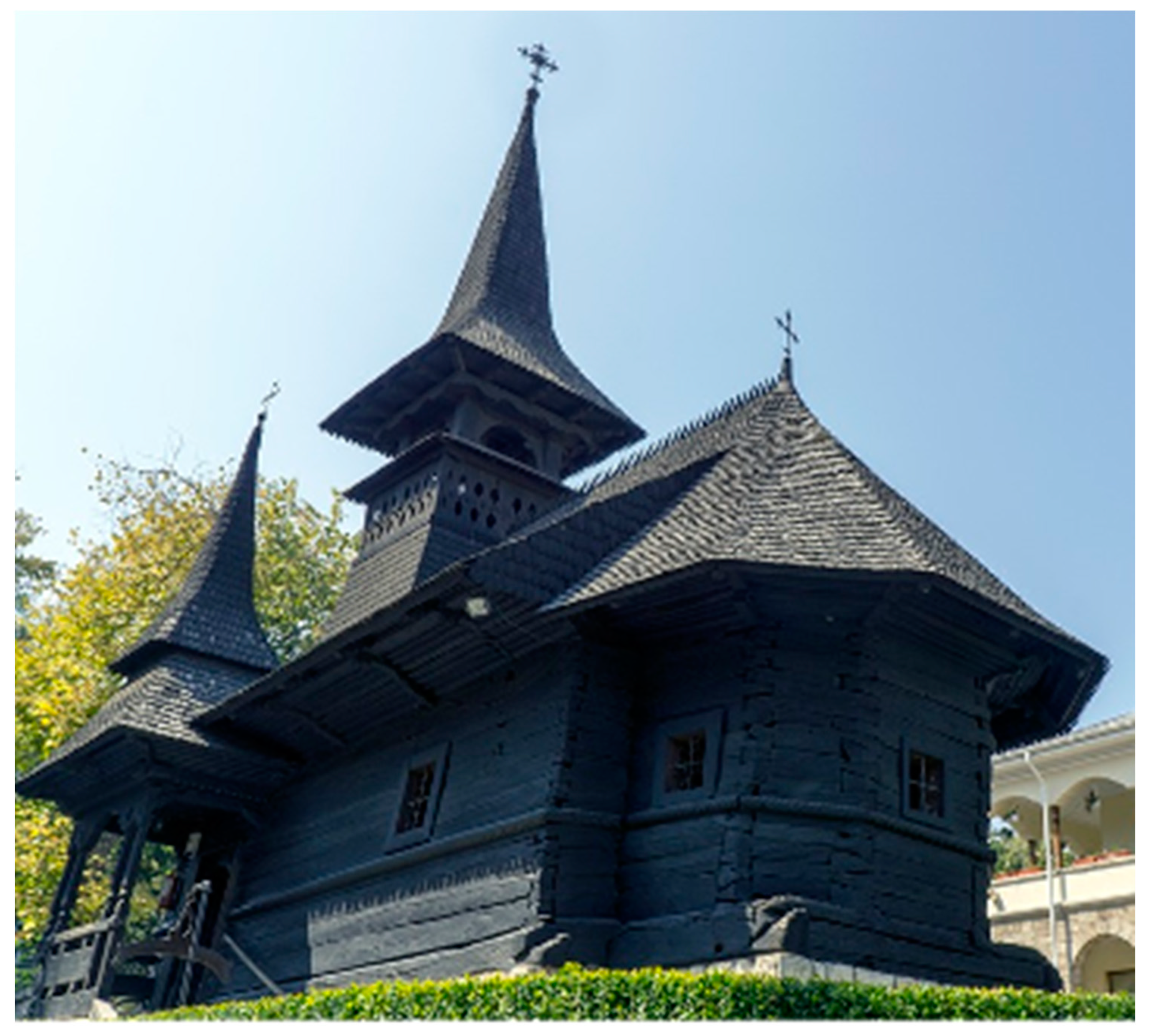
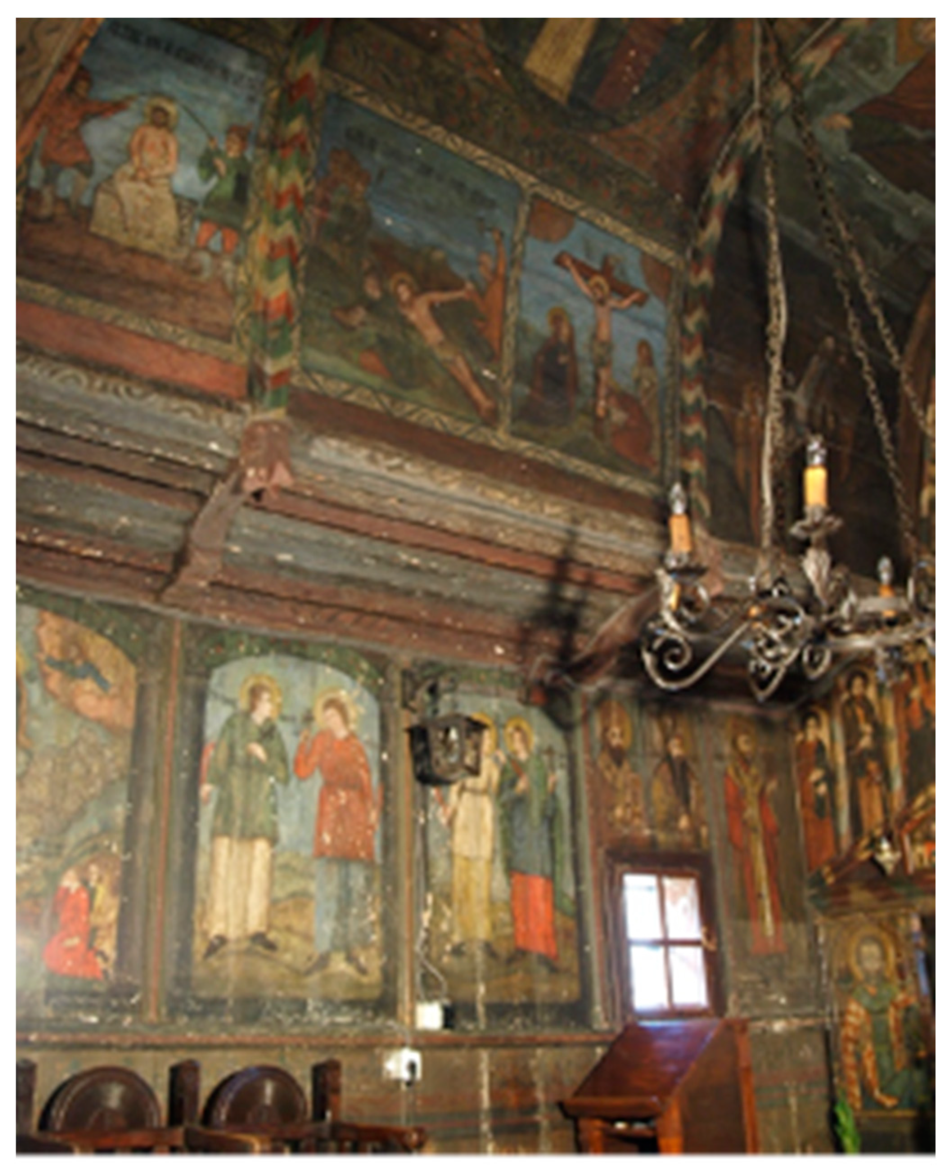
2.1. The Church Painting Samples
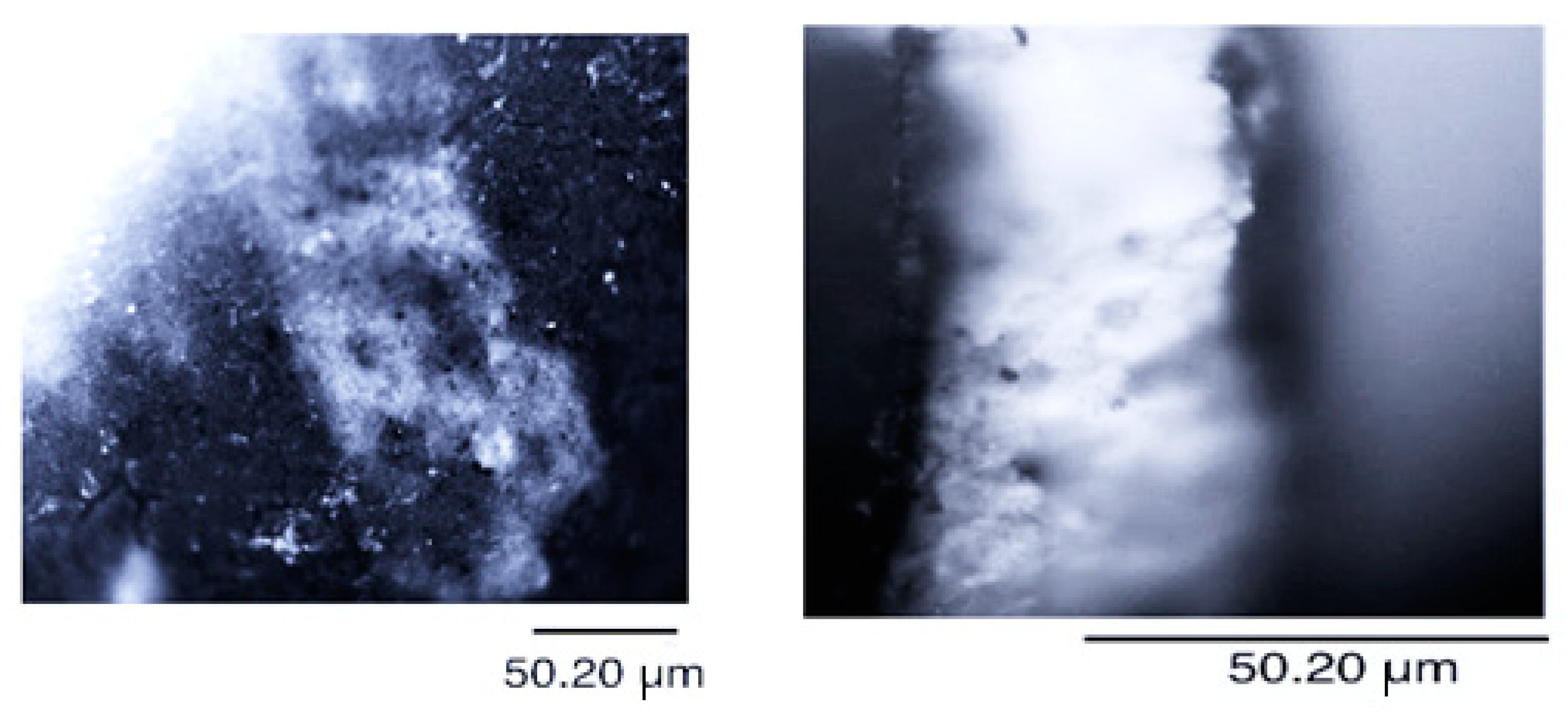
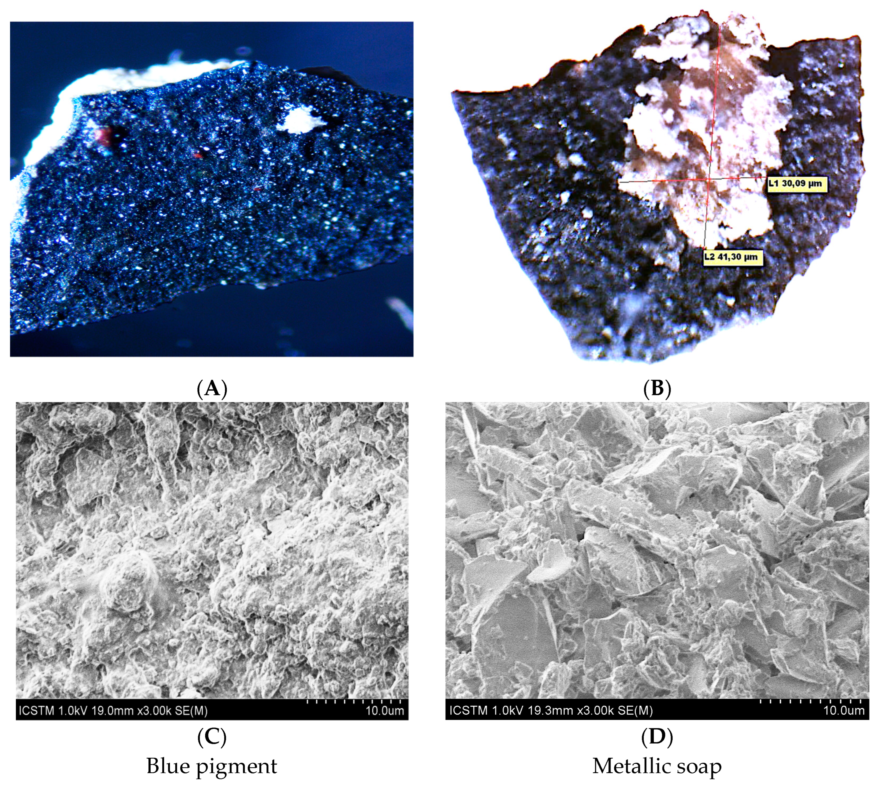
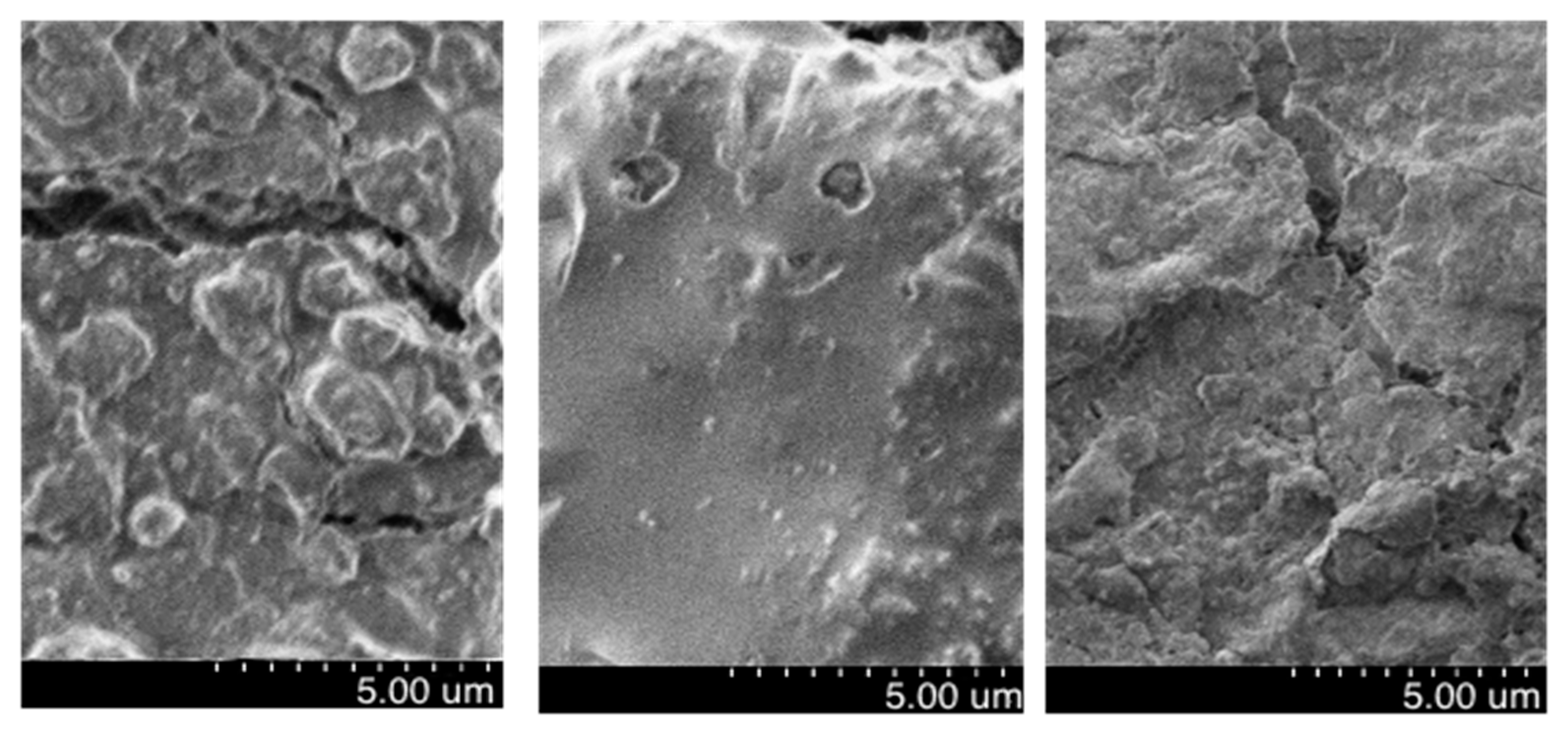
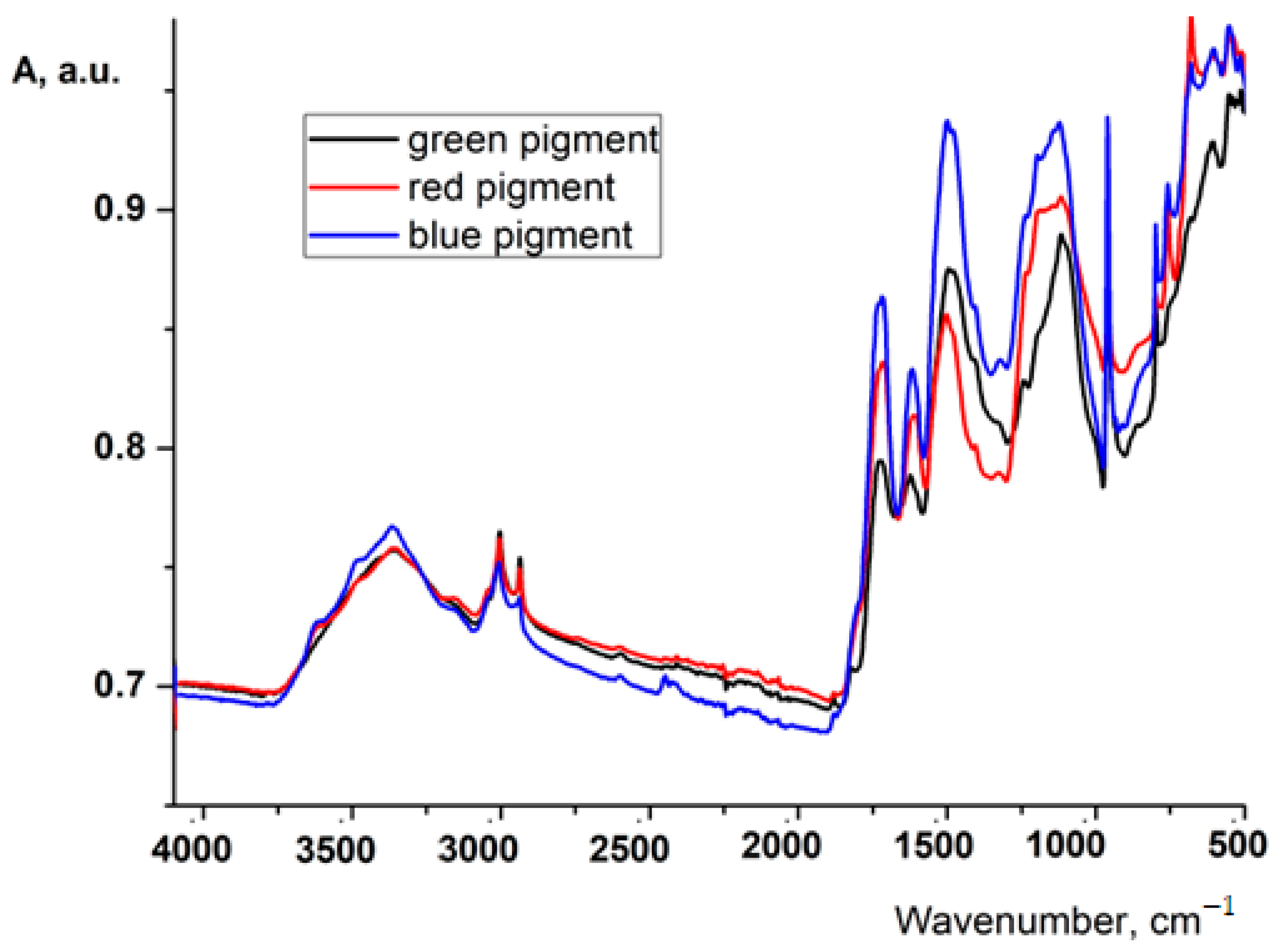
2.2. Characterization Methods
- L*-brightness (L* = 0 (black) to 100 (white);
- a*-red/green variation (a*-red/green coordinate, with + a* meaning red and-a* meaning green);
- b*-yellow/blue variation (b*-yellow/blue coordinate, with + b* meaning yellow and-b* meaning blue).
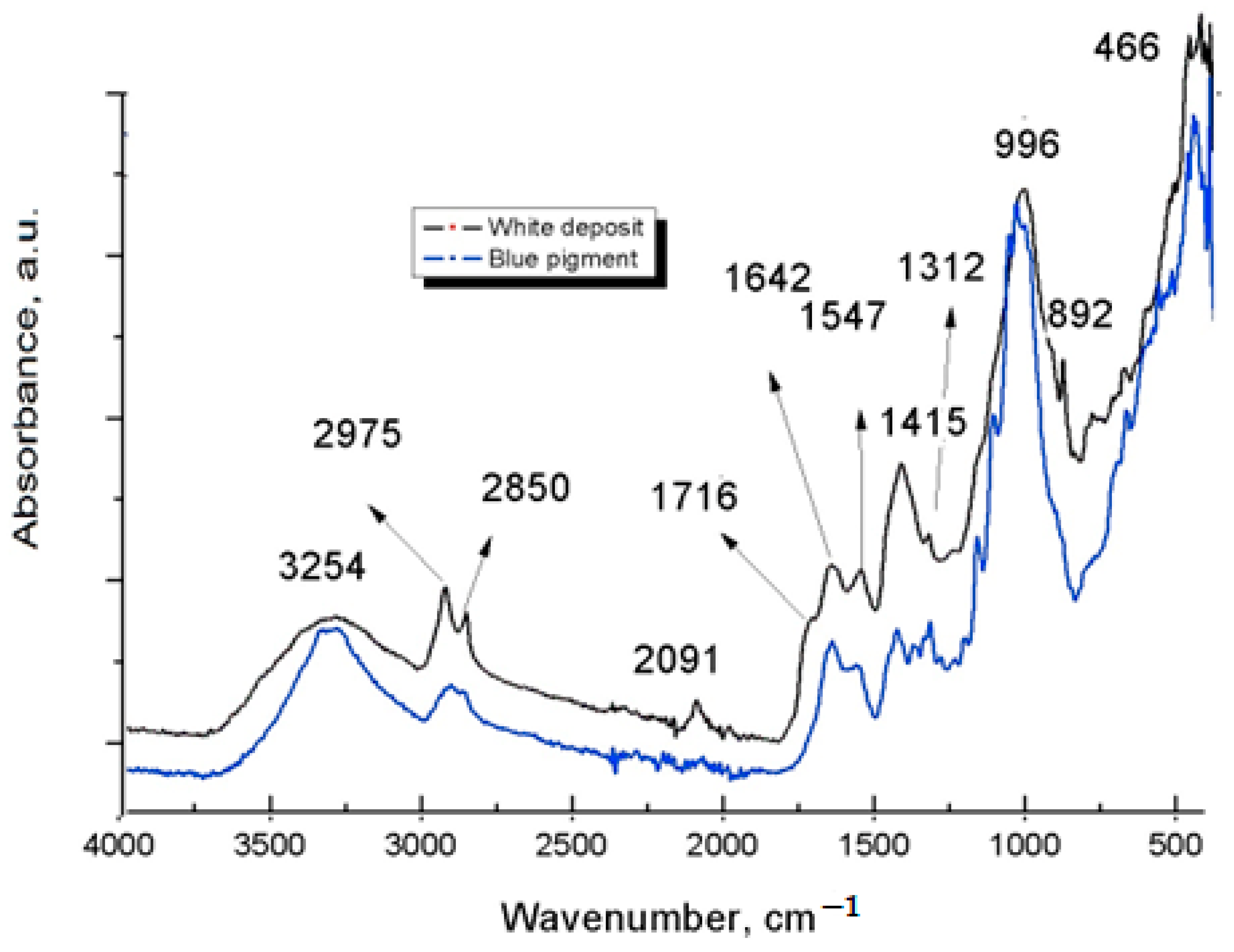
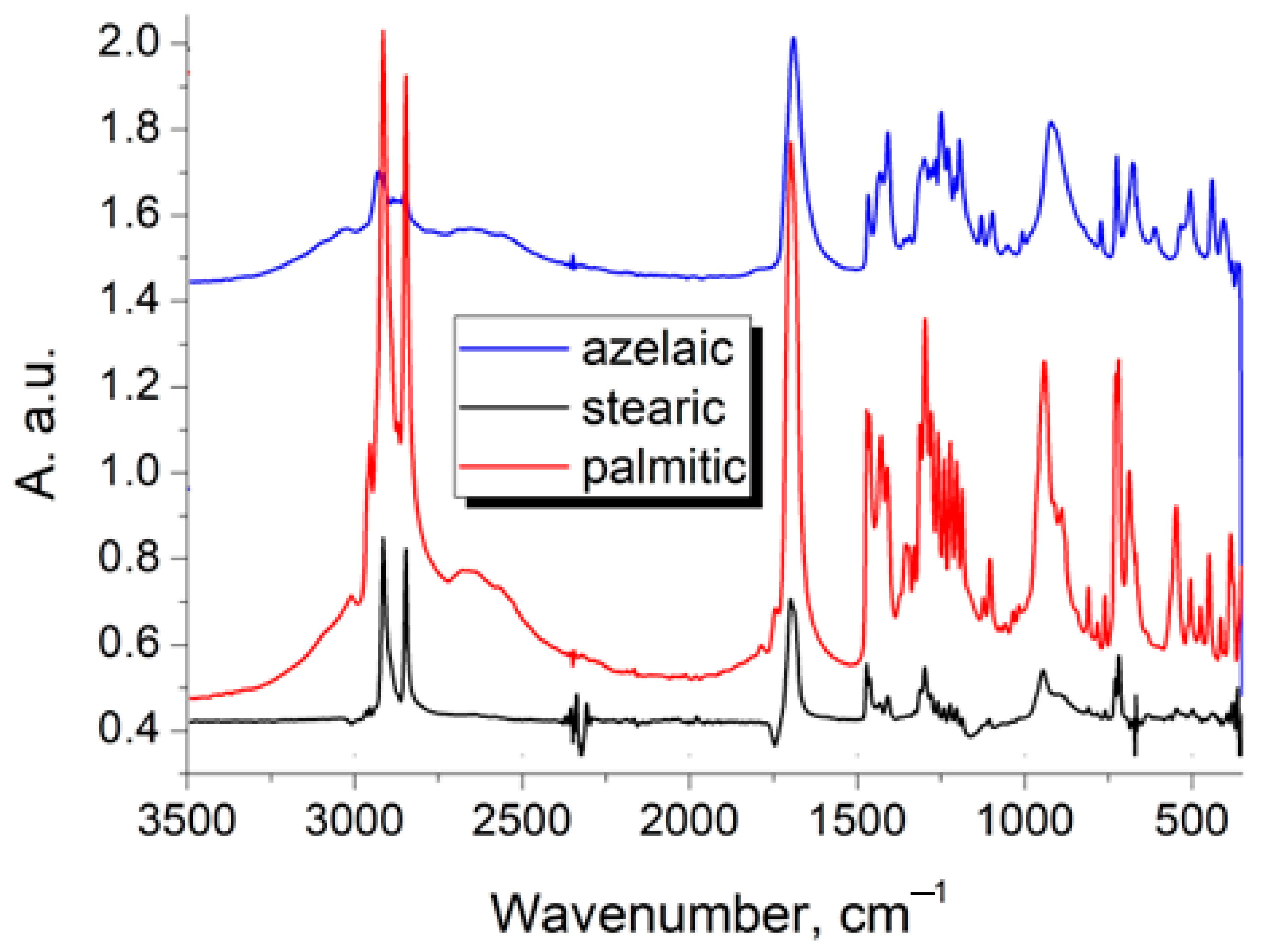
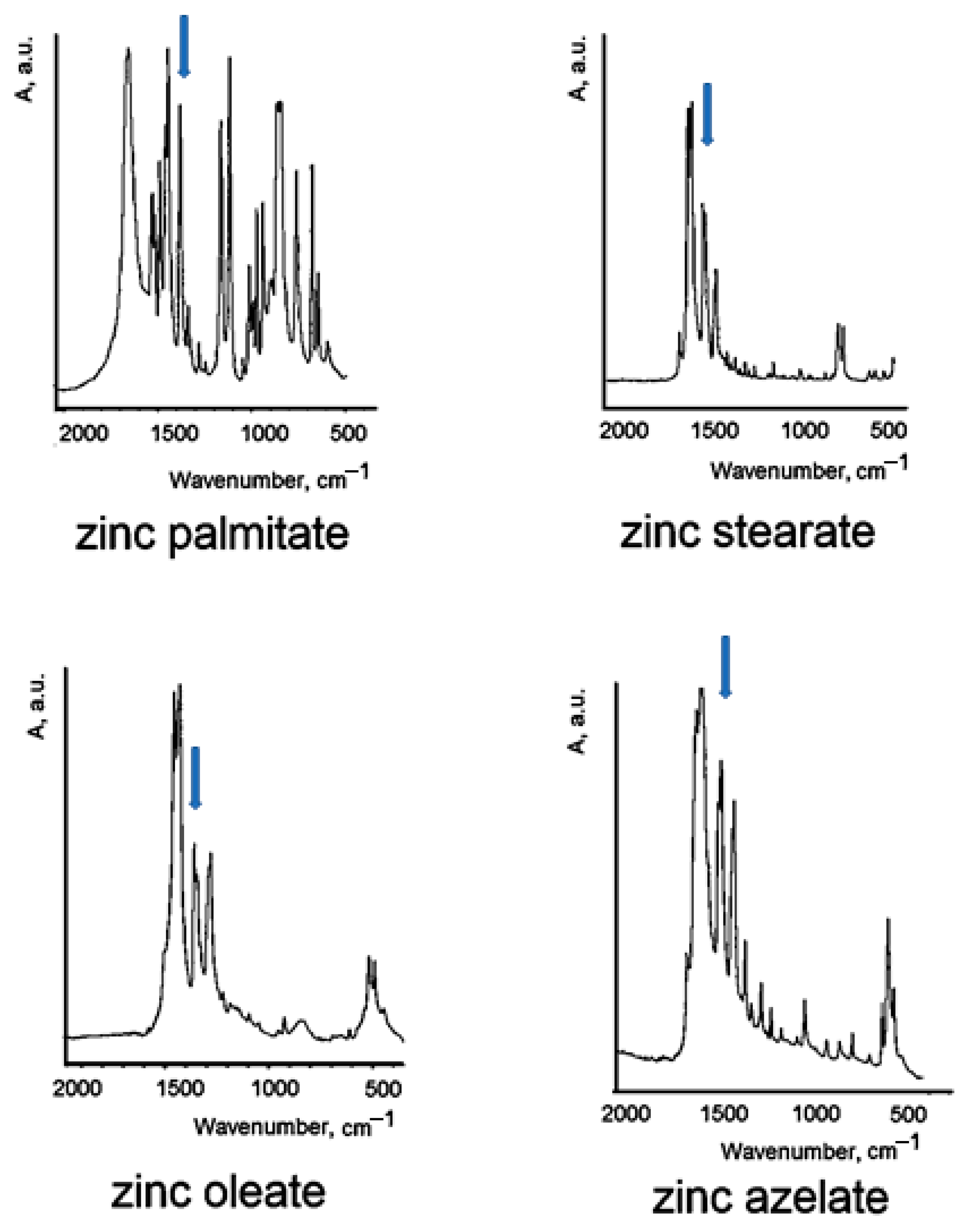
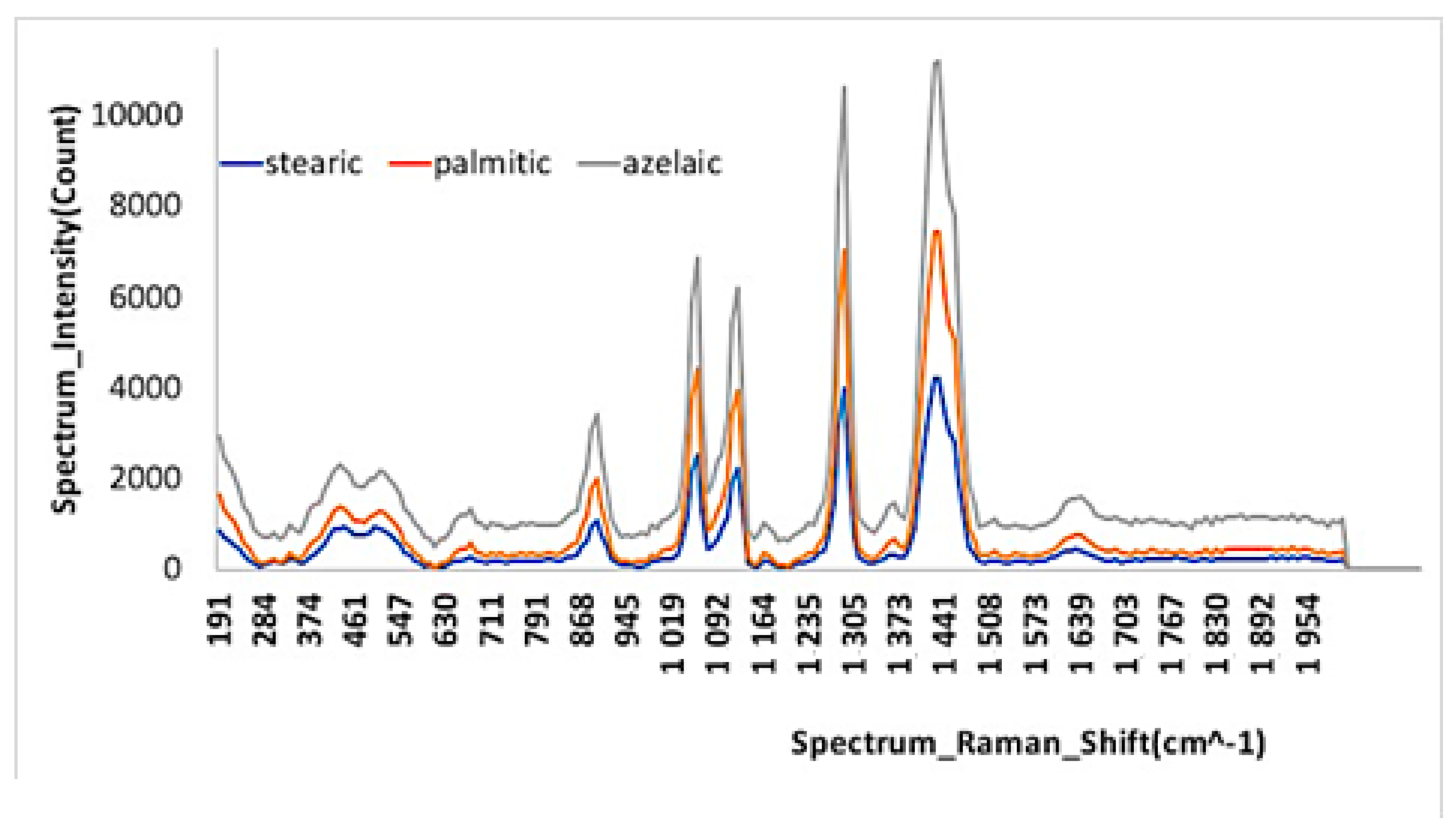
3. Results and Discussion
4. Conclusions
Author Contributions
Funding
Institutional Review Board Statement
Informed Consent Statement
Data Availability Statement
Acknowledgments
Conflicts of Interest
References
- Mayer, R. The Artist’s Handbook of Materials and Techniques, 4th ed.; Viking Penguin Inc.: New York, NY, USA, 1985; p. 215. [Google Scholar]
- Horie, V. Materials for Conservation. Organic Consolidants, Adhesives and Coatings, 2nd ed.; Routledge: New York, NY, USA, 2010. [Google Scholar]
- Domenech-Carbo, M.T.; Silva, M.F.; Aura-Castro, E.M.; Fuster-Lopez, L.; Martinez-Bazan, M.L.; Mas-Barbera, X.; Mecklenburg, M.F.; Osete-Cortina, L.; Domenech, A.; Gimeno-Adelantado, J.V.; et al. Study of behaviour on simulated daylight ageing of artists’ acrylic and poly(vinyl acetate) paint films. Anal. Bioanal. Chem. 2011, 399, 2921–2937. [Google Scholar] [CrossRef]
- Ion, R.M.; Nyokong, T.; Nwahara, N.; Suica-Bunghez, R.; Iancu, L.; Teodorescu, S.; Dulama, I.D.; Stirbescu, R.M.; Gheboianu, A.; Grigorescu, R.M. Wood preservation with gold hydroxyapatite system. Herit. Sci. 2018, 6, 37. [Google Scholar] [CrossRef]
- Nilson, T.; Rowell, R. Historical wood-structure and properties. J. Cult. Herit. 2012, 13, S5–S9. [Google Scholar] [CrossRef]
- Casadio, F.; Keune, K.; Noble, P.; van Loon, A.; Hendriks, E.; Centeno, S.; Osmond, G. Metal Soaps in Art; Springer International Publishing: Berlin/Heidelberg, Germany, 2018. [Google Scholar]
- Akerlund, L. Efflorescence: An investigation of selected paintings from the 19th to the 21st century with a preliminary experimental study of the role of moisture in the development of efflorescence. Final year project dissertation, Department of Conservation & Technology, Courtauld Institute of Art. 2012. [Google Scholar]
- Church, A.H. The Chemistry of Paints and Painting, 3rd ed.; Seeley and Co.: London, UK, 1901. [Google Scholar]
- Ferreira, E.S.B.; Boon, J.J.; Marone, F.; Stampanoni, M. Study of the mechanism of formation of calcium soaps in an early 20th century easel painting with correlative 2D and 3D microscopy. In Proceedings of the ICOM Committee for Conservation 16th Triennial Meeting, Lisbon, Portugal, 19–23 September 2011; Bridgland, J., Ed.; James & James Publisher: Lisbon, Portugal, 2011; p. 1604. [Google Scholar]
- Noble, P.; Boon, J.J. Metal soap degradation of oil paintings: Aggregates, increased transparency and efflorescence. In Proceedings of the AIC Paintings Specialty Group Postprints, Washington, DC, USA, 16–19 June 2006; AIC: Washington, DC, USA, 2007; Volume 19, pp. 1–15. [Google Scholar]
- Chiantore, O.; Scalarone, D. The Macro- and Microassessment of Physical and Ageing Properties in Modern Paints, Modern PAINTS Uncovered: Proceedings from the Modern Paints Uncovered Symposium May 2006; Learner, T.J.S., Smithen, P., Krueger, J.W., Schilling, M.R., Eds.; Getty Conservation Institute: Los Angeles, CA, USA, 2007; pp. 96–104. [Google Scholar]
- Chiantore, O.; Rava, A. Conserving Contemporary Art: Issues, Methods, Materials, and Research, 1st ed.; Getty Conservation Institute: Santa Monica, CA, USA, 2013. [Google Scholar]
- Learner, T.J.S. Analysis of Modern Paints; Getty Conservation Institute: Los Angeles, CA, USA, 2004. [Google Scholar]
- Hermans, J.J.; Keune, K.; Van Loon, A.; Iedema, P.D. , In Metal Soaps in Art. Cultural Heritage Science; Casadio, F., Keune, K., Noble, P., Van Loon, A., Hendriks, E., Centeno, S.A., Osmond, G., Eds.; Springer: Cham, Switzerland, 2019. [Google Scholar]
- Noble, P.; Boon, J.J.; Wadum, J. Dissolution, Aggregation, and Protrusion. Lead Soap Formation in 17th-century Grounds and Paint Layers. Art Matters 2003, 1, 46–61. [Google Scholar]
- Plater, M.J.; De Silva, B.; Gelbrich, T.; Hursthouse, M.B.; Higgitt, C.L.; Saunders, D.R. The characterisation of lead fatty acid soaps in ‘protrusions’ in aged traditional oil paint. Polyhedron 2003, 22, 3171–3179. [Google Scholar] [CrossRef]
- Izzo, F.C.; Kratter, M.; Nevin, E.; Zendri, A. A Critical Review on the Analysis of Metal Soaps in Oil Paintings. ChemistryOpen 2021, 10, 904–921. [Google Scholar] [CrossRef] [PubMed]
- Hermans, J.J. Metal Soaps in Oil Paint, Structure, Mechanisms and Dynamics, Amsterdam. Ph.D. Thesis, Faculty of Science, Van’t Hoff Institute for Molecular Sciences. University of Amsterdam, Amsterdam, The Netherlands, 2017. [Google Scholar]
- Cotte, M.; Checroun, E.; De Nolf, W.; Taniguchi, Y.; De Viguerie, L.; Burghammer, M.; Walter, P.; Rivard, C.; Salomé, M.; Janssens, K.; et al. Lead soaps in paintings: Friends or foes? Stud. Conserv. 2017, 62, 223–330. [Google Scholar] [CrossRef]
- Schilling, M.R.; Khanjian, H.P. Gas Chromatographic Determination of the Fatty Acid and Glycerol Content of Lipids: I. The Effects of Pigments and Aging on the Composition of Oil Paints. In ICOM Committee for Conservation, 11th Triennial Meeting, Preprints; Bridgland, J., Ed.; James & James (Science Publishers): London, UK, 1996; pp. 220–227. [Google Scholar]
- Robinet, L.; Corbeil, M.-C. The Characterization of Metal Soaps. Stud. Conserv. 2003, 48, 23–40. [Google Scholar] [CrossRef]
- Higgitt, C.; Spring, M.; Saunders, D. Pigment-medium interactions in oil paint films containing red lead or lead-tin yellow. Natl. Gallery Tech. Bull. 2003, 24, 75–95. [Google Scholar]
- Keune, K.; Boon, J.J. Analytical imaging studies of cross-sections of paintings affected by lead soap aggregate formation. Stud. Conserv. 2007, 52, 161–176. [Google Scholar] [CrossRef]
- Boon, J.; Keune, K.; Zucker, J. Imaging analytical studies of lead soaps aggregating in preprimed canvas used by the Hudson River School painter F. E. Church. Microsci. Microanal. 2005, 11 (Suppl. 2), 444–445. [Google Scholar]
- Ordonez, E.; Twilley, J. Clarifying the haze: Efflorescence on works of art. WAAC Newsl. 1998, 20, 1. 1997, 69, A416–A422. [Google Scholar]
- Gardner, H.A. Paint Researches and Their Practical Application; Press of Judd and Detweiler, Inc.: Washington, DC, USA, 1917. [Google Scholar]
- Carlyle, L. The Artist’s Assistant: Oil Painting Instruction Manuals and Handbooks in Britain 1800–1900 with Reference to Selected Eighteenth-Century Sources; Archetype Publications: London, UK, 2001. [Google Scholar]
- Townsend, J.; Carlyle, L.; Khandekar, N.; Woodcock, S. Later nineteenth century pigments: Evidence for additions and substitutions. Conservator 1995, 19, 65–78. [Google Scholar] [CrossRef]
- Burnstock, A.; Jones, C. Scanning electron microscopy techniques for imaging materials from paintings. Radiat. Art Archeometry 2000, 202–231. [Google Scholar] [CrossRef]
- Tammekivi, E.; Vahur, S.; Vilbaste, M.; Leito, I. Quantitative GC-MS Analysis of Artificially Aged Paints with Variable Pigment and Linseed Oil Ratios. Molecules 2021, 26, 2218. [Google Scholar] [CrossRef]
- van der Weerd, J.; van Loon, A.; Boon, J.J. FTIR studies of the effects of pigments on the aging of oil. Stud. Conserv. 2005, 50, 3–22. [Google Scholar]
- Otero, V.; Sanches, D.; Montagner, C.; Vilarigues, M.; Carlyle, L.; Lopes, J.A.; Melo, M.J. Characterisation of Metal Carboxylates by Raman and Infrared Spectroscopy in Works of Art. J. Raman Spectrosc. 2014, 45, 1197–1206. [Google Scholar] [CrossRef]
- Shugar, A.N.; Mass, J.L. Handheld XRF for Art and Archaeology; Leuven University Press: Leuven, Belgium, 2012; Volume 3. [Google Scholar]
- Lau, D.; Hay, D.; Wright, N. Micro X-ray diffraction for painting and pigment analysis. AICCM Bull. 2007, 30, 38–43. [Google Scholar] [CrossRef]
- Hradil, D.; Bezdicka, P.; Hradilova, J.; Vašutová, V. Microanalysis of clay-based pigments in paintings by XRD techniques. Microchem. J. 2016, 125, 10–20. [Google Scholar] [CrossRef]
- CIE S 014-1:2006-[ISO 11664-1:2007]; Colorimetry Part 1. CIE Standard Colorimetric Observers. ISO: Geneva, Switzerland, 2007.
- CIE S 014-2:2006-[ISO 11664-2:2007]; Colorimetry Part 2. CIE Standard Illuminants. ISO: Geneva, Switzerland.
- Hermans, J.J.; Keune, K.; Van Loon, A.; Iedema, P.D. Toward a complete molecular model for the formation of metal soaps in oil paints. In Metal Soaps in Art: Conservation and Research; Casadio, F., Keune, K., Noble, P., Van Loon, A., Hendriks, E., Centeno, S., Osmond, G., Eds.; Springer: Cham, Switzerland, 2019; pp. 47–65. [Google Scholar]
- Keune, K. Binding Medium, Pigments, and Metal Soaps Characterised and Localised in Paint Cross-Sections. Ph.D. Thesis, University of Amsterdam, Amsterdam, The Netherlands, 2005. [Google Scholar]
- Mullins, O.C.; Sheu, E.Y.; Hammani, A.; Marshall, A.G. Asphaltenes, Heavy Oils, and Petroleomics; Springer: Berlin/Heidelberg, Germany, 2007. [Google Scholar]
- Artesani, A. Zinc oxide instability in drying oil paint. Mater. Chem. Phys. 2020, 255, 123640. [Google Scholar] [CrossRef]
- Hermans, J.J.; Keune, K.; van Loon, A.; Iedema, P.D. The crystallization of metal soaps and fatty acids in oil paint model systems. Phys. Chem. Chem. Phys. 2016, 18, 10896–10905. [Google Scholar] [CrossRef]
- Harley, R.D. Artists’ Pigments c.1600–1835: A Study in English Documentary Sources; Elsevier Pub. Co.: New York, NY, USA, 1970. [Google Scholar]
- Picollo, M.; Bacci, M.; Magrini, D.; Radicati, B.; Trumpy, G.; Tsukada, M.; Kunzelman, D. Modern White Pigments: Their Identification by Means of Noninvasive Ultraviolet, Visible, and Infrared Fiber Optic Reflectance Spectroscopy. In Modern Paints Uncovered; Getty Conservation Institute: Los Angeles, CA, USA, 2006. [Google Scholar]
- Coccato, A.; Caggiani, M.C.; Occhipinti, R.; Mazzoleni, P.; D’Alessio, A.; Russo, A.; Barone, G. The Irreplaceable Contribution of Cross Sections Investigation: Painted Plasters from the Sphinx Room (Domus Aurea, Rome). Minerals 2021, 11, 4. [Google Scholar] [CrossRef]
- Desnica, V.; Furic, K.; Hochleitner, B.; Mantler, M. A comparative analysis of five chrome green pigments based on different spectroscopic techniques. Spectrochim. Acta Part B At. Spectrosc. 2003, 58, 681–687. [Google Scholar] [CrossRef]
- Pfaff, G. Inorganic Pigments; Walter de Gruyter GmbH: Berlin, Germany; Boston, MA, USA, 2017. [Google Scholar]
- Christie, R. Colour Chemistry, 2nd ed.; Royal Society of Chemistry: Cambridge, UK, 2015. [Google Scholar]
- Slansky, B. Technique of Painting, Part I. Painting and Conservation Material; Technika Malby: Prague, Czech Republic, 1953. (In Czech) [Google Scholar]
- Madejová, J. FTIR techniques in clay mineral studies. Vib. Spectrosc. 2003, 31, 1–10. [Google Scholar] [CrossRef]
- Mazzeo, R.; Prati, S.; Quaranta, M.; Joseph, E.; Kendix, E.; Galeotti, M. Attenuated total reflection micro FTIR characterization of pigment–binder interaction in reconstructed paint films. Anal. Bioanal. Chem. 2008, 392, 65–76. [Google Scholar] [CrossRef]
- Müller, C.M.; Pejcic, B.; Esteban, L.; Delle Piane, C.; Raven, M.; Mizaikoff, B. Infrared attenuated total reflectance spectroscopy: An innovative strategy for analyzing mineral components in energy relevant systems. Sci. Rep. 2014, 4, 6764. [Google Scholar] [CrossRef]
- Feller, R.L. Barium sulfate-natural and synthetic. In Artists’ Pigments; A Handbook of Their History and Characteristics; National Gallery of Art: Washington, DC, USA, 1986; Volume 1, pp. 47–64, M00FCller. [Google Scholar]
- Jacobsen, A.; Gardner, W.H. Zinc Soaps in Paints. Zinc Oleates. Ind. Eng. Chem. 1941, 33, 1254–1256. [Google Scholar] [CrossRef]
- Berkesi, O.; Katona, T.; Dreveni, I.; Andor, J.A.; Mink, J. Temperature-dependent Fourier transform infrared and differential scanning calorimetry studies of zinc carboxylates. Vib. Spectrosc. 1995, 8, 167–174. [Google Scholar] [CrossRef]
- Keune, K.; Van Loon, A.; Boon, J. SEM Backscattered-Electron Images of Paint Cross Sections as Information Source for the Presence of the Lead White Pigment and Lead-Related Degradation and Migration Phenomena in Oil Paintings. Microsc. Microanal. 2011, 17, 696–701. [Google Scholar] [CrossRef]
- Domenech-Carbo, M.T.; Domenech-Carbo, A.; Gimeno-Adelantado, J.V.; Bosch-Reig, F. Identification of synthetic resins used in works of art by Fourier Transform Infrared Spectroscopy. Appl. Spectrosc. 2001, 55, 1590–1602. [Google Scholar] [CrossRef]
- Criado, M.; Fernández-Jiménez, A.; Palomo, A. Alkali activation of fly ash: Effect of the SiO2/Na2O ratio Part I: FTIR study. Microporous Mesoporous Mater. 2007, 106, 180–191. [Google Scholar] [CrossRef]
- Yang, J.X.; Wang, S.M.; Zhao, X.; Tian, Y.P.; Zhang, S.Y.; Jin, B.K.; Hao, X.P.; Xu, X.Y.; Tao, X.T.; Jiang, M.H. Preparation and characterization of ZnS nanocrystal from Zn(II) coordination polymer and ionic liquid. J. Cryst. Growth 2008, 310, 4358. [Google Scholar] [CrossRef]
- Rema Devi, B.S.; Raveendran, R.; Vaidyan, A.V. Synthesis and characterization of Mn2+ doped ZnS nanoparticles. Pramana-J. Phy. 2007, 68, 679. [Google Scholar] [CrossRef]
- She, Y.Y.; Juan, Y.A.N.G.; Qiu, K.Q. Synthesis of ZnS nanoparticles by solid liquid chemical reaction with ZnO and Na2S under ultrasonic bath. Trans. Non Ferr. Met. Soc. China 2011, 20, 211. [Google Scholar] [CrossRef]
- Sutherland, K. Gas chromatography/mass spectrometry techniques for the characterisation of organic materials in works of art. Phys. Sci. Rev. 2018, 4. [Google Scholar] [CrossRef]
- Meilunas, R.J.; Bentsen, J.G.; Steinberg, A. Analysis of aged paint binders by FTIR spectroscopy. Stud. Conserv. 1990, 35, 33–51. [Google Scholar]
- Keune, K.; Boevé-Jones, G. Its Surreal: Zinc-Oxide Degradation and Misperceptions in Salvador Dalí’s Couple with Clouds in Their Heads, 1936. In Issues in Contemporary Oil Paint; Springer: Cham, Switzerland, 2014; pp. 283–294. [Google Scholar]
- Guineau, B. L’tude des pigments par les moyens de la microspectrométrie Raman. In Daiation-caractérisation des pariétales et murals. PACT 1987, 17, 259–294. [Google Scholar]
- Devezaux De Lavergne, E.; Diatre, O.; Et Vanhuong, P. Caractérisation des pigments picturaux d’uneenluminure médiévale par microspectrométrie Raman. In Pigments et Colorants; éditions du CNRS: Paris, France, 1990; pp. 143–150. [Google Scholar]
- Best, S.P.; Clark, R.J.H.; Daniels, M.; et Withnall, R. A Bible laid open. Chem. Br. FEBR 1993, 29, 118–122. [Google Scholar]
- Yufera, J.F.; Ruiz-Moreno, S.; Mansaneola, M.J.; Munoz, S.; et Jawhari, T. Raman spectroscopy for pigment analysis. In Proceedings of the LACONA I: Lasers in the Conservation of Artworks, Crete, Greece, 4–6 October 1995. [Google Scholar]
- Bonaduce, I.; Carlyle, L.; Colombini, M.P.; Duce, C.; Ferrari, C.; Ribechini, E.; Selleri, P.; Tinè, M.R. New Insights into the Ageing of Linseed Oil Paint Binder: A Qualitative and Quantitative Analytical Study. PLoS ONE 2012, 7, e49333. [Google Scholar] [CrossRef]
- Mills, J.S. The Gas Chromatographic Examination of Paint Media. Part I. Fatty Acid Composition and Identification of Dried Oil Films. Stud. Conserv. 1966, 11, 92–107. [Google Scholar]
- Colombini, M.P.; Andreotti, A.; Bonaduce, I.; Modugno, F.; Ribechini, E. Analytical Strategies for Characterizing Organic Paint Media Using Gas Chromatography/Mass Spectrometry. Acc. Chem. Res. 2010, 43, 715–727. [Google Scholar] [CrossRef]
- Colombini, M.P.; Modugno, F.; Ribechini, E. GC/MS in the Characterization of Lipids. In Organic Mass Spectrometry in Art and Archaeology; Colombini, M.P., Modugno, F., Eds.; John Wiley & Sons: Hoboken, NJ, USA, 2009; pp. 191–213. [Google Scholar]
- Casoli, A.; Palla, G.; Tavlaridis, J. Gas-Chromatography/Mass Spectrometry of Works of Art: Characterization of Binding Media in Post-Byzantine Icons. Stud. Conserv. 1998, 43, 150. [Google Scholar] [CrossRef]
- Osmond, G.; Boon, J.; Puskar, L.; Drennan, J. Metal Stearate Distributions in 6odern Artists’ Oil Paints: Surface and Cross-Sectional Investigation of Reference Paint Films Using Conventional and Synchrotron Infrared Microspectroscopy. Appl. Spectrosc. 2012, 66, 1136–1144. [Google Scholar] [CrossRef]
- Dietemann, P.; Kälin, M.; Zumbühl, S.; Knochenmuss, R.; Wülfert, S.; Zenobi, R. A Mass Spectrometry and Electron Paramagnetic Resonance Study of Photochemical and Thermal Aging of Triterpenoid Varnishes. Anal. Chem. 2001, 73, 2087–2096. [Google Scholar] [CrossRef]
- Zumbühl, S.; Soulier, B.; Zindel, C. Varnish Technology during the 16th–18th Century: The Use of Pumice and Bone ash as Solid Driers. J. Cult. Herit. 2021, 47, 59–68. [Google Scholar] [CrossRef]


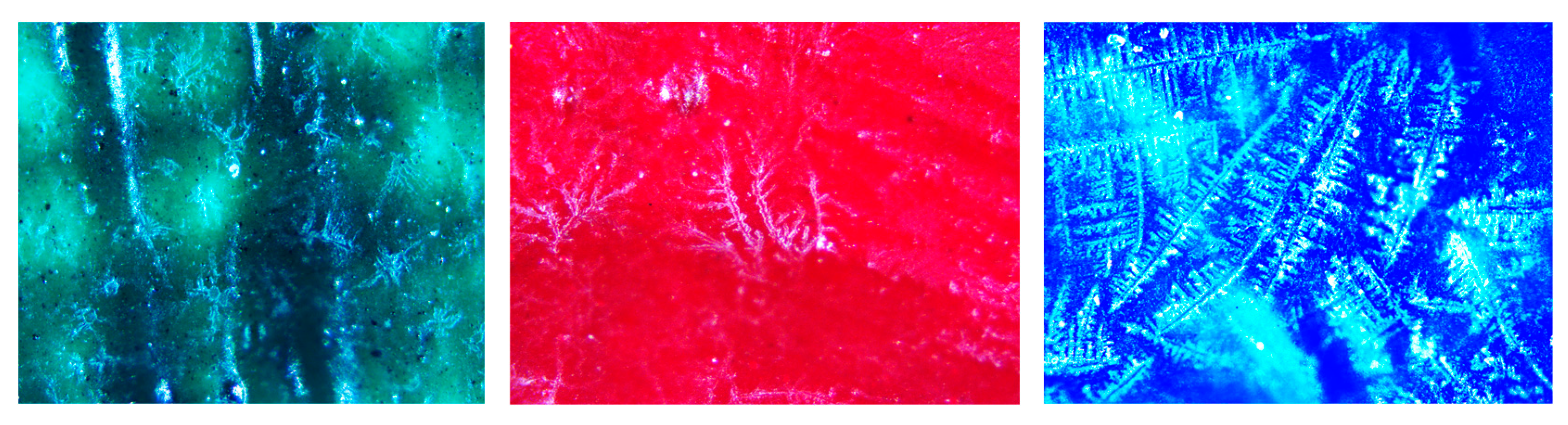
| Sampling Site | Sampling Point | Sample Name, Description and Analysis |
|---|---|---|
| St. Nichifor and St. Dimitrie Icon 1 (L*= 48.12; a* = 5.15; b* = 27.67) |  | Green decoration FTIR, SEM-EDS, OM, GC-MS, colorimetry |
| Pilda Slăbănogului Icon 2 (L*= 19.06; a* = −4.01; b* = 14.27) |  | Red decoration FTIR, SEM-EDS, OM, GC-MS, colorimetry 3 (L*= 40; a* = 35.92; b* = 36.60) 4 (L*= 69.88; a* = 4.25; b* = 37.61) |
| St. Nichifor and St. Dimitrie Icon |  | Blue decoration FTIR, SEM-EDS, OM, GC-MS, colorimetry 5 (L*= 27.49; a* = −2.73; b* = 10.92) 6 (L*= 46.29; a* = 28.05; b* = −29.76) |
| Element | Sample | ||
|---|---|---|---|
| Green | Red | Blue | |
| C | 43.43 ± 0.21 | 25.25 ± 0.13 | 38.26 ± 0.19 |
| O | 42.17 ± 0.73 | 49.21 ± 0.25 | 39.15 ± 0.56 |
| Na | 0.34 ± 0.01 | ||
| Mg | 0.20 ± 0.01 | 0.03 ± 0.01 | 0.25 ± 0.01 |
| Al | 0.56 ± 0.01 | 0.13 ± 0.01 | 0.64 ± 0.01 |
| Si | 1.09 ± 0.01 | 0.28 ± 0.01 | 1.35 ± 0.01 |
| P | 0.35 ± 0.01 | 0.74 ± 0.01 | |
| S | 1.09 ± 0.01 | 11.31 ± 0.04 | 1.60 ± 0.01 |
| Cl | 0.19 ± 0.01 | 0.19 ± 0.01 | 0.14 ± 0.01 |
| K | 0.14 ± 0.01 | 0.18 ± 0.01 | 0.15 ± 0.01 |
| Ca | 6.84 ± 0.03 | 11.10 ± 0.05 | 5.17 ± 0.03 |
| Ti | 0.32 ± 0.02 | 0.05 ± 0.01 | |
| Cr | 0.70 ± 0.02 | 0.15 ± 0.01 | |
| Fe | 1.68 ± 0.03 | 0.17 ± 0.01 | 4.83 ± 0.04 |
| Zn | 0.73 ± 0.05 | 7.54 ± 0.07 | |
| Ba | 1.92 ± 0.05 | 0.44 ± 0.02 | |
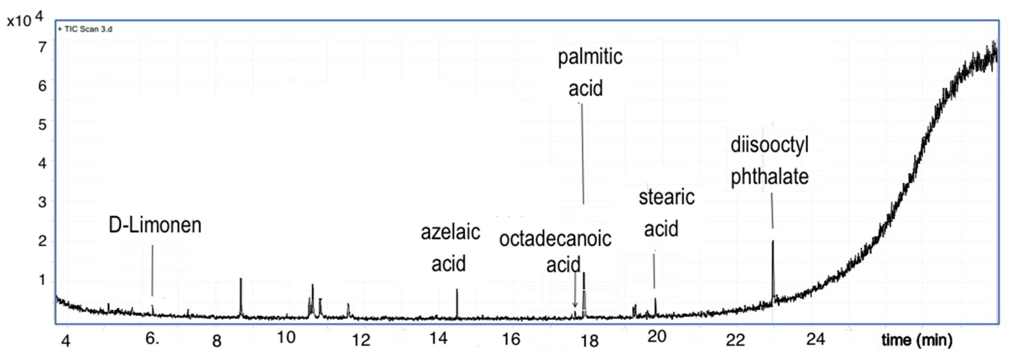
| Sample | Lipid Material | Drying Oil | Pine Resin Markers | Related Pine Resin Compounds | Phthalate Derivatives | Long Chain Hydroxycarboxylic Acids |
|---|---|---|---|---|---|---|
| Green | yes | Yes | Yes | Yes | yes | yes |
| Red | Yes | Yes | No | No | Yes | yes |
| Blue | yes | yes | no | no | yes | yes |
Disclaimer/Publisher’s Note: The statements, opinions and data contained in all publications are solely those of the individual author(s) and contributor(s) and not of MDPI and/or the editor(s). MDPI and/or the editor(s) disclaim responsibility for any injury to people or property resulting from any ideas, methods, instructions or products referred to in the content. |
© 2023 by the authors. Licensee MDPI, Basel, Switzerland. This article is an open access article distributed under the terms and conditions of the Creative Commons Attribution (CC BY) license (https://creativecommons.org/licenses/by/4.0/).
Share and Cite
Ion, R.-M.; Iancu, L.; Grigorescu, R.M.; Slamnoiu-Teodorescu, S.; Dulama, I.D.; Bucurica, I.A. Degradation Products Assessment of the Wooden Painted Surfaces from a XVIIth Heritage Monastery. Appl. Sci. 2023, 13, 2124. https://doi.org/10.3390/app13042124
Ion R-M, Iancu L, Grigorescu RM, Slamnoiu-Teodorescu S, Dulama ID, Bucurica IA. Degradation Products Assessment of the Wooden Painted Surfaces from a XVIIth Heritage Monastery. Applied Sciences. 2023; 13(4):2124. https://doi.org/10.3390/app13042124
Chicago/Turabian StyleIon, Rodica-Mariana, Lorena Iancu, Ramona Marina Grigorescu, Sofia Slamnoiu-Teodorescu, Ioana Daniela Dulama, and Ioan Alin Bucurica. 2023. "Degradation Products Assessment of the Wooden Painted Surfaces from a XVIIth Heritage Monastery" Applied Sciences 13, no. 4: 2124. https://doi.org/10.3390/app13042124