Review

The Role of PET and SPECT Imaging in Prostate Cancer Targeted Alpha Therapy: When and How?

1
Department of Radiological Sciences, Oncology and Anatomo-Pathology, Sapienza, University of Rome, 00161 Rome, Italy
2
Nuclear Medicine Unit, Cardarelli Hospital, 86100 Campobasso, Italy
3
Nuclear Medicine Unit, St. Salvatore Hospital, 67100 L’Aquila, Italy
4
Department of Nuclear Medicine, Santa Maria Goretti Hospital, 04100 Latina, Italy
*
Author to whom correspondence should be addressed.
Appl. Sci. 2023, 13(3), 1890; https://doi.org/10.3390/app13031890
Submission received: 24 November 2022 / Revised: 20 January 2023 / Accepted: 28 January 2023 / Published: 1 February 2023
(This article belongs to the Special Issue Developments of Diagnostic Imaging Applied in Radiotherapy)

Abstract

:
This review discusses the current state of Targeted Alpha Therapy (TAT) in prostate cancer, particularly in mCRPCT (metastatic castration-resistant prostate cancer). This review describes the widely used Radium-223 and the novel trend in the TAT field with a special focus on prostate-specific membrane antigen (PSMA)-based alpha therapy. With this in-depth discussion on the growing field of PSMA-based alpha therapy, we aim also to analyze the most useful diagnostic tools in the patient selection and in the treatment monitoring. We explored the diagnostic tools used in clinical practice and in research settings in order to clarify the imaging procedures that may support the PSMA-based TAT management, including both the patient’s selection and the therapy response monitoring, with a special focus on diagnostic PSMA-PET/CT imaging. Further multicenter trials are needed, but a better understanding of the strengths and limitations of molecular imaging in PSMA-based TAT management may help in creating an effective therapeutic algorithm for mCRPC and designing a rational approach to treatment.

1. Introduction

1.1. Prostate Cancer: Incidence and Survival

Prostate cancer (PCa) is the fourth most common malignancy worldwide in 2020, with 1,414,259 new cases (7.3% of all cancers) after breast cancer (11.7%), lung cancer (11.4%) and colorectum cancer (10%) [1]. It represents the third leading cause of cancer-related death in men. In fact, prostate cancer is not the primary cause of decease, since it is characterized by a slow growth and usually death occurs because of metastases spreading in the pelvic and retroperitoneal lymph nodes, spinal cord, bladder, rectum, bone, and brain. To confirm that we can observe the survival as a function of the stage, localized PCa is indolent and has a 5-year survival rate of nearly 100%, while it decreases to 30% in metastatic PCa [2].

1.2. Androgen Deprivation Therapy

Around 80–90% of PCa is androgen-dependent, so androgen deprivation therapy (ADT) is generally the first therapeutic choice whose aim is suppressing serum testosterone to “castrate” levels (defined as <50 ng/mL) and inhibiting androgen receptors (AR) [2,3]. ADT finds application in intermediate and high-risk PCa as neoadjuvant and adjuvant therapy in association with radiotherapy or in a salvage setting, as adjuvant treatment for nodal metastases after prostatectomy in patients with biochemical recurrence and short doubling times and, thirdly, in metastatic PCa [4]. ADT can be obtained with different medical approaches. First of all, GnRH agonists (leuprolide, goserelin, buserelin, triptorelin) and antagonists (degarelix, relugolix) inhibit the pituitary and reduce the androgen production via the HPG axis. Since 10% of androgen is converted in the adrenal gland from androgen precursors such as pregnenolone and DHEA, Abiraterone acetate is used to inhibit the conversion of precursors in adrenal gland as well as in testes and prostate-tumor tissues. In locally advanced and metastatic prostate cancer, antiandrogens such as Bicalutamide and Flutamide are used to interfere with the signaling of AR and can be combined with ADT in a therapeutic asset known as Combined Androgen Blockade (CAB) [5].

1.3. Epithelial to Mesenchymal Transition and Anoikis

The cytoskeleton plays a pivotal role in epithelial to mesenchymal transition (EMT) and so in progression. During the EMT process, the cells acquire a more fibroblastic appearance, becoming more invasive and motile. Given the crucial role of EMT, Anoikis-inducing agents have been proposed as potential therapy since they can stabilize the cytoskeleton, restrict the movements of cancer cells, and inhibit the intracellular signals involved in tumoral survival [6,7].

1.4. Metastatic Prostate Cancer’s Therapy

Unfortunately, in 20–30% of cases, the tumor goes toward progression even with hematic testosterone level below 20 ng/dL [2] and become castration-resistant PCa (CRPCa). Researchers demonstrated that the resistance to ADT occurs after approximately 10–15 months [8]. However, the overall survival in these patients has improved over time. In fact, the reactivation of AR signaling is seen in CRPCa. AR is targeted by androgen synthesis inhibitors (abiraterone) and AR-ligand inhibitors (enzalutamide, apalutamide, and daroglutamide). However, when a tumor becomes highly glycolytic and responsive to 18F-fluorodoxyglucose (18F-FDG), resistance to these agents and progression to aggressive disease can be observed. Another mechanism of resistance concerns the differentiation of AR-indifferent carcinoma such as neuroendocrine, which has a significant 18F-FDG uptake and leads to the loss of AR signaling [9]. Moreover, some studies demonstrated how androgen ablation itself can reactivate AR signaling pathways. Finally, the AR heterogeneity expression that pre-exists in treatment-naive primary tumors could explain the resistance to AR signaling inhibitors (ARSI) [10]. These are the basis that explains patients’ relapse and therapeutic failure. The therapeutic resistance can be overcome using cytotoxic agents targeting the microtubules as a first-line chemotherapy. For metastatic castration-resistant PCa (mCRPC), Docetaxel, a taxane chemotherapy, was the first drug to be approved in 2004, which was followed by another taxane chemotherapy, Cabazitaxel. In 2010, the FDA approved in the United States Sipuleucel-T, a vaccine therapy targeting prostatic acid phosphatase (PAP), for asymptomatic and minimally symptomatic patients with mCRPC. The FDA authorized the anti-PD1 immune checkpoint inhibitor pembrolizumab in 2017 for the treatment of unresectable or metastatic solid cancers that have progressed after receiving conventional therapy and have microsatellite instability (MSI-H) or a defect in mismatch repair (dMMR) [11,12]. In 2020, Olaparib and rucaparib, which target PARP, were approved by the FDA for mCRPC with genomic mutations on homologous recombination (HR) DNA repair genes which are present in up to 20% of mCRPC patients [4]. PTEN loss is present in 40–60% of mCRPC tumors and leads to hyperactivation of the PI3K–Akt–mTOR signaling pathway. Since there has been evidence of a cross-talk between the PI3K–Akt–mTOR pathway and AR signaling, the Phase 3 IPATential150 study with first-line mCRPC patients examined the combination of abiraterone with the Akt inhibitor ipatasertib. Patients taking abiraterone with ipatasertib saw a significant radiological PFS advantage, according to the research [13].

1.5. Nuclear Medicine Therapy and Prostate Cancer: What Is Known

For the treatment of adult patients with prostate-specific membrane antigen (PSMA)-positive mCRPC who have already had androgen receptor (AR) pathway inhibition and taxane-based chemotherapy, the FDA has authorized PLUVICTOTM (Lutetium 177Lu vipivotide [14]. Additionally, the FDA has authorized the use of Locametz® (East Hanover, NJ, USA) (gallium 68Ga gozetotide), a diagnostic imaging agent, to detect PSMA-positive lesions using positron emission tomography (PET). In this perspective, the two radiopharmaceuticals are employed according to the so-called theranostic approach, consisting of the sequential administration of two molecules with identical or very similar characteristics: the first one is bound to a radionuclide emitting photons or positrons for diagnostic purposes, while the second is conjugated with a radionuclide emitting particles to exert an anti-tumor effect, as shown in Figure 1 [15]. Targeted alpha therapy (TAT) represents an optimal weapon against cancer cells because it can selectively deliver a high burden of radiation to cancer cells and spares normal surrounding tissue thanks to the short range of alpha particles [16,17]. The great energy deposition results in the possibility of obtaining irreparable damage in DNA which is oxygen independent and hence eliminates the major mechanism of therapeutic resistance [18]. In the landscape of targeted alpha therapy, Radium-223 (223Ra) is a cornerstone. The double-stranded DNA breaks caused by this alpha emitter in cancer cells are incorporated into osteoblastic metastatic lesions. For mCRPC patients with symptomatic bone metastases but no visceral metastases, the FDA authorized it in 2013 [19]. New agents have been proposed such as Thorium 227 (227Th). It has been conjugated to BAY2315497, a human anti-PSMA antibody, which demonstrated a preferential tumor uptake. The combination with AR antagonist increased the sensibility to growth inhibition. This seemed to be due to the improved PSMA expression induced by AR antagonist [20]. Actinium-225 PSMA (225Ac-PSMA), indeed, has been proposed for “superscan” patients because the short tissue penetration range of its alpha particle could result in a more favorable microdosimetry in case of red marrow infiltration [21]. Another alpha emitter example is Astatine-211 (211At) linked with PSMA, which was recently investigated for the treatment of PCa [18].

1.6. Radiomedicine

The expansion of nanomedicine has seen the rise of radionanomedicine, which consists of using radionuclides conjugated to nanomaterials for both diagnosis and therapy purposes [22]. An example is nanoparticles of 198 Gold (198Au). The radioactive properties of 198Au such the beta ray of 0.96 MeV and the component gamma ray of 411 KeV make it an ideal candidate for use in diagnosis and therapy. 198Au functionalized with Gum arabic glycoprotein (GA) showed a therapeutic effect in a mouse prostate cancer model after intratumoral injection [23], while 198Au NP-EGCg in a prostate cancer mouse model demonstrated a 72% nanoparticles retention in the tumor after 24 h and 80% reduction in the tumor volume after 28 days [24]. There is an open-word in the therapy and diagnosis of prostate cancer. In this scenario, nuclear medicine and in particular alpha target therapy represent a promising field of research in terms of the therapeutic efficacy, specificity and sensibility of the procedures. The aim of the present review is to cover the applications of molecular and metabolic imaging through SPECT and PET tracers for patients’ selection and response assessment to targeted alpha therapy.

2. Molecular and Metabolic Imaging with PET and SPECT Technology

Since the introduction of personalized medicine, the primary focus of imaging has shifted from detection and diagnosis to tissue characterization, prognosis determination, treatment efficacy prediction, and treatment response measurement. The study of biological processes at the molecular and cellular level is known as molecular imaging. The fast advancement of molecular imaging in recent years has enabled the development of specificity and quantification that are helpful for the early diagnosis and follow-up of the disease. Currently, single photon emission tomography (SPECT) and positron emission tomography (PET) are used for this imaging approach. PET and SPECT radiotracers, which are excellent tools for many medical imaging applications, such as early diagnosis and therapy monitoring in cancer, are becoming more prevalent. The advantage of PET and SPECT is that they provide non-invasive molecular imaging of the entire body, assessing various illness locations [25]. Additionally, serial scanning can be performed, enabling the measurement of functional changes over time during therapeutic interventions.

2.1. Differences between Single Photon Emission Tomography (SPECT) and Positron Emission Tomography (PET)

The main difference between SPECT and PET is the type of radioactive tracer used: while SPECT is based on gamma rays, the decay of the tracers used in PET generates infinitesimal particles known as positrons which decay into two photons emitted in opposite directions. In SPECT, only the direct radiation perpendicular to the detector is recorded; in PET, two detectors hit simultaneously by photons with an oblique direction to the cylinder axis can equally record the radiation. This complex of conditions makes PET faster than SPECT and with higher resolution. The speed of execution is an essential requirement because the radioisotopes used in PET generally have a shorter half-life than those used in SPECT. More and more technological innovations follow one another that allow for progress in the medical field.

2.2. Technologic Progress

The use of WB-SPECT/CT has recently been introduced [26], in which the precision of the CT is added to the function of the SPECT [27,28]. In recent years, digital PET has been introduced through the implementation of silicon photomultipliers (SiMP)-based detectors [29]. This innovative PET detection technology ensures significant improvements over analog technology in terms of better sensitivity and increased spatial and contrast resolution [30,31]. All this technological progress allows for ever more personalized medicine.

3. Targeted Alpha Therapy

Surgery, chemotherapy, and external beam irradiation are frequently utilized as forms of selective therapy in medicine. However, because emitting radionuclides have more precise cell-killing capabilities, there has been some interest in using them in therapy. Targeted radionuclide therapy offers the potential to deliver extremely lethal radiation to cancer cells [31]. This form of treatment can boost the harmful radiobiological effects on cancer cells while decreasing the negative effects of radiation on healthy tissues.

3.1. Physical Characteristics of Alpha Particles and Advantages

The use of alpha particle emitters can help to explain why targeted alpha treatment (TaT) is successful. Such particles have a much higher LET than β particle emitters (50–230 keV/μm vs. 0.1–1.0 keV/μm) with an average energy deposition of 100 keV/μm. The range of α particles in tissues is short, which limits radiation deposition to the target cell and closely neighboring cells [32]. Cellular DNA is the main molecular target of high-LET particles because of the effective production of double-stranded DNA breaks. As a result, TaT can target tumor cells and the tumor microenvironment with high targeted radiation doses while causing the least amount of harm to normal cells. Alpha-emitting particles are preferred over beta particle emitters and external beam radiation for the treatment of malignancies due to all of these advantages. Due to the fact that stable radionuclide sequestration in vivo is an essential part of targeted radiotherapy, the increased interest in TaT has resulted in the development of new chelating agents. Many radionuclides are known which emit one or more α particles during the decay into stable nuclides. Only a small number of them, nevertheless, have been utilized in research projects that examined the effectiveness of targeted radionuclide treatment [33].

3.2. Synthetic Lethality

Not least, the TAT could have a fundamental role for the phenomenon of synthetic lethality [34]. It is defined as “a lethal phenomenon in which the occurrence of a single genetic event is tolerable for cell survival, while the occurrence of multiple genetic events causes cell death”. The variability and complexity of tumor biology, a lack of knowledge of the connections that cause synthetic lethality, drug resistance, and difficulties with clinical screening and translation are the key barriers to synthetic lethality. Recent studies have shown that cancer cells with altered DNA repair mechanisms are ideal candidates for therapies based on synthetic lethality approaches. According to this viewpoint, pharmacological combinations are created with the intention of ultimately curing the condition, but more typically, synergy leads to considerable improvements in treatment outcomes. In a study, Wera and collaborators demonstrated that radiation-induced synthetic lethality could broaden the therapeutic window, thus extending the use of poly(ADP-ribose) polymerase (PARP) inhibitors to patients without BRCAness [35]. In terms of biological tactics, the aim behind synthetic lethality is to take advantage of cancer cells’ reliance on DNA repair in order to maximize their reaction to radiation. They worked within the framework of radiation-induced synthetic lethality; i.e., they used PARPi and RAD51i at concentrations that led to limited cytotoxicity (alone or in combination) but to increased cell death when cells were irradiated with protons or X-rays. The use of TAT could therefore also play a crucial role in implementing a synthetic lethality strategy.

4. 223Radium-dichloride (Xofigo)

In 2013, 223Radium-dichloride (223Ra-Cl2) obtained the Food and Drug Administration (FDA) approval for the treatment of metastatic castration-resistant prostate cancer (mCRPC) in patients with symptomatic bone metastases, no visceral metastases and without lymph nodes greater than 30 mm [36,37]. 223Ra is an alpha-emitter with a half-life of 11.4 days [38]; 223Ra-Cl2 acts as a calcium analog and is taken up into sites of increased bone remodeling, such as bone metastasis, binding bone hydroxyapatite [39,40]: in skeletal metastasis, 223Ra induces apoptosis by means of double-stranded DNA breaks [41]. As regards posology and the method of administration of this radiopharmaceutical, the European Association of Nuclear Medicine (EANM) recommends a dose of 55 KBq per Kg body weight administered every 4 weeks for 6 injections [42]. As described in the EANM guidelines, relative contraindications for the treatment with 223Ra-Cl2 are represented by a Karnofsky score <50%, a limited bone marrow reserve, and fecal incontinence. The most common side effects are represented by thrombocytopenia, vomiting, diarrhea, and nausea. Pancytopenia, neutropenia, and leukopenia are common side effects as well [42]. Treatment with 223Ra-Cl2 extends the overall survival in patients with mCRPC, as demonstrated in the ALSYMPCA trial [19], in which 921 patients with mCRPC (these patients presented symptomatic bone metastasis and no visceral lesions) received six injections of 223Ra-Cl2 every 4 weeks: compared with placebo, treatment with 223Ra-dichloride improved overall survival (median, 14.0 months vs. 11.2 months). Moreover, this type of treatment is effective, regardless of previous chemotherapies [43]. A retrospective analysis conducted by Jarvis et al. demonstrated an important survival benefit especially when 223Ra-Cl2 is used earlier in the treatment pathway of patients with mCRPC [44]. Factors associated with survival in the treatment with 223Ra-Cl2 are represented by a baseline alkaline phosphatase <115 U/L, ≤5 bone metastases, and no prior chemotherapy [45,46]. Furthermore, the patients’ quality of life (QoL) in the pretreatment phase is an important predictor for the overall survival [47].

4.1. Imaging for Patients’ Selection and Follow-Up after 223RaCl2

4.1.1. Bone Scintigraphy and 18F-Fluoride PET

99mTc-bisphosphonates bind bone hydroxyapatite such as 223Ra-Cl2: therefore, bone scinitigraphy 99mTc-bisphosphonates is used in the patient selection for 223Ra-Cl2 treatment [48], identifying sites of pathological and increased osteoblastic activity [49]. Moreover, bone scintigraphy has been used for the evaluation of the response to treatment with 223Ra-Cl2 [50]. In these contexts, an alternative to bone scintigraphy may be represented by 18F-Fluoride PET/CT, since 18F-Fluoride binds bone hydroxyapatite with superior pharmacokinetic characteristics in comparison to 99mTc-bisphosphonates [51]; moreover, 18F-Fluoride PET/CT evaluates bone metabolism with high spatial resolution [52]. Nevertheless, the expensive costs of this imaging technique [53] limit its use in clinical practice. It is important to highlight that the skeletal burden on 18F-Fluoride PET/CT may predict the risk of bone marrow failure after 223Ra-Cl2 treatment, providing important prognostic information [54].

4.1.2. 18F-FDG PET

As reported by Bauckneht et al., PET/CT with 18F-Fluorodeoxyglucose (18F-FDG) may improve the patients’ selection for 223Ra-Cl2 treatment. Patients with bone metastasis have a higher likelihood of being FDG avid: for this reason, 18F-FDG PET may provide a prognostic stratification of patients at baseline [55]. Moreover, the authors demonstrated a longer overall survival in patients with partial metabolic response to 223Ra-Cl2 treatment [55]. The same authors, in a retrospective analysis, highlighted the potential role of PET parameters for the prediction of overall survival: in particular, the metabolic tumor volume (MTV) of lesions at baseline was able to determinate patients with worse prognosis.

4.1.3. 18F-choline PET

18F-choline PET may be useful in patients’ selection for 223Ra-Cl2 treatment, especially in terms of prognosis, and there is evidence in the literature that underline this potential role. In 2018, Vija Racaru and collaborators demonstrated that parameters such as metabolically active bone tumor volume (MBTV) and total bone lesion activity (TLA) at baseline may predict hematological toxicity after treatment [56]. The prognostic role of TLA in 18F-choline PET was highlighted in a study conducted by Filippi et al.: evaluating eleven patients with mCRPC treated with 223Ra-Cl2, the reduction in TLA between pretreatment and post-treatment scan was correlated with a longer survival of patients [57]. Another important study by Garcia Vicente et al. highlighted the potential role of interim and end-treatment 18F-choline PET, in which an important adverse factor for overall survival is represented by progression disease at interim 18F-choline PET [58].

4.1.4. PSMA PET

There is little evidence regarding PSMA PET and patients selection and follow-up after 223Ra-Cl2 treatment; PSMA PET may be useful in patients’ selection for 223Ra-Cl2 treatment: in fact, this imaging technique may provide additional information regarding visceral metastasis, leading to a change in the therapeutic management in some cases [59,60]. Moreover, Ahmadzadehfar et al., evaluating 63 patients who underwent 223Ra-Cl2 treatment, demonstrated a significant correlation between PSA changes and therapeutic response reported by PSMA-PET, thus highlighting the possible effectiveness of PSMA-PET in the evaluation of response to 223Ra-Cl2 treatment [61]. Figure 2 shows the case of a patient affected by mCRPC, whose overall tumor burden before 223Ra-therapy was evaluated by both 18F-choline and 68Ga-PSMA-11 PET/CT.

5. 225Ac/213Bi-PSMA TAT

Only a small number of clinical trials, early dosimetry attempts, and some retrospective observational studies have examined PSMA-targeting alpha-particle therapy (TAT) [62]. These include anti-PSMA antibodies such as J591 and PSMA targeted small molecules ligands such as MIP-1095, PSMA I and T, and PSMA-617 labeled with alpha particle-emitting isotopes [63]. Alpha emitters present several advantages for cancer therapy. Its usage in clinical practice is advised solely for patients without visceral metastases since 223Ra primarily targets skeletal lesions. Multiple attempts have been made to create radiopharmaceuticals that are appropriate for TAT and focused on the biomarker PSMA, which is overexpressed by both skeletal and visceral metastases from mCRPC, in order to work around this constraint [64]. Imaging with PSMA ligands indicates the specificity of the interaction [65]. Several PSMA small molecules (PSMA-617, PSMA I&T, MIP-1095) have been used with beta-emitters such as lutetium-177 (Lu) or iodine-131 [66]. Numerous groups from all over the world have published extensive reports on the safety and effectiveness of 177Lu-PSMA for the treatment of mCRPC [66]. Despite the impressive response of mCRPC to 177Lu-PSMA-617, a sizable percentage of patients may not benefit from the treatment. Ref. [67] or may demonstrate an initial response followed by a disease progression [68]. This has, therefore, led to an interest in the evaluation of the safety and efficacy of PSMA-based alpha therapy as a therapeutic alternative for mCRPC patients who may be unsuitable for or resistant to 177Lu-PSMA-617. The physical limitation of β-particle-emitting radiopharmaceuticals as 177Lu-labeled is their long energy emission range, which may lead to damages in surrounding healthy cells. Therefore, β-particle-emitting therapies are consequently less appropriate for smaller cancerous foci [69]. Theoretically, TAT, on the other hand, deposits energy within a few cell diameters, resulting in a less toxic treatment and a very high relative biological efficacy with an improved cytotoxic potential [70,71]. Therefore, a growing scientific interest is focused on alpha-emitting agents such as Actinium (Ac) or Bismuth (Bi) and others that can be delivered to PSMA, expressing tumors regardless of their metastatic location [72]. In fact, PSMA-based TAT holds the promise of becoming a powerful tool for the management of mCRPC, but several issues are still a matter of debate, especially concerning TAT toxicity, TAT monitoring, and the eventual necessity of a personalized approach. Clearly, further trials focused on toxicity and dosimetric findings of PSMA-based TAT are needed to clarify these aspects. However, there are not many alpha emitters with sufficient half-lives between hours and days that can be used often in clinical settings. It has already been shown in patients with neuroendocrine tumors that beta-resistance to 90Y/177Lu-DOTATOC can be overcome by alpha-emitting Bi-DOTATOC [73]. The primary pharmacokinetic characteristics of PSMA-617, the main substance in the literature overview, such as its rapid clearance of unbound ligand, high internalization rate, prolonged tumor retention, and fast tumor uptake are highly advantageous for combination with an alpha emitter with a half-life of several days [74]. The estimated Dosimetry of 213Bi-PSMA-617 is in a range traditionally considered reasonable for clinical application [75], which was evaluated in favorable pre-clinical papers [76]. No clinical studies demonstrating the efficacy and safety of Bi-J591 in the treatment of mCRPC have been reported yet, but the first-in-human treatment concept with 213Bi-PSMA-617 in a patient with mCRPC that was progressive under conventional therapy showed a remarkable molecular imaging and biochemical response [77]. Nevertheless, according to a previous paper, compared to 225Ac-PSMA-617, it suffers from higher perfusion-dependent off-target radiation and a longer biological half-life of PSMA-617 in dose-limiting organs than the physical half-life of 213Bi, rendering this nuclide as a second-choice radiolabel for targeted alpha therapy of prostate cancer [75]. Therefore, ongoing studies are mainly focused on 225Ac rather than 213Bi. An overview of ongoing studies focused on PSMA-based TAT registered on Clinicaltrials.org is provided in Table 1. Nevertheless, without comparative trials, there is no proof of whether, applied in identical clinical situations, 225Ac-PSMA-617 is more efficient than Lu-PSMA-617 or vice versa. However, there is some good rationale that PSMA-based TAT might have advantages in specific clinical indications [62], such as in 177Lu non-responding cancers. 225Ac-PSMA-617/177Lu-PSMA-617 tandem therapy was reported as an effective treatment option in patients who exhibited progress or an insufficient response to 177Lu-PSMA-617 monotherapy [78,79,80] with low treatment-related toxicities [79,81]. Formal study of this combination is warranted. Molecular imaging response and biochemical PSA response were mostly concordant, but molecular imaging response reflecting the change in total viable tumor burden appears to be superior to PSA change in estimating survival outcome after 225Ac-PSMA-617/177Lu-PSMA-617 tandem therapy [78]. The typical candidate for TAT with 225Ac-PSMA will have prostate cancer that has been histologically proven, is castration-resistant, and has progressed in standard therapy. The choice to administer 225Ac-PSMA to a patient should be decided in a multidisciplinary environment [68]. Based on the known toxicity of 225Ac-PSMA therapy, a special focus on bone marrow and kidneys functionalities should be considered in the patient’s selection. The growing scientific data will improve the selection’s criteria for TAT, but according to a recent comprehensive review [68], an acceptable organ reserve commonly applied in routine clinical practice includes:
-
Bone marrow function: hemoglobin level >8 g/dL; platelet count >75 × 10/L, white cell count >3 × 10/L;
-
Renal function: serum creatinine <2× the upper limit of normal.
Table 1. An overview of ongoing studies in clinical trials (“alpha emitter” in “prostate cancer”, “225Ac” in “prostate cancer”, “213Bi” in “prostate cancer” as registered on the ClinicalTrials.gov platform (https://clinicaltrials.gov/; last accessed on 20 September 2022).
Alpha EmitterCompoundClinicalTrials.govIdentifier TitleBrief Summary
223Ra NCT02141438Observational Study for the Evaluation of Long-term Safety of Radium-223 Used for the Treatment of Metastatic Castration Resistant Prostate Cancer (REASSURE)An observational research to assess the short- and long-term safety profile of Radium-223 in patients with metastatic castration-resistant prostate cancer and to assess the risk of acquiring secondary malignancies was conducted in the context of ordinary clinical practice.
225Ac225Ac-J591 + pembrolizumab + androgen receptor pathway inhibitor (ARPI)NCT04946370Maximizing Responses to Anti-PD1 Immunotherapy With PSMA-Targeted Alpha Therapy in mCRPCPhase I/II study investigating the combination of 225Ac-J591 with pembrolizumab. This study will assess whether 225Ac-J591 + pembrolizumab + androgen receptor pathway inhibitor (ARPI) is more effective against prostate cancer than pembrolizumab + ARPI alone.
225Ac225Ac-J591NCT03276572Phase I Trial of 225Ac-J591 in Patients With mCRPCThis is an open-label, single-center Phase I dose escalation study designed to determine the dose-limiting toxicity (DLT) and the maximum tolerated dose (MTD) of 225Ac-J591 in a single dose regimen.
225Ac225Ac-J591 administered together with 177Lu-PSMA-I and TNCT04886986225Ac-J591 Plus 177Lu-PSMA-I and T for mCRPCThe first phase of the study (phase I) will determine the highest dose of the study drug that can be safely given. The second phase of the study (phase II) will determine the effectiveness of the drug combination in patients with prostate cancer.
225Ac225Ac-J591
Diagnostic Test: 68Ga-PSMA-HBED-CC injection
NCT04506567Fractionated and Multiple Dose 225Ac-J591 for Progressive mCRPCThe purpose of the initial (phase I) portion of this study is to find a dose level and administration schedule of the study drug, 225Ac-J591, that can be given without severe side effects.
225Ac225Ac-PSMA-I and TNCT05219500Targeted Alpha Therapy With 225Actinium-PSMA-I and T of Castration-resISTant Prostate Cancer (TATCIST)The treatment regimen will consist of 4 doses of 225Ac-PSMA-I and T.
225Ac225Ac-PSMANCT04225910Clinical Trial of Ac225-PSMA Radioligand Therapy of Metastatic Castration-resistant Prostate CancerMetastatic castration-resistant prostate cancer is mostly to blame for prostate cancer patients’ deaths. Although various treatments have been tried to extend the lives of individuals with mCRPC, there has been a problem with medication resistance. For the treatment of these individuals, the PSMA RLT has undergone effectiveness and safety testing. A novel PSMA ligand that has been tagged with 225Ac will be employed in our clinical study. A prospective pilot clinical study will be conducted here. In this clinical trial, 20 mCRPC patients who were unable to receive chemotherapy or a second ADT will be included. After administration, the effectiveness and safety of 225Ac-PSMA will be assessed.
225Ac225Ac-PSMA-617
Diagnostic: 68Ga-PSMA-11
NCT04597411Study of 225Ac-PSMA-617 in Men With PSMA-positive Prostate CancerThis is a Phase 1, open-label, international, dose escalation study to evaluate the safety of [225Ac]Ac-PSMA-617 (225Ac-PSMA-617) in men with PSMA-positive prostate cancer who have and have not had prior exposure to [177Lu]Lu-PSMA-617 (177Lu-PSMA-617) or [177Lu]Lu-PSMA I&T (177Lu-PSMA I and T).
225Ac225Ac-J591NCT04576871Re-treatment 225Ac-J591 for mCRPCThe purpose of this study is to find out if re-treatment with 225Ac-J591 can be given without severe side effects.
223Ra,
177Lu
225Ac
90Y
223Ra,
177LuPSMA,
225AcPSMA, 90Y microspheres
NCT04833517Prospective REgistry of Targeted RadionucLide TherapY in Patients With mCRPC (REALITY Study)Through a prospective registry, individuals with advanced prostate cancer will be evaluated for the effectiveness and side effects of targeted radionuclide therapy. While liver-directed radioembolization and prostate-specific membrane antigen (PSMA)-targeted radioligand therapy are the main therapeutic modalities under investigation, alternative radionuclide treatments such as 223Ra and these others are also mentioned. According to the researchers, prospectively evaluated long-term outcome data on the use of radionuclide therapy, particularly in the palliative setting of advanced mCRPC, aid in better defining the true benefits and risks of the various treatment modalities for patients in terms of survival and quality of life.
225AcJNJ-69086420, an Actinium-225-Labeled Antibody Targeting Human Kallikrein-2 (hK2)NCT04644770A Study of JNJ-69086420, an Actinium-225-Labeled Antibody Targeting Human Kallikrein-2 (hK2) for Advanced Prostate CancerThe purpose of this study is to determine the recommended Phase 2 dose(s) (RP2D[s]) of JNJ-69086420 in Part 1 (Dose Escalation) and to determine safety and and preliminary signs of clinical activity at the RP2D(s) in Part 2 (Dose Expansion).
There are several 225Ac-PSMA-related side effects (anorexia, dyspepsia, nausea and vomiting, fatigue, etc.), but the vast majority of publications report on the main treatment-related toxicities, which are xerostomia, hematological toxicity, liver and renal toxicity [63]. Concerning the treatment response, the assessment of treatment efficacy should be made in three domains: PSA response, radiological response, and clinical response [82]. The following paragraphs will explore the role of nuclear imaging tools in the management of PSMA-based TAT.

5.1. Nuclear Medicine Imaging for Patients’ Selection and Follow-Up after PSMA TAT
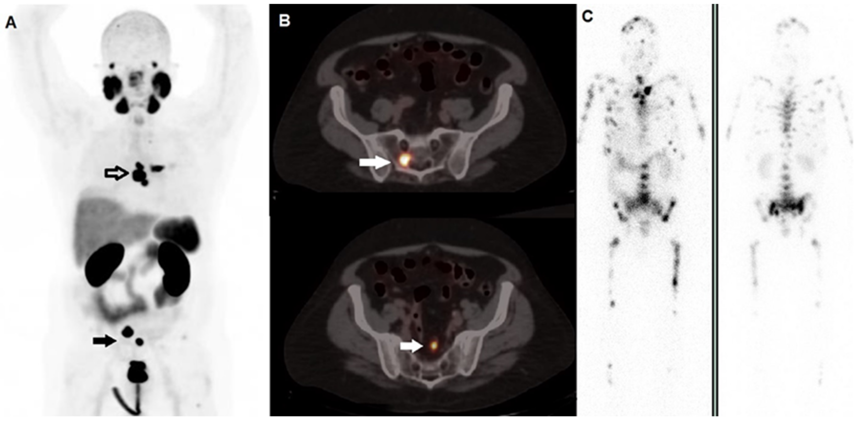
5.1.1. Bone Scan

The usage of bone scan in patients’ selection is a matter of debate. However, in cases of diffuse metastases of prostate cancer to the axial skeleton in a manner typical of superscan, it may be useful to use the 225Ac-PSMA due to its shorter tissue range to preserve the functional marrow [83] and maximize the crossfire effect [68]. However, the usability of a bone scan in patients’ selection is not clarified yet, and PSMA imaging (SPECT and PET) may provide this baseline assessment [84]. Moreover, the PSMA PET/CT is superior and can potentially replace a bone scan in the evaluation for skeletal metastases in the clinical and trial setting because of its ability to detect lytic and bone marrow metastases [85]. Interestingly, the gamma photon emitted during Bi decay (440 keV, emission probability of 26.1%) could be potentially useful for SPECT imaging for biodistribution and dosimetry studies [68].

5.1.2. 68Ga-PSMA/18F-PSMA

Interestingly, a recent consensus paper has recently been published by a multidisciplinary team of urologists, radiologists, and nuclear medicine physicians, reporting that PSMA PET imaging can be used at baseline and after local and systemic treatment in patients with metastatic disease, in order to evaluate the response to treatment. Ideally, PSMA PET/CT imaging criteria should categorize patients as responders (partial and complete response), patients with stable disease, or as non-responders, with a special consideration of specific clinical scenarios such as oligometastatic or poly-metastatic disease [86]. Beyond the specific therapy, the criteria to classify PSMA response into one of the four categories are the following [87]:
-
Complete response: Disappearance of any lesion with tracer uptake;
-
Partial response: Reduction in uptake tumor PET volume by >30%;
-
Stable disease: Change of uptake and tumor PET volume by ±≤30% and no new lesions documented;
-
Progressive disease: Appearance of ≥2 new lesions or increase in uptake or tumor volume ≥30%.
For the scope of the project, PSMA PET/CT refers to PET/CT imaging performed with several PSMA-targeting radioligands (e.g., 68Ga-PSMA-11, 18F-PSMA-1007, 18F-DCFPyL) [86]. Concerning the different radio-compounds and isotopes used in PSMA-based TAT, 68Gallium is a radioisotope with favorable pharmacokinetic characteristics with a commercial accessibility due to germanium-68/gallium-68 (Ge/Ga) generators and more recently produced by cyclotron, which allows a widespread diffusion [63]. In addition, in the PSMA field, the 68Ga labeled tracers have been widely validated and used to define PSMA-positive lesions before 177Lu-PSMA-617 in a previous study [88,89]. Concerning the PSMA-based TAT monitoring, some recent studies indicate that 68Ga-PSMA PET/CT could be a useful tool for the evaluation of response to 177Lu-PSMA-617 radioligand therapy within a theranostic framework [90,91] and that it could enhance RECIST criteria, specifically concerning its limitations for sclerotic bone lesions [92]. 68Ga-PSMA PET/CT may find greater application in response assessment of prostate cancer therapy, as it has already been shown to be a suitable replacement for conventional imaging, providing superior accuracy to the combined findings of CT and bone scanning in a randomized multicentric trial [93]. The optimal setting to use the PSMA-based TAT is still a matter of debate. Currently, PSMA-based TAT is performed in mCRPC non-responders to other treatments. In patients’ selection, there should be adequate uptake on the PSMA ligand based on a diagnostic scan. Even if a clear protocol for eligibility has not been provided, some previous papers concerning beta-emitting-therapy describe the 68Ga-PSMA uptake in lesions as a specific criterion, which should be 1.5× higher than liver uptake [67,94]. However, in the VISION trial, an international open-label phase 3 trial evaluating 177Lu-PSMA-617 in mCRPC [89], the presence of PSMA-positive lesions was defined as 68Ga-PSMA-11 uptake greater than that of liver parenchyma in one or more metastatic lesions of any size in any organ system. However, concerning the optimal setting for baseline assessment pre-treatment with 177Lu-PSMA-617 in NCCN Guidelines [95], the panel discussed the fact that the FDA-approved PSMA imaging agents—68Ga-PSMA-11 and 18F-piflufolastat PSMA—share the same PSMA binding motif with each other and with 177Lu-PSMA-617, suggesting that all approved PSMA PET imaging agents should be acceptable. Therefore, even if further clinical trials are needed, it seems reasonable to extend this assessment also to PSMA-based TAT. In fact, ongoing trials, described in Table 1, are using 68Ga-PSMA as a diagnostic tool for PSMA-based TAT monitoring (NCT04597411, NCT04506567). However, according to a recent paper, in patients who experience PSA progression on PSMA-based radioligand therapy, PET imaging with 68Ga-NODAGA that accumulates in the region of bone turnover surrounding the PCa metastatic deposit in the bone and provides an indirect assessment of bone metastasis is a more suitable imaging modality for the detection of skeletal lesions not expressing PSMA [96], which is probably due to the mechanisms by which mCRPC becomes resistant to PSMA-based radioligand therapy with alpha- or beta-emitting radionuclides: that is, by downregulating PSMA expression that influences 68Ga-PSMA uptake [68]. Further papers are needed in order to clearly define a dedicated protocol in patients’ selection and PSMA-based TAT monitoring. The fluorine-based agents developed for prostate cancer imaging include 18F-DCFBC, F-DCFPyl, 18F-rhPSMA and 18F-PSMA-1007. The half-life makes 18F more attractive for commercial development because it can be produced centrally and distributed to satellite sites [63]. However, certain PSMA ligands such as PSMA-1007 have high hepatic background activity compared with most other PSMA ligands [97]. In the patients’ selection, when 18F-PSMA-1007 PET/CT is obtained for baseline imaging, caution must be exercised in using hepatic background activity as reference standard to define sufficient PSMA expression in mCRPC lesions by considering mediastinal blood-pool as alternative internal reference.

5.1.3. 99mTc-PSMA

There are a host of 99Tc labeled PSMA ligands (Tc-MIP-1404, Tc I and S PSMA, Tc-HYNIC-PSMA) that have been evaluated for imaging of prostate cancer. 99mTc-PSMA-I and S have been tested at various clinical stages with detection rates that are clearly inferior to data reported for PET-imaging. However, in the case of unavailable PET imaging, 99mTc-PSMA-I and S may provide useful information, showing at low PSA levels inferior detection rates, whereas at higher PSA levels, it provides higher detection rates [98]. Similarly, further papers describe a significant relationship between quantitative 99mTc-MIP-1404 uptake, PSA level, and Gleason score [99,100], but the correlation with PSA has not been confirmed in a further paper. Comparable to 99mTc-MDP in terms of detecting bone metastases, 99mTc-PSMA also showed the advantage of giving details on visceral illness. In our resource-constrained setting, 99mTc-PSMA may be a superior option to 99mTc-MDP for the staging, restaging, and evaluation of patients with biochemical progression following radical PCa treatment. It may also help identify patients who are candidates for PSMA-labeled radioligand therapy [101]. A comparative study defined that Tc-99m HYNIC PSMA may be useful in the imaging of prostate cancer, although with a lower sensitivity for lesion detection compared to 68Ga PSMA PET/CT [102]. Even if the tracers used for PSMA-PET/CT have been shown to have greater imaging properties and results, there are some advantages in the usage of 99mTc-PSMA ligands, which are mainly linked to costs and logistic aspects. Moreover, the 99mTc-PSMA ligand may have a potential role in radioguided surgery with intraoperative probe localization [103,104,105].

6. Discussion

TAT holds the promise to represent a powerful weapon in oncology, especially as far as it concerns the management of mCRPC. However, it has to be highlighted that to date, 223Ra therapy is the first and only alpha-emitter agent approved by the regulatory authorities (i.e., FDA and EMA) for therapeutic use. It is noteworthy that TAT is not something new in the history of nuclear medicine, since many decades of research on alpha-emitters paved the way for the implementation of such a therapeutic regimen in clinical practice [106]. In 2018, according to a specific note issued by the EMA (EMA/500948/2018) on the indications for 223Ra therapy in a review of an ongoing phase 3 trial by the EMA’s Pharmacovigilance Risk Assessment Committee, 223Ra therapy was practically placed as a third-line therapy in mCRPC management. This indication led to the enrollment of heavily pre-treated patients, bearing high tumor burden metastatic disease, in contrast with published data showing a relatively more favorable outcome in subjects with lower metabolic tumor volume at baseline [55,107]. From this perspective, an accurate patients’ stratification before TAT enrollment plays a crucial role [108].
The amazing therapeutic success of 225Ac-PSMA-617 in the treatment of mCRPC reported in recent papers has greatly increased interest in the clinical use of targeted alpha therapy. The use of 225Ac-PSMA-617 highlights the potential applicability of the idea of targeted alpha treatment in cancer and offers a viable therapeutic alternative for prostate cancer [74]. Most of these agents are still in the preclinical phase of investigation, and 213Bi-PSMA is one of the few to have been tested clinically with no definitive results. However, the possibility to use different alpha emitters labeled with PSMA molecules and antibodies, which present different half-lives and physical properties, may open a new chapter of the prostate cancer management, especially considering the novel trends in TAT combination therapies, which are already described in the clinical setting for the combination 177Lu-PSMA/225Ac-PSMA therapy [78,79,80]. Beyond the evidence that supports the introduction of PSMA-based TAT in the therapeutic schema of mCRPC, there is a need for clinical trials to better define the timing of the application of 225Ac-PSMA in the treatment sequence of mCRPC [63]. In fact, the results are not heterogeneous and, according to some authors, the risk of a shorter progression-free survival in PSMA-based TAT exists in patients already treated with 177Lu-PSMA, suggesting that prior beta-emitter therapy may induce resistance to radiation [109]. As more centers gain access to 225Ac-PSMA, more data are becoming available. To our knowledge, we could not find a clinical study that includes the bone scan in the PSMA-based TAT management and therapy assessment algorithm. Of course, the bone scan may provide information concerning the bone involvement, but for PSMA-based TATA, therapeutic planning seems not mandatory: the only useful aspect of bone scan regarding the TAT is the superscan findings, which can be provided by different and more specific methodologies [84]. Imaging professionals have been forced to adapt and develop techniques or algorithms for reporting follow-up imaging as a result of the increase and extension of the use of PSMA-based ligands in imaging and therapy. Particularly in cases of bone-dominant illness, the molecular response evaluation is preferred above morphological response criteria in individuals with mCRPC. Actually, the PSMA PET/CT imaging criteria is considered the dominant tool for the patient’s selection and for PSMA-based TAT monitoring, allowing also the classification of patients as responders, stable disease, and non-responders [86]. PSMA PET/CT refers to PET/CT imaging performed with several PSMA-targeting radioligands (e.g., 68Ga-PSMA-11, 18F-PSMA-1007, 18F-DCFPyL), and it is the most validated tool in the molecular imaging field [86]. Further papers are needed in order to define the specific characteristics of each PSMA-labeled PET tracer. However, the 68Ga-labeled tracers have been the most validated, which is also due to favorable commercial aspects of 68Ga [88]. On the other hand, 18F-labeled tracers appear attractive due to logistic advantages. Potentially, future comparative trials may provide specific indications for the usage of different tracers in specific conditions also in consideration of different tracers biodistribution and different physical properties. 99mTc-PSMA ligands can be useful in the eligibility process of patients that should undergo PSMA-based TAT [101]. However, tracers used for PET/CT have shown superior properties in terms of imaging, but the potential role of 99mTc-PSMA ligands should not be underestimated, because of the possibility to use it in settings without PET equipment or in remote areas. Further data are needed about the possible inclusion of 99mTc-PSMA scans in the PSMA-based TAT therapy assessment algorithm.

Author Contributions

Writing—original draft preparation, V.F., M.R., A.C. and M.C.; writing—review and editing, L.F. and M.S.D.F.; supervision, G.D.V. All authors have read and agreed to the published version of the manuscript.

Funding

This research received no external funding.

Institutional Review Board Statement

Not applicable.

Informed Consent Statement

Not applicable.

Data Availability Statement

Not applicable.

Conflicts of Interest

The authors declare no conflict of interest.

References

  1. Sung, H.; Ferlay, J.; Siegel, R.L.; Laversanne, M.; Soerjomataram, I.; Jemal, A.; Bray, F. Global Cancer Statistics 2020: GLOBOCAN Estimates of Incidence and Mortality Worldwide for 36 Cancers in 185 Countries. CA Cancer J. Clin. 2021, 71, 209–249. [Google Scholar] [CrossRef] [PubMed]
  2. Aurilio, G.; Cimadamore, A.; Mazzucchelli, R.; Lopez-Beltran, A.; Verri, E.; Scarpelli, M.; Massari, F.; Cheng, L.; Santoni, M.; Montironi, R. Androgen Receptor Signaling Pathway in Prostate Cancer: From Genetics to Clinical Applications. Cells 2020, 9, 2653. [Google Scholar] [CrossRef] [PubMed]
  3. Ricci, M.; Frantellizzi, V.; Bulzonetti, N.; De Vincentis, G. Reversibility of castration resistance status after Radium-223 dichloride treatment: Clinical evidence and Review of the literature. Int. J. Radiat. Biol. 2018, 95, 554–561. [Google Scholar] [CrossRef] [PubMed]
  4. Yamada, Y.; Beltran, H. The treatment landscape of metastatic prostate cancer. Cancer Lett. 2021, 519, 20–29. [Google Scholar] [CrossRef]
  5. Crowley, F.; Sterpi, M.; Buckley, C.; Margetich, L.; Handa, S.; Dovey, Z. A Review of the Pathophysiological Mechanisms Underlying Castration-resistant Prostate Cancer. Res. Rep. Urol. 2021, 13, 457–472. [Google Scholar] [CrossRef]
  6. Martin, S.K.; Kamelgarn, M.; Kyprianou, N. Cytoskeleton targeting value in prostate cancer treatment. Am. J. Clin. Exp. Urol. 2014, 2, 15–26. [Google Scholar]
  7. Deschesnes, R.G.; Patenaude, A.; Rousseau, J.L.; Fortin, J.S.; Ricard, C.; Côté, M.F.; Huot, J.; C.-Gaudreault, R.; Petitclerc, E. Microtubule-destabilizing agents induce focal adhesion structure disorganization and anoikis in cancer cells. J. Pharm. Exp. 2007, 320, 853–864. [Google Scholar] [CrossRef]
  8. Sweeney, C.J.; Chen, Y.H.; Carducci, M.; Liu, G.; Jarrard, D.F.; Eisenberger, M.; Wong, Y.N.; Hahn, N.; Kohli, M.; Cooney, M.M.; et al. Chemohormonal Therapy in Metastatic Hormone-Sensitive Prostate Cancer. N. Engl. J. Med. 2015, 373, 737–746. [Google Scholar] [CrossRef]
  9. Uo, T.; Sprenger, C.C.; Plymate, S.R. Androgen Receptor Signaling and Metabolic and Cellular Plasticity During Progression to Castration Resistant Prostate Cancer. Front. Oncol. 2020, 10, 580617. [Google Scholar] [CrossRef]
  10. Jamroze, A.; Chatta, G.; Tang, D.G. Androgen receptor (AR) heterogeneity in prostate cancer and therapy resistance. Cancer Lett. 2021, 518, 1–9. [Google Scholar] [CrossRef]
  11. Kantoff, P.W.; Higano, C.S.; Shore, N.D.; Berger, E.R.; Small, E.J.; Penson, D.F.; Redfern, C.H.; Ferrari, A.C.; Dreicer, R.; Sims, R.B.; et al. Sipuleucel-T immunotherapy for castration-resistant prostate cancer. N. Engl. J. Med. 2010, 363, 411–422. [Google Scholar] [CrossRef] [Green Version]
  12. Marcus, L.; Lemery, S.J.; Keegan, P.; Pazdur, R. FDA Approval Summary: Pembrolizumab for the Treatment of Microsatellite Instability-High Solid Tumors. Clin. Cancer Res. 2019, 25, 3753–3758. [Google Scholar] [CrossRef]
  13. De Bono, J.S.; De Giorgi, U.; Rodrigues, D.N.; Massard, C.; Bracarda, S.; Font, A.; Arranz Arija, J.A.; Shih, K.C.; Radavoi, G.D.; Xu, N.; et al. Randomized Phase II Study Evaluating Akt Blockade with Ipatasertib, in Combination with Abiraterone, in Patients with Metastatic Prostate Cancer with and without PTEN Loss. Clin. Cancer Res. 2019, 25, 928–936. [Google Scholar] [CrossRef]
  14. FDA Approves Pluvicto/Locametz for Metastatic Castration-Resistant Prostate Cancer. J. Nucl. Med. 2022, 63, 13N.
  15. Filippi, L.; Chiaravalloti, A.; Schillaci, O.; Cianni, R.; Bagni, O. Theranostic approaches in nuclear medicine: Current status and future prospects. Expert Rev. Med. Devices 2020, 17, 331–343. [Google Scholar] [CrossRef]
  16. De Vincentis, G.; Gerritsen, W.; Gschwend, J.E.; Hacker, M.; Lewington, V.; O’Sullivan, J.M.; Oya, M.; Pacilio, M.; Parker, C.; Shore, N.; et al. Advances in targeted alpha therapy for prostate cancer. Ann. Oncol. 2019, 30, 1728–1739. [Google Scholar] [CrossRef]
  17. Frantellizzi, V.; Cosma, L.; Brunotti, G.; Pani, A.; Spanu, A.; Nuvoli, S.; De Cristofaro, F.; Civitelli, L.; De Vincentis, G. Target Alpha Therapy with Thorium-227. Cancer Biother. Radiopharm. 2020, 35, 437–445. [Google Scholar] [CrossRef]
  18. Tafreshi, N.K.; Doligalski, M.L.; Tichacek, C.J.; Pandya, D.N.; Budzevich, M.M.; El-Haddad, G.; Khushalani, N.I.; Moros, E.G.; McLaughlin, M.L.; Wadas, T.J.; et al. Development of Targeted Alpha Particle Therapy for Solid Tumors. Molecules 2019, 24, 4314. [Google Scholar] [CrossRef]
  19. Parker, C.; Nilsson, S.; Heinrich, D.; Helle, S.I.; O’Sullivan, J.M.; Fossa, S.D.; Chodacki, A.; Wiechno, P.; Logue, J.; Seke, M.; et al. Alpha emitter radium-223 and survival in metastatic prostate cancer. N. Engl. J. Med. 2013, 369, 213–223. [Google Scholar] [CrossRef]
  20. Hammer, S.; Hagemann, U.B.; Zitzmann-Kolbe, S.; Larsen, A.; Ellingsen, C.; Geraudie, S.; Grant, D.; Indrevoll, B.; Smeets, R.; von Ahsen, O.; et al. Preclinical Efficacy of a PSMA-Targeted Thorium-227 Conjugate (PSMA-TTC), a Targeted Alpha Therapy for Prostate Cancer. Clin. Cancer Res. 2020, 26, 1985–1996. [Google Scholar] [CrossRef]
  21. Kratochwil, C.; Fendler, W.P.; Eiber, M.; Baum, R.; Bozkurt, M.F.; Czernin, J.; Delgado Bolton, R.C.; Ezziddin, S.; Forrer, F.; Hicks, R.J.; et al. EANM procedure guidelines for radionuclide therapy with 177Lu-labelled PSMA-ligands (177Lu-PSMA-RLT). Eur. J. Nucl. Med. Mol. Imaging 2019, 46, 2536–2544. [Google Scholar] [CrossRef] [PubMed]
  22. Frantellizzi, V.; Verrina, V.; Raso, C.; Pontico, M.; Petronella, F.; Bertana, V.; Ballesio, A.; Marasso, S.L.; Miglietta, S.; Rosa, P.; et al. 99mTc-labeled keratin gold-nanoparticles in a nephron-like microfluidic chip for photo-thermal therapy applications. Mater. Today Adv. 2022, 16, 100286. [Google Scholar] [CrossRef]
  23. Lee, D.S.; Im, H.-J.; Lee, Y.-S. Radionanomedicine: Widened perspectives of molecular theragnosis. Nanomed. Nanotechnol. Biol. Med. 2015, 11, 795–810. [Google Scholar] [CrossRef] [PubMed]
  24. Shukla, R.; Chanda, N.; Zambre, A.; Upendran, A.; Katti, K.; Kulkarni, R.R.; Nune, S.K.; Casteel, S.W.; Smith, C.J.; Vimal, J.; et al. Laminin receptor specific therapeutic gold nanoparticles (198AuNP-EGCg) show efficacy in treating prostate cancer. Proc. Natl. Acad. Sci. USA 2012, 109, 12426–12431. [Google Scholar] [CrossRef] [PubMed]
  25. Hod, N.; Levin, D.; Tiktinsky, K.; Ezroh Katzap, D.; Lantsberg, S. Nuclear Medicine and Molecular Imaging: From Basic Science to the Front in Innovative Imaging and Treatment. Harefuah 2021, 160, 448–454. [Google Scholar]
  26. Alqahtani, M.M.; Willowson, K.P.; Constable, C.; Fulton, R.; Kench, P.L. Optimization of (99m) Tc whole-body SPECT/CT image quality: A phantom study. J. Appl. Clin. Med. Phys. 2022, 23, e13528. [Google Scholar] [CrossRef]
  27. Filippi, L.; Biancone, L.; Petruzziello, C.; Schillaci, O. Tc-99m HMPAO-labeled leukocyte scintigraphy with hybrid SPECT/CT detects perianal fistulas in Crohn disease. Clin. Nucl. Med. 2006, 31, 541–542. [Google Scholar] [CrossRef]
  28. Frantellizzi, V.; Pontico, M.; Letizia, C.; De Vincentis, G. Bladder wall paraganglioma located using (123)I-mIBG SPECT and CT imaging. Rev. Esp. De Med. Nucl. E Imagen Mol. 2018, 37, 253–254. [Google Scholar] [CrossRef]
  29. Del Guerra, A.; Belcari, N.; Giuseppina Bisogni, M.; Corsi, F.; Foresta, M.; Guerra, P.; Marcatili, S.; Santos, A.; Sportelli, G. Silicon Photomultipliers (SiPM) as novel photodetectors for PET. Nucl. Instrum. Methods Phys. Res. Sect. A Accel. Spectrometers Detect. Assoc. Equip. 2011, 648, S232–S235. [Google Scholar] [CrossRef]
  30. Alberts, I.; Hünermund, J.-N.; Sachpekidis, C.; Mingels, C.; Fech, V.; Bohn, K.P.; Rominger, A.; Afshar-Oromieh, A. The influence of digital PET/CT on diagnostic certainty and interrater reliability in [68Ga]Ga-PSMA-11 PET/CT for recurrent prostate cancer. Eur. Radiol. 2021, 31, 8030–8039. [Google Scholar] [CrossRef]
  31. Filippi, L.; Bagni, O.; Schillaci, O. Digital PET/CT with 18F-FACBC in early castration-resistant prostate cancer: Our preliminary results. Expert Rev. Med. Devices 2022, 19, 591–598. [Google Scholar] [CrossRef]
  32. Baidoo, K.E.; Yong, K.; Brechbiel, M.W. Molecular pathways: Targeted alpha-particle radiation therapy. Clin. Cancer Res. 2013, 19, 530–537. [Google Scholar] [CrossRef]
  33. Dahle, J.; Borrebaek, J.; Jonasdottir, T.J.; Hjelmerud, A.K.; Melhus, K.B.; Bruland, O.S.; Press, O.W.; Larsen, R.H. Targeted cancer therapy with a novel low-dose rate alpha-emitting radioimmunoconjugate. Blood 2007, 110, 2049–2056. [Google Scholar] [CrossRef] [Green Version]
  34. Topatana, W.; Juengpanich, S.; Li, S.; Cao, J.; Hu, J.; Lee, J.; Suliyanto, K.; Ma, D.; Zhang, B.; Chen, M.; et al. Advances in synthetic lethality for cancer therapy: Cellular mechanism and clinical translation. J. Hematol. Oncol. 2020, 13, 118. [Google Scholar] [CrossRef]
  35. Wéra, A.C.; Lobbens, A.; Stoyanov, M.; Lucas, S.; Michiels, C. Radiation-induced synthetic lethality: Combination of poly(ADP-ribose) polymerase and RAD51 inhibitors to sensitize cells to proton irradiation. Cell Cycle 2019, 18, 1770–1783. [Google Scholar] [CrossRef]
  36. Wale, D.J.; Viglianti, B.L.; Gross, M.D.; Ferretti, A.; Rubello, D.; Wong, K.K. Nuclear Medicine Therapy With 223Radium-dichloride for Osseous Metastases in Prostate Carcinoma. Am. J. Clin. Oncol. 2019, 42, 99–106. [Google Scholar] [CrossRef]
  37. Brito, A.E.; Etchebehere, E. Radium-223 as an Approved Modality for Treatment of Bone Metastases. Semin. Nucl. Med. 2020, 50, 177–192. [Google Scholar] [CrossRef]
  38. Collins, S.M.; Pearce, A.K.; Ferreira, K.M.; Fenwick, A.J.; Regan, P.H.; Keightley, J.D. Direct measurement of the half-life of (223)Ra. Appl. Radiat. Isot. 2015, 99, 46–53. [Google Scholar] [CrossRef]
  39. Abramenkovs, A.; Hariri, M.; Spiegelberg, D.; Nilsson, S.; Stenerlöw, B. Ra-223 induces clustered DNA damage and inhibits cell survival in several prostate cancer cell lines. Transl. Oncol. 2022, 26, 101543. [Google Scholar] [CrossRef]
  40. Den, R.B.; George, D.; Pieczonka, C.; McNamara, M. Ra-223 Treatment for Bone Metastases in Castrate-Resistant Prostate Cancer: Practical Management Issues for Patient Selection. Am. J. Clin. Oncol. 2019, 42, 399–406. [Google Scholar] [CrossRef]
  41. Du, Y.; Carrio, I.; De Vincentis, G.; Fanti, S.; Ilhan, H.; Mommsen, C.; Nitzsche, E.; Sundram, F.; Vogel, W.; Oyen, W.; et al. Practical recommendations for radium-223 treatment of metastatic castration-resistant prostate cancer. Eur. J. Nucl. Med. Mol. Imaging 2017, 44, 1671–1678. [Google Scholar] [CrossRef] [PubMed]
  42. Poeppel, T.D.; Handkiewicz-Junak, D.; Andreeff, M.; Becherer, A.; Bockisch, A.; Fricke, E.; Geworski, L.; Heinzel, A.; Krause, B.J.; Krause, T.; et al. EANM guideline for radionuclide therapy with radium-223 of metastatic castration-resistant prostate cancer. Eur. J. Nucl. Med. Mol. Imaging 2018, 45, 824–845. [Google Scholar] [CrossRef] [PubMed]
  43. Hoskin, P.; Sartor, O.; O’Sullivan, J.M.; Johannessen, D.C.; Helle, S.I.; Logue, J.; Bottomley, D.; Nilsson, S.; Vogelzang, N.J.; Fang, F.; et al. Efficacy and safety of radium-223 dichloride in patients with castration-resistant prostate cancer and symptomatic bone metastases, with or without previous docetaxel use: A prespecified subgroup analysis from the randomised, double-blind, phase 3 ALSYMPCA trial. Lancet. Oncol. 2014, 15, 1397–1406. [Google Scholar] [CrossRef] [PubMed]
  44. Jarvis, P.; Ho, A.; Sundram, F. Radium-223 therapy for metastatic castration-resistant prostate cancer: Survival benefit when used earlier in the treatment pathway. Nucl. Med. Commun. 2021, 42, 332–336. [Google Scholar] [CrossRef] [PubMed]
  45. Wong, W.W.; Anderson, E.M.; Mohammadi, H.; Daniels, T.B.; Schild, S.E.; Keole, S.R.; Choo, C.R.; Tzou, K.S.; Bryce, A.H.; Ho, T.H.; et al. Factors Associated with Survival Following Radium-223 Treatment for Metastatic Castration-resistant Prostate Cancer. Clin. Genitourin. Cancer 2017, 15, e969–e975. [Google Scholar] [CrossRef]
  46. Frantellizzi, V.; Monari, F.; Mascia, M.; Costa, R.; Rubini, G.; Spanu, A.; Di Rocco, A.; Lodi Rizzini, E.; Cindolo, L.; Licari, M.; et al. Validation of the 3-variable prognostic score (3-PS) in mCRPC patients treated with (223)Radium-dichloride: A national multicenter study. Ann. Nucl. Med. 2020, 34, 772–780. [Google Scholar] [CrossRef]
  47. Frantellizzi, V.; De Feo, M.S.; Di Rocco, A.; Pontico, M.; Pani, A.; Farcomeni, A.; Cosma, L.; Lazri, J.; De Vincentis, G. Baseline quality of life predicts overall survival in patients with mCRPC treated with (223)Ra-dichloride. Hell. J. Nucl. Med. 2020, 23, 12–20. [Google Scholar] [CrossRef]
  48. Frantellizzi, V.; Pani, A.; Ippoliti, M.D.; Farcomeni, A.; Aloise, I.; Colosi, M.; Polito, C.; Pani, R.; Vincentis, G. Scintigraphic load of bone disease evaluated by DASciS software as a survival predictor in metastatic castration-resistant prostate cancer patients candidates to 223RaCl treatment. Radiol. Oncol. 2019, 54, 40–47. [Google Scholar] [CrossRef]
  49. Ballinger, J.R. Theranostic radiopharmaceuticals: Established agents in current use. Br. J. Radiol. 2018, 91, 20170969. [Google Scholar] [CrossRef]
  50. García Vicente, A.M.; Soriano Castrejón, Á.; Alvarez Cabellos, R.; Sanchez Gil, B.; Mohedano Mohedano, N. Response Assessment of 223Ra Treatment: Should a Fluorocholine PET/CT Be Performed? Clin. Nucl. Med. 2017, 42, 761–765. [Google Scholar] [CrossRef]
  51. Even-Sapir, E. ¹⁸F-fluoride PET/computed tomography imaging. PET Clin. 2014, 9, 277–285. [Google Scholar] [CrossRef]
  52. Piert, M.; Zittel, T.T.; Jahn, M.; Stahlschmidt, A.; Becker, G.A.; Machulla, H.J. Increased sensitivity in detection of a porcine high-turnover osteopenia after total gastrectomy by dynamic 18F-fluoride ion PET and quantitative CT. J. Nucl. Med. 2003, 44, 117–124. [Google Scholar]
  53. Gauthé, M.; Zarca, K.; Aveline, C.; Lecouvet, F.; Balogova, S.; Cussenot, O.; Talbot, J.-N.; Durand-Zaleski, I. Comparison of 18F-sodium fluoride PET/CT, 18F-fluorocholine PET/CT and diffusion-weighted MRI for the detection of bone metastases in recurrent prostate cancer: A cost-effectiveness analysis in France. BMC Med. Imaging 2020, 20, 25. [Google Scholar] [CrossRef]
  54. Etchebehere, E.C.; Araujo, J.C.; Milton, D.R.; Erwin, W.D.; Wendt, R.E., 3rd; Swanston, N.M.; Fox, P.; Macapinlac, H.A.; Rohren, E.M. Skeletal Tumor Burden on Baseline 18F-Fluoride PET/CT Predicts Bone Marrow Failure After 223Ra Therapy. Clin. Nucl. Med. 2016, 41, 268–273. [Google Scholar] [CrossRef]
  55. Bauckneht, M.; Capitanio, S.; Donegani, M.I.; Zanardi, E.; Miceli, A.; Murialdo, R.; Raffa, S.; Tomasello, L.; Vitti, M.; Cavo, A.; et al. Role of Baseline and Post-Therapy 18F-FDG PET in the Prognostic Stratification of Metastatic Castration-Resistant Prostate Cancer (mCRPC) Patients Treated with Radium-223. Cancers 2019, 12, 31. [Google Scholar] [CrossRef]
  56. Vija Racaru, L.; Sinigaglia, M.; Kanoun, S.; Ben Bouallègue, F.; Tal, I.; Brillouet, S.; Bauriaud-Mallet, M.; Zerdoud, S.; Dierickx, L.; Vallot, D.; et al. Fluorine-18-fluorocholine PET/CT parameters predictive for hematological toxicity to radium-223 therapy in castrate-resistant prostate cancer patients with bone metastases: A pilot study. Nucl. Med. Commun. 2018, 39, 672–679. [Google Scholar] [CrossRef]
  57. Filippi, L.; Basile, P.; Schillaci, O.; Bagni, O. The Relationship Between Total Lesion Activity on (18)F Choline Positron Emission Tomography-Computed Tomography and Clinical Outcome in Patients with Castration-Resistant Prostate Cancer Bone Metastases Treated with (223)Radium. Cancer Biother. Radiopharm. 2020, 35, 398–403. [Google Scholar] [CrossRef]
  58. García Vicente, A.M.; Amo-Salas, M.; Cassinello Espinosa, J.; Gómez Díaz, R.; Soriano Castrejón, Á. Interim and end-treatment (18)F-Fluorocholine PET/CT and bone scan in prostate cancer patients treated with Radium 223 dichloride. Sci. Rep. 2021, 11, 7389. [Google Scholar] [CrossRef]
  59. Bräuer, A.; Rahbar, K.; Konnert, J.; Bögemann, M.; Stegger, L. Diagnostic value of additional (68)Ga-PSMA-PET before (223)Ra-dichloride therapy in patients with metastatic prostate carcinoma. Nuklearmedizin. Nucl. Med. 2017, 56, 14–22. [Google Scholar] [CrossRef]
  60. Bode, A.; Rahbar, K.; Konnert, J.; Bögemann, M.; Stegger, L. Benefit of 68Ga-PSMA-PET/CT in Patients Considered for 223Ra-Dichloride Therapy. Clin. Nucl. Med. 2016, 41, 951–952. [Google Scholar] [CrossRef]
  61. Ahmadzadehfar, H.; Azgomi, K.; Hauser, S.; Wei, X.; Yordanova, A.; Gaertner, F.C.; Kürpig, S.; Strunk, H.; Essler, M. (68)Ga-PSMA-11 PET as a Gatekeeper for the Treatment of Metastatic Prostate Cancer with (223)Ra: Proof of Concept. J. Nucl. Med. 2017, 58, 438–444. [Google Scholar] [CrossRef] [PubMed]
  62. Kratochwil, C.; Haberkorn, U.; Giesel, F.L. (225)Ac-PSMA-617 for Therapy of Prostate Cancer. Semin. Nucl. Med. 2020, 50, 133–140. [Google Scholar] [CrossRef] [PubMed]
  63. Mokoala, K.; Lawal, I.; Lengana, T.; Kgatle, M.; Giesel, F.L.; Vorster, M.; Sathekge, M. PSMA Theranostics: Science and Practice. Cancers 2021, 13, 3904. [Google Scholar] [CrossRef] [PubMed]
  64. Filippi, L.; Chiaravalloti, A.; Schillaci, O.; Bagni, O. The potential of PSMA-targeted alpha therapy in the management of prostate cancer. Expert Rev. Anticancer 2020, 20, 823–829. [Google Scholar] [CrossRef]
  65. Lütje, S.; Heskamp, S.; Cornelissen, A.S.; Poeppel, T.D.; van den Broek, S.A.; Rosenbaum-Krumme, S.; Bockisch, A.; Gotthardt, M.; Rijpkema, M.; Boerman, O.C. PSMA Ligands for Radionuclide Imaging and Therapy of Prostate Cancer: Clinical Status. Theranostics 2015, 5, 1388–1401. [Google Scholar] [CrossRef]
  66. Emmett, L.; Willowson, K.; Violet, J.; Shin, J.; Blanksby, A.; Lee, J. Lutetium (177) PSMA radionuclide therapy for men with prostate cancer: A review of the current literature and discussion of practical aspects of therapy. J. Med. Radiat. Sci. 2017, 64, 52–60. [Google Scholar] [CrossRef]
  67. Hofman, M.S.; Violet, J.; Hicks, R.J.; Ferdinandus, J.; Thang, S.P.; Akhurst, T.; Iravani, A.; Kong, G.; Ravi Kumar, A.; Murphy, D.G.; et al. [(177)Lu]-PSMA-617 radionuclide treatment in patients with metastatic castration-resistant prostate cancer (LuPSMA trial): A single-centre, single-arm, phase 2 study. Lancet. Oncol. 2018, 19, 825–833. [Google Scholar] [CrossRef]
  68. Sathekge, M.M.; Bruchertseifer, F.; Vorster, M.; Morgenstern, A.; Lawal, I.O. Global experience with PSMA-based alpha therapy in prostate cancer. Eur. J. Nucl. Med. Mol. Imaging 2021, 49, 30–46. [Google Scholar] [CrossRef]
  69. Kleynhans, J.; Sathekge, M.; Ebenhan, T. Obstacles and Recommendations for Clinical Translation of Nanoparticle System-Based Targeted Alpha-Particle Therapy. Materials 2021, 14, 4784. [Google Scholar] [CrossRef]
  70. Majkowska-Pilip, A.; Gawęda, W.; Żelechowska-Matysiak, K.; Wawrowicz, K.; Bilewicz, A. Nanoparticles in Targeted Alpha Therapy. Nanomaterials 2020, 10, 1366. [Google Scholar] [CrossRef]
  71. Roscher, M.; Bakos, G.; Benešová, M. Atomic Nanogenerators in Targeted Alpha Therapies: Curie’s Legacy in Modern Cancer Management. Pharmaceuticals 2020, 13, 76. [Google Scholar] [CrossRef]
  72. Sartor, O.; Sharma, D. Radium and other alpha emitters in prostate cancer. Transl. Androl. Urol. 2018, 7, 436–444. [Google Scholar] [CrossRef]
  73. Dos Santos, J.C.; Schäfer, M.; Bauder-Wüst, U.; Lehnert, W.; Leotta, K.; Morgenstern, A.; Kopka, K.; Haberkorn, U.; Mier, W.; Kratochwil, C. Development and dosimetry of (203)Pb/(212)Pb-labelled PSMA ligands: Bringing "the lead" into PSMA-targeted alpha therapy? Eur. J. Nucl. Med. Mol. Imaging 2019, 46, 1081–1091. [Google Scholar] [CrossRef]
  74. Morgenstern, A.; Apostolidis, C.; Kratochwil, C.; Sathekge, M.; Krolicki, L.; Bruchertseifer, F. An Overview of Targeted Alpha Therapy with (225)Actinium and (213)Bismuth. Curr. Radiopharm. 2018, 11, 200–208. [Google Scholar] [CrossRef]
  75. Kratochwil, C.; Schmidt, K.; Afshar-Oromieh, A.; Bruchertseifer, F.; Rathke, H.; Morgenstern, A.; Haberkorn, U.; Giesel, F.L. Targeted alpha therapy of mCRPC: Dosimetry estimate of (213)Bismuth-PSMA-617. Eur. J. Nucl. Med. Mol. Imaging 2018, 45, 31–37. [Google Scholar] [CrossRef]
  76. Nonnekens, J.; Chatalic, K.L.; Molkenboer-Kuenen, J.D.; Beerens, C.E.; Bruchertseifer, F.; Morgenstern, A.; Veldhoven-Zweistra, J.; Schottelius, M.; Wester, H.J.; van Gent, D.C.; et al. (213)Bi-Labeled Prostate-Specific Membrane Antigen-Targeting Agents Induce DNA Double-Strand Breaks in Prostate Cancer Xenografts. Cancer Biother. Radiopharm. 2017, 32, 67–73. [Google Scholar] [CrossRef]
  77. Sathekge, M.; Knoesen, O.; Meckel, M.; Modiselle, M.; Vorster, M.; Marx, S. (213)Bi-PSMA-617 targeted alpha-radionuclide therapy in metastatic castration-resistant prostate cancer. Eur. J. Nucl. Med. Mol. Imaging 2017, 44, 1099–1100. [Google Scholar] [CrossRef]
  78. Rosar, F.; Hau, F.; Bartholomä, M.; Maus, S.; Stemler, T.; Linxweiler, J.; Ezziddin, S.; Khreish, F. Molecular imaging and biochemical response assessment after a single cycle of [(225)Ac]Ac-PSMA-617/[(177)Lu]Lu-PSMA-617 tandem therapy in mCRPC patients who have progressed on [(177)Lu]Lu-PSMA-617 monotherapy. Theranostics 2021, 11, 4050–4060. [Google Scholar] [CrossRef]
  79. Khreish, F.; Ebert, N.; Ries, M.; Maus, S.; Rosar, F.; Bohnenberger, H.; Stemler, T.; Saar, M.; Bartholomä, M.; Ezziddin, S. (225)Ac-PSMA-617/(177)Lu-PSMA-617 tandem therapy of metastatic castration-resistant prostate cancer: Pilot experience. Eur. J. Nucl. Med. Mol. Imaging 2020, 47, 721–728. [Google Scholar] [CrossRef]
  80. Rosar, F.; Krause, J.; Bartholomä, M.; Maus, S.; Stemler, T.; Hierlmeier, I.; Linxweiler, J.; Ezziddin, S.; Khreish, F. Efficacy and Safety of [(225)Ac]Ac-PSMA-617 Augmented [(177)Lu]Lu-PSMA-617 Radioligand Therapy in Patients with Highly Advanced mCRPC with Poor Prognosis. Pharmaceutics 2021, 13, 722. [Google Scholar] [CrossRef]
  81. Yadav, M.P.; Ballal, S.; Sahoo, R.K.; Tripathi, M.; Seth, A.; Bal, C. Efficacy and safety of (225)Ac-PSMA-617 targeted alpha therapy in metastatic castration-resistant Prostate Cancer patients. Theranostics 2020, 10, 9364–9377. [Google Scholar] [CrossRef] [PubMed]
  82. Scher, H.I.; Morris, M.J.; Stadler, W.M.; Higano, C.; Basch, E.; Fizazi, K.; Antonarakis, E.S.; Beer, T.M.; Carducci, M.A.; Chi, K.N.; et al. Trial Design and Objectives for Castration-Resistant Prostate Cancer: Updated Recommendations from the Prostate Cancer Clinical Trials Working Group 3. J. Clin. Oncol. 2016, 34, 1402–1418. [Google Scholar] [CrossRef] [PubMed]
  83. Haberkorn, U.; Giesel, F.; Morgenstern, A.; Kratochwil, C. The Future of Radioligand Therapy: α, β, or Both? J. Nucl. Med. 2017, 58, 1017–1018. [Google Scholar] [CrossRef] [PubMed]
  84. Lawal, I.; Vorster, M.; Boshomane, T.; Ololade, K.; Ebenhan, T.; Sathekge, M. Metastatic Prostate Carcinoma Presenting as a Superscan on 68Ga-PSMA PET/CT. Clin. Nucl. Med. 2015, 40, 755–756. [Google Scholar] [CrossRef] [Green Version]
  85. Lengana, T.; Lawal, I.O.; Boshomane, T.G.; Popoola, G.O.; Mokoala, K.M.G.; Moshokoa, E.; Maes, A.; Mokgoro, N.P.; Van de Wiele, C.; Vorster, M.; et al. (68)Ga-PSMA PET/CT Replacing Bone Scan in the Initial Staging of Skeletal Metastasis in Prostate Cancer: A Fait Accompli? Clin. Genitourin. Cancer 2018, 16, 392–401. [Google Scholar] [CrossRef]
  86. Fanti, S.; Goffin, K.; Hadaschik, B.A.; Herrmann, K.; Maurer, T.; MacLennan, S.; Oprea-Lager, D.E.; Oyen, W.J.; Rouvière, O.; Mottet, N.; et al. Consensus statements on PSMA PET/CT response assessment criteria in prostate cancer. Eur. J. Nucl. Med. Mol. Imaging 2021, 48, 469–476. [Google Scholar] [CrossRef]
  87. Fanti, S.; Minozzi, S.; Morigi, J.J.; Giesel, F.; Ceci, F.; Uprimny, C.; Hofman, M.S.; Eiber, M.; Schwarzenbock, S.; Castellucci, P.; et al. Development of standardized image interpretation for 68Ga-PSMA PET/CT to detect prostate cancer recurrent lesions. Eur. J. Nucl. Med. Mol. Imaging 2017, 44, 1622–1635. [Google Scholar] [CrossRef]
  88. Schaeffer, E.; Srinivas, S.; Antonarakis, E.S.; Armstrong, A.J.; Bekelman, J.E.; Cheng, H.; D’Amico, A.V.; Davis, B.J.; Desai, N.; Dorff, T.; et al. NCCN Guidelines Insights: Prostate Cancer, Version 1.2021. J. Natl. Compr. Cancer Netw. 2021, 19, 134–143. [Google Scholar] [CrossRef]
  89. Sartor, O.; de Bono, J.; Chi, K.N.; Fizazi, K.; Herrmann, K.; Rahbar, K.; Tagawa, S.T.; Nordquist, L.T.; Vaishampayan, N.; El-Haddad, G.; et al. Lutetium-177-PSMA-617 for Metastatic Castration-Resistant Prostate Cancer. N. Engl. J. Med. 2021, 385, 1091–1103. [Google Scholar] [CrossRef]
  90. Heinzel, A.; Boghos, D.; Mottaghy, F.M.; Gaertner, F.; Essler, M.; von Mallek, D.; Ahmadzadehfar, H. (68)Ga-PSMA PET/CT for monitoring response to (177)Lu-PSMA-617 radioligand therapy in patients with metastatic castration-resistant prostate cancer. Eur. J. Nucl. Med. Mol. Imaging 2019, 46, 1054–1062. [Google Scholar] [CrossRef]
  91. Grubmüller, B.; Senn, D.; Kramer, G.; Baltzer, P.; D’Andrea, D.; Grubmüller, K.H.; Mitterhauser, M.; Eidherr, H.; Haug, A.R.; Wadsak, W.; et al. Response assessment using (68)Ga-PSMA ligand PET in patients undergoing (177)Lu-PSMA radioligand therapy for metastatic castration-resistant prostate cancer. Eur. J. Nucl. Med. Mol. Imaging 2019, 46, 1063–1072. [Google Scholar] [CrossRef]
  92. Grubmüller, B.; Rasul, S.; Baltzer, P.; Fajkovic, H.; D’Andrea, D.; Berndl, F.; Maj-Hes, A.; Grubmüller, K.H.; Mitterhauser, M.; Wadsak, W.; et al. Response assessment using [(68) Ga]Ga-PSMA ligand PET in patients undergoing systemic therapy for metastatic castration-resistant prostate cancer. Prostate 2020, 80, 74–82. [Google Scholar] [CrossRef]
  93. Hofman, M.S.; Lawrentschuk, N.; Francis, R.J.; Tang, C.; Vela, I.; Thomas, P.; Rutherford, N.; Martin, J.M.; Frydenberg, M.; Shakher, R.; et al. Prostate-specific membrane antigen PET-CT in patients with high-risk prostate cancer before curative-intent surgery or radiotherapy (proPSMA): A prospective, randomised, multicentre study. Lancet 2020, 395, 1208–1216. [Google Scholar] [CrossRef]
  94. Hofman, M.S.; Emmett, L.; Sandhu, S.; Iravani, A.; Joshua, A.M.; Goh, J.C.; Pattison, D.A.; Tan, T.H.; Kirkwood, I.D.; Ng, S.; et al. [177Lu]Lu-PSMA-617 versus cabazitaxel in patients with metastatic castration-resistant prostate cancer (TheraP): A randomised, open-label, phase 2 trial. Lancet 2021, 397, 797–804. [Google Scholar] [CrossRef]
  95. Schaeffer, E.M.; Srinivas, S.; Adra, N.; An, Y.; Barocas, D.; Bitting, R.; Bryce, A.; Chapin, B.; Cheng, H.H.; D’Amico, A.V.; et al. NCCN Guidelines® Insights: Prostate Cancer, Version 1.2023. J. Natl. Compr. Cancer Netw. 2022, 20, 1288–1298. [Google Scholar] [CrossRef]
  96. Lawal, I.O.; Mokoala, K.M.G.; Mahapane, J.; Kleyhans, J.; Meckel, M.; Vorster, M.; Ebenhan, T.; Rösch, F.; Sathekge, M.M. A prospective intra-individual comparison of [(68)Ga]Ga-PSMA-11 PET/CT, [(68)Ga]Ga-NODAGA(ZOL) PET/CT, and [(99m)Tc]Tc-MDP bone scintigraphy for radionuclide imaging of prostate cancer skeletal metastases. Eur. J. Nucl. Med. Mol. Imaging 2021, 48, 134–142. [Google Scholar] [CrossRef]
  97. Giesel, F.L.; Will, L.; Lawal, I.; Lengana, T.; Kratochwil, C.; Vorster, M.; Neels, O.; Reyneke, F.; Haberkon, U.; Kopka, K.; et al. Intraindividual Comparison of (18)F-PSMA-1007 and (18)F-DCFPyL PET/CT in the Prospective Evaluation of Patients with Newly Diagnosed Prostate Carcinoma: A Pilot Study. J. Nucl. Med. 2018, 59, 1076–1080. [Google Scholar] [CrossRef]
  98. Werner, P.; Neumann, C.; Eiber, M.; Wester, H.J.; Schottelius, M. [99cmTc]Tc-PSMA-I&S-SPECT/CT: Experience in prostate cancer imaging in an outpatient center. EJNMMI Res. 2020, 10, 45. [Google Scholar] [CrossRef]
  99. Schmidkonz, C.; Hollweg, C.; Beck, M.; Reinfelder, J.; Goetz, T.I.; Sanders, J.C.; Schmidt, D.; Prante, O.; Bäuerle, T.; Cavallaro, A.; et al. (99m) Tc-MIP-1404-SPECT/CT for the detection of PSMA-positive lesions in 225 patients with biochemical recurrence of prostate cancer. Prostate 2018, 78, 54–63. [Google Scholar] [CrossRef]
  100. Schmidkonz, C.; Cordes, M.; Beck, M.; Goetz, T.I.; Schmidt, D.; Prante, O.; Bäuerle, T.; Uder, M.; Wullich, B.; Goebell, P.; et al. SPECT/CT With the PSMA Ligand 99mTc-MIP-1404 for Whole-Body Primary Staging of Patients with Prostate Cancer. Clin. Nucl. Med. 2018, 43, 225–231. [Google Scholar] [CrossRef]
  101. Kabunda, J.; Gabela, L.; Kalinda, C.; Aldous, C.; Pillay, V.; Nyakale, N. Comparing 99mTc-PSMA to 99mTc-MDP in Prostate Cancer Staging of the Skeletal System. Clin. Nucl. Med. 2021, 46, 562–568. [Google Scholar] [CrossRef] [PubMed]
  102. Lawal, I.O.; Ankrah, A.O.; Mokgoro, N.P.; Vorster, M.; Maes, A.; Sathekge, M.M. Diagnostic sensitivity of Tc-99m HYNIC PSMA SPECT/CT in prostate carcinoma: A comparative analysis with Ga-68 PSMA PET/CT. Prostate 2017, 77, 1205–1212. [Google Scholar] [CrossRef] [PubMed]
  103. Robu, S.; Schottelius, M.; Eiber, M.; Maurer, T.; Gschwend, J.; Schwaiger, M.; Wester, H.J. Preclinical Evaluation and First Patient Application of 99mTc-PSMA-I&S for SPECT Imaging and Radioguided Surgery in Prostate Cancer. J. Nucl. Med. 2017, 58, 235–242. [Google Scholar] [CrossRef] [PubMed]
  104. Maurer, T.; Robu, S.; Schottelius, M.; Schwamborn, K.; Rauscher, I.; van den Berg, N.S.; van Leeuwen, F.W.B.; Haller, B.; Horn, T.; Heck, M.M.; et al. (99m)Technetium-based Prostate-specific Membrane Antigen-radioguided Surgery in Recurrent Prostate Cancer. Eur. Urol. 2019, 75, 659–666. [Google Scholar] [CrossRef]
  105. Filippi, L.; Palumbo, B.; Frantellizzi, V.; Nuvoli, S.; De Vincentis, G.; Spanu, A.; Schillaci, O. Prostate-specific membrane antigen-directed imaging and radioguided surgery with single-photon emission computed tomography: State of the art and future outlook. Expert Rev. Med. Devices 2022, 19, 815–824. [Google Scholar] [CrossRef]
  106. Brechbiel, M.W. Targeted α-Therapy. Cancer Biother. Radiopharm. 2020, 35, 397. [Google Scholar] [CrossRef]
  107. Bauckneht, M.; Rebuzzi, S.E.; Ponzano, M.; Borea, R.; Signori, A.; Frantellizzi, V.; Lodi Rizzini, E.; Mascia, M.; Lavelli, V.; Miceli, A.; et al. Prognostic Value of the BIO-Ra Score in Metastatic Castration-Resistant Prostate Cancer Patients Treated with Radium-223 after the European Medicines Agency Restricted Use: Secondary Investigations of the Multicentric BIO-Ra Study. Cancers 2022, 14, 1744. [Google Scholar] [CrossRef]
  108. Frantellizzi, V.; Costa, R.; Mascia, M.; Spanu, A.; Farcomeni, A.; Licari, M.; Cindolo, L.; Nuvoli, S.; Pontico, M.; De Vincentis, G. Primary Radical Prostatectomy or Ablative Radiotherapy as Protective Factors for Patients With mCRPC Treated with Radium-223 Dichloride: An Italian Multicenter Study. Clin. Genitourin. Cancer 2020, 18, 185–191. [Google Scholar] [CrossRef]
  109. Sathekge, M.; Bruchertseifer, F.; Vorster, M.; Lawal, I.O.; Knoesen, O.; Mahapane, J.; Davis, C.; Reyneke, F.; Maes, A.; Kratochwil, C.; et al. Predictors of Overall and Disease-Free Survival in Metastatic Castration-Resistant Prostate Cancer Patients Receiving (225)Ac-PSMA-617 Radioligand Therapy. J. Nucl. Med. 2020, 61, 62–69. [Google Scholar] [CrossRef]
Figure 1. A 59-year-old man affected by prostate cancer, diagnosed in 2016 and submitted to prostatectomy (Gleason score 4 + 5, ISUP 5, pT3b pN1), subsequently treated with adjuvant androgen deprivation therapy. After 2 years, progressively increasing values of PSA were registered, and the patient underwent 2nd generation anti-androgen therapy with abiraterone and, due to progressive skeletal disease, chemotherapy with taxanes. (A) Whole Body 68Ga-PSMA-11 PET/CT demonstrated highly increased tracer incorporation within metastases in the thorax (black bordered arrow) and pelvis (black arrow). (B) Fused PET/CT axial images well depicted PSMA-avid lesions in sacrum (upper row, white arrow) and in a pelvic node (lower row, white arrow). Two months later, he underwent radioligand therapy with 177Lu-PSMA-617; PSMA PET/CT was not repeated due to the shortage of tracer’s availability. (C) Whole planar images (left side; anterior view, right side: posterior view) carried out by scintigraphy on 177Lu’s photopeaks after the 1st cycle of 177Lu-PSMA-617 showed multiple sites of tracer incorporation with a much more extensive skeletal metastatization than that revealed by PET/CT. This discrepancy might be explained by the too long interval of time occurred between the diagnostic phase and the therapeutic procedure in a subject with rapidly progressive disease.
Applsci 13 01890 g001
Figure 2. A 71-year-old man affected by prostate cancer, diagnosed in 2009 and submitted to prostatectomy (Gleason score 4 + 3, ISUP 3, pT2 pN0). After 8 years, progressively increasing values of PSA were registered, and the patient underwent a sequence of therapeutic regimens (antiandrogen deprivation therapy, 2nd generation antiandrogen therapy with enzalutamide, chemotherapy with taxanes). In 2022, due to progressive disease at a skeletal level, he was submitted to PET/CT with 18F-choline and 68Ga-PSMA-11 before enrollment for 223Ra-therapy. (A) Whole body (left side) and fused sagittal 18F-choline PET/CT (right side) demonstrated highly increased tracer incorporation in the thoracic spine (white bordered arrows). (B) Whole body (left side) and fused sagittal 68Ga-PSMA-11-PET/CT (right side) depicted multiple PSMA-avid lesions in the cervical, thorax and lumbar spine (white arrows). Note that the extension of skeletal involvement shown by 68Ga-PSMA PET/CT is much more relevant with respect to that demonstrated by 18F-choline.
Applsci 13 01890 g002
Disclaimer/Publisher’s Note: The statements, opinions and data contained in all publications are solely those of the individual author(s) and contributor(s) and not of MDPI and/or the editor(s). MDPI and/or the editor(s) disclaim responsibility for any injury to people or property resulting from any ideas, methods, instructions or products referred to in the content.

