Study Protocol

Rationale and Design of a Wearable Cardiopulmonary Monitoring System for Improving the Efficiency of Critical Care Monitoring

1
Department of Convergence IT Engineering (CiTE), Pohang University of Science and Technology (POSTECH), Pohang 37673, Gyeongsangbuk-do, Republic of Korea
2
Department of Cardiology, St. Vincent’s Hospital, The Catholic University of Korea, Seoul 16247, Republic of Korea
3
Catholic Research Institute for Intractable Cardiovascular Disease (CRID), College of Medicine, The Catholic University of Korea, Seoul 06591, Republic of Korea
4
Department of Electrical Engineering, Pohang University of Science and Technology (POSTECH), Pohang 37673, Gyeongsangbuk-do, Republic of Korea
5
Department of Mechanical Engineering, Pohang University of Science and Technology (POSTECH), Pohang 37673, Gyeongsangbuk-do, Republic of Korea
*
Authors to whom correspondence should be addressed.
Appl. Sci. 2023, 13(24), 13101; https://doi.org/10.3390/app132413101
Submission received: 8 November 2023 / Revised: 4 December 2023 / Accepted: 5 December 2023 / Published: 8 December 2023

Abstract

:
Despite the recent development of wearable cardiopulmonary monitoring devices and their necessity in clinical settings, the evidence regarding their application in real-world intensive care units (ICUs) is limited. These devices have notable problems, such as inefficient manufacturing and cumbersome hardware for medical staff and patients. In this study, we propose a simplified cardiopulmonary monitoring system and present a protocol for a single-center prospective study to evaluate the efficacy of the proposed system compared with those from the conventional monitoring system. The system was designed to continuously measure electrocardiogram, respiration rate, and oxygen saturation in a stand-alone device with an intuitive data visualization platform and automatic data collection. The accuracy of the data measured from the proposed device will be pre-validated by comparing them with those from the reference device. Medical staff from the St. Vincent’s Hospital ICU will complete a five-point Likert-type scale questionnaire regarding their experience with conventional ICU monitoring systems. The result will be compared with the second questionnaire conducted after deploying the system. Since this is a study proposal paper, we do not have any data on this study yet. However, compared with the conventional patient monitoring system, the proposed device should be a promising method to relieve medical staff fatigue and that of the patients who must wear and attach the monitoring device for a long time.

1. Introduction

Monitoring various human vital signs offers valuable insights into pathological status, improving the accuracy and effectiveness of clinical diagnosis and treatment [1,2,3,4,5,6]. Especially in critical care management, monitoring cardiopulmonary functions is crucial for assessing disease status and determining appropriate treatment. Typical cardiopulmonary monitoring equipment comprises continuous electrocardiogram (ECG), oxygen saturation (SpO2) monitoring, and respiration monitoring, providing real-time vital signs [7,8,9,10,11,12]. These parameters offer comprehensive insight into cardiopulmonary function, allowing medical staff to detect any abnormalities in real time and promptly respond with appropriate interventions.
Despite the benefits of cardiopulmonary monitoring, the complexity of the currently available clinical devices poses a critical limitation. Currently, separate individual sensors are required for acquiring each signal, leading to complex cabling between each sensor and signal acquisition electronics. Moreover, all current systems require a power line for operation, further complicating the arrangement of cables and power lines from other devices, such as ventilator monitors and infusion pumps. Due to this complexity of setup, the differences in experience between users may cause a difference in the quality of the acquired data [13,14,15]. So far, several previous researchers have proposed to improve wearable cardiopulmonary monitoring devices. For example, the wearable device with flexible electrodes, which consist of porous graphene [16], nanowires [17], and nanofiber [18], achieved high permeability to enhance their long-term usage and signal-to-noise ratio without any obstructive cables. However, the manufacturing of the devices needs high-cost materials and a low-throughput process. Some devices are still bulky and have poor portability [19,20], so they are unsuitable for full-day or bedside monitoring. Moreover, to enhance the ease of wear and device integrity, the T-shirt-type wearable sensor arrays [21,22] also emerged to improve the comfort of wear and device integration. Nevertheless, synchronizing the signal from each sensor and parallel usage of other life support equipment has remained an unresolved problem. Although such recent developments in material and device integration technology have created a new era for wearable and flexible healthcare sensors that include cardiopulmonary monitoring devices [10,12,23,24,25,26,27,28,29,30,31], more practical and simplified devices with evidence regarding their use in the real-world intensive care unit (ICU) are needed.
Herein, we propose a simplified cardiopulmonary monitoring system with a user-friendly interface (UI) and wireless data transmission. Unlike existing monitoring devices, the proposed system removes obstructive cables. The relocation and arrangement of cables are often cumbersome during patient transport and urgent treatments in ICUs, endangering the critical care environment [22,32]. This device also integrates several stand-alone units, conventionally used as separate equipment to measure vital signals across the patient’s body [33,34]. The manufacturing process of the proposed device involves only PCB fabrication and direct lasing of commercial polymer, a method that is significantly more time- and cost-effective than those used in other recently developed monitoring devices incorporating new materials [35,36,37].(Table 1).
Accordingly, we present the protocol of our study, which aims to provide (1) a rationale for the necessity of a simplified monitoring system for ICU medical staff evaluated by a survey and (2) feedback for a proposed system comprising a user interface developed to acquire and visualize the data and an integrated device that can continuously measure ECG, respiration rate (RR), and SpO2 for comprehensively monitoring the cardiopulmonary function of patients in the ICU. The study protocol and the questionnaire were originated by Y.-M.H., and a novel device was designed and tested by J.L. and S.-M.P. The original draft was written by Y.-M.H. and J.L. and supervised by Y.-M.H. and S.-M.P. The developed sensor was also compared with the conventional monitoring system used in the ICU.

2. Materials and Methods

2.1. Study Design

This single-center prospective study aims to evaluate the necessity of a wearable ICU cardiopulmonary monitoring system and compare the accuracy and efficacy of conventional and proposed monitoring systems. Participants will comprise ICU medical staff and patients admitted to the ICU due to various cardiovascular diseases. Patients who are cognitively sound and have cardiovascular disease who require ICU monitoring will be included after providing informed consent to participate. Patients who disagree or those with active wounds or discharge in the trunk will be excluded. This study (version 1.0) was approved by the Institutional Review Board (IRB) of the Catholic University of Korea St. Vincent’s Hospital (Suwon, Republic of Korea; Institutional Review Board No. VC22OISI0030). The IRB will independently monitor the study protocol, communicate with the researchers, and process and audit the study every six months, beginning with the first patient enrollment. This study will be conducted according to the Declaration of Helsinki.

2.2. Questionnaire for Intensive Care Unit Medical Staff

Medical staff in the St. Vincent’s Hospital ICU were requested to complete a first questionnaire (Table 2) regarding their prior experience with conventional ICU monitoring systems before deploying the proposed system. After implementing the proposed system, a second questionnaire (Table 3) related to the experience with the new device will be requested from staff members.

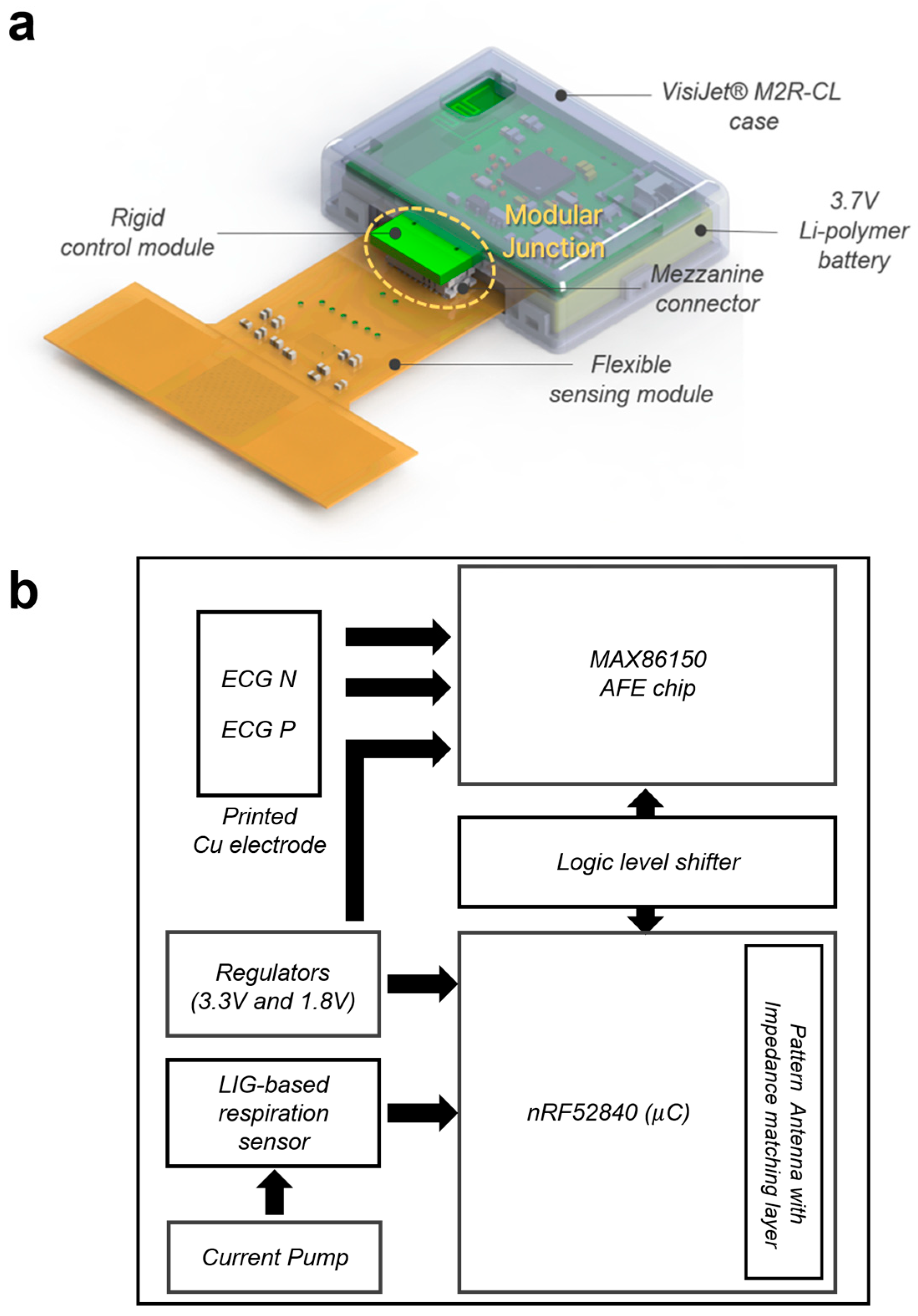
2.3. Developing an Integrated Wearable Cardiopulmonary Device for a Patient Monitoring System

This report proposes a cable-less patch-type multimodal biometric system that combines photoplethysmography (PPG) for SpO2, ECG for cardiac monitoring, and a respiration sensor for RR. Additionally, the system has automated data collection and a visualization interface. The proposed patient monitoring system is designed to overcome the problems of conventional clinical cardiopulmonary monitoring systems, which cause fatigue and discomfort for patients and medical staff due to the complex arrangement of cables and powerlines from various medical equipment and generate a significant workload for medical staff due to the unintegrated system that lacks compatibility with electronic medical record (EMR) systems. Using a modularized and cable-less design, the proposed system aims to alleviate these issues and provide a more efficient and user-friendly approach to the attachment, usage, and interpretation of patient monitoring systems in the ICU.
A modular approach will be employed when designing the device. Here modularization refers to dividing a complex system or device into smaller units that can be independently developed, manufactured, and replaced. In this context, the proposed device will comprise two separate modules (units): the sensing module that measures the vital signal and the control module that processes and transfers the acquired data. This modularized structure allows excellent reusability, easy replacement, and personalization of the device and, thus, provides the most optimal solution for usability and cost efficiency.
The modularized devices will comprise a semi-permanent control module that includes electronics on a rigid printed circuit board (PCB) and an expendable sensing module built on a flexible PCB (Figure 1a). The modular structure of the device offers cost-effectiveness by enabling the reuse of the control module, which contains high-cost electronics. Furthermore, it can prevent cross-infection among patients with skin-transmitted infectious diseases by using personalized sensing modules for each patient.
These two modules will be easily combined with a mezzanine connector, and the modular junction is marked with a dashed circle in Figure 1a.
The control module has dimensions of 30 × 40 mm. It will be powered by a rechargeable Li-polymer battery with a nominal voltage of 3.7 V. For the microcontroller, we have selected the nRF52840 (Nordic Semiconductor, Norway, distributed by Mouser Electronics) built on the powerful ARM® Cortex™-M4 CPU (Cambridge, UK). This microcontroller offers various digital interfaces and protocol support for BLE, which is essential for establishing wired and wireless communication within the system. The overall circuit configuration is demonstrated in Figure 1b.
The sensing module will comprise a respiration sensor, printed Cu electrodes for the ECG subsystem, and an MAX86150 biosensor module. As a sensing material, the respiration sensor will utilize Laser-Induced Graphene (LIG), a 3D porous graphene that was found to exhibit high sensitivity and biocompatibility in our previous study [5]. LIG is an attractive option for the practical fabrication and application of wearable sensors owing to its simple synthesis process, controllable design and morphology, and excellent electrical and mechanical properties [38,39,40]. Compared with conventional graphene synthesis processes, which include complex chemical reactions and multi-stage mechanical mechanisms [41,42], direct lasing of polymer substrates such as polyimide (PI) converts the layer into graphene through a photothermal effect without the requirement of additional chemicals or high-cost equipment. As shown in Figure 2a, laser irradiation on the exposed PI layer of FPCB makes a stable electrical connection between copper traces and fabricated LIG sensing area, and another LIG will be formed on a single PI film stacked on the top of the LIG area on FPCB (Figure 2b,c). As the final step of the fabrication process, the LIG sensor with a double-layer structure will be covered with an additional layer of elastomeric silicon to enhance its biocompatibility and protect it from external damage. The fabricated sensor will measure the respiratory signal by pressure sensing. It will be able to measure the patient’s respiratory motion, detecting the subtle changes in pressure caused by the movement of the chest during inhalation or exhalation, which provides reliable data for respiratory monitoring.
One of the challenges facing wearable sensor development is the issue of the reliable connection between the sensor and its front-end circuit [43,44]. In this study, we propose a solution to overcome this challenge by utilizing a silk-screened area of FPCB and direct lasing of the exposed PI layer during sensor fabrication. This approach allows for a straightforward and stable connection between the sensor and its front-end circuit, ensuring reliable and accurate data acquisition.
MAX86150, which operates on a 1.8 V supply voltage and includes two internal LEDs with different wavelengths, a photodetector, and an ECG subsystem optimized for dry electrode operation, will be placed on a platform to record PPG and ECG signals. The dry electrodes will be integrated into FPCB as part of the Cu signal traces, eliminating the need for separate electrode attachments. Conventional disposable gel electrodes, a type of wet electrode, are most commonly used in the ICU and show excellent adhesion and signal quality; however, their long-term usage may cause skin irritation (e.g., tickle, skin redness, and allergic reaction) [45] and discomfort from the long cables that come out of the electrodes. Moreover, the long-term use of gel electrodes triggers the problem of signal degradation caused by the dehydration of the electrolyte gel, which may lead to inaccurate measurements and reduced monitoring system reliability [46].
Conventionally, the high electrode–skin impedance of dry electrodes makes the clinical usage of dry electrodes challenging. The absence of conductive gel makes it difficult to form a stable interface between the skin and the electrode, increasing impedance and inhibiting conformality between the electrode and skin [47,48]. To address this issue, the proposed device will exploit soft silicon caps on each electrode to help form conformal contact by maintaining a stable electrode–skin contact area.
For communication between MAX86150 and the nRF52840 microcontroller, a standard I2C-compatible interface will be used. The data will be transferred through wireless Bluetooth Low Energy and periodically saved. A Python-based graphic user interface will be developed to visualize the transmitted data in real time.

2.4. Data Collection

Before the study, a questionnaire (Table 2) for intensive care unit medical staff will be completed regarding their experience with the conventional monitoring system. After obtaining documentation of informed written consent from the participating patients by the corresponding investigator (Y.-M.H.), continuous ECG monitoring will be attained from both systems only during participants’ ICU hospitalization. Oxygen saturation data are conventionally monitored continuously. However, saturation data are recorded to the EMR every 1 to 4 h, while the newer device monitors and records them continuously and simultaneously. Participants who want to discontinue the study will be excluded from the analysis. To compare the correlation and accuracy between the two monitoring systems, ten patients with at least 20 recordings per patient are planned. After the study, a questionnaire will be completed by intensive care unit medical staff regarding their experience with the new device (Table 3).
Before the clinical experiment, the pre-validation process of the signal will be conducted. The complexity of single-cycle ECG waveforms will be analyzed to validate the reliability of the ECG signal from the proposed device. As a quantitative indicator, the time-domain heart rate variability (HRV), such as the mean RR peak interval (mean heart rate), the standard deviation of normal-to-normal intervals (SDNN), the root-mean-square of successive differences of normal-to-normal intervals (RMSSD), and the frequency-domain HRV such as a power spectral analysis will be analyzed. After measuring the HRV data from each device, the correlation coefficient (R) value will be calculated to measure the accuracy of the proposed device compared with that of the reference devices. To evaluate the performance of the respiration sensor, the respiration rate per minute and the duration of inhalation and exhalation for each respiration will be compared. After the pre-validation process in the lab, the clinical experiment with ten patients in the ICU/surgical ICU patients will be conducted. An ECG Holter recorder and patient monitoring device will be attached to all test patients in advance. In the clinical experiment, vital signs such as heart rate, respiration, and SpO2 will be acquired from the proposed device and compared with those from the conventional patient monitoring systems currently used in the ICU (Figure 3). All files and data generated by this protocol will be stored in a secured safe box or as copied files with passwords with only the investigators’ accessibility. Data from this study will not be further openly accessible because of the personal identifier or sensitive information included. Since the questionnaires proposed in this study needed to confirm their reliability, we performed an interobserver agreement test with each of the questionnaires to two ICU physicians unrelated to this study. Cohen’s kappa was 0.98 and 0.99 for the pre-questionnaire (Table 2) and post-questionnaire (Table 3).
Based on the intensive care unit medical staff survey, the primary end point is to assess the necessity of simplified ICU monitoring devices. Secondary end points include an accuracy comparison of ECG, saturation measured by the conventional system, and the device developed.

2.5. Statistical Analysis

Continuous variables will be expressed as mean ± standard deviation. The categorical variables attained by the survey will be described using numbers and frequencies and multiple-choice analysis, as appropriate. A paired t-test will be applied for conventional and proposed monitoring system measurements. Moreover, intraclass correlation measurements will be determined. All tests will be two-sided, and a p-value of p < 0.05 will be considered statistically significant. Statistical analyses will be performed using R version 4.05 statistical software (R Foundation for Statistical Computing, Vienna, Austria).

3. Discussion

Flexible device technologies such as wearable sensors have significantly advanced in recent years, enabling continuous monitoring of various physiological parameters. Although these technologies have yet to be demonstrated in critical care management, their potential to revolutionize the clinical setting is considerable. In this study, we propose a wearable cardiopulmonary monitoring system that can be used in the ICU environment where healthcare workers and patients are prone to fatigue and error due to equipment complexity. The proposed system with a cable-less design is suggested to overcome the limitation of the existing monitoring system caused by the complex arrangement of multiple electrode cables and power lines. By eliminating the numerous wires and power lines, the proposed device system is anticipated to relieve patients’ discomfort, promote their mobility, and improve the work efficiency of medical staff. This cable-less design should also reduce the risk of accidents and disconnections caused by the tangling of cables. This system integrates all ECG, SpO2, and respiratory monitoring subsystems into a single device with its user interface. The BLE profile streamlines the monitoring process, enhancing convenience and eliminating the necessity for multiple separate monitoring devices. The modularized structure of the device should offer cost-effectiveness through the reusability of high-cost modules and enable the replacement and customization of sensing modules. To see the usefulness, we will compare the measured ECG parameters, including HRV, RR changes, and SpO2 measured in this novel device, to conventionally measured data. Then, we will use the questionnaires to prove the ICU workflow’s efficacy indirectly. Therefore, the proposed system has the potential to provide both economical and practical benefits, offering a more integrated and adaptable approach to patient monitoring in clinical situations for both medical staff and patients.
Meanwhile, arrhythmias, ventricular tachycardia, or atrial fibrillation during hospitalization or clinical treatment are well-known predictors of poor prognoses, such as sudden cardiac death or rehospitalization, highlighting the necessity of periodic post-discharge monitoring for patients with CVDs [49,50,51]. The conventional monitoring system has significant limitations for post-discharge monitoring, including difficulties in self-use by patients. Such difficulties come from its lack of portability and integrity between several measurement devices with their cumbersome design accompanied by numerous cables. However, the proposed device integrates all measurement functions into a stand-alone device mounted on a single measurement site and measures vital signals without obstructive cables. Patients are expected to be able to mount or remove the device themselves easily, and the signals from the integrated device will be monitored through their personal PC or smartphone in real time. Thus, it may be seamlessly used for patients’ care pathways from hospitalization to post-discharge monitoring.

4. Conclusions

Our study aims to redefine the necessity of an improved patient monitoring system and develop a simplified and efficient monitoring system for real-world critical care units. The accuracy of the signal from the proposed system will be pre-evaluated through the comparison analysis of the signals with those from the reference devices, and the usefulness and effectiveness of the proposed device will be validated in clinical situations. The study will also advance the current ICU setup by exploring how the device can be integrated into a critical care environment to improve patient care and clinical workflow. Because of recent developments and awareness of wearable devices, healthcare service has been revolutionized, especially in the context of cardiovascular diseases. Without invasive procedures, this easily attachable novel device can reliably offer various forms of ECG information with respiratory monitoring or activity tracking, and these can all be integrated with smartphone apps. This can provide early detection and management of arrhythmic events or heart failure aggravation. This device can remotely monitor any patient who needs acute or chronic medical attention. By proving its clinical reliability and ease of use, the proposed device will provide sufficient benefits in a real-world clinical environment, enabling it to perform as a potential solution for critical and post-discharge monitoring while reducing the inconvenience of existing monitoring equipment.

Author Contributions

Y.-M.H.: conceptualization, data curation, methodology, writing original draft, and supervision; J.L.: writing initial draft and validation; and S.-M.P.: methodology, formal analysis, verification, and supervision. All authors have read and agreed to the published version of the manuscript.

Funding

This work was supported by the Po-Ca Networking Groups funded by The Postech-Catholic Biomedical Engineering Institute(PCBMI) (No. 5-2023-B0001-00044). This research was also supported by the Pioneer Research Center Program and National R&D Program through the National Research Foundation of Korea, funded by the Ministry of Science, ICT & Future Planning (grant numbers 2022M3C1A3081294 and 2021M3H4A1A03049084).

Institutional Review Board Statement

This study was approved by the Institutional Review Board of the Catholic University of Korea St. Vincent’s Hospital (Suwon, Republic of Korea; Institutional Review Board No. VC22OISI0030) and conducted in accordance with the Declaration of Helsinki.

Informed Consent Statement

Only patients who provide informed consent will be included in the study.

Data Availability Statement

Due to the sensitive nature of the data, information created during and analyzed during the current study is available from the corresponding author (Y.-M.H., youmi0607@naver.com) upon reasonable request to bona fide researchers. After publishing, the study protocol will be opened to the public with full accessibility.

Conflicts of Interest

The authors declare that the research was conducted without any commercial or financial relationships that could be construed as a potential conflict of interest. The funders had no role in the study’s design, in the collection, analyses, or interpretation of data, in the writing of the manuscript, or in the decision to publish the results.

References

  1. Kang, S.; Pradana Rachim, V.; Baek, J.H.; Lee, S.Y.; Park, S. A flexible patch-type strain sensor based on polyaniline for continuous monitoring of pulse waves. IEEE Access 2020, 8, 152105–152115. [Google Scholar] [CrossRef]
  2. Ding, X.; Cao, H.; Zhang, X.; Li, M.; Liu, Y. Large scale triboelectric nanogenerator and self-powered flexible sensor for human sleep monitoring. Sensors 2018, 18, 1713. [Google Scholar] [CrossRef] [PubMed]
  3. Bariya, M.; Nyein, H.Y.Y.; Javey, A. Wearable sweat sensors. Nat. Electron. 2018, 1, 160–171. [Google Scholar] [CrossRef]
  4. Yoon, S.; Sim, J.K.; Cho, Y.H. A flexible and wearable human stress monitoring patch. Sci. Rep. 2016, 6, 23468. [Google Scholar] [CrossRef] [PubMed]
  5. Rachim, V.P.; Lee, J.; Kim, Y.C.; Oh, J.; Jeong, U.; Park, S. A scalable laser-centric fabrication of an epidermal cardiopulmonary patch. Adv. Mater. Technol. 2022, 7, 2200242. [Google Scholar] [CrossRef]
  6. Liu, Y.; Norton, J.J.S.; Qazi, R.; Zou, Z.; Ammann, K.R.; Liu, H.; Yan, L.; Tran, P.L.; Jang, K.-I.; Lee, J.W.; et al. Epidermal mechano-acoustic sensing electronics for cardiovascular diagnostics and human-machine interfaces. Sci. Adv. 2016, 2, e1601185. [Google Scholar] [CrossRef] [PubMed]
  7. Piepoli, M.F.; Hoes, A.W.; Agewall, S.; Albus, C.; Brotons, C.; Catapano, A.L.; Cooney, M.-T.; Corrà, U.; Cosyns, B.; Deaton, C.; et al. European Guidelines on cardiovascular disease prevention in clinical practice: The Sixth Joint Task Force of the European Society of Cardiology and other Societies on cardiovascular Disease Prevention in Clinical Practice (constituted by representatives of 10 societies and by invited experts) Developed with the special contribution of the European Association for Cardiovascular Prevention and Rehabilitation (EACPR). Eur. Heart J. 2016, 37, 2315–2381. [Google Scholar] [CrossRef]
  8. Breteler, M.J.M.; KleinJan, E.J.; Dohmen, D.A.J.; Leenen, L.P.H.; van Hillegersberg, R.; Ruurda, J.P.; van Loon, K.; Blokhuis, T.J.; Kalkman, C.J. Vital signs monitoring with wearable sensors in high-risk surgical patients: A clinical validation study. Anesthesiology 2020, 132, 424–439. [Google Scholar] [CrossRef] [PubMed]
  9. Downey, C.; Randell, R.; Brown, J.; Jayne, D.G. Continuous versus intermittent vital signs monitoring using a wearable, wireless patch in patients admitted to surgical wards: Pilot cluster randomized controlled trial. J. Med. Internet Res. 2018, 20, e10802. [Google Scholar] [CrossRef]
  10. Downey, C.L.; Chapman, S.; Randell, R.; Brown, J.M.; Jayne, D.G. The impact of continuous versus intermittent vital signs monitoring in hospitals: A systematic review and narrative synthesis. Int. J. Nurs. Stud. 2018, 84, 19–27. [Google Scholar] [CrossRef]
  11. Cardona-Morrell, M.; Prgomet, M.; Lake, R.; Nicholson, M.; Harrison, R.; Long, J.; Westbrook, J.; Braithwaite, J.; Hillman, K. Vital signs monitoring and nurse-patient interaction: A qualitative observational study of hospital practice. Int. J. Nurs. Stud. 2016, 56, 9–16. [Google Scholar] [CrossRef]
  12. Bøgh, N.; Agger, P.; Omann, C.; Skov, M.N.; Laustsen, C.; Wang, T.; Pedersen, M. New device for noninvasive telemetric monitoring of vital signs in healthy and newly operated piglets. J. Am. Assoc. Lab. Anim. Sci. 2020, 59, 90–93. [Google Scholar] [CrossRef]
  13. Poncette, A.S.; Wunderlich, M.M.; Spies, C.; Heeren, P.; Vorderwülbecke, G.; Salgado, E.; Kastrup, M.; Feufel, M.; Balzer, F. Patient monitoring alarms in an intensive care unit: Observational study with do-it-yourself instructions. J. Med. Internet Res. 2021, 23, e26494. [Google Scholar] [CrossRef]
  14. Asadi, N.; Salmani, F.; Asgari, N.; Salmani, M. Alarm fatigue and moral distress in ICU nurses in COVID-19 pandemic. BMC Nurs. 2022, 21, 125. [Google Scholar] [CrossRef]
  15. Akturan, S.; Güner, Y.; Tuncel, B.; Üçüncüoğlu, M.; Kurt, T. Evaluation of alarm fatigue of nurses working in the COVID-19 Intensive Care Service: A mixed methods study. J. Clin. Nurs. 2022, 31, 2654–2662. [Google Scholar] [CrossRef]
  16. Sun, B.; McCay, R.N.; Goswami, S.; Xu, Y.; Zhang, C.; Ling, Y.; Lin, J.; Yan, Z. Gas-Permeable, Multifunctional On-Skin Electronics Based on Laser-Induced Porous Graphene and Sugar-Templated Elastomer Sponges. Adv. Mater. 2018, 30, e1804327. [Google Scholar] [CrossRef]
  17. Yang, X.; Wang, S.; Liu, M.; Li, L.; Zhao, Y.; Wang, Y.; Bai, Y.; Lu, Q.; Xiong, Z.; Feng, S.; et al. All-Nanofiber-Based Janus Epidermal Electrode with Directional Sweat Permeability for Artifact-Free Biopotential Monitoring. Small 2022, 18, 2106477. [Google Scholar] [CrossRef]
  18. Zhou, W.; Yao, S.; Wang, H.; Du, Q.; Ma, Y.; Zhu, Y. Gas-Permeable, Ultrathin, Stretchable Epidermal Electronics with Porous Electrodes. ACS Nano 2020, 14, 5798–5805. [Google Scholar] [CrossRef]
  19. Chowdhury, M.E.H.; Khandakar, A.; Alzoubi, K.; Mansoor, S.; MTahir, A.; Reaz, M.B.I.; Al-Emadi, N. Real-Time Smart-Digital Stethoscope System for Heart Diseases Monitoring. Sensors 2019, 19, 2781. [Google Scholar] [CrossRef]
  20. Nakamura, Y.; Matsuda, Y.; Arakawa, Y.; Yasumoto, K. WaistonBelt X: A Belt-Type Wearable Device with Sensing and Intervention toward Health Behavior Change. Sensors 2019, 19, 4600. [Google Scholar] [CrossRef]
  21. Roudjane, M.; Bellemare-Rousseau, S.; Drouin, E.; Bélanger-Huot, B.; Dugas, M.; Miled, A.; Messaddeq, Y. Smart T-Shirt Based on Wireless Communication Spiral Fiber Sensor Array for Real-Time Breath Monitoring: Validation of the Technology. IEEE Sens. J. 2020, 20, 10841–10850. [Google Scholar] [CrossRef]
  22. Tscholl, D.W.; Handschin, L.; Rössler, J.; Weiss, M.; Spahn, D.R.; Nöthiger, C.B. It’s not you, it’s the design—Common problems with patient monitoring reported by anesthesiologists: A mixed qualitative and quantitative study. BMC Anesthesiol. 2019, 19, 87. [Google Scholar] [CrossRef]
  23. Lubitz, S.A.; Faranesh, A.Z.; Atlas, S.J.; McManus, D.D.; Singer, D.E.; Pagoto, S.; Pantelopoulos, A.; Foulkes, A.S. Rationale and design of a large population study to validate software for the assessment of atrial fibrillation from data acquired by a consumer tracker or smartwatch: The Fitbit heart study. Am. Heart J. 2021, 238, 16–26. [Google Scholar] [CrossRef]
  24. Rush, B.; Celi, L.A.; Stone, D.J. Applying machine learning to continuously monitored physiological data. J. Clin. Monit. Comput. 2019, 33, 887–893. [Google Scholar] [CrossRef]
  25. Han, C.; Song, Y.; Lim, H.S.; Tae, Y.; Jang, J.-H.; Lee, B.T.; Lee, Y.; Bae, W.; Yoon, D. Automated detection of acute myocardial infarction using asynchronous electrocardiogram signals-preview of implementing artificial intelligence with multichannel electrocardiographs obtained from smartwatches: Retrospective study. J. Med. Internet Res. 2021, 23, e31129. [Google Scholar] [CrossRef]
  26. Yoon, S.; Kim, T.; Roh, T.; Chang, H.; Hwang, S.Y.; Yoon, H.; Shin, T.G.; Sim, M.S.; Jo, I.J.; Cha, W.C. Twelve-lead electrocardiogram acquisition with a patchy-type wireless device in ambulance transport: Simulation-based randomized controlled trial. JMIR MHealth UHealth 2021, 9, e24142. [Google Scholar] [CrossRef]
  27. Morgado Areia, C.; Santos, M.; Vollam, S.; Pimentel, M.; Young, L.; Roman, C.; Ede, J.; Piper, P.; King, E.; Gustafson, O.; et al. A Chest patch for continuous vital sign monitoring: Clinical validation study during movement and controlled hypoxia. J. Med. Internet Res. 2021, 23, e27547. [Google Scholar] [CrossRef]
  28. Gardner, M.; Randhawa, S.; Malouf, G.; Reynolds, K. A wearable ballistocardiography device for estimating heart rate during positive airway pressure therapy: Investigational study among the general population. JMIR Cardio 2021, 5, e26259. [Google Scholar] [CrossRef]
  29. Nazir, S.; Pateau, V.; Bert, J.; Clement, J.; Fayad, H.; L’Her, E.; Visvikis, D. Surface imaging for real-time patient respiratory function assessment in intensive care. Med. Phys. 2021, 48, 142–155. [Google Scholar] [CrossRef]
  30. Khunlertkit, A.; Carayon, P. Contributions of tele-intensive care unit (Tele-ICU) technology to quality of care and patient safety. J. Crit. Care 2013, 28, 315.e1–315.e12. [Google Scholar] [CrossRef]
  31. Ha, T.; Tran, J.; Liu, S.; Jang, H.; Jeong, H.; Mitbander, R.; Huh, H.; Qiu, Y.; Duong, J.; Wang, R.L.; et al. A chest-laminated ultrathin and stretchable e-tattoo for the measurement of electrocardiogram, seismocardiogram, and cardiac time intervals. Adv. Sci. 2019, 6, 1900290. [Google Scholar] [CrossRef]
  32. Poncette, A.S.; Spies, C.; Mosch, L.; Schieler, M.; Weber-Carstens, S.; Krampe, H.; Balzer, F. Clinical Requirements of Future Patient Monitoring in the Intensive Care Unit: Qualitative Study. JMIR Med. Inform. 2019, 7, e13064. [Google Scholar] [CrossRef]
  33. Shahidul Islam, M.; Islam, M.T.; Almutairi, A.F.; Beng, G.K.; Misran, N.; Amin, N. Monitoring of the Human Body Signal through the Internet of Things (IoT) Based LoRa Wireless Network System. Appl. Sci. 2019, 9, 1884. [Google Scholar] [CrossRef]
  34. Sanfilippo, F.; Pettersen, K.Y. A sensor fusion wearable health-monitoring system with haptic feedback. In Proceedings of the 2015 11th International Conference on Innovations in Information Technology (IIT), Dubai, United Arab Emirates, 1–3 November 2015; pp. 262–266. [Google Scholar] [CrossRef]
  35. Ren, Y.; Sun, X.; Liu, J. Advances in Liquid Metal-Enabled Flexible and Wearable Sensors. Micromachines 2020, 11, 200. [Google Scholar] [CrossRef]
  36. Okutani, C.; Yokota, T.; Someya, T. Interconnected Heat-Press-Treated Gold Nanomesh Conductors for Wearable Sensors. ACS Appl. Nano Mater. 2020, 3, 200. [Google Scholar] [CrossRef]
  37. Myers, A.C.; Huang, H.; Zhu, Y. Wearable silver nanowire dry electrodes for electrophysiological sensing. RSC Adv. 2015, 5, 1848–1854. [Google Scholar] [CrossRef]
  38. Huang, L.; Su, J.; Song, Y.; Ye, R. Laser-induced graphene: En route to smart sensing. Nanomicro. Lett. 2020, 12, 157. [Google Scholar] [CrossRef]
  39. Ye, R.; James, D.K.; Tour, J.M. Laser-induced graphene: From discovery to translation. Adv. Mater. 2019, 31, e1803621. [Google Scholar] [CrossRef]
  40. Ye, R.; James, D.K.; Tour, J.M. Laser-induced graphene. Acc. Chem. Res. 2018, 51, 1609–1620. [Google Scholar] [CrossRef]
  41. Yi, M.; Shen, Z. A review on mechanical exfoliation for the scalable production of graphene. J. Mater. Chem. A 2015, 3, 11700–11715. [Google Scholar] [CrossRef]
  42. Parvez, K.; Yang, S.; Feng, X.; Müllen, K. Exfoliation of graphene via wet chemical routes. Synth. Met. 2015, 210, 123–132. [Google Scholar] [CrossRef]
  43. Jeerapan, I.; Poorahong, S. Review—Flexible and stretchable electrochemical sensing systems: Materials, energy sources, and integrations. J. Electrochem. Soc. 2020, 167, 037573. [Google Scholar] [CrossRef]
  44. Choi, J.; Ahmed, B.; Gutierrez-Osuna, R. Development and evaluation of an ambulatory stress monitor based on wearable sensors. IEEE Trans. Inf. Technol. Biomed. 2012, 16, 279–286. [Google Scholar] [CrossRef]
  45. Meziane, N.; Webster, J.G.; Attari, M.; Nimunkar, A.J. Dry electrodes for electrocardiography. Physiol. Meas. 2013, 34, R47–R69. [Google Scholar] [CrossRef] [PubMed]
  46. Oh, J.; Jang, S.G.; Moon, S.; Kim, J.; Park, H.K.; Kim, H.S.; Park, S.; Jeong, U. Air-permeable waterproofing electrocardiogram patch to monitor full-day activities for multiple days. Adv. Healthc. Mater 2022, 11, e2102703. [Google Scholar] [CrossRef]
  47. Chen, Y.H.; De Beeck, M.O.; Vanderheyden, L.; Carrette, E.; Mihajlović, V.; Vanstreels, K.; Grundlehner, B.; Gadeyne, S.; Boon, P.; Van Hoof, C. Soft, comfortable polymer dry electrodes for high quality ECG and EEG recording. Sensors 2014, 14, 23758–23780. [Google Scholar] [CrossRef]
  48. Yamamoto, Y.; Yamamoto, D.; Takada, M.; Naito, H.; Arie, T.; Akita, S.; Takei, K. Efficient skin temperature sensor and stable gel-less sticky ECG sensor for a wearable flexible healthcare patch. Adv. Healthc. Mater. 2017, 6. [Google Scholar] [CrossRef] [PubMed]
  49. Hwang, Y.M.; Sung, M.K.; Kim, S.O. Impact of de novo tachyarrhythmias in patients with prior acute coronary syndrome. Medicine 2022, 101, e29685. [Google Scholar] [CrossRef]
  50. Orvin, K.; Eisen, A.; Goldenberg, I.; Gottlieb, S.; Kornowski, R.; Matetzky, S.; Golovchiner, G.; Kuznietz, J.; Gavrielov-Yusim, N.; Segev, A.; et al. Outcome of contemporary acute coronary syndrome complicated by ventricular tachyarrhythmias. Europace 2016, 18, 219–226. [Google Scholar] [CrossRef]
  51. Holden, J.; Fernie, G.R.; Soto, M. An assessment of a system to monitor the activity of patients in a rehabilitation programme. Prosthet. Orthot. Int. 1979, 3, 99–102. [Google Scholar] [CrossRef]
Figure 1. Modularized device for the simplified patient monitoring system. (a) A 3D drawing of the modularized device for the proposed patient monitoring system. (b) Configuration of the overall circuit components.
Applsci 13 13101 g001
Figure 2. Fabrication process of the LIG-based mechano-acoustic sensor. (a) laser irradiation on the exposed PI layer of FPCB to make a stable electrical connection between copper traces and fabricated LIG sensing area; (b,c) another LIG will be formed on a single PI film stacked on the top of the LIG area on FPCB; (d) Integrated version of the proposed sensor.
Applsci 13 13101 g002
Figure 3. Experimental setup for the pre-validation process of the signal from the proposed device. (a) Experimental setup for the pre-validation process and measurement site of the reference ECG, PPG, and respiration signal. (b) Photograph of the manufactured device attached to the human chest during the pre-validation process.
Applsci 13 13101 g003
Table 1. Comparison between the conventional patient monitoring system and the proposed monitoring system.
ConventionalProposed
Weight Bulky
(~tens of kilograms)
Lightweight
(~5 g)
PortabilityHard (non-portable)Easy
Presence of cablesYesNo
IntegrityNoYes
(single device mounted on the chest)
Synchronization of signalsHard
(signals are obtained from separate devices)
Easy
Table 2. Questionnaire for intensive care unit medical staff regarding their experience of the conventional monitoring system.
Survey for ICU Medical StaffStrongly Agree AgreeNeutral Disagree Strongly Disagree
Awareness of ICU monitoring equipment currently in use
I think the current ICU monitoring equipment is optimized for patients and medical staff.
It is convenient to use the current ICU monitoring equipment.
I am satisfied with the current ICU monitoring system; there is nothing to supplement.
The inconvenience of the currently used ICU monitoring equipment
There are many unnecessary alarms (device noise, false alarms, incorrect settings, and so on).
Many cables are connected to the sensor, which is inconvenient when applied.
Interlocking or compatibility with other devices is difficult (ventilator, DC device, and EMR linkage).
It is difficult to use patient monitoring equipment (it takes learning time, responding to alarms is inconvenient, and so on).
I hope that ICU monitoring equipment will be improved as follows.
Using a wireless sensor (without cable)
Unnecessary alarms are reduced.
In-hospital remote monitoring system (mobile phone/tablet PC)
Other ideas?
What are your thoughts on using new digital equipment to make ICU monitoring more straightforward and accurate?
I want to try it as soon as possible.
I would like to apply new equipment more slowly (skilled use seems complicated).
New digital equipment is unlikely to be trusted yet.
Table 3. Questionnaire for intensive care unit medical staff regarding their experience with the new device.
Survey for ICU Medical Staff Strongly Agree Agree Neutral Disagree Strongly Disagree
Ease of use of the proposed device
The overall system was easy to use.
The user interface of the system was intuitive and easy to understand.
It was easy to learn how to use the system.
It was easy to attach/detach the device.
Effectiveness and satisfaction
The usage of the system reduced the workload and fatigue.
The usage of the system was time-efficient.
I would use the system again.
I would recommend the system to other medical staff.
Disclaimer/Publisher’s Note: The statements, opinions and data contained in all publications are solely those of the individual author(s) and contributor(s) and not of MDPI and/or the editor(s). MDPI and/or the editor(s) disclaim responsibility for any injury to people or property resulting from any ideas, methods, instructions or products referred to in the content.

